Case Report

A Relapsed AML Case Featuring MYC and MECOM Rearrangements

1
Medical Scientist Training Program, Stony Brook University Renaissance School of Medicine, Stony Brook, NY 11794, USA
2
Department of Pathology, Stony Brook University Hospital, Stony Brook, NY 11794, USA
3
Department of Hematology and Oncology, Stony Brook University Hospital, Stony Brook, NY 11794, USA
*
Author to whom correspondence should be addressed.
Diagnostics 2025, 15(18), 2410; https://doi.org/10.3390/diagnostics15182410
Submission received: 11 August 2025 / Revised: 15 September 2025 / Accepted: 17 September 2025 / Published: 22 September 2025

Abstract

Background/Objectives: Relapsed acute myeloid leukemia (AML) is often characterized by clonal evolution and acquired genomic abnormalities, which can inform prognosis and direct therapeutic decisions. The emergence of high-risk chromosomal rearrangements during relapse is of particular significance, yet the impact of rare and complex events remains poorly understood. This report details a case of relapsed AML that demonstrated rare MYC and MECOM rearrangements and additional features that were not observed at initial diagnosis, emphasizing the clinical relevance of serial cytogenetic assessments. Case Description: A 70-year-old man was initially diagnosed with AML, exhibiting monocytic differentiation, an 11q23 deletion involving KMT2A loss, and a U2AF1 mutation. After achieving remission with azacitidine and venetoclax, the patient relapsed within ten months, necessitating reevaluation and modification of therapy. Repeat cytogenetic analysis at relapse revealed a distinct t(3;8)(q26.2;q24.3) exhibiting MYC and MECOM rearrangements, features that were absent at initial diagnosis. Conclusions: This case underscores the importance of serial cytogenetic and molecular profiling in relapsed AML. The emergence of new abnormalities upon relapse suggested underlying genomic instability and clonal evolution. MYC rearrangements are notably rare in AML, especially with concurrent MECOM rearrangements, highlighting a unique feature of this case. The identification of novel abnormalities at relapse may carry prognostic and therapeutic significance and may be used to refine risk stratification. Thus, ongoing cytogenetic monitoring is essential to adapt management approaches in evolving disease contexts.

1. Introduction

Acute myeloid leukemia (AML) is a heterogeneous group of leukemias that develop from dysregulated proliferation of myeloid cells, which can be driven by a broad variety of genetic mechanisms [1,2]. Genomic alterations are commonly observed in AML and may be readily detected by conventional cytogenetic techniques, including Giemsa-banded karyotyping and molecular cytogenetic techniques such as fluorescence in situ hybridization (FISH) and next-generation sequencing (NGS) [3]. The prognostic value of identifying these alterations motivates the routine use of cytogenetic assays to characterize each patient’s disease course and guide therapy [2,4]. Crucially, relapses of AML are often found to have acquired new cytogenetic abnormalities from the initial diagnosis, possibly representing unique clonal expansion that is selected for by cytotoxic therapy [5,6,7].
In this report, we describe a case of relapsed AML that developed a cytogenetic rearrangement involving MYC and MECOM (also known as EVI1) that was not present on initial diagnosis. Notably, MYC translocations are rare in AML, and concurrent MECOM rearrangements are even rarer, with only a few cases described in the literature; thus, this report may contribute insight into a distinct mechanism that drives AML relapse [8,9,10,11]. We summarize the clinical presentation, diagnostic workup, management, and surveillance, with an emphasis on the timeline of cytogenetic changes. We conclude with a discussion of proposed mechanisms of the genomic aberrations seen in this case, along with a review of the literature focusing on similar AML cases involving MYC and MECOM events.

2. Case Report

2.1. Case Presentation

A 70-year-old man presented with six months of progressive exertional dyspnea. Initial labs revealed severe anemia with hemoglobin of 6.1 g/dL and moderate leukocytosis of 19.3 × 10⁹/L, prompting hospitalization and further workup. Peripheral blood smear showed increased monocytes, and flow cytometry also demonstrated a small population of circulating myeloblasts representing 0.3% of total events. Bone marrow biopsy demonstrated greater than 80% immature monocyte blasts on histology, diagnostic of AML with monocytic differentiation (Figure 1; Table 1). Additionally, prominent trilineage dysplasia was present. On flow cytometry, abnormal myeloid maturation with downregulated CD10 and CD16 was seen, and monocytes (65% of total) demonstrated aberrant downregulation of CD14 (34% of total). Cytogenetic analysis demonstrated a karyotype with chromosome 12p deletion in all cells and 11p deletion in a subset of cells, and FISH analysis detected KMT2A loss without gene rearrangement. NGS panel showed U2AF1 c.101C>T (S34F) mutation with variant allele frequency of 50%. Notably, the U2AF1 mutation indicated adverse risk based on the European Leukemia Network 2022 AML classification [2].
During the hospitalization, the patient’s anemia was managed with packed red blood cell transfusions. Given the patient’s significant debility, the patient was not felt to be a candidate for intensive induction and subsequently began treatment with a combination of azacitidine 75mg/m2 on days 1–7 and venetoclax 400mg daily in a 28-day cycle, with a ramp-up dose of venetoclax used. A repeat bone marrow biopsy after two cycles demonstrated complete morphologic remission, corroborated by flow cytometry finding no increased blasts or monocytes. Measurable residual disease was suggested by the presence of persistent KMT2A abnormality in a subset of cells. With his AML in remission, the patient continued this chemotherapy regimen for seven cycles. Given the risk of disease relapse, he was followed closely and was recommended for allogeneic bone marrow transplant but deferred during this time for personal reasons.
Ten months after diagnosis, the patient experienced recurrent fatigue; he was found again to be anemic, with hemoglobin of 6.0 g/dL. Repeat bone marrow biopsy revealed 14% CD34-positive blasts along with increased hypolobated megakaryocytes, signifying AML relapse. Molecular cytogenetic testing again demonstrated the previously seen loss of KMT2A and U2AF1 mutation but also demonstrated MYC and MECOM rearrangements and loss of ETV6, findings that were not described at initial diagnosis. Collectively, these new findings indicated a complex karyotype with genomic instability and increased risk compared to the initial disease.
The patient was then initiated on high-intensity induction therapy with cladribine 5mg/m2 on days 1–5, cytarabine 20mg every 12 hours on days 1–5, and venetoclax 100mg on days 10–14, and was admitted for supportive therapy and transfusions. Post-induction bone marrow biopsy demonstrated residual disease, implying insufficient response to this regimen. The patient was then admitted for salvage reinduction with FLAG-IDA (fludarabine 30mg/m2 on days 2–5, cytarabine 2000mg/m2 on days 2–6, idarubicin 10mg/m2 on days 2–4, and filgrastim 300 mcg/m2 on days 1–5). Most recently, post-salvage bone marrow assessment demonstrated persistent AML. The patient remains transfusion-dependent and is undergoing enrollment for trial with cellular therapy.

2.2. Materials and Methods

2.2.1. Conventional Cytogenetics

Chromosome analysis was performed using conventional cytogenetics protocols, including Giemsa (G-band) staining of metaphase cells. Results were annotated according to the International System for Human Cytogenomic Nomenclature 2024 standards.

2.2.2. Molecular Cytogenetics

Fluorescence in situ hybridization (FISH) was performed with the XL MECOM (3q26) (MetaSystems, Medford, MA, USA), the LSI 5q EGR1/D5S23, D5S721 (5q31, 5p15.2), the LSI D7S486/CEP 7 (7q31/7p11.1-q11.1(D7Z1)), the LSI RUNX1/RUNX1T1(8q21.3, 21q22), the LSI MYC DC BAR (8q24), the LSI BCR/ABL1 DC DF (9q34, 22q11.2) (Abbott, Des Plaines, IL, USA), the XL KMT2A BA (11q23.3) the XL ETV6 BA (12q13.2) (MetaSystems, Medford, MA, USA), the LSI CBFB (16q22) DC BAR, and the LSI D20S108 (20q12) (Abbott, Des Plaines, IL, USA) probes. Probes were used following the manufacturer’s instructions for signal interpretation.

2.3. Cytogenetic Results

2.3.1. Conventional Cytogenetics

At initial diagnosis, karyotype analysis of twenty cells revealed an abnormal male karyotype with segmental deletion at 12p13-12p11.2 in all cells and segmental deletion at 11q21-11q25 in nine of twenty cells, indicating evolution of a side clone containing two abnormalities. This karyotype was reported as 46,XY,del(12)(p13p11.2) [20]/46,XY,del(11)(q21q25),del(12)(p13p11.2)[9].
Upon relapse, a more complex karyotype emerged in a set of fifteen cells, including apparent translocations of chromosomes 3q26 and 8q24, in addition to the previously noted chromosome 11 and chromosome 12 deletions. This karyotype was reported as 46,XY,t(3;8)(q26;q24),del(11)(q21q25),add(12)(p13).

2.3.2. Molecular Cytogenetics

At diagnosis, FISH was performed to examine two hundred interphase cells and metaphase cells using a comprehensive panel of commercial probes. FISH analysis revealed loss of KMT2A in 13.5% of cells, consistent with 11q23 deletion. Additional FISH markers were negative for common alterations involving RUNX1/RUNX1T1 (t(8;21)(q22;q22)), PML/RARA (t(15;17)(q24;q21)), CBFB (16q22), CSF1R/RPS14 (5q33), and MDFIC (7q31). Mutation testing for FLT3, IDH1, IDH2, CEBPA, NPM1, PML::RARA, and cKIT were also negative. The isolated finding of KMT2A loss was consistent with the del(11)(q21q25) reported by conventional cytogenetics. Further, a targeted NGS panel identified a U2AF1 c.101C>T (S34F) mutation with variant allele frequency of 50%, which was denoted a Tier 1 variant of strong clinical significance. A variant of unknown significance with KMT2A c.7240T>A was also identified.
After two cycles of induction treatment, FISH analysis again demonstrated one copy of KMT2A in 28.5% of cells, while three copies of KMT2A were identified in 5.0% of cells, suggesting minimal residual disease that was not detected on histological and immunohistochemical examination or flow cytometry.
Upon relapse, additional cytogenetic changes were noted. FISH again demonstrated monosomy of KMT2A in 91.5% out of two hundred cells, suggesting expansion of the previously identified clones with KMT2A loss. Because conventional karyotyping suggested newly acquired chromosomal changes involving t(3;8)(q26;q24), break-apart FISH analysis on metaphase cells was essential to delineate the precise rearrangements of MYC and MECOM (Figure 2). Specifically, we observed a clone with MECOM rearrangement from one homolog of chromosome 3 onto one homolog of chromosome 8 with apparently intact MYC, evidenced by the 3′ (distal) signal of MECOM (green) on a derivative 8 that retained intact MYC and the 5′ (proximal) signal of MECOM (red) that was retained on one of the derivative 3; simultaneously, we observed MYC rearrangement from the other homolog of chromosome 8 to the other homolog of chromosome 3 with apparently intact MECOM, as evidenced by the 3′ (distal) signal of MYC (green) on the other derivative 3 that retained MECOM and 5’ (proximal) signal of MYC retained on the other derivative 8. These findings revealed that both copies of both chromosomes 3 and 8 were abnormal in this specific clone. Monosomy of ETV6 was also present on FISH, consistent with 12p13 deletion. Additionally, NGS panel again identified a U2AF1 c.101C>T (S34F) mutation with variant allele frequency 31.5%. Incorporating these FISH findings, the final karyotype of the relapse was reported as 46,XY,t(3;8)(q26.2;q24.3),del(11)(q23),add(12)(p13),der(3)(3′MECOM+,5′MECOM-),der(3)(3′MYC+),der(8)(5′MYC+,3′MYC-),der(8)(5′MECOM+).

3. Discussion

This case highlights the progressive nature of cytogenetic evolution in AML. At diagnosis, our patient had a deletion of 11q23 involving monosomic KMT2A loss without rearrangement, which is considered an intermediate-risk feature, and U2AF1 mutation, which is considered an adverse-risk myelodysplasia-related gene mutation [2]. Despite initial rapid remission with cytotoxic therapy, the patient’s disease relapsed within a year. On relapse, the disease appeared to have undergone clonal evolution, retaining the KMT2A deletion and U2AF1 mutation while acquiring several high-risk features in a subset of cells, including rarely observed concurrent rearrangements of MYC and MECOM. Critically, these features raised the possibility of genomic instability and clonal evolution driving leukemic progression and relapse. The relapse was treated with a shift in chemotherapy regimen, a strategy that was informed by emerging data suggesting that some patients with prior venetoclax exposure may still derive benefit from cytarabine-based reinduction regimens [4,12]. Given the acquisition of high-risk cytogenetics and insufficient response to the second-line therapy, the patient was subsequently transitioned to high-dose reinduction chemotherapy and was evaluated for further treatment options, including allogeneic hematopoietic stem cell transplantation as well as cellular-based therapy [4,13].
Mechanistically, this case implicates several genes with diverse mechanisms. The loss of one copy of KMT2A, a histone methyltransferase and tumor suppressor, likely contributed to the initial disease through activation of proliferation genes and disruption of the DNA damage response [14,15]. The proto-oncogene transcription factor MYC is often implicated as a central driver gene in AML, wherein MYC amplification is typically seen due to duplications, promoter mutations, or other gene-regulatory shifts, rather than chromosomal rearrangements [16,17,18]. Other studies have also implicated PVT1, a long non-coding RNA with a genomic locus that is directly downstream to MYC, as a key regulator of MYC activity involved in complex transcriptional feedback regulation, wherein disruption of local chromosome architecture may negate topological chromatin insulation, which normally prevents MYC activation by PVT1 enhancer regions, thereby inducing MYC overexpression and driving proliferation [19,20]. Like MYC, MECOM is another transcription factor which can activate proliferation gene programs, and translocations of MECOM have recently been included as adverse risk factors in leukemia classification guidelines [2,21,22]. In our case, the distal 3q arm containing MECOM and distal 8q arm containing MYC and PVT1 were rearranged to different homologs; a few prior cases of MECOM::PVT1 rearrangement have been reported, though not in context of functional studies [10,23,24]. ETV6 is another transcription factor involved in hematopoeisis, for which loss has been observed in AML [25]. Finally, U2AF1 S34F mutation represents a specific splicing factor mutation that is known to be linked to poor outcomes [26,27].

Review of the Literature

The phenomenon of clonal evolution is a well-established feature of relapsed and therapy-related AML, wherein selective pressure from prior cytotoxic therapy may lead to the emergence of new cytogenetic and molecular abnormalities. A review by Vosberg and Greif (2019) noted that a significant proportion of relapsed AML cases acquire new genetic abnormalities that are not present at initial diagnosis, thereby signifying underlying genomic instability and selection of resistant subclones [5]. Clonal evolution studies further support the view that such rearrangements may emerge during treatment, contributing to disease progression and therapy resistance [6,7]. Our case provides a striking example of this process, with the emergence of several high-risk genomic features at relapse.
Rearrangements involving MYC and MECOM represent rare but clinically meaningful events in AML, particularly in relapsed or therapy-related cases. MECOM, a complex gene locus encoding the EVI1 transcription factor, is a well-established adverse prognostic marker associated with chemoresistance and poor survival [28,29,30]. MYC abnormalities are more commonly associated with lymphoid malignancies yet remain rare in AML [16,17]. When MYC rearrangements do occur in myeloid neoplasms, they frequently correlate with aggressive disease [31]. Although both MYC and MECOM rearrangements are individually recognized in hematologic malignancies, their co-occurrence in AML is exceedingly rare. The first reported t(3;8)(q26.2;q24) translocation in myelodysplastic syndrome (MDS) was identified in 1987 [32]. A review of the recent literature identified a small number of case reports and case series in which simultaneous rearrangements of the MYC and MECOM loci were documented (Table 2). These cases tended to emerge in the context of disease progression or therapy-related AML and were frequently associated with adverse-risk cytogenetic features.
Liu and Tirado (2018) reported a case of polycythemia vera progressing to AML and exhibiting t(3;8)(q26.2;q24) [8]. This finding was established by chromosome analysis and confirmed by break-apart FISH. The authors further queried the Mitelman Database of Chromosome Aberrations and Gene Fusions in Cancer to identify 36 additional cases of AML and 4 cases of MDS which harbored a t(3;8) translocation and examined common co-occurring abnormalities, the most common of which was monosomy 7. Furthermore, the authors highlighted an incomplete understanding of the molecular mechanism of this translocation in driving AML, noting that some cases did not exhibit PVT1 involvement, as others had hypothesized [8,33].
Tang et al. (2019) reported twenty cases of t(3;8)(q26.2;q24) resulting in MYC and MECOM rearrangement in patients from a single institution, including six previously reported cases [9]. These cases, diagnosed over a 20-year period, encompassed therapy-related AML and MDS, as well as blast phases of chronic myeloid leukemia and myeloproliferative neoplasms. One-third of cases showed megakaryocyte hypoplasia, as in this case. Karyotypes were analyzed, and MYC and MECOM involvement were confirmed with break-apart FISH probes. Additional sequencing panel analysis identified a few cases with mutations in ABL1, ASXL1, BRINP3, CBFB, GATA2, NRAS, RUNX1, SRSF2, and TET2; notably, FLT3 and KRAS mutations were not detected in any of these cases. The authors further characterized survival outcomes, noting a median overall survival of 6 months and inferior survival compared to a control cohort of 44 cases without t(3;8), indicating an aggressive disease subtype with poor outcomes [9].
Smith et al. (2020) reported two additional cases of MDS with confirmed MYC and MECOM rearrangements [10]. Both cases utilized break-apart FISH probes to diagnose MYC and MECOM translocations. The authors note that their MYC break-apart probe was insufficient to distinguish PVT1 involvement; however, they reference a key study that demonstrated MYC and PVT1 overexpression in cases with t(3;8) rearrangements to support the hypothesis of both genes being involved [23]. They additionally reviewed 33 previously reported cases in a 10-year retrospective search of the Human Genetics Laboratory database, encompassing AML, MDS, and blast-phase chronic myeloid leukemia. They again identified monosomy 7 in 45% of these cases, while noting a single occurrence of monosomy 5, which is more common in inv(3) and t(3;3) cases [34]. Mechanistically, the authors proposed that the translocation activated MECOM while disrupting MYC regulatory control by PVT1, contributing to disease progression [10].
In addition to FISH and sequencing techniques, recent advances in optical genome mapping (OGM) have enhanced the detection of subtle genetic rearrangements [11,35]. McGinnis et al. (2024) demonstrated this technology on 90 cases from a 10-month period encompassing AML and lymphoblastic leukemias [11]. The authors identified several rearrangements that standard cytogenetics, including FISH, failed to detect, even describing a MECOM::MYC rearrangement, although further specifics on the case or cases were not provided. This study emphasized the sensitivity of OGM for high-resolution genomic profiling to capture low-frequency or subtle-to-detect events, highlighting promising benefits from the evolving techniques of cytogenetic surveillance if practically implemented [36].
Lastly, emerging evidence suggests that AML with monocytic differentiation, as in this case, may respond poorly to venetoclax and azacitidine; a single-institution cohort of 86 AML patients distinguished poorer treatment response and survival in cases with greater than 10% monoblasts [37]. Cytogenetic information, including U2AF1 mutation status, has been studied in AML to stratify and predict the response to venetoclax [38]. Notably, novel therapies are emerging in the field of AML, such as menin inhibitors for KMT2A rearrangements (not deletions as in this case) [39]. Formulated combination treatments such as liposomal CPX-351 (cytarabine and daunorubicin) show promise for AML cases with high-risk features [40]. Hence, the prognostic information provided by serial cytogenetic analysis may better guide management decisions, as well as support trials investigating alternative therapies in AML [41].
Collectively, the current literature provides insight into MYC and MECOM rearrangements as rare yet critical late-acquired events in AML, reflecting clonal evolution and conferring adverse prognosis. Detection of these genetic abnormalities is enabled by advanced cytogenetic techniques like FISH and OGM. Moreover, this line of research raises critical questions regarding the functional consequences of MYC and MECOM rearrangement, particularly whether these rearrangements represent cooperating leukemogenic events. Our case contributes to this sparse but growing literature by documenting concurrent MYC and MECOM rearrangements acquired at relapse and reinforces the diagnostic value of cytogenetic evaluation in identifying and monitoring complex cytogenetic events.

4. Conclusions

This case underscores the importance of serial cytogenetic and molecular characterization, particularly at relapse, where the emergence of new abnormalities can significantly alter prognosis and guide critical treatment decisions. In relapsed AML, especially with adverse-risk cytogenetics, prompt intervention with modified therapy based on prior treatment history is imperative. AML cases exhibiting complex karyotypes with genomic instability pose a distinct challenge and merit research attention to unravel the effects of co-occurring rearrangements and mutations. Thus, improved cytogenetics techniques and characterization will continue to enhance our clinical understanding of genomic alterations driving AML, ultimately working towards the goal of improving patient outcomes.

Author Contributions

Conceptualization, K.A.M. and C.A.T.; investigation, K.A.M.; data curation, K.A.M. and J.R.; writing—original draft preparation, K.A.M.; writing—review and editing, P.M.III, L.Z.L., T.A. and C.A.T.; visualization, J.R.; supervision, M.S., T.A. and C.A.T.; methodology, C.A.T.; project administration, C.A.T.; resources, M.S., T.A. and C.A.T. All authors have read and agreed to the published version of the manuscript.

Funding

This research received no external funding.

Institutional Review Board Statement

The study was conducted in accordance with the Declaration of Helsinki. Ethical review and approval were waived for this study due to at Stony Brook University a case report does not meet the definition of research requiring IRB approval.

Informed Consent Statement

Written informed consent has been obtained from the patient to publish this paper.

Data Availability Statement

No new data were created or analyzed in this study. Data sharing is not applicable to this article.

Acknowledgments

We thank the Stony Brook University Hospital Departments of Pathology and Hematology-Oncology for their clinical and technical support. We also acknowledge the cytogenetics laboratory staff for their assistance in generating and interpreting FISH and karyotype data.

Conflicts of Interest

The authors declare no conflicts of interest.

Abbreviations

AML—acute myeloid leukemia; FISH—fluorescent in situ hybridization; t(3;8)—translocation of chromosomes 3 and 8.

References

  1. Chakraborty, S.; Park, C.Y. Pathogenic Mechanisms in Acute Myeloid Leukemia. Curr. Treat. Options Oncol. 2022, 23, 1522–1534. [Google Scholar] [CrossRef]
  2. Döhner, H.; Wei, A.H.; Appelbaum, F.R.; Craddock, C.; DiNardo, C.D.; Dombret, H.; Ebert, B.L.; Fenaux, P.; Godley, L.A.; Hasserjian, R.P. Diagnosis and Management of AML in Adults: 2022 Recommendations from an International Expert Panel on Behalf of the ELN. Blood J. Am. Soc. Hematol. 2022, 140, 1345–1377. [Google Scholar] [CrossRef]
  3. Ozkan, E.; Lacerda, M.P. Genetics, Cytogenetic Testing and Conventional Karyotype; StatPearls Publishing: Treasure Island, FL, USA, 2020. [Google Scholar]
  4. National Comprehensive Cancer Network (NCCN). Clinical Practice Guidelines in Oncology (NCCN Guidelines®): Acute Myeloid Leukemia; Version 2; NCCN: Plymouth Meeting, PA, USA, 2025; Available online: https://www.nccn.org/professionals/physician_gls/pdf/aml.pdf (accessed on 11 May 2025).
  5. Vosberg, S.; Greif, P.A. Clonal Evolution of Acute Myeloid Leukemia from Diagnosis to Relapse. Genes. Chromosomes Cancer 2019, 58, 839–849. [Google Scholar] [CrossRef]
  6. Rapaport, F.; Neelamraju, Y.; Baslan, T.; Hassane, D.; Gruszczynska, A.; Robert de Massy, M.; Farnoud, N.; Haddox, S.; Lee, T.; Medina-Martinez, J. Genomic and Evolutionary Portraits of Disease Relapse in Acute Myeloid Leukemia. Leukemia 2021, 35, 2688–2692. [Google Scholar] [CrossRef]
  7. Kim, N.; Hahn, S.; Choi, Y.J.; Cho, H.; Chung, H.; Jang, J.E.; Lyu, C.J.; Lee, S.-T.; Choi, J.R.; Cheong, J.-W. Comprehensive Insights into AML Relapse: Genetic Mutations, Clonal Evolution, and Clinical Outcomes. Cancer Cell Int. 2024, 24, 174. [Google Scholar] [CrossRef] [PubMed]
  8. Liu, K.; Tirado, C.A. A t (3; 8)(Q26. 2; Q24) Involving the EVI1 (MECOM) Gene. J. Assoc. Genet. Technol. 2018, 44, 92–99. [Google Scholar]
  9. Tang, G.; Hu, S.; Wang, S.A.; Xie, W.; Lin, P.; Xu, J.; Toruner, G.; Zhao, M.; Gu, J.; Doty, M. T(3;8)(Q26.2;Q24) Often Leads to MECOM/MYC Rearrangement and Is Commonly Associated with Therapy-Related Myeloid Neoplasms and/or Disease Progression. J. Mol. Diagn. 2019, 21, 343–351. [Google Scholar] [CrossRef]
  10. Smith, S.C.; Qdaisat, T.Z.S.; Althof, P.A.; Dave, B.J.; Sanmann, J.N. MECOM Rearrangement Involving the MYC Locus: Two Additional Patients with the Rare Translocation, t(3;8)(Q26.2;Q24), and Molecular Review. Leuk. Res. 2020, 95, 106387. [Google Scholar] [CrossRef]
  11. McGinnis, E.; Stubbins, R.J.; Li, D.; Hamadeh, Z.; Spence, T.J. Optical Genome Mapping As Standard-of-Care in Acute Leukemia: Diagnostic and Clinical Impacts 10 Months Post-Implementation. Blood 2024, 144, 1538. [Google Scholar] [CrossRef]
  12. Cheung, E.; Schumann, C.; Zhang, T.Y.; Fakhri, B.; Gotlib, J.; Liedtke, M.; Shomali, W.; Mannis, G. Cladribine and Low-Dose Cytarabine-Based Salvage Therapy for Relapsed/Refractory AML in a Predominantly Venetoclax-Exposed Cohort. Blood 2023, 142, 5927. [Google Scholar] [CrossRef]
  13. Delia, M.; Pastore, D.; Carluccio, P.; Pasciolla, C.; Ricco, A.; Rossi, A.R.; Casieri, P.; Mestice, A.; Albano, F.; Specchia, G. FLAG-Ida Regimen as Bridge Therapy to Allotransplantation in Refractory/Relapsed Acute Myeloid Leukemia Patients. Clin. Lymphoma Myeloma Leuk. 2017, 17, 767–773. [Google Scholar] [CrossRef]
  14. Issa, G.C.; Zarka, J.; Sasaki, K.; Qiao, W.; Pak, D.; Ning, J.; Short, N.J.; Haddad, F.; Tang, Z.; Patel, K.P. Predictors of Outcomes in Adults with Acute Myeloid Leukemia and KMT2A Rearrangements. Blood Cancer J. 2021, 11, 162. [Google Scholar] [CrossRef]
  15. Iacobucci, I.; Mullighan, C.G. KMT2A-Rearranged Leukemia: The Shapeshifter. Blood J. Am. Soc. Hematol. 2022, 140, 1833–1835. [Google Scholar] [CrossRef]
  16. Boddu, P.; Pemmaraju, N.; Tang, G. MYC Rearrangements in Myeloid Neoplasms. Blood 2017, 130, 5077. [Google Scholar]
  17. Tang, R.; Cheng, A.; Guirales, F.; Yeh, W.; Tirado, C.A. C-MYC Amplification in AML. J. Assoc. Genet. Technol. 2021, 47, 202–212. [Google Scholar]
  18. Call, S.G.; Duren, R.P.; Panigrahi, A.K.; Nguyen, L.; Freire, P.R.; Grimm, S.L.; Coarfa, C.; Conneely, O.M. Targeting Oncogenic Super Enhancers in MYC-Dependent AML Using a Small Molecule Activator of NR4A Nuclear Receptors. Sci. Rep. 2020, 10, 2851. [Google Scholar] [CrossRef]
  19. Cho, S.W.; Xu, J.; Sun, R.; Mumbach, M.R.; Carter, A.C.; Chen, Y.G.; Yost, K.E.; Kim, J.; He, J.; Nevins, S.A. Promoter of LncRNA Gene PVT1 Is a Tumor-Suppressor DNA Boundary Element. Cell 2018, 173, 1398–1412. [Google Scholar] [CrossRef]
  20. Tolomeo, D.; Agostini, A.; Visci, G.; Traversa, D.; Storlazzi, C.T. PVT1: A Long Non-Coding RNA Recurrently Involved in Neoplasia-Associated Fusion Transcripts. Gene 2021, 779, 145497. [Google Scholar] [CrossRef]
  21. Lugthart, S.; Gröschel, S.; Beverloo, H.B.; Kayser, S.; Valk, P.J.M.; van Zelderen-Bhola, S.L.; Jan Ossenkoppele, G.; Vellenga, E.; van den Berg-de Ruiter, E.; Schanz, U. Clinical, Molecular, and Prognostic Significance of WHO Type Inv(3)(Q21q26.2)/t(3;3)(Q21;Q26.2) and Various Other 3q Abnormalities in Acute Myeloid Leukemia. J. Clin. Oncol. 2010, 28, 3890–3898. [Google Scholar] [CrossRef]
  22. Ottema, S.; Mulet-Lazaro, R.; Beverloo, H.B.; Erpelinck, C.; van Herk, S.; van der Helm, R.; Havermans, M.; Grob, T.; Valk, P.J.M.; Bindels, E. Atypical 3q26/MECOM Rearrangements Genocopy Inv(3)/t(3;3) in Acute Myeloid Leukemia. Blood J. Am. Soc. Hematol. 2020, 136, 224–234. [Google Scholar] [CrossRef]
  23. Lennon, P.A.; Abruzzo, L.V.; Medeiros, L.J.; Cromwell, C.; Zhang, X.; Yin, C.C.; Kornblau, S.M.; Konopieva, M.; Lin, P. Aberrant EVI1 Expression in Acute Myeloid Leukemias Associated with the t(3;8)(Q26;Q24). Cancer Genet. Cytogenet. 2007, 177, 37–42. [Google Scholar] [CrossRef]
  24. Pei Lin T(3;8)(Q26;Q24) PVT1/MECOM. Available online: http://atlasgeneticsoncology.org/haematological/1463/t(3;8)(q26;q24)-pvt1-mecom (accessed on 12 May 2025).
  25. Papadopoulou, V.; Schoumans, J.; Scarpelli, I.; Blum, S. Description of an Institutional Cohort of Myeloid Neoplasms Carrying ETV6-Locus Deletions or ETV6 Rearrangements. Acta Haematol. 2023, 146, 401–407. [Google Scholar] [CrossRef]
  26. Yip, B.H.; Steeples, V.; Repapi, E.; Armstrong, R.N.; Llorian, M.; Roy, S.; Shaw, J.; Dolatshad, H.; Taylor, S.; Verma, A. The U2AF1 S34F Mutation Induces Lineage-Specific Splicing Alterations in Myelodysplastic Syndromes. J. Clin. Investig. 2017, 127, 2206–2221. [Google Scholar] [CrossRef]
  27. Badar, T.; Vanegas, Y.A.M.; Nanaa, A.; Foran, J.M.; Al-Kali, A.; Mangaonkar, A.; Murthy, H.; Alkhateeb, H.B.; Viswanatha, D.; He, R. U2AF1 Pathogenic Variants in Myeloid Neoplasms and Precursor States: Distribution of Co-Mutations and Prognostic Heterogeneity. Blood Cancer J. 2023, 13, 149. [Google Scholar] [CrossRef]
  28. Hu, Z.; Medeiros, L.J.; Wang, W.; Chen, Z.; Tang, G.; Hodjat, P.; Yang, S.; Fang, L.; Li, Y.; Verstovsek, S. 3q26. 2/EVI1 Rearrangement Is Associated with Poor Prognosis in Classical Philadelphia Chromosome-Negative Myeloproliferative Neoplasms. Mod. Pathol. 2017, 30, 940–951. [Google Scholar] [CrossRef]
  29. Tang, Z.; Tang, G.; Hu, S.; Patel, K.P.; Yin, C.C.; Wang, W.; Lin, P.; Toruner, G.A.; Ok, C.Y.; Gu, J. Deciphering the Complexities of MECOM Rearrangement-Driven Chromosomal Aberrations. Cancer Genet. 2019, 233, 21–31. [Google Scholar] [CrossRef]
  30. Liu, K.; Tirado, C.A. MECOM: A Very Interesting Gene Involved Also in Lymphoid Malignancies. J. Assoc. Genet. Technol. 2019, 45, 109–114. [Google Scholar]
  31. Ohanian, M.; Rozovski, U.; Kanagal-Shamanna, R.; Abruzzo, L.V.; Loghavi, S.; Kadia, T.; Futreal, A.; Bhalla, K.; Zuo, Z.; Huh, Y.O. MYC Protein Expression Is an Important Prognostic Factor in Acute Myeloid Leukemia. Leuk Lymphoma 2019, 60, 37–48. [Google Scholar] [CrossRef]
  32. Mertens, F.; Johansson, B.; Billström, R.; Engquist, L.; Mitelman, F. A Case of Myelodysplastic Syndrome with High Platelet Counts and at (3; 8)(Q26; Q24). Cancer Genet. Cytogenet. 1987, 27, 1–4. [Google Scholar] [CrossRef]
  33. Baldazzi, C.; Luatti, S.; Zuffa, E.; Papayannidis, C.; Ottaviani, E.; Marzocchi, G.; Ameli, G.; Bardi, M.A.; Bonaldi, L.; Paolini, R. Complex Chromosomal Rearrangements Leading to MECOM Overexpression Are Recurrent in Myeloid Malignancies with Various 3q Abnormalities. Genes Chromosomes Cancer 2016, 55, 375–388. [Google Scholar] [CrossRef]
  34. Kendrick, T.S.; Buic, D.; Fuller, K.A.; Erber, W.N. Abnormalities in Chromosomes 5 and 7 in Myelodysplastic Syndrome and Acute Myeloid Leukemia. Ann. Lab. Med. 2025, 45, 133–145. [Google Scholar] [CrossRef] [PubMed]
  35. Levy, B.; Baughn, L.B.; Akkari, Y.; Chartrand, S.; LaBarge, B.; Claxton, D.; Lennon, P.A.; Cujar, C.; Kolhe, R.; Kroeger, K. Optical Genome Mapping in Acute Myeloid Leukemia: A Multicenter Evaluation. Blood Adv. 2023, 7, 1297–1307. [Google Scholar] [CrossRef]
  36. Levy, B.; Kanagal-Shamanna, R.; Sahajpal, N.S.; Neveling, K.; Rack, K.; Dewaele, B.; Olde Weghuis, D.; Stevens-Kroef, M.; Puiggros, A.; Mallo, M. A Framework for the Clinical Implementation of Optical Genome Mapping in Hematologic Malignancies. Am. J. Hematol. 2024, 99, 642–661. [Google Scholar] [CrossRef]
  37. Zhao, L.-P.; Dumas-Rivero, T.; Barette, L.; Aguinaga, L.; Cheffai, A.; Chauvel, C.; Dal Bello, R.; Raffoux, E.; Clappier, E.; Duchmann, M. Prognostic Significance of Monocytic-like Phenotype in Patients with AML Treated with Venetoclax and Azacytidine. Blood Adv. 2025, 9, 3556–3565. [Google Scholar] [CrossRef]
  38. Döhner, H.; Pratz, K.W.; DiNardo, C.D.; Wei, A.H.; Jonas, B.A.; Pullarkat, V.A.; Thirman, M.J.; Récher, C.; Schuh, A.C.; Babu, S. Genetic Risk Stratification and Outcomes among Treatment-Naive Patients with AML Treated with Venetoclax and Azacitidine. Blood 2024, 144, 2211–2222. [Google Scholar] [CrossRef]
  39. Issa, G.C.; Aldoss, I.; Thirman, M.J.; DiPersio, J.; Arellano, M.; Blachly, J.S.; Mannis, G.N.; Perl, A.; Dickens, D.S.; McMahon, C.M. Menin Inhibition with Revumenib for KMT2A-Rearranged Relapsed or Refractory Acute Leukemia (AUGMENT-101). J. Clin. Oncol. 2025, 43, 75–84. [Google Scholar] [CrossRef]
  40. Cluzeau, T.; Guolo, F.; Chiche, E.; Minetto, P.; Rahme, R.; Bertoli, S.; Fianchi, L.; Micol, J.-B.; Gottardi, M.; Peterlin, P. Long-Term Real-World Evidence of CPX-351 of High-Risk Patients with AML Identified High Rate of Negative MRD and Prolonged OS. Blood Adv. 2025, 9, 752–758. [Google Scholar] [CrossRef] [PubMed]
  41. Leisch, M.; Jansko, B.; Zaborsky, N.; Greil, R.; Pleyer, L. Next Generation Sequencing in AML—on the Way to Becoming a New Standard for Treatment Initiation and/or Modulation? Cancers 2019, 11, 252. [Google Scholar] [CrossRef] [PubMed]
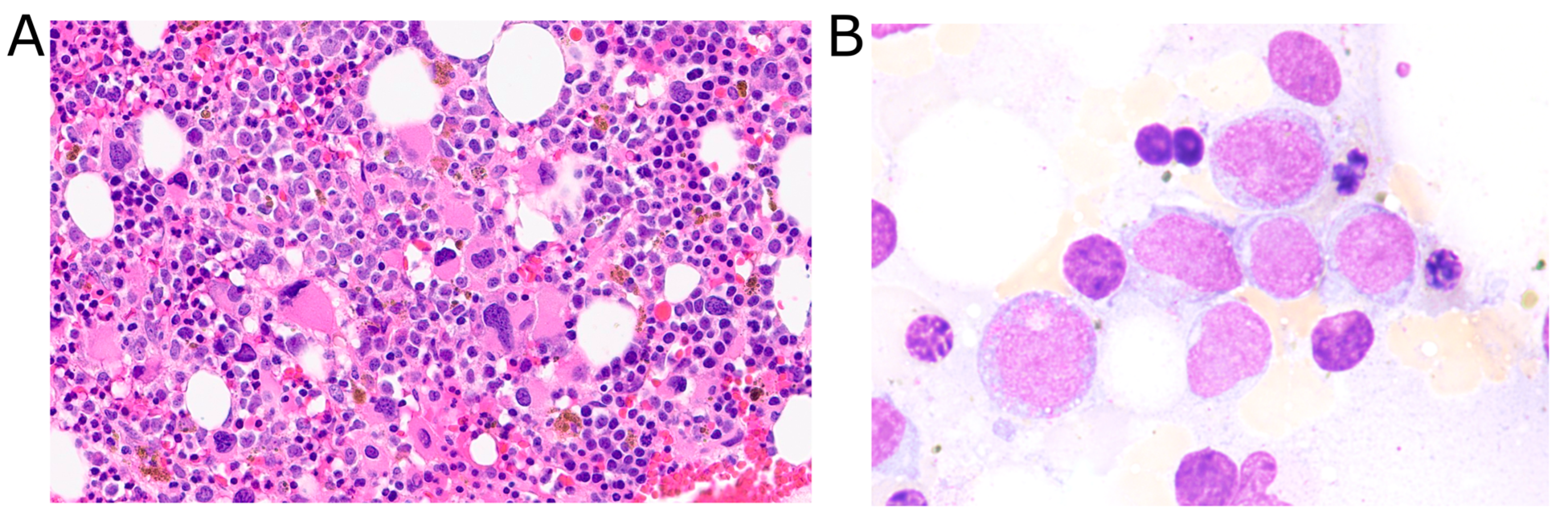
Figure 1. Bone marrow histology after AML relapse. (A): Representative image (40× magnification) of bone marrow core biopsy demonstrating increased blasts and hypolobated megakaryocytes. (B): Representative image (100× magnification) of bone marrow aspirate smear demonstrating blasts with abnormal morphology including large, moderate cytoplasm and vacuoles, along with features of erythroid dysplasia.
Diagnostics 15 02410 g001
Figure 2. Rearrangement events involving MYC and MECOM diagnosed by FISH. (A): G-banded karyotype including identification of derivative chromosomes 3 and 8. (B): MYC break-apart FISH, showing one distal 3′ MYC probe (green) translocated to the derivative chromosome 3 with intact MECOM (overlapping red and green probes, shown in panel (C)). (C): MECOM break-apart FISH, showing one distal 3′ MECOM probe (green) translocated to the derivative chromosome 8, which had intact MYC (overlapping red and green probes, shown in panel (B)).
Diagnostics 15 02410 g002
Table 1. Timeline of key clinical events and pathological findings. Bone marrow histology, flow cytometry, and cytogenetics findings are summarized at key timepoints when bone marrow biopsies were performed. Percentage of cells with abnormalities is reported. Abbreviations: VAF variant allele frequency.
TimeBone Marrow HistologyFlow CytometryCytogenetics
0 months: initial diagnosis>80–90% immature monocytes, trilineage dysplasiaAbnormal myeloid maturation, abnormal monocytes (65%)del(12p) (100%), del(11q) (45%), KMT2A loss (13.5%), U2AF1 mutation (VAF 50%)
3 months: post two cycles chemotherapyNo evidence of residual diseaseNo abnormal myeloid or blast populationsKMT2A loss (28.5%),
KMT2A gain (5.0%)
10 months: relapse14% blasts, decreased erythropoiesis, dysplastic megakaryocytesMild increase in myeloblasts (8%)t(3;8), del(11), del(12), MYC (68%) and MECOM (18%) rearrangements, KMT2A loss (91.5%), ETV6 loss, U2AF1 mutation (VAF 31.5%)
15 months: post salvage reinductionMinimal residual diseaseResidual AML blasts 2–3%t(3;8), del(11), MYC (16.5%) and MECOM (30%) rearrangements, KMT2A loss (93.5%)
Table 2. Summary of published cases exhibiting concurrent MYC and MECOM rearrangements. Summarized are the source publication, number of patients, abnormalities identified, age, sex, treatment(s), and outcome(s). Abbreviations: AWD—alive with disease; NS—not specified.
SourceN PatientsAbnormalities IdentifiedAgeSexTreatment(s)Outcome(s)
Liu and Tirado 2018 [8]1t(3;8)(q26.2;q23)68MaleRuxolitinibDeath
Tang et al. 2019 [9]20t(3;8)(q26.2;q24), various gene mutationsRange: 21–79
Median: 61
12 male, 8 femaleVariousDeath 90%, AWD 10%
Smith et al. 2020 [10]2Both: t(3;8)(q26.2;q24.1)
Case 1: der(5), IGH/CCND1 fusion
Case 2: del(7), del(13q14), loss of ASXL1, CSF3R, ETV6, and U2AF1
60; 68Both maleNS; RituximabDeath; AWD
McGinnis et al. 2024 [11]NS (at least 1)MECOM::MYC rearrangementNSNSNSNS
Present case1t(3;8)(q26.2;q24.3), del(11)(q23), del(12)(p13), ETV6 loss, U2AF1 c.101C>T (S34F) mutation70MaleVenetoclax + azacytidineIn remission
Disclaimer/Publisher’s Note: The statements, opinions and data contained in all publications are solely those of the individual author(s) and contributor(s) and not of MDPI and/or the editor(s). MDPI and/or the editor(s) disclaim responsibility for any injury to people or property resulting from any ideas, methods, instructions or products referred to in the content.