Article

Radiologic Predictors of Disease Recurrence in Nasopharyngeal Carcinoma: A Retrospective Evaluation of MRI and 18F-FDG-PET/CT Parameters

Departments of Radiology, Nuclear Medicine, Radiation Oncology, Otorhinolaryngology and Medical Oncology, Istanbul Medipol University Hospital, Istanbul 34214, Turkey
*
Author to whom correspondence should be addressed.
Diagnostics 2025, 15(13), 1646; https://doi.org/10.3390/diagnostics15131646
Submission received: 3 June 2025 / Revised: 25 June 2025 / Accepted: 25 June 2025 / Published: 27 June 2025
(This article belongs to the Special Issue Advances in the Diagnosis and Management of Head and Neck Disease)

Abstract

Background/Objectives: NPC is a radiosensitive malignancy with high recurrence rates despite therapeutic advances. This study aimed to identify radiologic and metabolic predictors of recurrence in newly diagnosed NPC by integrating MRI and 18F-FDG PET/CT parameters. Methods: Fifty-two patients with biopsy-proven, previously untreated NPC who underwent pre-treatment MRI and 18F-FDG PET/CT were retrospectively analyzed. Local tumor features, nodal status, and response patterns were evaluated using MRI and PET/CT-derived metrics: SUVmax, SUVmean, SUVpeak, MTV, and TLG. The post-treatment MRI response was categorized into six patterns. Univariate and multivariate analyses were performed to identify independent predictors. Results: Recurrence occurred in 27% of patients. Based on the multivariate analysis, PNI, extensive PPS invasion, GTV, and metastatic LN count were identified as independent predictors of recurrence (PNI: OR = 1.60, p = 0.029; PPS: OR = 1.23, p = 0.027; GTV: OR = 1.08, p = 0.042; LN count: OR = 1.20, p = 0.031). PNI and PPS invasion were significantly associated with local failure (HR = 8.21, p = 0.008 and HR = 3.52, p = 0.043, respectively). GTV was independently associated with an increased risk of local (HR = 1.14, p = 0.021) and distant recurrence (HR = 1.19, p = 0.024). The presence of metastatic disease at diagnosis (HR = 6.27, p = 0.027) and a higher LN count (HR = 1.17, p = 0.028) were also linked to distant failure. Conclusions: Imaging-derived predictors including GTV, PNI, LN burden, and MRI-based response patterns demonstrate prognostic value for disease recurrence in NPC and may guide risk-adapted treatment strategies.

1. Introduction

Nasopharyngeal carcinoma (NPC) is a malignant epithelial tumor arising from nasopharyngeal mucosa and accounted for 0.8% of cancer-related deaths worldwide in 2020 [1]. Despite its global rarity, NPC demonstrates a strikingly unbalanced geographic distribution, with more than 70% of new cases reported in East and Southeast Asia [2]. In endemic regions, it comprises approximately 28–29% of all head and neck malignancies [3,4,5]. However, NPC is biologically and clinically distinct from other head and neck epithelial tumors, exhibiting an insidious onset, a high propensity for early metastasis, and a unique association with Epstein–Barr virus (EBV) infection [2].
Due to its deep anatomical location, clinical presentation is often delayed and typically reflects the extent of the primary tumor or more frequently regional lymphatic involvement. Cervical lymphadenopathy is the most common initial finding, observed in approximately 75% of patients [6]. Fewer than 7% of patients present with stage I disease; most are diagnosed at a locoregionally advanced stage, and about 10–15% present with distant metastases at the time of diagnosis [7,8]. NPC’s aggressive and infiltrative growth within a complex anatomical region presents significant therapeutic challenges, often rendering complete surgical resection unfeasible. Its high radiosensitivity has made radiotherapy the cornerstone of treatment. Intensity-modulated radiotherapy (IMRT), with or without concurrent chemotherapy, remains the standard approach [2]. For early-stage disease, IMRT alone is considered sufficient, while concurrent chemoradiotherapy (CCRT) is the preferred strategy for locoregionally advanced disease [7,9].
The survival benefit of adding adjuvant chemotherapy following CCRT remains controversial [10]. In contrast, the addition of induction chemotherapy prior to CCRT has demonstrated improved overall survival, largely due to the better control of distant metastases [11]. In the era of IMRT, both strategies are endorsed in clinical guidelines and continue to play key roles in the management of advanced-stage NPC [12]. Nevertheless, despite these advances, 3–13% present with residual disease within 12 weeks post-treatment and 20–30% of patients still develop local recurrence or distant metastasis [13,14,15].
The 8th edition of the UICC/AJCC TNM staging system remains the most widely adopted and universally accepted standard for prognostic stratification in NPC [16]. MRI, CT, and 18F-FDG-PET/CT are the most commonly employed imaging modalities for this purpose. Owing to its significantly superior soft-tissue resolution, MRI is more effective in evaluating primary tumor extension and retropharyngeal lymph node involvement, while 18F-FDG-PET/CT is more accurate for detecting distant metastases and identifying small cervical lymph node metastases [17,18,19,20]. Unfortunately, clinical outcomes often vary among patients with the same TNM stage receiving similar treatment, highlighting the need for more precise prognostic markers and individualized therapeutic approaches.
This study investigates risk factors for disease recurrence in NPC by systematically evaluating local tumor characteristics, nodal involvement, and treatment response patterns through the integration of MRI findings with PET/CT-derived metabolic parameters.

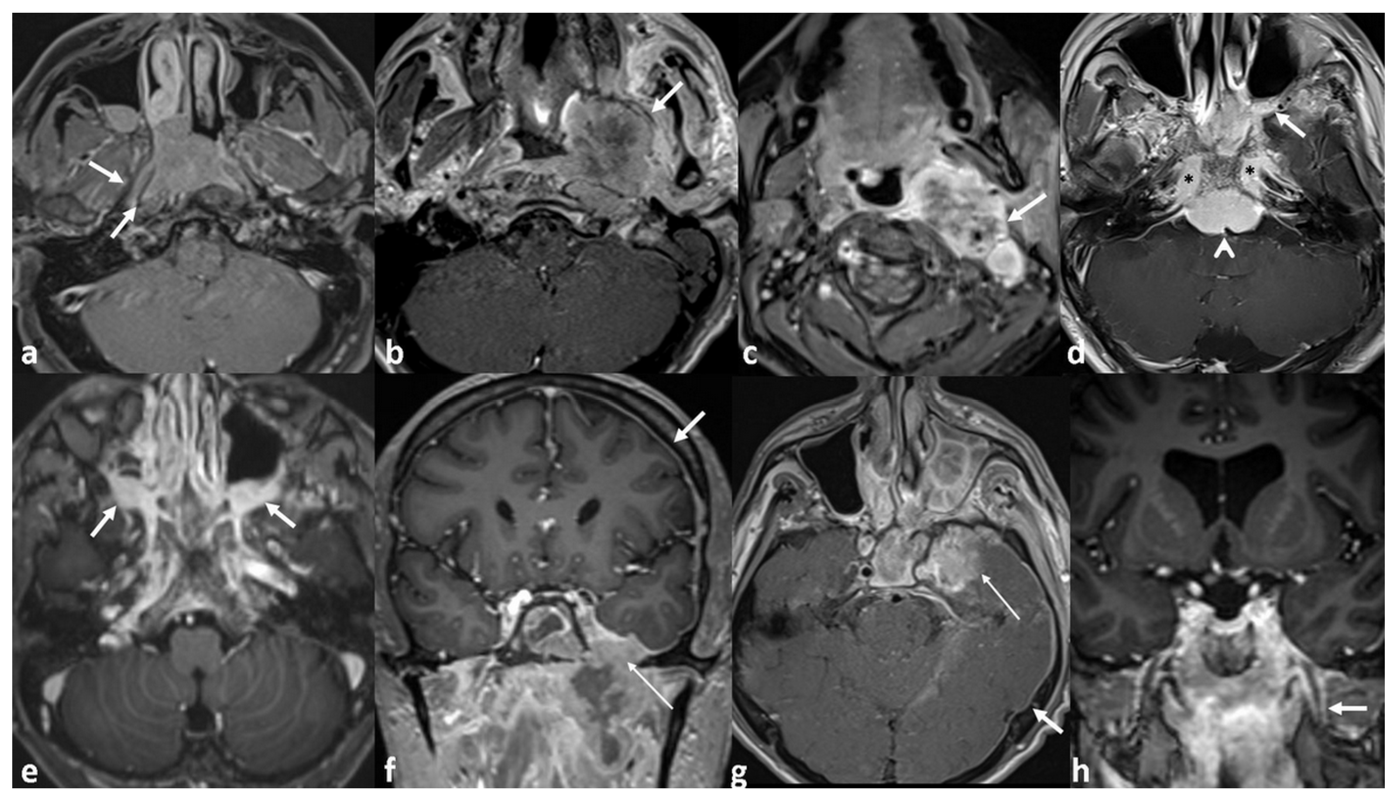
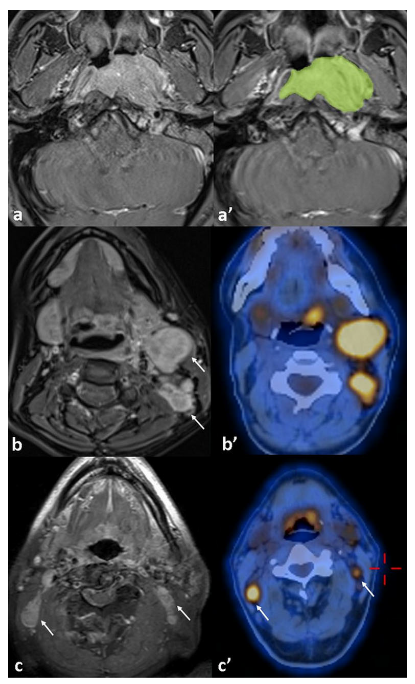
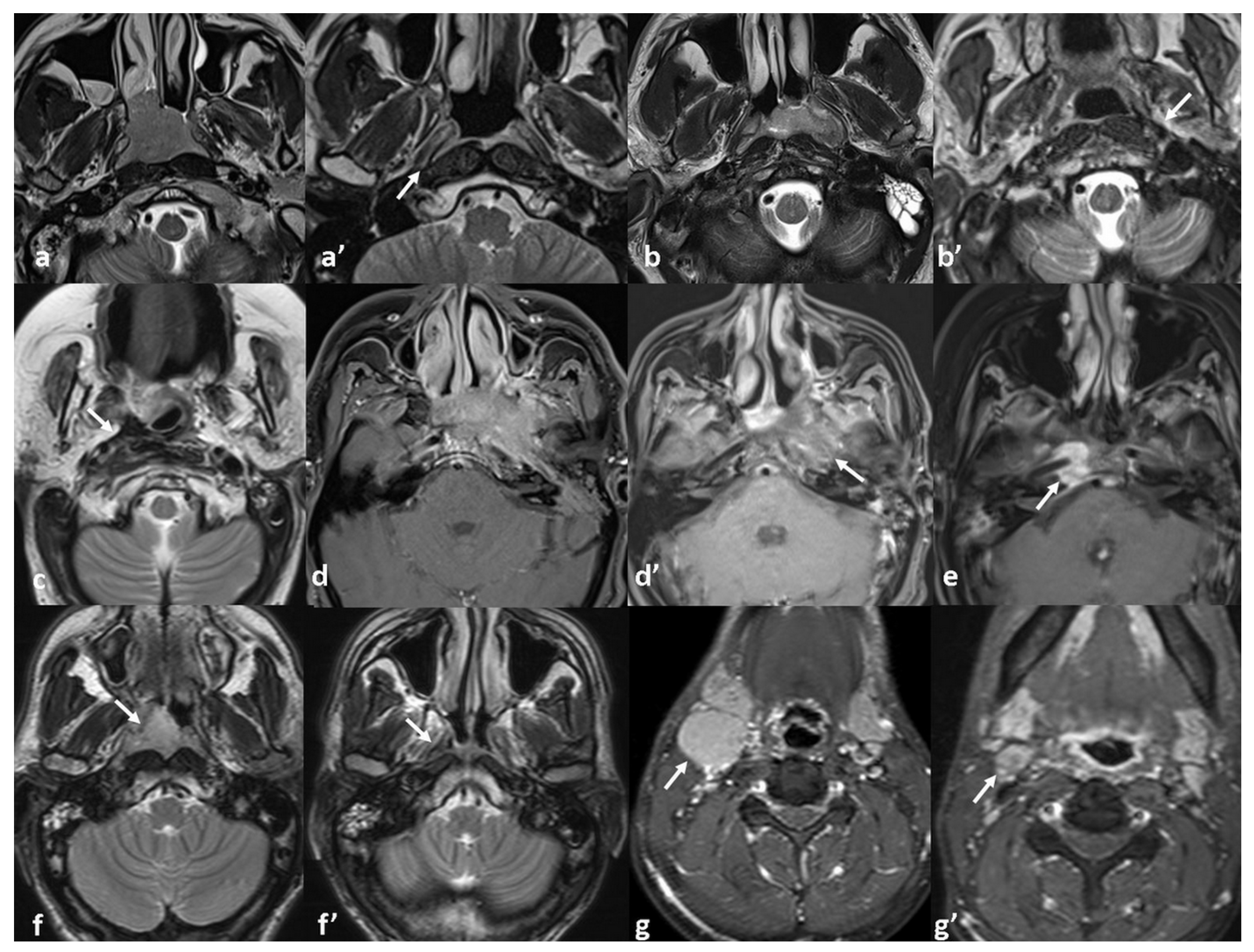
2. Materials and Methods

Medical records of 80 consecutive patients diagnosed with NPC between January 2018 and February 2022 at our institution were retrospectively reviewed. All patients had biopsy-confirmed, previously untreated disease. Patients with stage I disease (n = 5), those who lack follow-up (n = 12), or those with incomplete baseline imaging (n = 11) were excluded from this study. The final study cohort comprised 52 patients who had undergone comprehensive baseline imaging—including concurrent contrast-enhanced neck MRI and 18F-FDG-PET/CT—and maintained regular clinical and radiological follow-up for at least 3 years.
All patients were restaged according to the 8th edition of the AJCC staging system. Pre-treatment primary tumor volume, laterality of the tumor (unilateral or bilateral), and growth pattern (mass-forming vs. infiltrative) were documented. Tumor volume was manually contoured on each axial slice of pre-treatment contrast-enhanced T1-weighted MRI using the syngo.via® VB60 image analysis software (Siemens Healthineers, Erlangen, Germany). Segmentations were performed by a single radiologist with nine years of experience in head and neck imaging. After manual delineation, gross tumor volume (GTV) was automatically calculated by the syngo.via® VB60 software based on the contoured areas and the predefined slice reconstruction interval of 3 mm.
Tumor extension was evaluated in detail beyond AJCC staging; parapharyngeal space (PPS), skull base (SBI), and intracranial space (ICS) invasions were further categorized as ‘local’ or ‘extended’ based on the depth of anatomical involvement. Classification criteria are summarized in Table 1. Perineural invasion (PNI) was included as an independent predictor of disease recurrence. Figure 1 presents representative MRI examples illustrating various patterns of local tumor extension.
Lymph node metastases identified by 18F-FDG PET/CT were further assessed from concurrent MRI in addition to the AJCC nodal classification, including nodal dimensions (short and long axis), the presence of necrosis, and the number of metastatic lymph nodes. Nodal positivity was confirmed by complete resolution on follow-up imaging after chemoradiotherapy.
Baseline metabolic parameters derived from 18F-FDG-PET/CT—including peak standardized uptake value (SUV-peak), maximum SUV (SUV-max), mean SUV (SUV-mean), metabolic tumor volume (MTV), and total lesion glycolysis (TLG)—were measured for both the primary tumor (PT) and the most metabolically active metastatic lymph node (LN) to assess their association with disease recurrence. Figure 2 displays an example of tumor volume measurement, accompanied by imaging of lymph node involvement on 18F-FDG PET/CT and MRI.
All patients were treated according to a standardized institutional protocol, consistent with current international guidelines. All patients received CCRT with cisplatin and IMRT. Chemotherapy regimens were selected based on tumor stage and nodal burden, as determined during multidisciplinary tumor board discussions. Patients receiving induction chemotherapy were treated with cisplatin and gemcitabine, while those undergoing maintenance therapy received oral capecitabine. These treatment decisions were made according to a consistent institutional algorithm. Additionally, to account for potential treatment-related variability, clinical stage was included as a covariate in multivariate models. This approach helped reduce confounding and strengthened the internal validity of our findings.
Follow-up MRI and 18F-FDG-PET/CT scans were reviewed for all patients in the cohort to assess residual disease or recurrence. The optimal radiologic response to treatment was determined based on follow-up MRI findings. MRI characteristics at the time of best response were evaluated across T1WI, T2WI, STIR, ADC maps, and post-contrast T1WI sequences. Radiologic response patterns were classified into six categories: (1) complete tumor resolution (absence of any residual tumor or sequel signal abnormality), (2) thin fibrosis at the primary tumor site, (3) bulky fibrosis without discernible soft tissue or enhancement, (4) heterogeneous signal with subtle contrast enhancement, (5) residual enhancing soft tissue, and (6) complete response at the primary site with persistent nodal disease. Associations between response patterns and local tumor stage as well as local failure were subsequently analyzed.
All statistical analyses were performed using Stata version 18.0 (StataCorp LLC, College Station, TX, USA). Tumor characteristics derived from MRI and 18F-FDG-PET/CT metabolic parameters were compared to assess their association with disease recurrence. Chi-square tests were applied for categorical variables, and independent t-tests were used for continuous variables, as appropriate. Variables demonstrating statistical significance in univariate analyses (p < 0.05) were subsequently included in a multivariate logistic regression model to determine their independent predictive value for recurrence. The Chi-square test was conducted to evaluate the association between MRI treatment response patterns and recurrence risk. To assess the strength of association between post-treatment MRI response patterns and initial T-stage, Kendall’s Tau-b correlation was used. In addition, variables with p < 0.05 in univariable analysis were included in a multivariable Cox regression model to assess their independent association with time-to-recurrence outcomes. Hazard ratios (HRs) with 95% confidence intervals (CIs) were reported. A two-sided p value < 0.05 was considered statistically significant.
MR imaging was performed on 1.5-Tesla machine (Sola; Siemens). Routine sequences arranged for all neck imaging in our institution were used for the patients’ MRI scans. The sequences were acquired with a 20-array head coil and included the following data; axial and coronal T2-TSE-Dixon (3450/85; TR/TE, slice thickness 3 mm, matrix: 320 × 224; 4380/83; TR/TE, slice thickness 3 mm, matrix: 320 × 232), axial DWI-b1000 (4500/67; TR/TE, slice thickness 3.5 mm, matrix: 128 × 128) and pre-contrast axial and post-contrast 3 plan T1-TSE-Dixon (413/13; TR/TE, slice thickness 3 mm, matrix: 198 × 288; 440/13; TR/TE, slice thickness 3 mm, matrix: 217 × 320; 524/13; TR/TE, slice thickness 3 mm, matrix: 210 × 320).
PET/CT imaging was performed using a Siemens Biograph mCT scanner. Patients received an intravenous injection of approximately 370 MBq (10 mCi) of 18F-FDG and rested for 60 min before scanning. PET data were acquired in 3D mode over 7 bed positions, 3 min per position, and reconstructed using a 200 × 200 matrix with point spread function (PSF) and time-of-flight (TOF) modeling (3 iterations, 21 subsets). Low-dose CT (120 kV, ~57 mAs) was performed for attenuation correction and anatomical localization. Images were corrected for attenuation, scatter, and random events, and reviewed on a dedicated workstation.

3. Results

A total of 52 patients with newly diagnosed NPC were included in this study. The cohort comprised forty-three males and nine females, with a mean age of 46 ± 12 years (range: 20–69). All tumors were of the nonkeratinizing subtype, with 31 classified as differentiated and 21 as undifferentiated. According to the 8th edition of the AJCC staging system, 25% (thirteen out of fifty-two) were diagnosed with stage II, 29% (fifteen out of fifty-two) with stage III, 31% (16/52) with stage IVA, and 15% (eight out of fifty-two) with stage IVB disease.
Among the eight patients with metastatic disease, seven had bone involvement—two with isolated vertebral lesions and five with additional metastases including distant lymph nodes (n = two), liver (n = two), and lung (n = one). One patient had isolated lung metastasis.
Disease recurrence was observed in 14 patients (27%) and confirmed through biopsy or serial imaging. Three additional patients—two with skull base osteitis secondary to sphenoid sinusitis on MRI and one with mucositis demonstrating FDG avidity on PET/CT—were initially suspected of recurrence but were subsequently confirmed as false positives. Among the fourteen confirmed recurrences, one patient had residual enhancing soft tissue on MRI at 4 months post-treatment, which remained stable before progressing locally at 8 months. The remaining thirteen patients developed recurrence following an initial complete remission confirmed by both MRI and PET/CT: eight had local failure, five had distant failure, and one had both local and distant failure. Among the six patients with distant metastases, four (66.7%) had metastatic disease at initial diagnosis. Secondary relapse occurred in 57% (eight out of fourteen) of recurrent cases, and five of the them relapsed after a complete response. Demographic and tumor-specific characteristics are summarized in Table 2.
Seventeen patients demonstrated completely normal post-treatment MRI findings, with no evidence of residual disease or treatment-related sequelae. However, ten patients (19%) showed thin localized fibrosis, eleven (21%) had bulky non-enhancing fibrosis, ten (19%) exhibited heterogeneous signal with subtle enhancement, and four (8%) presented with residual enhancing soft tissue at the primary tumor site. Post-treatment MRI response patterns showed a moderate to strong correlation with the initial T-stage (Kendall’s tau-b = 0.43, p < 0.001). Advanced-stage tumors more frequently exhibited category 3–5 response patterns—bulky fibrosis, heterogeneous enhancement, or residual enhancing tissue—which were also significantly associated with disease recurrence (p = 0.004). Post-treatment MRI response patterns and the association with local tumor stage and recurrence status are detailed in Table 3. Figure 3 presents MRI findings demonstrating diverse patterns of tumor response.
The univariate analysis showed that the overall disease stage is a significant predictor of recurrence, with recurrence rates increasing across advancing stages (p = 0.044). Patients with metastatic disease had significantly higher recurrence than non-metastatic patients (62.5% vs. 20.4%, p = 0.014). Tumors with bilateral mucosal involvement had higher recurrence rates compared to unilateral cases (54.5% vs. 22.8%, p = 0.034), and infiltrative growth patterns were more likely to recur than well-defined, mass-forming tumors (62.5% vs. 20.4%, p = 0.014). Primary tumor stage (T-stage) did not reach statistical significance (p = 0.06); however, a detailed local analysis revealed that PNI was a strong predictor of recurrence (77.7% vs. 16.2%, p < 0.001). Extensive PPS invasion (62.5% vs. 11%, p = 0.001) and the presence of ICS invasion (63.6% vs. %17, p = 0.008) were also significantly associated with recurrence, whereas SBI was not (35% vs. %21.8, p = 0.109).
Among nodal parameters, only the number of metastatic lymph nodes was significantly associated with disease recurrence (p = 0.020). Recurrence was also more frequent in patients with necrotic nodes compared to those without (47% vs. 17.1%, p = 0.023), whereas the nodal stage and lymph node diameter showed no significant association.
Among PET/CT-derived metabolic parameters, only MTV (p = 0.049) and TLG (p = 0.045) demonstrated marginal significance for disease progression in the univariate analysis. However, none retained independent prognostic value in the multivariate model.
The multivariate analysis identified PNI, extensive PPS invasion, tumor volume, and the number of metastatic lymph nodes as independent predictors of recurrence. PNI was associated with a 60% increase in the odds of recurrence (OR = 1.60, p = 0.029), while extensive PPS invasion conferred 23% higher odds (OR = 1.23, p = 0.027). Each cm3 increase in tumor volume was linked to an 8% increase in the odds of recurrence (OR = 1.08, p = 0.042), and each additional metastatic lymph node raised the odds by 20% (OR = 1.20, p = 0.031). Results of the univariate and multivariate analyses are summarized in Table 4.
Both PNI and extensive PPS invasion were significantly associated with local recurrence (PNI: HR = 8.21; 95% CI: 3.73–28.37; p = 0.008; PPS: HR = 3.52; 95% CI: 1.39–11.9; p = 0.043). Tumor volume emerged as a common independent predictor linked to both local and distant recurrence (local: HR = 1.14; 95% CI: 1.02–1.29; p = 0.021; distant: HR = 1.19; 95% CI: 1.05–1.40; p = 0.024). The presence of metastatic disease at diagnosis (HR = 6.27; 95% CI: 1.23–31.9; p = 0.027) and a higher number of metastatic lymph nodes (HR = 1.17; 95% CI: 1.08–1.35; p = 0.028) were associated with increased risk of distant failure.
Local recurrences were managed with CyberKnife radiosurgery, while distant metastases were treated with systemic chemotherapy. By the end of follow-up, seven patients (13%) had died from cancer-related causes, forty-three were alive, and two were lost to follow-up after three years.

4. Discussion

This study identified PNI, extensive PPS invasion, tumor volume, and the number of metastatic lymph nodes as significant predictors of disease recurrence in NPC. PNI and extensive PPS invasion emerged as independent predictors of recurrence and were significantly associated with an increased risk of local failure. Prior studies have also highlighted the adverse prognostic implications of spatial tumor spread. Liu X. et al. reported MRI-detected cranial nerve invasion as an independent predictor of local recurrence (HR = 2.605, p = 0.032), consistent with our findings regarding PNI [21]. Although carotid space invasion was not directly assessed in our cohort, it is classified under extensive PPS involvement and has been identified as a high-risk feature in previous studies [22]. For example, Huang et al. proposed subclassifying PPS invasion based on its extent to reduce heterogeneity within the T3 category, citing its association with aggressive tumor behavior [23]. Similarly, Zhang et al. demonstrated that invasion beyond the lateral pterygoid muscle was associated with an increased risk of distant failure (HR = 1.734, p = 0.006) [24]. These findings highlight the clinical importance of the imaging-based subclassification of spatial tumor extension and the identification of PNI to facilitate early risk stratification and guide treatment planning.
The absence of a significant association between skull base invasion with recurrence may seem inconsistent with the prior literature. However, this discrepancy likely stems from an ambiguous categorization of bone invasion in previous papers. In our analysis, skull base invasion was strictly defined as bony infiltration, whereas PNI was evaluated independently and reported as a separate variable. Cheng et al. reported a significant prognostic impact of skull base invasion in an advanced subgroup that included neural foraminal involvement [25]. Similarly, Feng et al.’s study identified extended skull base invasion as an independent predictor of recurrence; however, as in Cheng et al.’s work, extended invasion included neural foramina [26]. Including neural foraminal invasion as part of extended skull base invasion may lead to misattributing a poor prognosis to bone involvement, when the adverse outcomes may, in fact, reflect the contribution of perineural spread.
Tumor volume was found to be a significant independent predictor of recurrence in the multivariate model (p = 0.042). This finding is consistent with previous studies that underscore the prognostic relevance of tumor burden in NPC. Weiqiong et al. reported that GTV was the only independent predictor of local control (HR = 5.24; 95% CI: 1.074–25.57; p = 0.041) [27]. Tian et al. similarly demonstrated that patients with GTV > 38 cm3 had a significantly increased risk of local failure compared to those with smaller volumes (p < 0.01), advocating for corresponding revisions to T-category classification [28]. Although Qin et al. primarily focused on survival endpoints, they also observed poorer outcomes in patients with GTV > 33 mL, indicating the broader prognostic impact of tumor burden even when stratified by T or clinical stage [29]. Collectively, these findings support incorporating tumor volume into future refinements of the TNM staging system to enhance prognostic stratification. However, a wide range of GTV cut-off values has been reported across studies, likely due to differences in cohort characteristics and imaging methodology [30,31,32,33]. Tumor volume demonstrated predictive value for both local and distant recurrence; however, no definitive cut-off was proposed to avoid overgeneralization from a single-center dataset. These findings support the recommendation to incorporate tumor volume into future TNM staging revisions and underscore the need for meta-analytic studies to establish standardized, evidence-based thresholds.
18F-FDG PET/CT enables the non-invasive functional assessment of both the primary tumors (PTs) and metastatic lymph nodes (LN). Elevated standardized uptake values (SUVmax, SUVmean, SUV-peak), along with TLG and MTV, reflect either increased glucose metabolism or cellular density. Guo et al. investigated the relationship between 18F-FDG PET metabolic parameters (SUVmax, SUVmean, MTV, and TLG) and the Ki-67 labeling index. Their analysis revealed a statistically significant but weak correlation (r < 0.225) between metabolic activity and proliferative status [34]. Aktan et al. demonstrated the potential predictive value of SUVmax, reporting significantly elevated values in patients with unfavorable outcomes (PT ≥ 13, p = 0.017; LN ≥ 9, p = 0.013) [35]. In another study, Jin et al. identified SUVmax-N as the only independent prognostic factor associated with distant failure in locoregionally advanced NPC [36]. Although higher MTV and TLG values of the primary tumor were observed in pairwise comparisons, these did not remain statistically significant predictors of recurrence in the multivariate analysis. This suggests that the prognostic value of volumetric metabolic parameters may vary across patient populations and methodological settings, limiting their current utility as universal markers for risk stratification.
The prognostic relevance of various characteristics of metastatic lymph nodes in NPC has been previously investigated. Huang et al. identified maximal nodal diameter as a significant prognostic factor, with nodal size ≥ 4 cm associated with more aggressive disease behavior [37]. Chen et al. reported that the co-existence of bilateral retropharyngeal and cervical lymph node metastases independently predicted disease progression and increased the risk of distant failure (p < 0.001) [38]. Jiang et al. demonstrated that the involvement of posterior level V nodes was significantly associated with a higher likelihood of distant spread [39]. In our analysis, necrotic lymph node metastasis emerged as a notable risk factor for disease recurrence, consistent with previous studies that have linked nodal necrosis to unfavorable clinical outcomes [40,41,42]. However, this feature did not remain an independent predictor in the multivariate analysis after adjusting for other significant confounding variables. Given its potential clinical value, we recommend that the presence of nodal necrosis be routinely documented in baseline MRI evaluations.
Among the nodal characteristics evaluated, including N stage, maximum nodal diameter, bilateral involvement, and presence of necrosis, only the number of metastatic lymph nodes remained an independent predictor of recurrence risk (OR = 1.20, p = 0.031). Furthermore, each additional metastatic lymph node was significantly associated with an increased risk of distant failure (p = 0.028). In line with this, Ma et al. reported that patients with ≥9 positive LNs exhibited significantly worse outcomes compared to those classified as N3 [43]. Consistent with our findings, Zhu et al. emphasized the prognostic significance of the metastatic nodal count, identifying it as an independent and superior predictor of distant failure compared to other nodal characteristics [44]. This highlights the prognostic relevance of metastatic nodal burden in NPC, which may surpass traditional indicators such as nodal size or laterality. Collectively, these findings support the integration of the LN count into the NPC staging framework, as is currently implemented in several gastrointestinal cancers.
In this context, the identification of imaging-based markers becomes increasingly important for early risk stratification and treatment planning. Although extensive PPS invasion and PNI are established markers of locally aggressive disease for which induction or adjuvant chemotherapy may be considered, tumor volume and nodal burden—despite their strong association with recurrence and survival—are not yet incorporated into standard treatment criteria. In our retrospective cohort, these MRI-based features were not used to guide therapy. However, our findings emphasize their clinical relevance and suggest they may warrant inclusion in future risk stratification models to support treatment intensification in high-risk patients.
To date, MRI-based treatment response patterns in NPC have not been systematically characterized. One study by Lin et al. investigated this topic by assessing the relationship between post-radiotherapy fibrosis and recurrence, demonstrating a significantly higher recurrence rate in patients with MRI-detected fibrosis 3–6 months after treatment (p = 0.021) [45]. However, their analysis was confined to the presence of fibrosis and did not explore the broader range of post-treatment MRI features or their prognostic implications. In our study, the degree of local tumor invasiveness appeared to influence distinct MRI response patterns. Among patients with advanced-stage disease, features such as bulky fibrosis, heterogeneous signal intensity with subtle enhancement, and residual soft tissue components were observed more frequently and were significantly associated with recurrence risk (p = 0.004). Although preliminary, these findings underscore the potential prognostic relevance of MRI-based response assessment in NPC. Prospective validation in larger, multicenter cohorts is needed to confirm these imaging patterns and to determine their clinical utility in identifying patients who may benefit from adjuvant or maintenance chemotherapy.
This study has several limitations that warrant consideration. Its retrospective design may introduce selection bias and limits the ability to establish causal relationships. Conducting the analysis at a single institution may affect the generalizability of the findings across diverse clinical settings and populations. The relatively small sample size may reduce statistical power, particularly in subgroup analyses. Additionally, Epstein–Barr virus (EBV) DNA data were not available, preventing the evaluation of a well-established biomarker that could have enhanced risk stratification. Despite these limitations, the study provides meaningful insights by comprehensively assessing recurrence risk factors in NPC. Unlike many prior studies that examined these variables in isolation, our integrative approach offers a more comprehensive understanding of the interplay between clinical, radiologic, and metabolic parameters.

5. Conclusions

This study identified PNI, extensive PPS invasion, tumor volume, and the number of metastatic lymph nodes as significant predictors of disease recurrence in NPC. PNI and extensive PPS invasion were strong indicators of local failure, while tumor volume was independently associated with both local and distant recurrence. The presence of metastatic disease at diagnosis and a higher metastatic nodal burden were also linked to an increased risk of distant failure. Although our findings are consistent with previous studies, they provide a meaningful contribution by specifically addressing the prognostic value of these factors in the Caucasian population. This population-specific validation strengthens the generalizability of prior conclusions and highlights the value of imaging-based stratification across different demographic groups. Importantly, distinct MRI-based treatment response patterns were also associated with recurrence risk, suggesting their potential role in identifying patients who may benefit from adjuvant or maintenance therapy.

Author Contributions

Conceptualization, Ö.Y.; methodology, B.K. and T.Ç.; formal analysis, B.K. and Ö.Y.; investigation, M.S.T., Ö.Y. and E.K.; data curation, B.K. and T.Ç.; writing—original draft preparation, B.K.; writing—review and editing, T.Ç. and Ö.Y.; visualization, B.K. and T.Ç.; supervision, E.K. All authors have read and agreed to the published version of the manuscript.

Funding

This research received no specific grant from any funding agency in the public, commercial, or not-for-profit sectors.

Institutional Review Board Statement

This study was conducted in accordance with the Declaration of Helsinki and approved by the Institutional Review Board of Istanbul Medipol University (Approval No. 796; date of approval 29 August 2024).

Informed Consent Statement

Written informed consent was obtained from all participants, and patient data were anonymized and de-identified prior to analysis.

Data Availability Statement

The data presented in this study are available on request from the corresponding author due to ethical and privacy restrictions.

Conflicts of Interest

The authors declare that they have no conflicts of interest.

Abbreviations

The following abbreviations are used in this manuscript:
NPCNasopharyngeal carcinoma
EBVEpstein–Barr virus
IMRTIntensity-modulated radiotherapy
CCRTConcurrent chemoradiotherapy
PPSParapharyngeal space
SBISkull base invasion
ICSIntracranial space
PNIPerineural invasion
SUVStandardized uptake value
TLGTotal lesion glycolysis
MTVMetabolic tumor volume
PTPrimary Tumor
LNLymph node
DFSDisease-free survival
LRRFSLocoregional recurrence-free survival
DMFSDistant metastasis-free survival
OSOverall survival
GTVGross tumor volume

References

  1. Sung, H.; Ferlay, J.; Siegel, R.L.; Laversanne, M.; Soerjomataram, I.; Jemal, A.; Bray, F. Global cancer statistics 2020: GLOBOCAN estimates of incidence and mortality worldwide for 36 cancers in 185 countries. CA A Cancer J. Clin. 2021, 71, 209–249. [Google Scholar] [CrossRef] [PubMed]
  2. Chen, Y.P.; Chan, A.T.C.; Le, Q.T.; Blanchard, P.; Sun, Y.; Ma, J. Nasopharyngeal carcinoma. Lancet 2019, 394, 64–80. [Google Scholar] [CrossRef] [PubMed]
  3. Abdulamir, A.; Hafidh, R.; Abdulmuhaimen, N.; Abubakar, F.; Abbas, K. The distinctive profile of risk factors of nasopharyngeal carcinoma in comparison with other head and neck cancer types. BMC Public Health 2008, 8, 400. [Google Scholar] [CrossRef] [PubMed]
  4. Adham, M.; Kurniawan, A.N.; Muhtadi, A.I.; Roezin, A.; Hermani, B.; Gondhowiardjo, S.; Tan, I.B.; Middeldorp, J.M. Nasopharyngeal carcinoma in Indonesia: Epidemiology, incidence, signs, and symptoms at presentation. Chin. J. Cancer 2012, 31, 185. [Google Scholar] [CrossRef]
  5. Chang, E.T.; Ye, W.; Zeng, Y.-X.; Adami, H.-O. The evolving epidemiology of nasopharyngeal carcinoma. Cancer Epidemiol. Biomark. Prev. 2021, 30, 1035–1047. [Google Scholar] [CrossRef]
  6. Chua, M.L.K.; Wee, J.T.S.; Hui, E.P.; Chan, A.T.C. Nasopharyngeal carcinoma. Lancet 2016, 387, 1012–1024. [Google Scholar] [CrossRef]
  7. Jen, C.-W.; Tsai, Y.-C.; Wu, J.-S.; Chen, P.-L.; Yen, J.-H.; Chuang, W.-K.; Cheng, S.H.-C. Prognostic classification for patients with nasopharyngeal carcinoma based on American Joint Committee on Cancer staging system T and N categories. Ther. Radiol. Oncol. 2020, 4, 2. [Google Scholar] [CrossRef]
  8. Guo, L.-L.; Wang, H.-Y.; Zheng, L.-S.; Wang, M.-D.; Cao, Y.; Li, Y.; Liu, Z.-J.; Peng, L.-X.; Huang, B.-J.; Shao, J.-Y. Metastasis of nasopharyngeal carcinoma: What we know and do not know. Vis. Cancer Med. 2021, 2, 4. [Google Scholar] [CrossRef]
  9. Xu, C.; Zhang, L.-H.; Chen, Y.-P.; Liu, X.; Zhou, G.-Q.; Lin, A.-H.; Sun, Y.; Ma, J. Chemoradiotherapy versus radiotherapy alone in stage II nasopharyngeal carcinoma: A systemic review and meta-analysis of 2138 patients. J. Cancer 2017, 8, 287. [Google Scholar] [CrossRef]
  10. Chen, L.; Hu, C.-S.; Chen, X.-Z.; Hu, G.-Q.; Cheng, Z.-B.; Sun, Y.; Li, W.-X.; Chen, Y.-Y.; Xie, F.-Y.; Liang, S.-B. Adjuvant chemotherapy in patients with locoregionally advanced nasopharyngeal carcinoma: Long-term results of a phase 3 multicentre randomised controlled trial. Eur. J. Cancer 2017, 75, 150–158. [Google Scholar] [CrossRef]
  11. Chen, Y.-P.; Tang, L.-L.; Yang, Q.; Poh, S.-S.; Hui, E.P.; Chan, A.T.; Ong, W.-S.; Tan, T.; Wee, J.; Li, W.-F. Induction chemotherapy plus concurrent chemoradiotherapy in endemic nasopharyngeal carcinoma: Individual patient data pooled analysis of four randomized trials. Clin. Cancer Res. 2018, 24, 1824–1833. [Google Scholar] [CrossRef] [PubMed]
  12. Colevas, A.D.; Yom, S.S.; Pfister, D.G.; Spencer, S.; Adelstein, D.; Adkins, D.; Brizel, D.M.; Burtness, B.; Busse, P.M.; Caudell, J.J. NCCN guidelines insights: Head and neck cancers, version 1.2018. J. Natl. Compr. Cancer Netw. 2018, 16, 479–490. [Google Scholar] [CrossRef] [PubMed]
  13. Juarez-Vignon Whaley, J.J.; Afkhami, M.; Onyshchenko, M.; Massarelli, E.; Sampath, S.; Amini, A.; Bell, D.; Villaflor, V.M. Recurrent/metastatic nasopharyngeal carcinoma treatment from present to future: Where are we and where are we heading? Curr. Treat. Options Oncol. 2023, 24, 1138–1166. [Google Scholar] [CrossRef] [PubMed]
  14. Chen, S.; Yang, D.; Liao, X.; Lu, Y.; Yu, B.; Xu, M.; Bin, Y.; Zhou, P.; Yang, Z.; Liu, K. Failure patterns of recurrence and metastasis after intensity-modulated radiotherapy in patients with nasopharyngeal carcinoma: Results of a multicentric clinical study. Front. Oncol. 2022, 11, 693199. [Google Scholar] [CrossRef]
  15. Huang, Y.-Y.; Zhou, J.-Y.; Zhan, Z.-J.; Ke, L.-R.; Xia, W.-X.; Cao, X.; Cai, Z.-C.; Deng, Y.; Chen, X.; Zhang, L.-L. Tumor residue in patients with stage II–IVA nasopharyngeal carcinoma who received intensity-modulated radiation therapy: Development and validation of a prediction nomogram integrating postradiotherapy plasma Epstein–Barr virus deoxyribonucleic acid, clinical stage, and radiotherapy dose. BMC Cancer 2023, 23, 410. [Google Scholar]
  16. Zanoni, D.K.; Patel, S.G.; Shah, J.P. Changes in the 8th edition of the American Joint Committee on Cancer (AJCC) staging of head and neck cancer: Rationale and implications. Curr. Oncol. Rep. 2019, 21, 52. [Google Scholar] [CrossRef]
  17. Chen, W.-S.; Li, J.-J.; Hong, L.; Xing, Z.-B.; Wang, F.; Li, C.-Q. Comparison of MRI, CT and 18F-FDG PET/CT in the diagnosis of local and metastatic of nasopharyngeal carcinomas: An updated meta analysis of clinical studies. Am. J. Transl. Res. 2016, 8, 4532. [Google Scholar]
  18. Chan, S.-C.; Yeh, C.-H.; Yen, T.-C.; Ng, S.-H.; Chang, J.T.-C.; Lin, C.-Y.; Yen-Ming, T.; Fan, K.-H.; Huang, B.-S.; Hsu, C.-L. Clinical utility of simultaneous whole-body 18 F-FDG PET/MRI as a single-step imaging modality in the staging of primary nasopharyngeal carcinoma. Eur. J. Nucl. Med. Mol. Imaging 2018, 45, 1297–1308. [Google Scholar] [CrossRef]
  19. Xie, H.J.; Sun, X.S.; Zhang, X.; Xiao, B.B.; Lin, D.F.; Lin, X.P.; Lv, X.F.; Liu, L.Z.; Han, F.; Zou, R.H.; et al. Head and neck MRI-based T stage and [18F]FDG PET/CT-based N/M stage improved prognostic stratification in primary nasopharyngeal carcinoma. Eur. Radiol. 2023, 33, 7952–7966. [Google Scholar] [CrossRef]
  20. Peng, H.; Chen, L.; Tang, L.-L.; Li, W.-F.; Mao, Y.-P.; Guo, R.; Zhang, Y.; Liu, L.-Z.; Tian, L.; Zhang, X. Significant value of 18 F-FDG-PET/CT in diagnosing small cervical lymph node metastases in patients with nasopharyngeal carcinoma treated with intensity-modulated radiotherapy. Chin. J. Cancer 2017, 36, 95. [Google Scholar] [CrossRef]
  21. Liu, X.; Liu, L.; Mao, Y.; Chen, L.; Tang, L.; Zhou, G.; Sun, Y.; Yue, D.; Lin, A.; Li, L. Prognostic value of magnetic resonance imaging-detected cranial nerve invasion in nasopharyngeal carcinoma. Br. J. Cancer 2014, 110, 1465–1471. [Google Scholar] [CrossRef] [PubMed]
  22. Quan, T.; Guan, W.; Huang, W.; Cui, C.; Li, H.; Ruan, G.; Liu, L.; Zhao, Q.; Ma, H. Carotid space involvement is a prognostic factor and marker for induction chemotherapy in patients with nasopharyngeal carcinoma. Oral Oncol. 2022, 135, 106230. [Google Scholar] [CrossRef] [PubMed]
  23. Huang, W.; Quan, T.; Zhao, Q.; Li, S.; Cai, Y.; Zhou, J.; Luo, C.; Ruan, G.; Cui, C.; Liang, S. MRI of nasopharyngeal carcinoma: Parapharyngeal subspace involvement has prognostic value and influences T-staging in the IMRT era. Eur. Radiol. 2022, 32, 262–271. [Google Scholar] [CrossRef]
  24. Zhang, G.Y.; Huang, Y.; Hu, X.F.; Chen, X.P.; Xu, T.; Liu, L.Z.; Wei, W.H.; Huang, G.S.; Zhou, M.M.; Huang, Z.L.; et al. Prognostic value of classifying parapharyngeal extension in nasopharyngeal carcinoma based on magnetic resonance imaging. Biomed. Res. Int. 2015, 2015, 749515. [Google Scholar] [CrossRef]
  25. Cheng, Y.-K.; Liu, L.-Z.; Jiang, N.; Yue, D.; Tang, L.-L.; Zhang, F.; Lin, L.; Liu, X.; Chen, L.; Ma, J. MRI-detected skull-base invasion. Strahlenther. Onkol. 2014, 190, 905. [Google Scholar] [CrossRef]
  26. Feng, Y.; Cao, C.; Hu, Q.; Chen, X. Grading of MRI-detected skull-base invasion in nasopharyngeal carcinoma with skull-base invasion after intensity-modulated radiotherapy. Radiat. Oncol. 2019, 14, 10. [Google Scholar] [CrossRef]
  27. Ni, W.; Qi, W.; Xu, F.; Cao, W.; Xu, C.; Chen, J.; Gao, Y. Prognostic value of nasopharynx tumour volume in local-regional advanced nasopharyngeal carcinoma. Ann. Transl. Med. 2020, 8, 223. [Google Scholar] [CrossRef]
  28. Tian, Y.M.; Xiao, W.W.; Bai, L.; Liu, X.W.; Zhao, C.; Lu, T.X.; Han, F. Impact of primary tumor volume and location on the prognosis of patients with locally recurrent nasopharyngeal carcinoma. Chin. J. Cancer 2015, 34, 247–253. [Google Scholar] [CrossRef]
  29. Qin, L.; Wu, F.; Lu, H.; Wei, B.; Li, G.; Wang, R. Tumor volume predicts survival rate of advanced nasopharyngeal carcinoma treated with concurrent chemoradiotherapy. Otolaryngol. Head Neck Surg. 2016, 155, 598–605. [Google Scholar] [CrossRef]
  30. Sze, W.-M.; Lee, A.W.; Yau, T.-K.; Yeung, R.M.; Lau, K.-Y.; Leung, S.K.; Hung, A.W.; Lee, M.C.; Chappell, R.; Chan, K. Primary tumor volume of nasopharyngeal carcinoma: Prognostic significance for local control. Int. J. Radiat. Oncol. Biol. Phys. 2004, 59, 21–27. [Google Scholar] [CrossRef]
  31. Shen, C.; Lu, J.J.; Gu, Y.; Zhu, G.; Hu, C.; He, S. Prognostic impact of primary tumor volume in patients with nasopharyngeal carcinoma treated by definitive radiation therapy. Laryngoscope 2008, 118, 1206–1210. [Google Scholar] [CrossRef] [PubMed]
  32. Chen, C.; Fei, Z.; Pan, J.; Bai, P.; Chen, L. Significance of primary tumor volume and T-stage on prognosis in nasopharyngeal carcinoma treated with intensity-modulated radiation therapy. Jpn. J. Clin. Oncol. 2011, 41, 537–542. [Google Scholar] [CrossRef] [PubMed]
  33. Guo, R.; Sun, Y.; Yu, X.-L.; Yin, W.-J.; Li, W.-F.; Chen, Y.-Y.; Mao, Y.-P.; Liu, L.-Z.; Li, L.; Lin, A.-H. Is primary tumor volume still a prognostic factor in intensity modulated radiation therapy for nasopharyngeal carcinoma? Radiother. Oncol. 2012, 104, 294–299. [Google Scholar] [CrossRef]
  34. Guo, C.; Luo, J.; Liang, M.; Xiao, J. Correlation of 18F-FDG PET/CT metabolic parameters with Ki-67 expression and tumor staging in nasopharyngeal carcinoma. Nucl. Med. Commun. 2025, 46, 437–443. [Google Scholar] [CrossRef]
  35. Aktan, M.; Kanyilmaz, G.; Yavuz, B.B.; Koc, M.; Eryilmaz, M.A.; Adli, M. Prognostic value of pre-treatment (18)F-FDG PET uptake for nasopharyngeal carcinoma. Radiol. Med. 2017, 130, 4–12. [Google Scholar] [CrossRef]
  36. Jin, Y.-N.; Yao, J.-J.; Wang, S.-Y.; Zhang, W.-J.; Zhou, G.-Q.; Zhang, F.; Cheng, Z.-B.; Ma, J.; Mo, H.-Y.; Sun, Y. Prognostic value of primary gross tumor volume and standardized uptake value of 18F-FDG in PET/CT for distant metastasis in locoregionally advanced nasopharyngeal carcinoma. Tumor Biol. 2017, 39, 1010428317717843. [Google Scholar] [CrossRef]
  37. Huang, C.L.; Chen, Y.; Guo, R.; Mao, Y.P.; Xu, C.; Tian, L.; Liu, L.Z.; Lin, A.H.; Sun, Y.; Ma, J.; et al. Prognostic value of MRI-determined cervical lymph node size in nasopharyngeal carcinoma. Cancer Med. 2020, 9, 7100–7106. [Google Scholar] [CrossRef]
  38. Chen, B.; Zhan, Z.; Pan, J.; Xiao, Y.; Tang, L.; Guo, Q.; Xu, Y.; Zong, J.; Zhang, R.; Xu, H.; et al. Re-evaluation of the prognostic significance of retropharyngeal node metastasis in nasopharyngeal carcinoma patients treated with intensity-modulated radiotherapy. Asia Pac. J. Clin. Oncol. 2022, 18, e173–e181. [Google Scholar] [CrossRef]
  39. Jiang, C.; Gao, H.; Zhang, L.; Li, H.; Zhang, T.; Ma, J.; Liu, B. Distribution pattern and prognosis of metastatic lymph nodes in cervical posterior to level V in nasopharyngeal carcinoma patients. BMC Cancer 2020, 20, 667. [Google Scholar] [CrossRef]
  40. Wang, R.-Z.; Zhu, L.-R.; Xu, Y.-C.; Chen, M.-W.; Liang, Z.-G.; Chen, K.-H.; Li, L.; Zhu, X.-D. Development and Validation of a Nomogram Based on the Different Grades of Cervical Lymph Node Necrosis to Predict Overall Survival in Patients with Lymph Node-Positive Locally Advanced Nasopharyngeal Carcinoma. Acad. Radiol. 2025, 32, 3647–3658. [Google Scholar] [CrossRef]
  41. Fu, Y.C.; Liang, S.B.; Huang, W.J.; Chen, L.S.; Chen, D.M.; Liu, L.Z.; Luo, M.; Zhong, X.F.; Xu, X.Y. Prognostic Value of Lymph Node Necrosis at Different N Stages in Patients with Nasopharyngeal Carcinoma. J. Cancer 2023, 14, 2085–2092. [Google Scholar] [CrossRef] [PubMed]
  42. Bin, Y.; Meng, Z.; Huang, L.L.; Hu, X.Y.; Song, J.M.; Xie, Y.T.; Kang, M.; Wang, R.S. Prognostic value of the cervical lymph node necrosis ratio in nasopharyngeal carcinoma. Radiother. Oncol. 2022, 177, 185–190. [Google Scholar] [CrossRef] [PubMed]
  43. Ma, H.; Liang, S.; Cui, C.; Zhang, Y.; Xie, F.; Zhou, J.; Dong, A.; Chen, M.; Xie, C.; Li, H.; et al. Prognostic significance of quantitative metastatic lymph node burden on magnetic resonance imaging in nasopharyngeal carcinoma: A retrospective study of 1224 patients from two centers. Radiother. Oncol. 2020, 151, 40–46. [Google Scholar] [CrossRef]
  44. Zhu, Y.-L.; Deng, X.-L.; Zhang, X.-C.; Tian, L.; Cui, C.-Y.; Lei, F.; Xu, G.-Q.; Li, H.-J.; Liu, L.-Z.; Ma, H.-L. Predicting distant metastasis in nasopharyngeal carcinoma using gradient boosting tree model based on detailed magnetic resonance imaging reports. World J. Radiol. 2024, 16, 203. [Google Scholar] [CrossRef]
  45. Lin, G.W.; Wang, L.X.; Ji, M.; Qian, H.Z. The use of MR imaging to detect residual versus recurrent nasopharyngeal carcinoma following treatment with radiation therapy. Eur. J. Radiol. 2013, 82, 2240–2246. [Google Scholar] [CrossRef]
Figure 1. Post-contrast axial and coronal T1-weighted MR images showing patterns of tumor extension. (a) Tumor extends beyond the pharyngobasillary fascia but is confined to the tensor veli palatini. (b) Mass-forming left-sided tumor invading the lateral pterygoid muscle (arrow), indicating extended PPS invasion. (c) Extended PPS invasion with ICA encasement in the carotid space (arrow). (d) ICS and skull base invasion involving the foramen lacerum (stars), left pterygopalatine fossa (arrow), and prepontine cistern (arrowhead). (e) Infiltrative pattern with bilateral enlargement of pterygopalatine fossae. (f) CN V involvement with widening of the foramen ovale (thin arrow) and dural infiltration (thick arrow). (g) Parenchymal invasion of the medial temporal lobe, dural spread (arrows), and cavernous ICA invasion. (h) Perineural spread along CN V (arrow). CN, cranial nerve; ICA, internal carotid artery; PPS, parapharyngeal space; SB, skull base; ICS, intracranial space.
Diagnostics 15 01646 g001
Figure 2. (a) Axial contrast-enhanced T1-weighted MR image shows the primary tumor located on the left lateral wall of the nasopharynx. (a′) Gross tumor volume (GTV) measurement is demonstrated on the same axial slice using the image viewer in syngo.via® VB60 (Siemens Healthineers). The tumor was manually delineated on each axial slice where it was visible, and the software automatically calculated the total volume by summing the segmented areas across slices. The segmented tumor regions were automatically color-coded by the software (green area). (b,b’) Metastatic lymph nodes at left cervical levels IIb and Va are visualized on concurrent MRI and fused 18F-FDG PET/CT images (arrows). (c,c’) Bilateral small but pathological lymph nodes are demonstrated on both MRI and fused PET/CT images (arrows).
Diagnostics 15 01646 g002
Figure 3. Pre- and post-treatment MRI findings demonstrating response patterns. Original letters, e.g., (a), indicate pre-treatment images; apostrophes, e.g., (a’), indicate corresponding post-treatment images. Axial TSE-Dixon T2WIs (ac,f) and post-contrast fat-saturated axial TSE-Dixon T1WIs (d,e,g) are shown. (a,a’) Complete response with no residual or post-treatment changes. (b,b’) Linear fibrotic changes at the original tumor site (arrow). (c) Bulky fibrosis obliterating fat planes, without intermediate signal or soft tissue component (arrow). (d,d’) Heterogeneous signal with subtle enhancement, without soft tissue residue (arrow). (e) Residual enhancing soft tissue at the foramen lacerum (arrow). (f,f’) Complete local response (arrow) with (g,g’) residual level IIa lymph node (arrow).
Diagnostics 15 01646 g003
Table 1. Classification criteria for invasion of the parapharyngeal space, skull base, and intracranial structures.
Classification Number of Patients (%)Classification Number of Patients (%)
Limited PPS invasion
Parapharyngeal fat
Pre-styloid space
Medial pterygoid muscle
Retropharygeal space
Prevertebral space
Nasal cavity
Medial pterygoid plate
25 (48%)
2 (3%)
2 (3%)
4 (7%)
8 (15%)
3 (5%)
5 (5%)
Extensive PPS invasion
Carotid canal
Lateral pterygoid muscle
Infratemporal fossa
Temporal muscle
Lateral pterygoid plate
Pterygomaxillary fissure
Middle ear
Mastoid space
Paranasal sinus
Orbital cavity
Para-oropharyngeal space
13 (25%)
3 (5%)
4 (7%)
2 (5%)
5 (5%)
2 (3%)
1 (2%)
1 (2%)
2 (3%)
1 (2%)
1 (2%)
27/5216/52
Limited SB invasion
Base of sphenoid bone
Clivus
Petrous bone apex
Sphenoid bone greater wing medial border; bony structure around
-Vidian canal
-Foramen lacerum
13 (15%)
10 (19%)
2 (3%)
4 (7%)
Extensive SB invasion
Pterygopalatine fossa
Tegmen tympani
Sphenoid bone greater wing and bony structure around
-Foramen rotundum
-Foramen ovale
-Jugular foramen
-Hypoglossal canal
-Foramen magnum
4 (7%)
1 (2%)
6 (12%)
13/527/52
Limited ICS invasion
Cavernous sinus
Retro-clival space
Dura mater (around middle cranial fossa)
7 (13%)
2 (3%)
4 (7%)
Extensive ICS invasion
Orbital cavity through orbital fissure and canal
Distant dura mater (exp. lateral to temporal lobe)
Brain parenchyma
1 (2%)
3 (5%)
8/523/52
PPS: parapharyngeal space, SB: skull base, ICS: intracranial space; due to multifocal involvement, several patients exhibited invasion of more than one anatomical subsite within the same region. Consequently, the sum of individual subsite counts exceeds the total number of patients classified with extended PPS, SBI, or ICS invasion.
Table 2. Demographic and tumor-specific characteristics of the study cohort.
Patients’ CharacteristicsNo. of Patients (%)
Gender (M/F)43/9
Age (yrs), mean ± sd46 ± 12
Pathology (Nonkeratinizing SCC)
Differentiated 31
Undifferentiated21
T-Stage (%)
T17/52 (14%)
T224/52 (46%)
T311/52 (21%)
T410/52 (19%)
N-stage
N05/52 (9%)
N118/52 (35%)
N217/52 (33%)
N312/52 (23%)
Overall-Stage (TNM-AJCC 8th addition)
213/52 (25%)
315/52 (29%)
4a16/52 (31%)
4b8/52 (15%)
Metastasis (n)8/52 (15%)
Bone 2 (1 = Cervical 5, 1 = Thoracic 6)
Lung 1
Bone + distant LN2 (n = 1, axilla; n = 1, retroperitoneum)
Bone + Lung1
Bone + Liver2
MRI and 18F-FDGPET/CT
Time interval (d), mean ± sd3 ± 5
Treatment (n)
CCRT28 (54%)
Induction CT + CCRT10 (19%)
Induction CT + CCRT + maintenance CT14 (27%)
Disease recurrence14/52 (27%)
Local8/14 (57%)
Distant5/14 (36%)
Both1/14 (7%)
Second recurrence8/14 (57%)
Outcome
Death7 (13%)
Survive43 (83%)
Not known 2 (%4)
Table 3. Association of post-treatment MRI response patterns with local tumor stage and recurrence status.
MRI Response PatternsLocal Tumor StageTotalNo RecurrenceRecurrenceTotal
T1T2T3T4
Complete resolution without any sequel signal abnormality72%
(n = 5)
29%
(n = 7)
0%
(n = 0)
0%
(n = 0)
23%
(n = 12)
92%
(n = 11)
8%
(n = 1)
n = 12
Thin fibrosis at the primary tumor site14%
(n = 1)
21%
(n = 5)
36%
(n = 4)
0%
(n = 0)
19%
(n = 10)
90%
(n = 9)
10%
(n = 1)
n = 10
Bulky fibrosis without discernible soft tissue or enhancement0%
(n = 0)
21%
(n = 5)
46%
(n = 5)
10%
(n = 1)
21%
(n = 11)
73%
(n = 8)
27%
(n = 3)
n = 11
Heterogeneous signal with subtle contrast enhancement0%
(n = 0)
12%
(n = 3)
18%
(n = 2)
30%
(n = 3)
15%
(n = 8)
50%
(n = 4)
50%
(n = 4)
n = 8
Residual enhancing soft tissue0%
(n = 0)
0%
(n = 0)
0%
(n = 0)
60%
(n = 6)
12%
(n = 6)
17%
(n = 1)
83%
(n = 5)
n = 6
Complete resolution at the primary site with persistent nodal disease14%
(n = 1)
17%
(n = 4)
0%
(n = 0)
0%
(n = 0)
10%
(n = 5)
100%
(n = 5)
0%
(n = 0)
n = 5
Total100%
(n = 7)
100%
(n = 24)
100%
(n = 11)
100%
(n = 10)
100%
(n = 52)
73%
(n = 38)
27%
(n = 14)
100%
(n = 52)
p < 0.001 *
(tau-b = 0.43)
p = 0.004 *
2)
* indicates statistical significance.
Table 4. Univariate and multivariate analyses of recurrence risk.
Variables, n (%) Univariate Analysis pMultivariate Analysis p
T-stage 0.060
T11/7 (14%)
T24/24 (16.6%)
T33/11 (27.2%)
T46/10 (60%)
PT laterality 0.034 *0.692
Unilateral8/35 (22.8%)
Bilateral6/11 (54.5%)
Central0/6 (0%)
PT Growth Pattern 0.014 *0.897
Mass-forming9/44 (20.4%)
Infiltrative5/8 (62.5%)
PPS invasion 0.001 *0.027 *
OR: 1.23 95%CI [1.03–1.49]
No1/9 (11.1%)
Local3/27 (11.1%)
Extensive10/16 (62.5%)
SB invasion 0.521
No7/32 (21.8%)
Local5/13 (38.4%)
Extensive2/7 (28.5%)
ICS invasion 0.008 *0.076
No7/41 (17%)
Local5/8 (62.5%)
Extensive2/3 (66.6%)
Perineural invasion <0.001 *0.029 *
OR: 1.60 95%CI [1.05–2.43]
Positive7/43 (16.2%)
Negative7/9 (77.7%)
GTV cm3 (mean) 0.019 *0.042 *
OR: 1.08 95%CI [1.02–1.16]
No recurrence/recurrence11.6 (±1.6)/30.9 (±8.2)
N-stage 0.212
N02/5 (40%)
N12/18 (11.1%)
N27/17 (41.2%)
N33/12 (25%)
Nodal laterality 0.178
Unilateral inv.3/22 (14%)
Bilateral inv.9/25 (36%)
LN diameters (mean ± sd)
(No recurrence/recurrence)
Short axis22.5 (±2)/23.1 (±2.6)0.432
Long axis34.7 (±3.6)/34.4 (±3.9)0.481
LN necrosis 0.023 *0.443
Absent6/35 (17.1%)
Present8/17 (47%)
No. of Metastatic LN 0.020 *0.031 *
OR: 1.20 95%CI [0.02–0.43]
No recurrence/recurrence5 (±0.8)/9 (±1.6)
8th AJCC TNM Stage 0.004 *
Stage 21/13 (7.7%)
Stage 33/15 (20%)
Stage 4a5/16 (31.2%)
Stage 4b5/8 (62.5%)
18FDG-PET/CT (PT)
0.915
0.757
MTV21.7 (±3.1)/42.2 (±11)0.049 *
TLG164.4 (±27)/394.2 (±121)0.045 *
SUV-max14.9 (±1.1)/15.9 (±2)0.328
SUV-mean6.8 (±0.4)/7.6 (0.7)0.195
SUV-peak12.2 (±0.9)/13.4 (±2)0.281
18FDG-PET/CT (LN)
MTV19.7 (±4.8)/16.3 (±3.9)0.295
TLG149.4 (±38.8)/139.6 (±42.5)0.445
SUV-max12 (±1.1)/13.5 (±1.9)0.261
SUV-mean5.9 (±0.5)/6.4 (±0.9)0.314
SUV-peak9.4 (±1)/10.9 (±1.8)0.240
* indicates statistical significance.
Disclaimer/Publisher’s Note: The statements, opinions and data contained in all publications are solely those of the individual author(s) and contributor(s) and not of MDPI and/or the editor(s). MDPI and/or the editor(s) disclaim responsibility for any injury to people or property resulting from any ideas, methods, instructions or products referred to in the content.

