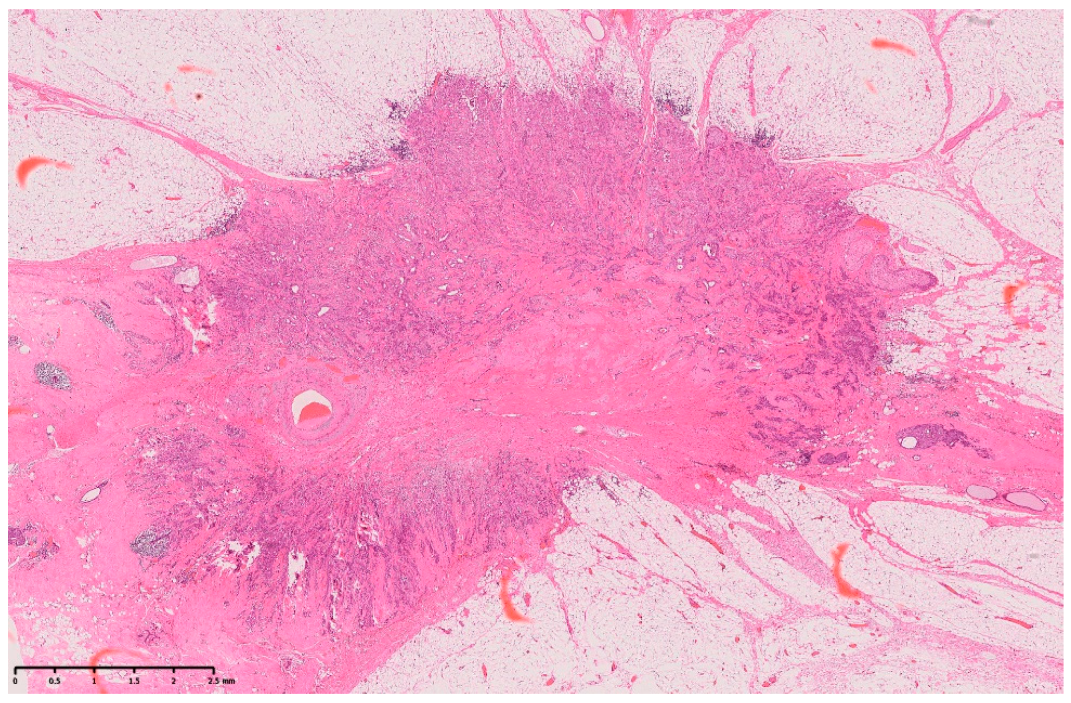
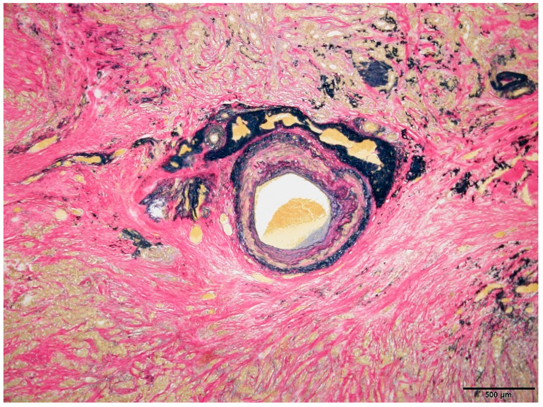
Arterial Calcification Disappearance in Breast Imaging: A Key Indicator for Transition to Invasive Ductal Carcinoma

 , , , ,
 Abstract
Share and Cite
Sato, A.; Fujioka, T.; Onishi, I.; Yamaga, E.; Katsuta, L.; Kubota, K.; Kumaki, Y.; Ishiba, T.; Oda, G.; Tateishi, U. Arterial Calcification Disappearance in Breast Imaging: A Key Indicator for Transition to Invasive Ductal Carcinoma. Diagnostics 2024, 14, 727. https://doi.org/10.3390/diagnostics14070727
Sato A, Fujioka T, Onishi I, Yamaga E, Katsuta L, Kubota K, Kumaki Y, Ishiba T, Oda G, Tateishi U. Arterial Calcification Disappearance in Breast Imaging: A Key Indicator for Transition to Invasive Ductal Carcinoma. Diagnostics. 2024; 14(7):727. https://doi.org/10.3390/diagnostics14070727
Chicago/Turabian StyleSato, Arisa, Tomoyuki Fujioka, Iichiroh Onishi, Emi Yamaga, Leona Katsuta, Kazunori Kubota, Yuichi Kumaki, Toshiyuki Ishiba, Goshi Oda, and Ukihide Tateishi. 2024. "Arterial Calcification Disappearance in Breast Imaging: A Key Indicator for Transition to Invasive Ductal Carcinoma" Diagnostics 14, no. 7: 727. https://doi.org/10.3390/diagnostics14070727
APA StyleSato, A., Fujioka, T., Onishi, I., Yamaga, E., Katsuta, L., Kubota, K., Kumaki, Y., Ishiba, T., Oda, G., & Tateishi, U. (2024). Arterial Calcification Disappearance in Breast Imaging: A Key Indicator for Transition to Invasive Ductal Carcinoma. Diagnostics, 14(7), 727. https://doi.org/10.3390/diagnostics14070727