


Noninvasive Visualization of Tumor Blood Vessels within Hepatocellular Carcinoma by Application of Superb Microvascular Imaging to Contrast-Enhanced Ultrasonography


Abstract
Author Contributions
Funding
Institutional Review Board Statement
Informed Consent Statement
Data Availability Statement
Acknowledgments
Conflicts of Interest
References
- Kudo, M.; Finn, R.S.; Qin, S.; Han, K.H.; Ikeda, K.; Piscaglia, F.; Baron, A.; Park, J.W.; Han, G.; Jassem, J.; et al. Lenvatinib versus sorafenib in first-line treatment of patients with unresectable hepatocellular carcinoma: A randomised phase 3 non-inferiority trial. Lancet 2018, 391, 1163–1173. [Google Scholar] [CrossRef] [Scilit]
- Itoh, S.; Toshida, K.; Morita, K.; Kurihara, T.; Nagao, Y.; Tomino, T.; Toshima, T.; Harada, N.; Mori, M.; Yoshizumi, T. Clinical effectiveness of surgical treatment after lenvatinib administration for hepatocellular carcinoma. Int. J. Clin. Oncol. 2022, 27, 1725–1732. [Google Scholar] [CrossRef] [Scilit]
- Wang, F.; Numata, K.; Komiyama, S.; Miwa, H.; Sugimori, K.; Ogushi, K.; Moriya, S.; Nozaki, A.; Chuma, M.; Ruan, L.; et al. Combination Therapy with Lenvatinib and Radiofrequency Ablation for Patients with Intermediate-Stage Hepatocellular Carcinoma beyond Up-to-Seven Criteria and Child-Pugh Class A Liver function: A Pilot Study. Front. Oncol. 2022, 12, 843680. [Google Scholar] [CrossRef] [Scilit] [PubMed]
- Peng, Z.; Fan, W.; Zhu, B.; Wang, G.; Sun, J.; Xiao, C.; Huang, F.; Tang, R.; Cheng, Y.; Huang, Z.; et al. Lenvatinib Combined With Transarterial Chemoembolization as First-Line Treatment for Advanced Hepatocellular Carcinoma: A Phase III, Randomized Clinical Trial (LAUNCH). J. Clin. Oncol. 2023, 41, 117–127. [Google Scholar] [CrossRef] [Scilit]
- Une, N.; Takano-Kasuya, M.; Kitamura, N.; Ohta, M.; Inose, T.; Kato, C.; Nishimura, R.; Tada, H.; Miyagi, S.; Ishida, T.; et al. The anti-angiogenic agent lenvatinib induces tumor vessel normalization and enhances radiosensitivity in hepatocellular tumors. Med. Oncol. 2021, 38, 60. [Google Scholar] [CrossRef] [Scilit]
- Yokoo, H.; Takahashi, H.; Hagiwara, M.; Iwata, H.; Imai, K.; Saito, Y.; Matsuno, N.; Furukawa, H. Successful hepatic resection for recurrent hepatocellular carcinoma after lenvatinib treatment: A case report. World J. Hepatol. 2020, 12, 1349–1357. [Google Scholar] [CrossRef] [Scilit] [PubMed]
- Tachiiri, T.; Nishiofuku, H.; Maeda, S.; Sato, T.; Toyoda, S.; Matsumoto, T.; Chanoki, Y.; Minamiguchi, K.; Taiji, R.; Kunichika, H.; et al. Vascular Normalization Caused by Short-Term Lenvatinib Could Enhance Transarterial Chemoembolization in Hepatocellular Carcinoma. Curr. Oncol. 2023, 30, 4779–4786. [Google Scholar] [CrossRef] [Scilit]
- Yoh, T.; Ishii, T.; Nishio, T.; Koyama, Y.; Ogiso, S.; Fukumitsu, K.; Uchida, Y.; Ito, T.; Seo, S.; Hata, K.; et al. A Conceptual Classification of Resectability for Hepatocellular Carcinoma. World J. Surg. 2023, 47, 740–748. [Google Scholar] [CrossRef] [Scilit] [PubMed]
- Lencioni, R.; Llovet, J.M. Modified RECIST (mRECIST) assessment for hepatocellular carcinoma. Semin. Liver Dis. 2010, 30, 52–60. [Google Scholar] [CrossRef] [Scilit] [PubMed]
- Ichida, A.; Arita, J.; Hatano, E.; Eguchi, S.; Saiura, A.; Nagano, H.; Shindoh, J.; Hashimoto, M.; Takemura, N.; Taura, K.; et al. A Multicenter Phase 2 Trial Evaluating the Efficacy and Safety of Preoperative Lenvatinib Therapy for Patients with Advanced Hepatocellular Carcinoma (LENS-HCC Trial). Liver Cancer, 2023; Online ahead of print. [CrossRef] [Scilit]
- Llovet, J.M.; Ricci, S.; Mazzaferro, V.; Hilgard, P.; Gane, E.; Blanc, J.F.; de Oliveira, A.C.; Santoro, A.; Raoul, J.L.; Forner, A.; et al. Sorafenib in advanced hepatocellular carcinoma. N. Engl. J. Med. 2008, 359, 378–390. [Google Scholar] [CrossRef] [Scilit] [PubMed]
- Finn, R.S.; Qin, S.; Ikeda, M.; Galle, P.R.; Ducreux, M.; Kim, T.Y.; Kudo, M.; Breder, V.; Merle, P.; Kaseb, A.O.; et al. Atezolizumab plus Bevacizumab in Unresectable Hepatocellular Carcinoma. N. Engl. J. Med. 2020, 382, 1894–1905. [Google Scholar] [CrossRef] [Scilit] [PubMed]
- Kudo, M. All Stages of Hepatocellular Carcinoma Patients Benefit from Systemic Therapy Combined with Locoregional Therapy. Liver Cancer 2023, 12, 395–404. [Google Scholar] [CrossRef] [Scilit] [PubMed]
- Marrero, J.A.; Kulik, L.M.; Sirlin, C.B.; Zhu, A.X.; Finn, R.S.; Abecassis, M.M.; Roberts, L.R.; Heimbach, J.K. Diagnosis, Staging, and Management of Hepatocellular Carcinoma: 2018 Practice Guidance by the American Association for the Study of Liver Diseases. Hepatology 2018, 68, 723–750. [Google Scholar] [CrossRef] [Scilit] [PubMed]
- European Association for the Study of the Liver. EASL Clinical Practice Guidelines: Management of hepatocellular carcinoma. J. Hepatol. 2018, 69, 182–236. [Google Scholar] [CrossRef] [Scilit]
- Ota, Y.; Aso, K.; Otake, S.; Okada, M.; Shukuda, K.; Sawada, K.; Yokoo, H.; Tanino, M.; Fujiya, M.; Okumura, T. Contrast-enhanced ultrasonography for the diagnosis of spontaneous necrosis of hepatocellular carcinoma: A report of 2 cases. Radiol. Case Rep. 2023, 18, 173–181. [Google Scholar] [CrossRef] [Scilit]
- Otake, S.; Ota, Y.; Aso, K.; Okada, M.; Hayashi, H.; Hasebe, T.; Nakajima, S.; Sawada, K.; Fujiya, M.; Okumura, T. Contrast-enhanced Ultrasonography Features for Diagnosing Pseudoprogression of Hepatocellular Carcinoma with Immunotherapy: A Case Report of the Response after Pseudoprogression. Intern. Med. 2023; Online ahead of print. [CrossRef] [Scilit]
- Kuroda, H.; Abe, T.; Fujiwara, Y.; Okamoto, T.; Yonezawa, M.; Sato, H.; Endo, K.; Oikawa, T.; Sawara, K.; Takikawa, Y. Change in arterial tumor perfusion is an early biomarker of lenvatinib efficacy in patients with unresectable hepatocellular carcinoma. World J. Gastroenterol. 2019, 25, 2365–2372. [Google Scholar] [CrossRef] [Scilit]
- Eso, Y.; Nakano, S.; Mishima, M.; Arasawa, S.; Iguchi, E.; Takeda, H.; Takai, A.; Takahashi, K.; Seno, H. A simplified method to quantitatively predict the effect of lenvatinib on hepatocellular carcinoma using contrast-enhanced ultrasound with perfluorobutane microbubbles. Quant. Imaging Med. Surg. 2021, 11, 2766–2774. [Google Scholar] [CrossRef] [Scilit]
- Kamachi, N.; Nakano, M.; Okamura, S.; Niizeki, T.; Iwamoto, H.; Shimose, S.; Shirono, T.; Noda, Y.; Kuromatsu, R.; Koga, H.; et al. Evaluating the therapeutic effect of lenvatinib against advanced hepatocellular carcinoma by measuring blood flow changes using contrast-enhanced ultrasound. Cancer Rep. 2022, 5, e1471. [Google Scholar] [CrossRef] [Scilit]
- Jiang, Z.Z.; Huang, Y.H.; Shen, H.L.; Liu, X.T. Clinical Applications of Superb Microvascular Imaging in the Liver, Breast, Thyroid, Skeletal Muscle, and Carotid Plaques. J. Ultrasound Med. 2019, 38, 2811–2820. [Google Scholar] [CrossRef] [Scilit] [PubMed]
- Artul, S.; Nseir, W.; Armaly, Z.; Soudack, M. Superb Microvascular Imaging: Added Value and Novel Applications. J. Clin. Imaging Sci. 2017, 7, 45. [Google Scholar] [CrossRef] [Scilit]
- Muraishi, N.; Kawamura, Y.; Akuta, N.; Shindoh, J.; Matsumura, M.; Okubo, S.; Fujiyama, S.; Hosaka, T.; Saitoh, S.; Sezaki, H.; et al. The Impact of Lenvatinib on Tumor Blood Vessel Shrinkage of Hepatocellular Carcinoma during Treatment: An Imaging-Based Analysis. Oncology 2023, 101, 134–144. [Google Scholar] [CrossRef] [Scilit]
- Matsuda, N.; Imai, N.; Kuzuya, T.; Yamamoto, K.; Ito, T.; Ishizu, Y.; Honda, T.; Ishigami, M.; Fujishiro, M. Progression after Molecular Targeted Agents: Hepatic Arterial Changes and Transarterial Chemoembolization in Hepatocellular Carcinoma. In Vivo 2021, 35, 1185–1189. [Google Scholar] [CrossRef] [Scilit] [PubMed]
- Yang, J.; Guo, Z.; Song, M.; Pan, Q.; Zhao, J.; Huang, Y.; Han, Y.; Ouyang, D.; Yang, C.; Chen, H.; et al. Lenvatinib improves anti-PD-1 therapeutic efficacy by promoting vascular normalization via the NRP-1-PDGFRbeta complex in hepatocellular carcinoma. Front. Immunol. 2023, 14, 1212577. [Google Scholar] [CrossRef] [Scilit] [PubMed]
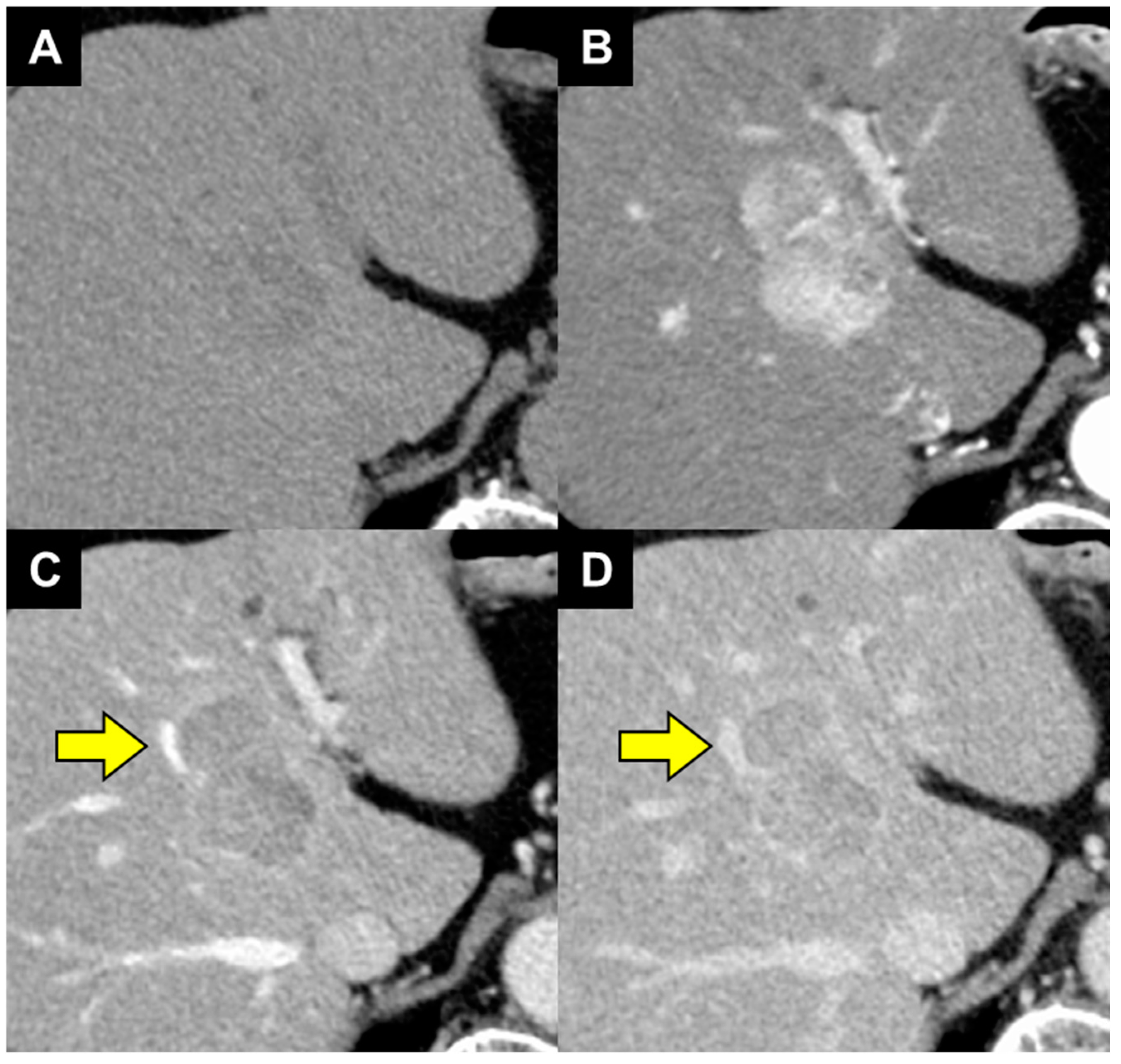
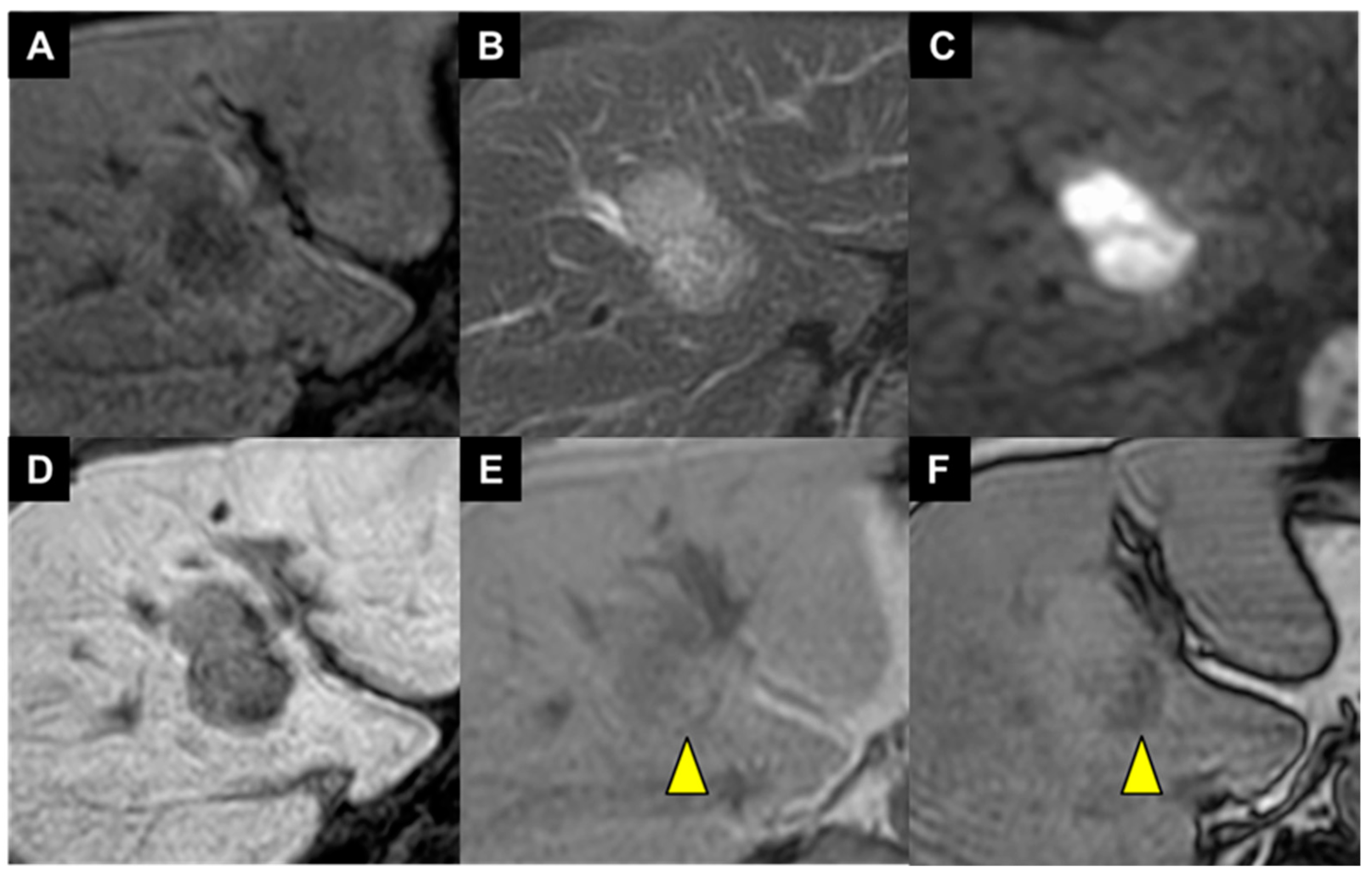
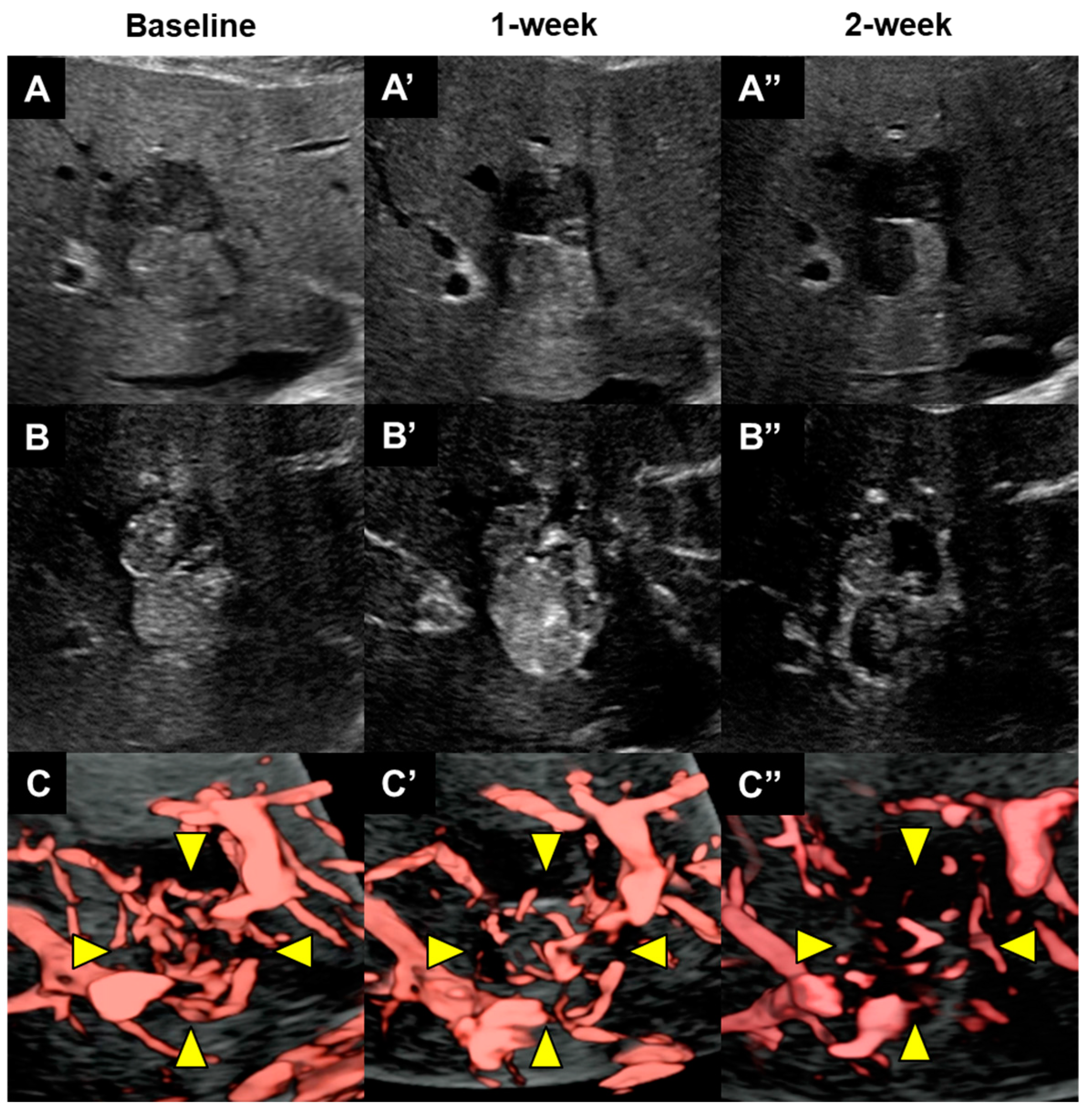
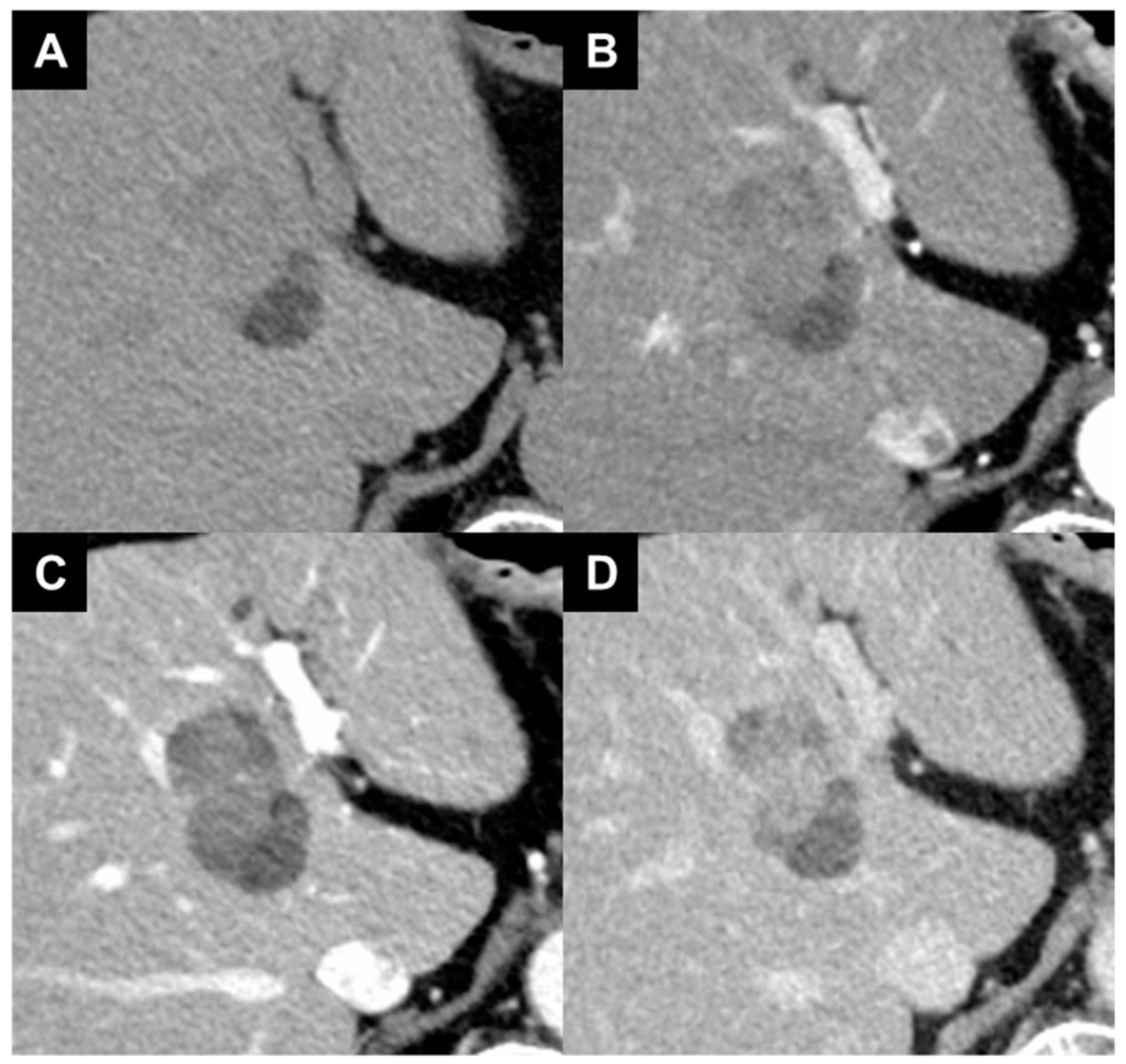
Disclaimer/Publisher’s Note: The statements, opinions and data contained in all publications are solely those of the individual author(s) and contributor(s) and not of MDPI and/or the editor(s). MDPI and/or the editor(s) disclaim responsibility for any injury to people or property resulting from any ideas, methods, instructions or products referred to in the content. |
© 2024 by the authors. Licensee MDPI, Basel, Switzerland. This article is an open access article distributed under the terms and conditions of the Creative Commons Attribution (CC BY) license (https://creativecommons.org/licenses/by/4.0/).
Share and Cite
Ota, Y.; Aso, K.; Yokoo, H.; Fujiya, M. Noninvasive Visualization of Tumor Blood Vessels within Hepatocellular Carcinoma by Application of Superb Microvascular Imaging to Contrast-Enhanced Ultrasonography. Diagnostics 2024, 14, 678. https://doi.org/10.3390/diagnostics14070678
Ota Y, Aso K, Yokoo H, Fujiya M. Noninvasive Visualization of Tumor Blood Vessels within Hepatocellular Carcinoma by Application of Superb Microvascular Imaging to Contrast-Enhanced Ultrasonography. Diagnostics. 2024; 14(7):678. https://doi.org/10.3390/diagnostics14070678
Chicago/Turabian StyleOta, Yu, Kazunobu Aso, Hideki Yokoo, and Mikihiro Fujiya. 2024. "Noninvasive Visualization of Tumor Blood Vessels within Hepatocellular Carcinoma by Application of Superb Microvascular Imaging to Contrast-Enhanced Ultrasonography" Diagnostics 14, no. 7: 678. https://doi.org/10.3390/diagnostics14070678
APA StyleOta, Y., Aso, K., Yokoo, H., & Fujiya, M. (2024). Noninvasive Visualization of Tumor Blood Vessels within Hepatocellular Carcinoma by Application of Superb Microvascular Imaging to Contrast-Enhanced Ultrasonography. Diagnostics, 14(7), 678. https://doi.org/10.3390/diagnostics14070678

