Comparison of Two Contemporary Quantitative Atherosclerotic Plaque Assessment Tools for Coronary Computed Tomography Angiography: Single-Center Analysis and Multi-Center Patient Cohort Validation
Abstract
1. Introduction
2. Materials and Methods
2.1. Study Design and Patient Population
2.2. CCTA Protocol
2.3. Plaque Burden Quantification Using Plaque Analysis Software
2.4. Time Spent and Image Quality
2.5. Statistical Analysis
3. Results
3.1. Demographic Data, Cardiac Medications, and CCTA Characteristics
3.2. Image Quality
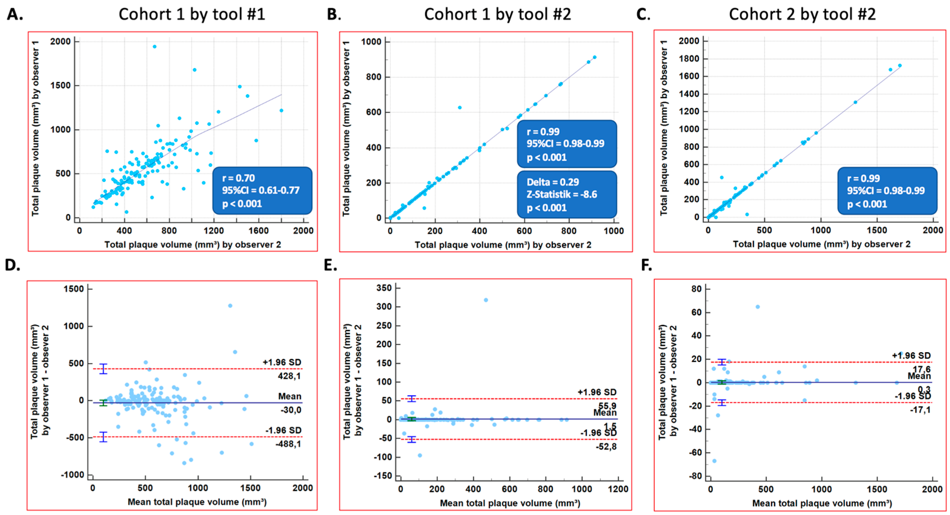
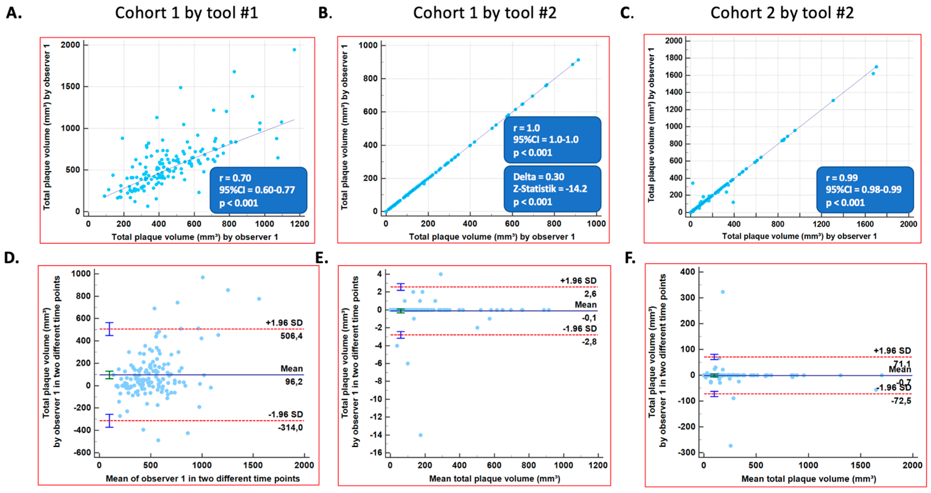
3.3. Inter- and Intra-Observer Variabilities for Total Plaque Volumes in All Three Coronary Vessels
3.4. Association of Increasing Automation with Lower Time Spent
4. Discussion
5. Conclusions
Study Limitations
Supplementary Materials
Author Contributions
Funding
Institutional Review Board Statement
Informed Consent Statement
Data Availability Statement
Acknowledgments
Conflicts of Interest
Abbreviations
| ACE | angiotensin-converting enzyme |
| AT2 | angiotensin 2 |
| CAD | coronary artery disease |
| CV | cardiovascular |
| DSCT | dual source CT |
| ECG | electrocardiogram |
| ICC | intraclass correlation |
| IQR | interquartile ranges |
| LAD | left anterior descending artery |
| LCX | left circumflex artery |
| LOA | limits of agreement |
| MDCT | multidetector computed tomography |
| PCI | percutaneous coronary intervention |
| RCA | right coronary artery |
References
- Naghavi, M.; Libby, P.; Falk, E.; Casscells, S.W.; Litovsky, S.; Rumberger, J.; Badimon, J.J.; Stefanadis, C.; Moreno, P.; Pasterkamp, G.; et al. From vulnerable plaque to vulnerable patient: A call for new definitions and risk assessment strategies: Part II. Circulation 2003, 108, 1772–1778. [Google Scholar] [CrossRef] [PubMed]
- Knuuti, J.; Wijns, W.; Saraste, A.; Capodanno, D.; Barbato, E.; Funck-Brentano, C.; Prescott, E.; Storey, R.F.; Deaton, C.; Cuisset, T.; et al. 2019 ESC Guidelines for the diagnosis and management of chronic coronary syndromes. Eur. Heart J. 2020, 41, 407–477. [Google Scholar] [CrossRef] [PubMed]
- Moss, A.J.; Williams, M.C.; Newby, D.E.; Nicol, E.D. The Updated NICE Guidelines: Cardiac CT as the First-Line Test for Coronary Artery Disease. Curr. Cardiovasc. Imaging Rep. 2017, 10, 15. [Google Scholar] [CrossRef] [PubMed]
- Gitsioudis, G.; Schüssler, A.; Nagy, E.; Maurovich-Horvat, P.; Buss, S.J.; Voss, A.; Hosch, W.; Hofmann, N.; Kauczor, H.-U.; Giannitsis, E.; et al. Combined Assessment of High-Sensitivity Troponin T and Noninvasive Coronary Plaque Composition for the Prediction of Cardiac Outcomes. Radiology 2015, 276, 73–81. [Google Scholar] [CrossRef] [PubMed]
- Williams, M.C.; Kwiecinski, J.; Doris, M.; McElhinney, P.; D’Souza, M.S.; Cadet, S.; Adamson, P.D.; Moss, A.J.; Alam, S.; Hunter, A.; et al. Low-Attenuation Noncalcified Plaque on Coronary Computed Tomography Angiography Predicts Myocardial Infarction: Results from the Multicenter SCOT-HEART Trial (Scottish Computed Tomography of the HEART). Circulation 2020, 141, 1452–1462. [Google Scholar] [CrossRef] [PubMed]
- Korosoglou, G.; Chatzizisis, Y.S.; Raggi, P. Coronary computed tomography angiography in asymptomatic patients: Still a taboo or precision medicine? Atherosclerosis 2021, 317, 47–49. [Google Scholar] [CrossRef] [PubMed]
- Gallone, G.; Bellettini, M.; Gatti, M.; Tore, D.; Bruno, F.; Scudeler, L.; Cusenza, V.; Lanfranchi, A.; Angelini, A.; de Filippo, O.; et al. Coronary Plaque Characteristics Associated with Major Adverse Cardiovascular Events in Atherosclerotic Patients and Lesions: A Systematic Review and Meta-Analysis. JACC Cardiovasc. Imaging 2023, 16, 1584–1604. [Google Scholar] [CrossRef]
- Jávorszky, N.; Homonnay, B.; Gerstenblith, G.; Bluemke, D.; Kiss, P.; Török, M.; Celentano, D.; Lai, H.; Lai, S.; Kolossváry, M.; et al. Deep learning-based atherosclerotic coronary plaque segmentation on coronary CT angiography. Eur. Radiol. 2022, 32, 7217–7226. [Google Scholar] [CrossRef]
- Denzinger, F.; Wels, M.; Hopfgartner, C.; Lu, J.; Schöbinger, M.; Maier, A.; Sühling, M. Coronary Plaque Analysis for CT Angiography Clinical Research. arXiv 2021, arXiv:2101.03799. [Google Scholar]
- Tesche, C.; Bauer, M.J.; Straube, F.; Rogowski, S.; Baumann, S.; Renker, M.; Fink, N.; Schoepf, U.J.; Hoffmann, E.; Ebersberger, U. Association of epicardial adipose tissue with coronary CT angiography plaque parameters on cardiovascular outcome in patients with and without diabetes mellitus. Atherosclerosis 2022, 363, 78–84. [Google Scholar] [CrossRef]
- Yan, C.; Zhou, G.; Yang, X.; Lu, X.; Zeng, M.; Ji, M. Image quality of automatic coronary CT angiography reconstruction for patients with HR >/= 75 bpm using an AI-assisted 16-cm z-coverage CT scanner. BMC Med. Imaging 2021, 21, 24. [Google Scholar] [CrossRef] [PubMed]
- Clouse, M.E.; Sabir, A.; Yam, C.-S.; Yoshimura, N.; Lin, S.; Welty, F.; Martinez-Clark, P.; Raptopoulos, V. Measuring noncalcified coronary atherosclerotic plaque using voxel analysis with MDCT angiography: A pilot clinical study. AJR Am. J. Roentgenol. 2008, 190, 1553–1560. [Google Scholar] [CrossRef] [PubMed]
- Korosoglou, G.; Mueller, D.; Lehrke, S.; Steen, H.; Hosch, W.; Heye, T.; Kauczor, H.-U.; Giannitsis, E.; Katus, H.A. Quantitative assessment of stenosis severity and atherosclerotic plaque composition using 256-slice computed tomography. Eur. Radiol. 2010, 20, 1841–1850. [Google Scholar] [CrossRef] [PubMed]
- Lee, M.S.; Chun, E.J.; Kim, K.J.; Kim, J.A.; Vembar, M.; Choi, S.I. Reproducibility in the assessment of noncalcified coronary plaque with 256-slice multi-detector CT and automated plaque analysis software. Int. J. Cardiovasc. Imaging 2010, 26 (Suppl. S2), 237–244. [Google Scholar] [CrossRef] [PubMed]
- Klass, O.; Kleinhans, S.; Walker, M.J.; Olszewski, M.; Feuerlein, S.; Juchems, M.; Hoffmann, M.H.K. Coronary plaque imaging with 256-slice multidetector computed tomography: Interobserver variability of volumetric lesion parameters with semiautomatic plaque analysis software. Int. J. Cardiovasc. Imaging 2010, 26, 711–720. [Google Scholar] [CrossRef] [PubMed]
- Papadopoulou, S.-L.; Garcia-Garcia, H.M.; Rossi, A.; Girasis, C.; Dharampal, A.S.; Kitslaar, P.H.; Krestin, G.P.; de Feyter, P.J. Reproducibility of computed tomography angiography data analysis using semiautomated plaque quantification software: Implications for the design of longitudinal studies. Int. J. Cardiovasc. Imaging 2013, 29, 1095–1104. [Google Scholar] [CrossRef]
- Symons, R.; Morris, J.Z.; Wu, C.O.; Pourmorteza, A.; Ahlman, M.A.; Lima, J.A.C.; Chen, M.Y.; Mallek, M.; Sandfort, V.; Bluemke, D.A. Coronary CT Angiography: Variability of CT Scanners and Readers in Measurement of Plaque Volume. Radiology 2016, 281, 737–748. [Google Scholar] [CrossRef]
- Meah, M.N.; Singh, T.; Williams, M.C.; Dweck, M.R.; Newby, D.E.; Slomka, P.; Adamson, P.D.; Moss, A.J.; Dey, D. Reproducibility of quantitative plaque measurement in advanced coronary artery disease. J. Cardiovasc. Comput. Tomogr. 2021, 15, 333–338. [Google Scholar] [CrossRef]
- Tzolos, E.; McElhinney, P.; Williams, M.C.; Cadet, S.; Dweck, M.R.; Berman, D.S.; Slomka, P.J.; Newby, D.E.; Dey, D. Repeatability of quantitative pericoronary adipose tissue attenuation and coronary plaque burden from coronary CT angiography. J. Cardiovasc. Comput. Tomogr. 2021, 15, 81–84. [Google Scholar] [CrossRef]
- Korosoglou, G.; Thiele, H.; Baldus, S.; Bohm, M.; Frey, N. Lessons learned from SCOT-HEART, DISCHARGE, and PRECISE: A patient-centered perspective with implications for the appropriate use of CCTA. Clin. Res. Cardiol. 2023, 112, 1347–1350. [Google Scholar] [CrossRef]
- Blackmon, K.N.; Streck, J.; Thilo, C.; Bastarrika, G.; Costello, P.; Schoepf, U.J. Reproducibility of automated noncalcified coronary artery plaque burden assessment at coronary CT angiography. J. Thorac. Imaging 2009, 24, 96–102. [Google Scholar] [CrossRef]
- Laqmani, A.; Klink, T.; Quitzke, M.; Creder, D.D.; Adam, G.; Lund, G. Accuracy of Coronary Plaque Detection and Assessment of Interobserver Agreement for Plaque Quantification Using Automatic Coronary Plaque Analysis Software on Coronary CT Angiography. Rofo 2016, 188, 933–939. [Google Scholar] [CrossRef] [PubMed]
- Groth, M.; Muellerleile, K.; Klink, T.; Säring, D.; Halaj, S.; Folwarski, G.; Kaul, M.; Bannas, P.; Adam, G.; Lund, G.K. Improved agreement between experienced and inexperienced observers using a standardized evaluation protocol for cardiac volumetry and infarct size measurement. Rofo 2012, 184, 1131–1137. [Google Scholar] [CrossRef] [PubMed]
- Karamitsos, T.D.; Hudsmith, L.E.; Selvanayagam, J.B.; Neubauer, S.; Francis, J.M. Operator induced variability in left ventricular measurements with cardiovascular magnetic resonance is improved after training. J. Cardiovasc. Magn. Reson. 2007, 9, 777–783. [Google Scholar] [CrossRef] [PubMed]
- Stolzmann, P.; Schlett, C.L.; Maurovich-Horvat, P.; Maehara, A.; Ma, S.; Scheffel, H.; Engel, L.-C.; Károlyi, M.; Mintz, G.S.; Hoffmann, U. Variability and accuracy of coronary CT angiography including use of iterative reconstruction algorithms for plaque burden assessment as compared with intravascular ultrasound-an ex vivo study. Eur. Radiol. 2012, 22, 2067–2075. [Google Scholar] [CrossRef]


| Single-Center Cohort #1, N = 50 | Multi-Center Cohort #2, N = 50 | p-Values | |
|---|---|---|---|
| Baseline data and risk factors | |||
| Age (yrs.) | 62.0 (IQR = 55.0–70.0) | 69.5 (IQR = 64.0–80.0) | <0.001 |
| Age > 60 yrs. | 28 (56.0%) | 40 (80.0%) | 0.01 |
| Female gender | 10 (20.0%) | 35 (70.0%) | <0.001 |
| Body mass index (kg/m2) | 28.3 (IQR = 24.5–30.3) | 25.7 (IQR = 23.0–30.5) | 0.04 |
| Arterial hypertension | 31 (62.0%) * | 32 (64.0%) | 0.83 |
| Hyperlipidemia | 34 (68.0%) ** | 20 (40.0%) | 0.005 |
| Diabetes mellitus | 4 (8.0%) * | 7 (14.0%) | 0.34 |
| Active or former smoking | 13 (26.0%) * | 8 (16.0%) | 0.22 |
| Family history of CAD Number of cardiovascular (CV) risk factors (0–6) | 21 (42.0%) * | 19 (38.0%) | 0.68 |
| 3.0 (IQR = 2.0–4.0) | 2.0 (IQR = 2.0–3.0) | 0.53 | |
| History of CAD, PCI, and arrhythmias | |||
| Prior cardiac catheterization | 20 (40.0%) | 8 (16.0%) | 0.008 |
| Prior PCI | 16 (32.0%) | 5 (10.0%) | 0.007 |
| Prior myocardial infarction | 8 (16.0%) | 4 (8.0%) | 0.22 |
| Atrial fibrillation | 7 (14.0%) | 10 (20.0%) | 0.43 |
| Baseline clinical presentation | |||
| Stable chest pain syndrome | 26 (52.0%) | 26 (52.0%) | 1.0 |
| Exertional dyspnea | 16 (32.0%) | 27 (54.0%) | 0.03 |
| Palpitations or other unspecific symptoms | 8 (16.0%) | 4 (8.0%) | 1.0 |
| Syncope | 0 (0.0%) | 1 (2.0%) | 0.32 |
| Baseline cardiac medications | |||
| Aspirin | 27 (54.0%) | 18 (36.0%) | 0.07 |
| ß-blockers | 22 (44.0%) | 21 (42.0%) | 0.84 |
| Calcium antagonists | 10 (20.0%) | 9 (18.0%) | 0.80 |
| Diuretics | 11 (22.0%) | 11 (22.0%) | 1.0 |
| ACE inhibitors or AT2 blockers | 13 (26.0%) | 25 (50.0%) | 0.01 |
| PCSK9 inhibitors | 9 (18.0%) | 0 (0.0%) | 0.002 |
| Statins (independent of intensity) | 35 (70.0%) | 22 (44.0%) | 0.009 |
| Ezetimibe | 5 (10.0%) | 3 (6.0%) | 0.46 |
| RCA | LAD | LCX | All 3 Coronary Arteries (per Patient) | |
|---|---|---|---|---|
| Cohort #1; Inter-observer variability using Tool #1 (%) | ||||
| Total plaque volume (mm3) | 19.46 | 21.34 | 27.62 | 22.81 |
| Calcified plaque volume (mm3) | 19.30 | 25.06 | 33.63 | 26.00 |
| Non-calcified plaque volume (mm3) | 24.41 | 18.99 | 22.70 | 22.03 |
| Cohort #1; Inter-observer variability using Tool #2 (%) | ||||
| Total plaque volume (mm3) | 1.79 | 0.07 | 4.98 | 2.28 |
| Calcified plaque volume (mm3) | 2.47 | 0.04 | 5.06 | 2.52 |
| Non-calcified plaque volume (mm3) | 1.43 | 5.17 | 4.98 | 3.86 |
| Cohort #1; Intra-observer variability using Tool #1 (%) | ||||
| Total plaque volume (mm3) | 18.72 | 15.38 | 25.10 | 19.73 |
| Calcified plaque volume (mm3) | 17.81 | 19.07 | 29.48 | 22.12 |
| Non-calcified plaque volume (mm3) | 21.70 | 19.89 | 22.73 | 21.44 |
| Cohort #1; Intra-observer variability using Tool #2 (%) | ||||
| Total plaque volume (mm3) | 0.27 | 0.00 | 0.28 | 0.18 |
| Calcified plaque volume (mm3) | 0.39 | 0.12 | 0.51 | 0.34 |
| Non-calcified plaque volume (mm3) | 0.07 | 0.07 | 0.10 | 0.08 |
| Cohort #2 (validation); Inter-observer variability using Tool #2 (%) | ||||
| Total plaque volume (mm3) | 1.83 | 0.34 | 6.58 | 2.92 |
| Calcified plaque volume (mm3) | 1.77 | 0.24 | 6.84 | 2.95 |
| Non-calcified plaque volume (mm3) | 1.67 | 0.37 | 6.12 | 2.72 |
| Cohort #2 (validation); Intra-observer variability using Tool #2 (%) | ||||
| Total plaque volume (mm3) | 6.46 | 1.20 | 3.79 | 3.82 |
| Calcified plaque volume (mm3) | 6.77 | 0.86 | 4.48 | 4.04 |
| Non-calcified plaque volume (mm3) | 5.46 | 1.99 | 3.76 | 3.74 |
| Number of Patients | Number of Vessels | Scanner Type | Plaque Analysis Software | Intra-Observer Reproducibility | Inter-Observer Reproducibility | |
|---|---|---|---|---|---|---|
| Symons et al. [17] | 80 | 667 | 320-Detektor-Zeilenscanner (Aquilion One Vision; Toshiba, Otawara, Japan) | QAngioCT, version 2.1.9.1 (Medis Medical Imaging Systems, Leiden, The Netherlands) | ICC: 0.96 | NA |
| Gitsioudis et al. [4] | 521 | 7690 | 256-detector row CT scanner (iCT; Philips Medical Systems, Best, The Netherlands) | Extended Brilliance Workspace 4.0 (Philips Medical Systems) | Variability: 9% | Variability: 13% LAO: 93% (k = 0.85) |
| Tzolos et al. [19] | 50 | 157 | 320-multidetector row scanners | Autoplaque 2.5 (Cedars-Sinai Medical Center) | ICC: 0.978 LAO ± 5.97% | ICC: 0.944 LOA ± 9.61% |
| Laqmani et al. [22] | 10 | 30 | 256-MDCT scanner (Brilliance iCT, Philips, Best, The Netherlands) | Comprehensive Cardiac Analysis, Extended Brilliance Workspace, V4.0 (Philips Healthcare, Best, The Netherlands) | NA | LOA: −3.3 ± 33.8% |
| Papadopoulou et al. [16] | 10 | 21 | 64-slice dual source CT scanner (Somatom Definition, Siemens Medical Solutions, Forchheim, Germany) | QAngioCT Research Edition v1.3.61 (Medis Medical Imaging Systems, Leiden, The Netherlands) | Variability: 1.30 ± 1.09% | Variability: 1.6% |
| Lee et al. [14] | 39 | 15 | Brilliance iCT (Philips Medical Systems, Best, The Netherlands) | Extended Brilliance Workspace V4.0; (Philips Healthcare, Cleveland, OH, USA) | LOA: −21.6 and 13.2 mm3 | LOA: 24.6 and 20.3 mm3 |
| Klass et al. [15] | 35 | 105 | Brilliance iCT (Philips Healthcare, Cleveland, OH, USA) | Cardiac Viewer and Comprehensive Cardiac Analysis, Brilliance Workspace (Philips Healthcare, Cleveland, OH, USA) | NA | Variability: 3.3% |
| Korosoglou et al. [13] | 27 | 81 | 256-slice Brilliance iCT (Philips Medical Systems) | Plaque SW version 4.0.2 (Extended Brilliance Workspace 4.0, Philips Medical Systems) | Variability: 9% | Variability: 13% |
| Meah et al. [18] | 20 | NA | 64-multidetector Biograph mCT (Siemens Medical Systems, Erlangen, Germany) | AutoPlaque, Version 2.5, Cedars-Sinai Medical Center, Los Angeles, CA, USA | Variability: 2.6% LCCC: 1.0 (1.0–1.0) | Variability: 13.5% LCCC: 0.97 (0.93–0.99) |
Disclaimer/Publisher’s Note: The statements, opinions and data contained in all publications are solely those of the individual author(s) and contributor(s) and not of MDPI and/or the editor(s). MDPI and/or the editor(s) disclaim responsibility for any injury to people or property resulting from any ideas, methods, instructions or products referred to in the content. |
© 2024 by the authors. Licensee MDPI, Basel, Switzerland. This article is an open access article distributed under the terms and conditions of the Creative Commons Attribution (CC BY) license (https://creativecommons.org/licenses/by/4.0/).
Share and Cite
Weichsel, L.; Giesen, A.; André, F.; Renker, M.; Baumann, S.; Breitbart, P.; Beer, M.; Maurovitch-Horvat, P.; Szilveszter, B.; Vattay, B.; et al. Comparison of Two Contemporary Quantitative Atherosclerotic Plaque Assessment Tools for Coronary Computed Tomography Angiography: Single-Center Analysis and Multi-Center Patient Cohort Validation. Diagnostics 2024, 14, 154. https://doi.org/10.3390/diagnostics14020154
Weichsel L, Giesen A, André F, Renker M, Baumann S, Breitbart P, Beer M, Maurovitch-Horvat P, Szilveszter B, Vattay B, et al. Comparison of Two Contemporary Quantitative Atherosclerotic Plaque Assessment Tools for Coronary Computed Tomography Angiography: Single-Center Analysis and Multi-Center Patient Cohort Validation. Diagnostics. 2024; 14(2):154. https://doi.org/10.3390/diagnostics14020154
Chicago/Turabian StyleWeichsel, Loris, Alexander Giesen, Florian André, Matthias Renker, Stefan Baumann, Philipp Breitbart, Meinrad Beer, Pal Maurovitch-Horvat, Bálint Szilveszter, Borbála Vattay, and et al. 2024. "Comparison of Two Contemporary Quantitative Atherosclerotic Plaque Assessment Tools for Coronary Computed Tomography Angiography: Single-Center Analysis and Multi-Center Patient Cohort Validation" Diagnostics 14, no. 2: 154. https://doi.org/10.3390/diagnostics14020154
APA StyleWeichsel, L., Giesen, A., André, F., Renker, M., Baumann, S., Breitbart, P., Beer, M., Maurovitch-Horvat, P., Szilveszter, B., Vattay, B., Buss, S. J., Marwan, M., Giannopoulos, A. A., Kelle, S., Frey, N., & Korosoglou, G. (2024). Comparison of Two Contemporary Quantitative Atherosclerotic Plaque Assessment Tools for Coronary Computed Tomography Angiography: Single-Center Analysis and Multi-Center Patient Cohort Validation. Diagnostics, 14(2), 154. https://doi.org/10.3390/diagnostics14020154


