The Supratrochlear Artery Sign—A New Piece in the Puzzle of Cerebral Vasospasm
 , , ,
, , ,
Abstract
1. Introduction
2. Materials and Methods
2.1. Study Population
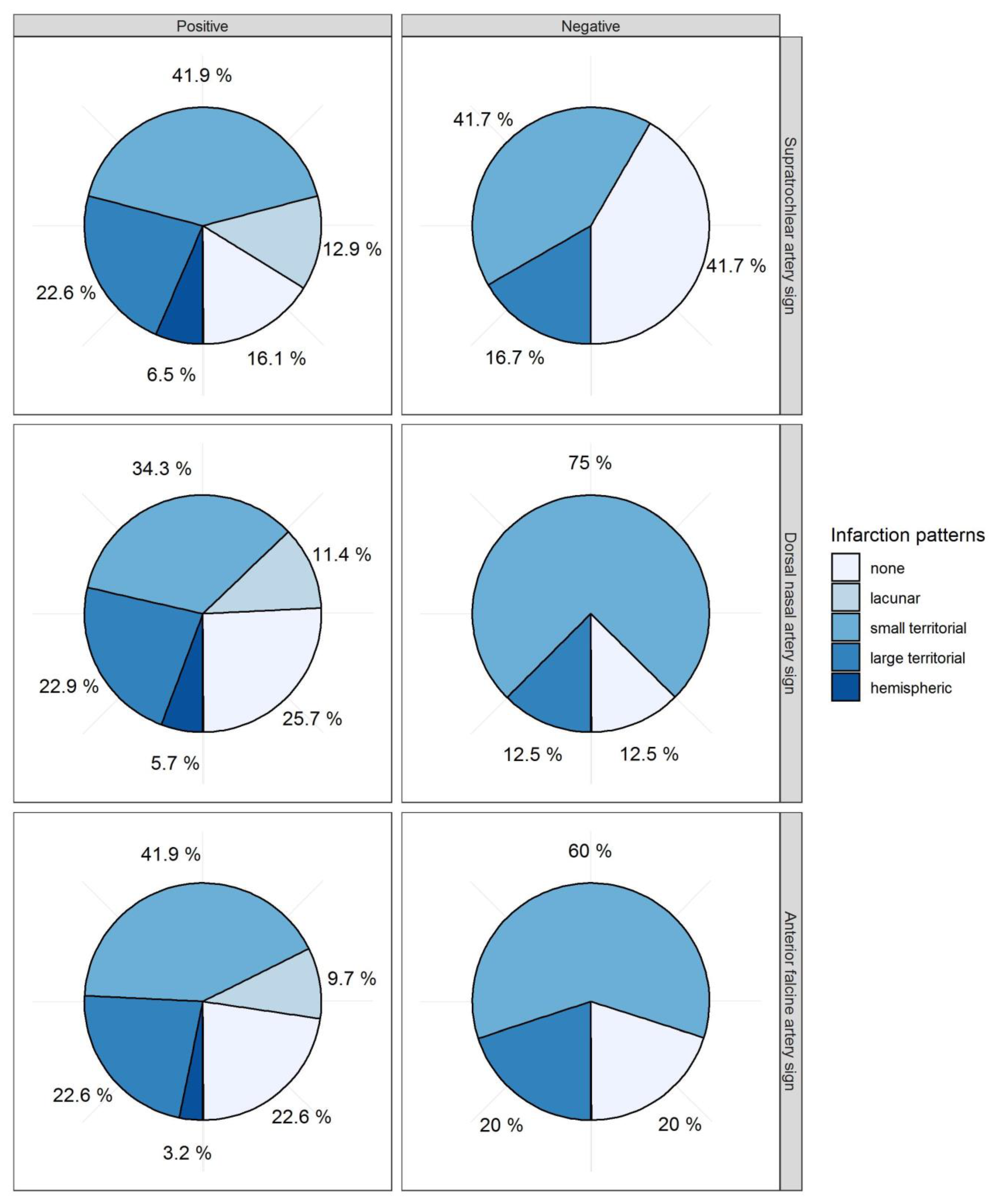
2.2. Infarction Pattern
2.3. Clinical Management
2.4. Diagnostic Cerebral Angiography and Intra-Arterial Spasmolysis
2.5. Vasospasm Classification
- CVSG 0—all intracranial vessels show a physiological shape;
- CVSG 1—Vasospasm affects the A2, A1, and M2 segments;
- CVSG 2—Vasospasm expands to the M1 and terminal segment of the ICA;
- CVSG 3—Severe reduction of the intradural internal carotid artery with filiform A1 and M1 segments, sometimes with a ghost-like fading appearance (ghost sign) [10].
2.6. Vessel Signs
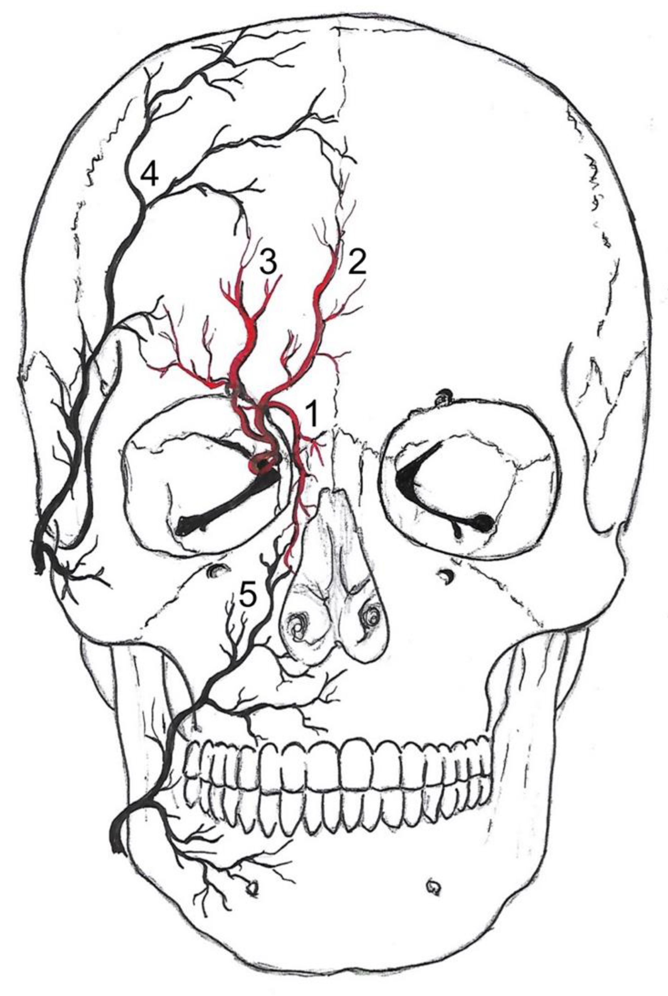
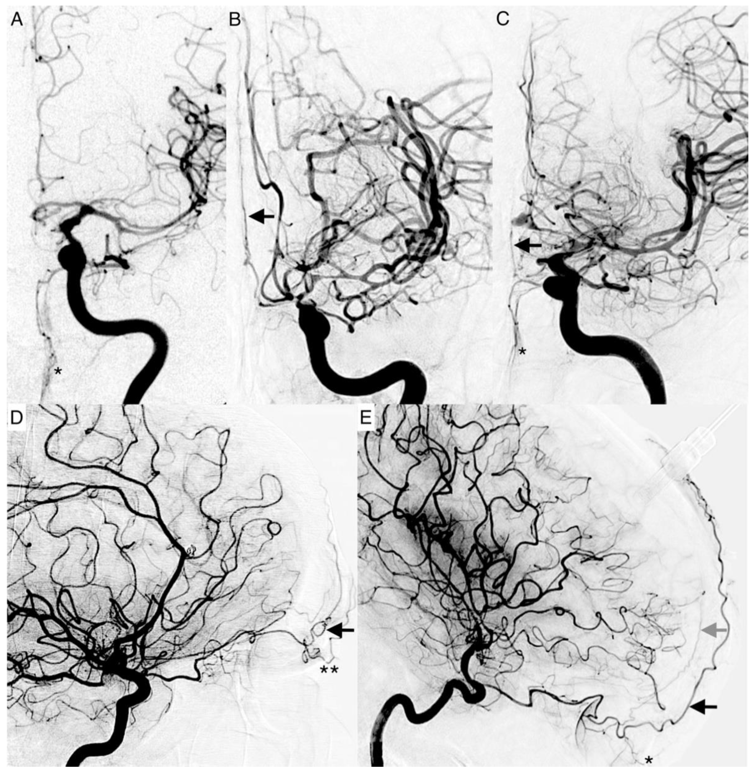
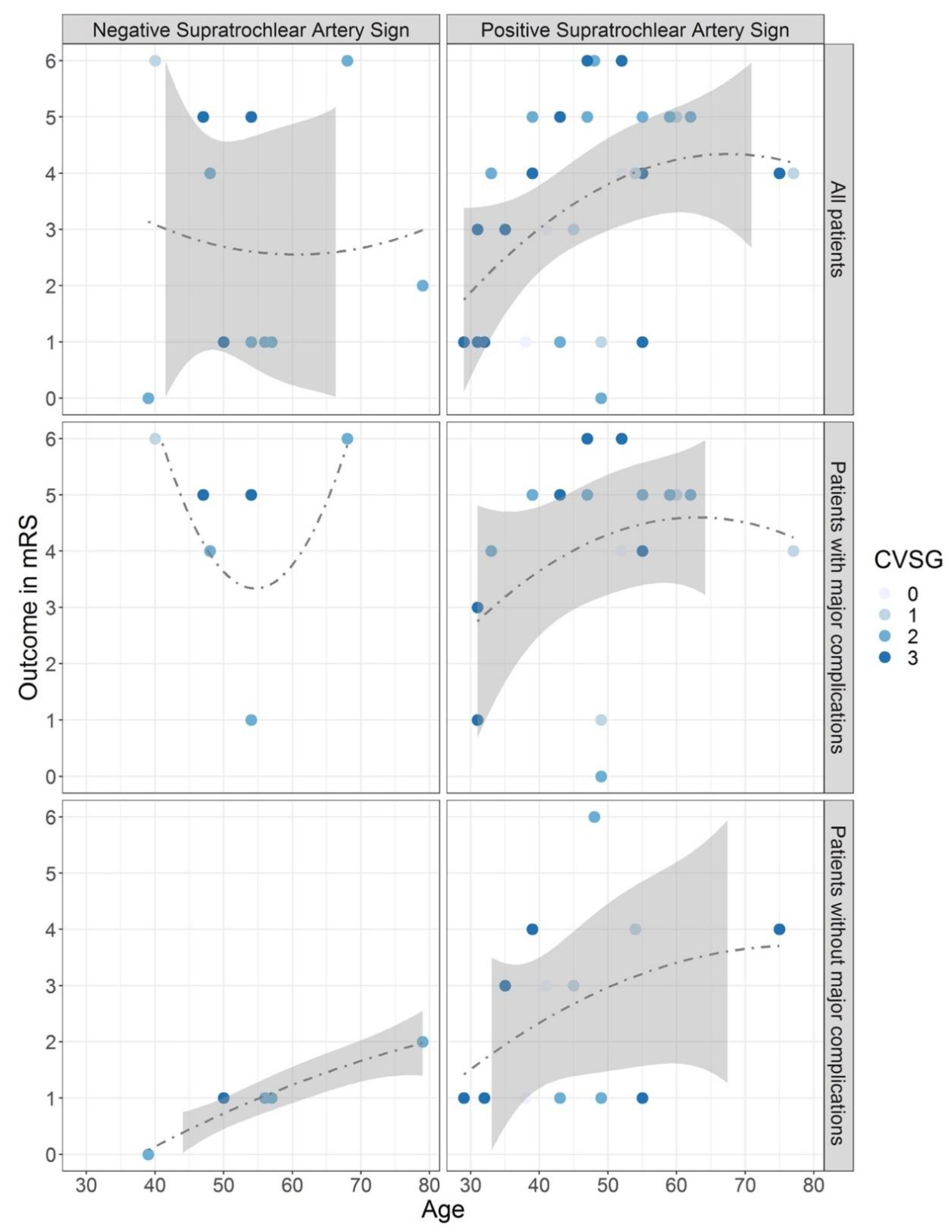
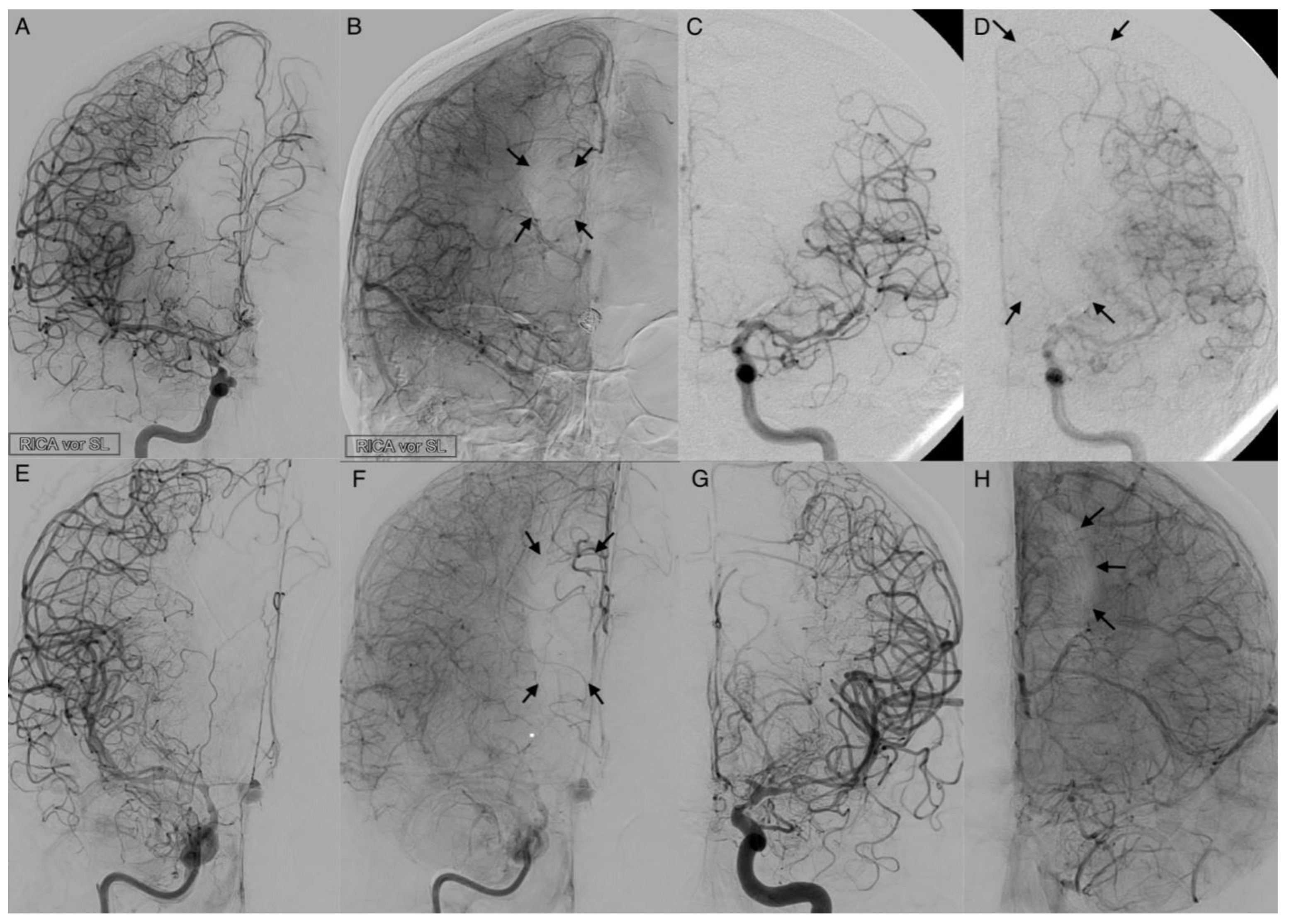
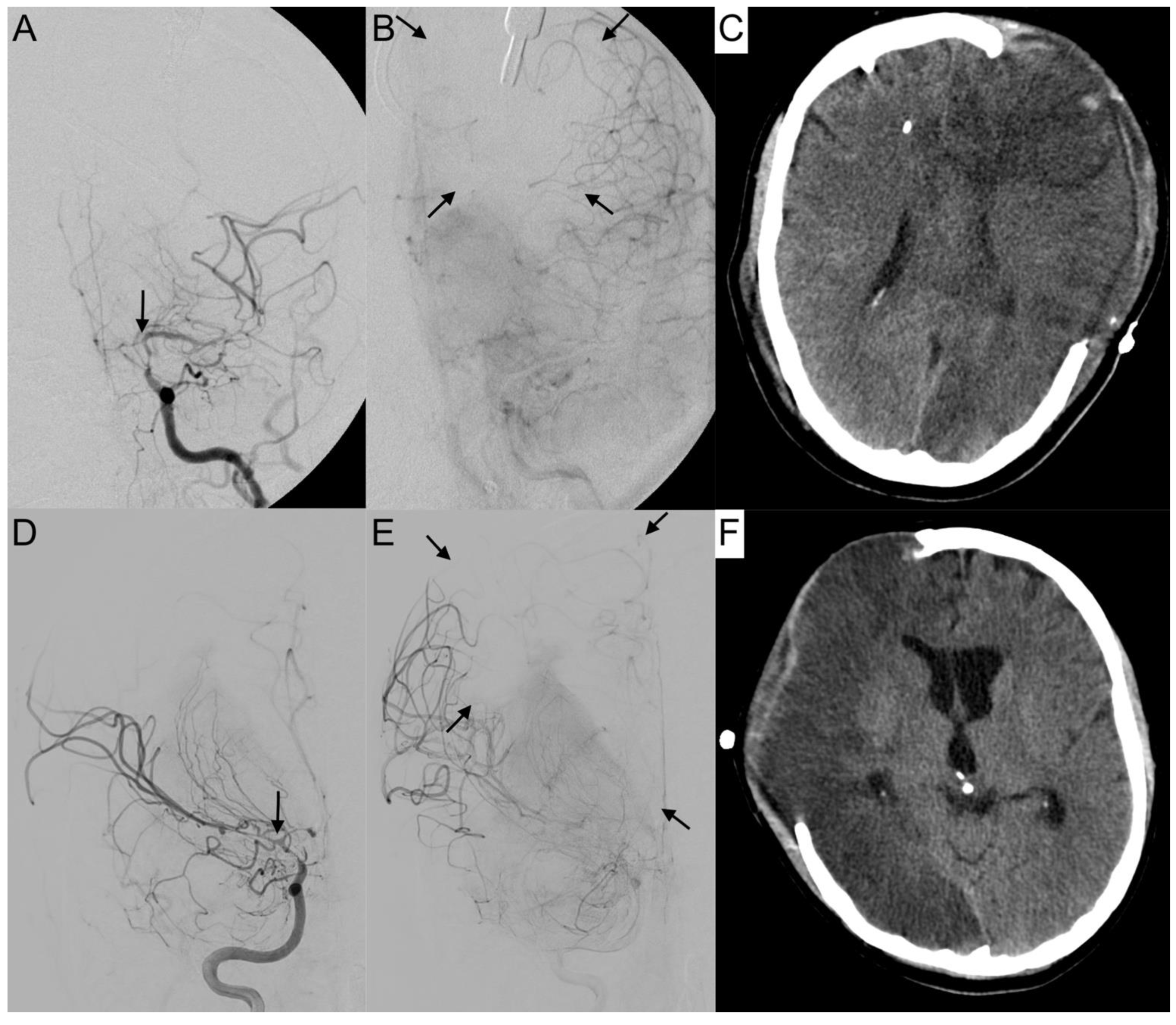
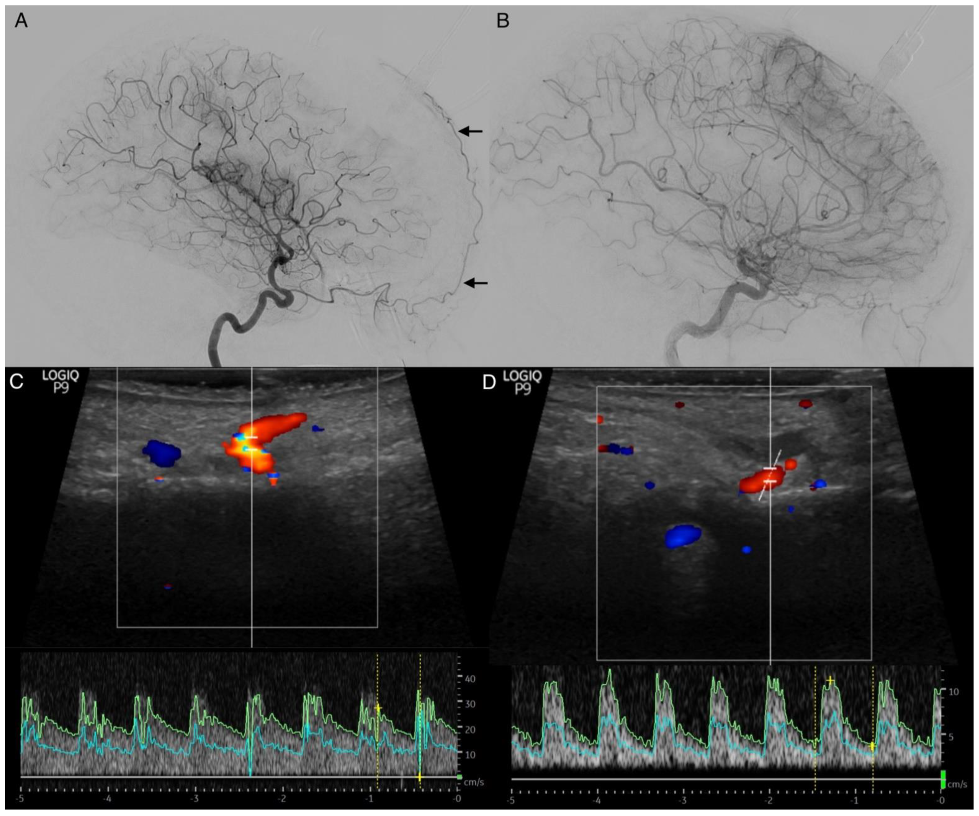
- The supratrochlear artery (STA) is one of the terminal branches of the ophthalmic artery. It pierces the septum orbitale at the medial corner of the orbit, superiorly. This artery supplies the skin, muscles, and the periosteum of the forehead. It anastomoses with the temporal artery and contralateral STA. It can be identified on an ICA angiogram in posterior–anterior (p.a.) and lateral projections. We considered the STA sign positive if it crossed the eyebrow in lateral projections. In p.a. projections, it had to be present at the paramedial zone (in the arterial or parenchymal phase) in parallel course with the A2 segment of the ACA. It can be easily mistaken for the supraorbital artery. The supraorbital artery is the smaller one, which runs laterally, and is often overlayed in p.a. projections. In lateral projections, it crosses the protruding part of the eyebrow (Figure 2D).
- The dorsal nasal artery (DNA) is the second terminal branch of the ophthalmic artery. It pierces the septum orbitale above the medial palpebral ligament to supply the skin on the roof of the nose and lacrimal sac region with the infraorbital artery. It anastomoses with the angular termination of the facial artery and the contralateral DNA. It can be identified with a parallel course to the nose on the ICA angiogram in p.a. projection. We considered the DNA sign positive if there was an artery (in arterial or parenchymal phase) in projection to the nose detectable.
- T-sign: The STA and the DNA branch from the same vessel origin forming a twisted “T” if both vessels are present on the ICA angiogram.
- The anterior falcine artery (AFA) is the meningeal branch of the anterior ethmoidal artery. It supplies the anterior portion of the falx cerebri with blood. The anterior ethmoidal artery arises from the third portion of the ophthalmic artery beneath the superior oblique muscle. It passes through the anterior ethmoidal canal and perforates the cribriform plate. On ICA angiograms, it is only detectable in lateral projections. We considered the AFA sign positive if there was a vessel on the inner side of the skullcap detectable (in the arterial or parenchymal phase).
2.7. Statistical Analysis
3. Results
3.1. Data Collection
3.2. Overall Outcome
3.3. Vessel Signs
3.4. Leptomeningeal Collaterals
4. Discussion
Limitations
5. Conclusions
Author Contributions
Funding
Institutional Review Board Statement
Informed Consent Statement
Data Availability Statement
Conflicts of Interest
References
- Thompson, J.C.; Chalet, F.-X.; Manalastas, E.J.; Hawkins, N.; Sarri, G.; Talbot, D.A. Economic and Humanistic Burden of Cerebral Vasospasm and Its Related Complications after Aneurysmal Subarachnoid Hemorrhage: A Systematic Literature Review. Neurol. Ther. 2022, 11, 597–620. [Google Scholar] [CrossRef] [Scilit] [PubMed]
- Alpers, B.J.; Berry, R.G.; Paddison, R.M. Anatomical Studies of the Circle of Willis in Normal Brain. Arch. Neurol. Psychiatry 1959, 81, 409–418. [Google Scholar] [CrossRef] [Scilit] [PubMed]
- Harrison, M.J.; Marshall, J. The variable clinical and CT findings after carotid occlusion: The role of collateral blood supply. J. Neurol. Neurosurg. Psychiatry 1988, 51, 269–272. [Google Scholar] [CrossRef] [Scilit] [PubMed]
- Hawkins, T.D. The collateral anastomoses in cerebro-vascular occlusion. Clin. Radiol. 1966, 17, 203–219. [Google Scholar] [CrossRef] [Scilit]
- Ginsberg, M.D.; Greenwood, S.A.; Goldberg, H.I. Noninvasive diagnosis of extracranial cerebrovascular disease: Oculoplethysmography-phonoangiography and directional Doppler ultrasonography. Neurology 1979, 29, 623. [Google Scholar] [CrossRef] [Scilit] [PubMed]
- Keller, H.M.; Meier, W.E.; Kumpe, D.A. Noninvasive angiography for the diagnosis of vertebral artery disease using Doppler ultrasound (vertebral artery Doppler). Stroke 1976, 7, 364–369. [Google Scholar] [CrossRef] [Scilit]
- Al-Mufti, F.; Witsch, J.; Manning, N.; Crimmins, M.; Amuluru, K.; Agarwal, S.; Park, S.; Willey, J.Z.; Kamel, H.; Connolly, E.S.; et al. Severity of cerebral vasospasm associated with development of collaterals following aneurysmal subarachnoid hemorrhage. J. NeuroInterv. Surg. 2018, 10, 638–643. [Google Scholar] [CrossRef] [Scilit]
- Fisher, C.M.; Kistler, J.P.; Davis, J.M. Relation of Cerebral Vasospasm to Subarachnoid Hemorrhage Visualized by Computerized Tomographic Scanning. Neurosurgery 1980, 6, 1–9. [Google Scholar] [CrossRef] [Scilit]
- van Swieten, J.C.; Koudstaal, P.J.; Visser, M.C.; Schouten, H.J.; van Gijn, J. Interobserver agreement for the assessment of handicap in stroke patients. Stroke 1988, 19, 604–607. [Google Scholar] [CrossRef] [Scilit]
- Merkel, H.; Lindner, D.; Gaber, K.; Ziganshyna, S.; Jentzsch, J.; Mucha, S.; Gerhards, T.; Sari, S.; Stock, A.; Vothel, F.; et al. Standardized Classification of Cerebral Vasospasm after Subarachnoid Hemorrhage by Digital Subtraction Angiography. J. Clin. Med. 2022, 11, 2011. [Google Scholar] [CrossRef] [Scilit] [PubMed]
- Liebeskind, D.S. Collateral Circulation. Stroke 2003, 34, 2279–2284. [Google Scholar] [CrossRef] [Scilit] [PubMed]
- Jung, S.; Wiest, R.; Gralla, J.; McKinley, R.; Mattle, H.; Liebeskind, D. Relevance of the cerebral collateral circulation in ischaemic stroke: Time is brain, but collaterals set the pace. Swiss Med. Wkly. 2017, 147, w14538. [Google Scholar] [CrossRef] [Scilit] [PubMed]
- Robert, T.; Cicciò, G.; Sylvestre, P.; Chiappini, A.; Weil, A.; Smajda, S.; Chaalala, C.; Blanc, R.; Reinert, M.; Piotin, M.; et al. Anatomic and Angiographic Analyses of Ophthalmic Artery Collaterals in Moyamoya Disease. Am. J. Neuroradiol. 2018, 39, 1121–1126. [Google Scholar] [CrossRef] [Scilit] [PubMed]
- Kwag, H.-J.; Jeong, D.-W.; Lee, S.H.; Kim, D.H.; Kim, J. Intracranial Hemodynamic Changes During Adult Moyamoya Disease Progression. J. Clin. Neurol. 2008, 4, 67–74. [Google Scholar] [CrossRef] [Scilit] [PubMed]
- Rosenkranz, K.; Langer, R.; Felix, R. Transcranial Doppler Sonography: Collateral Pathways in Internal Carotid Artery Obstructions. Angiology 1991, 42, 819–826. [Google Scholar] [CrossRef] [Scilit] [PubMed]
- Menon, B.K.; Smith, E.E.; Coutts, S.B.; Welsh, D.G.; Faber, J.E.; Goyal, M.; Hill, M.D.; Demchuk, A.M.; Damani, Z.; Cho, K.-H.; et al. Leptomeningeal collaterals are associated with modifiable metabolic risk factors. Ann. Neurol. 2013, 74, 241–248. [Google Scholar] [CrossRef] [Scilit] [PubMed]
- Benzel, E.C.; Kesterson, L. Extracranial-intracranial bypass surgery for the management of vasospasm after subarachnoid hemorrhage. Surg. Neurol. 1988, 30, 231–234. [Google Scholar] [CrossRef] [Scilit]
- Batjer, H.; Samson, D. Use of Extracranial-Intracranial Bypass in the Management of Symptomatic Vasospasm. Neurosurgery 1986, 19, 235–346. [Google Scholar] [CrossRef] [Scilit]
- Samson, D.S.; Boone, S. Extracranial-Intracranial (EC-IC) Arterial Bypass: Past performance and current concepts. Neurosurgery 1978, 3, 79–86. [Google Scholar] [CrossRef] [Scilit]
- Pollock, J.A.; Newton, T.H. The Anterior Falx Artery: Normal and Pathologic Anatomy. Radiology 1968, 91, 1089–1095. [Google Scholar] [CrossRef] [Scilit]
- Mount, L.A.; Taveras, J.M. Arteriographic Demonstration of the Collateral Circulation of the Cerebral Hemispheres. Arch. Neurol. Psychiatry 1957, 78, 235–253. [Google Scholar] [CrossRef] [Scilit] [PubMed]
- Vergouwen, M.D.; Vermeulen, M.; van Gijn, J.; Rinkel, G.J.; Wijdicks, E.F.; Muizelaar, J.P.; Mendelow, A.D.; Juvela, S.; Yonas, H.; Terbrugge, K.G.; et al. Definition of Delayed Cerebral Ischemia After Aneurysmal Subarachnoid Hemorrhage as an Outcome Event in Clinical Trials and Observational Studies: Proposal of a multidisciplinary research group. Stroke 2010, 41, 2391–2395. [Google Scholar] [CrossRef] [Scilit]
- Tjerkstra, M.A.; Verbaan, D.; Coert, B.A.; Post, R.; van den Berg, R.; Coutinho, J.M.; Horn, J.; Vandertop, W.P. Large practice variations in diagnosis and treatment of delayed cerebral ischemia after subarachnoid hemorrhage. World Neurosurg. 2022, 160, e412–e420. [Google Scholar] [CrossRef] [Scilit] [PubMed]
- Moftakhar, P.; Cooke, D.L.; Fullerton, H.J.; Ko, N.U.; Amans, M.R.; Narvid, J.A.; Dowd, C.F.; Higashida, R.T.; Halbach, V.V.; Hetts, S.W. Extent of collateralization predicting symptomatic cerebral vasospasm among pediatric patients: Correlations among angiography, transcranial Doppler ultrasonography, and clinical findings. J. Neurosurg. Pediatr. 2015, 15, 282–290. [Google Scholar] [CrossRef] [Scilit] [PubMed]
- Magge, S.N.; Chen, H.I.; Ramakrishna, R.; Cen, L.; Chen, Z.; Elliott, J.P.; Winn, H.R.; Le Roux, P.D. Association of a younger age with an increased risk of angiographic and symptomatic vasospasms following subarachnoid hemorrhage. J. Neurosurg. 2010, 112, 1208–1215. [Google Scholar] [CrossRef] [Scilit] [PubMed]


| Baseline Demographics | Subdivision | Outcome in mRS | |||||||
|---|---|---|---|---|---|---|---|---|---|
| n | 0 | 1 | 2 | 3 | 4 | 5 | 6 | ||
| Overall | 43 | 2 | 13 | 1 | 4 | 8 | 10 | 5 | |
| Fisher Score | 2 | 3 | 1 | 0 | 0 | 1 | 0 | 1 | 0 |
| 3 | 10 | 0 | 5 | 1 | 0 | 2 | 1 | 1 | |
| 4 | 30 | 1 | 8 | 0 | 3 | 6 | 8 | 4 | |
| Hunt and Hess Score | 1 | 5 | 1 | 0 | 0 | 1 | 2 | 1 | 0 |
| 2 | 10 | 0 | 4 | 0 | 2 | 0 | 2 | 2 | |
| 3 | 13 | 1 | 4 | 1 | 0 | 3 | 3 | 1 | |
| 4 | 7 | 0 | 4 | 0 | 0 | 1 | 1 | 1 | |
| 5 | 7 | 0 | 1 | 0 | 1 | 2 | 2 | 1 | |
| Localization of aneurysm | AComA | 0 | 1 | 4 | 0 | 3 | 2 | 1 | 0 |
| ACA | 0 | 0 | 1 | 0 | 0 | 1 | 1 | 1 | |
| MCA | 0 | 1 | 4 | 0 | 1 | 3 | 4 | 2 | |
| ICA/PComA | 0 | 0 | 4 | 1 | 0 | 1 | 1 | 0 | |
| VA/BA | 0 | 0 | 0 | 0 | 0 | 0 | 1 | 2 | |
| none | 0 | 0 | 0 | 0 | 0 | 1 | 0 | 0 | |
| Clipping | 5 | 0 | 1 | 0 | 0 | 1 | 1 | 2 | |
| Endovascular procedure | 35 | 2 | 12 | 1 | 3 | 6 | 7 | 3 | |
| Combined therapy | 3 | 0 | 0 | 0 | 1 | 0 | 2 | 0 | |
| Vessel signs | |||||||||
| Positive supratrochlear artery sign | at time of admission | 4 | 0 | 2 | 1 | 1 | 0 | 0 | 0 |
| (STA) | during vasospasm | 31 | 1 | 8 | 0 | 4 | 7 | 8 | 3 |
| Positive dorsal nasal artery sign (DNA) | at time of admission | 13 | 0 | 6 | 0 | 1 | 3 | 2 | 1 |
| during vasospasm | 35 | 1 | 11 | 0 | 4 | 7 | 8 | 4 | |
| Positive anterior falcine artery sign (AFA) | at time of admission | 9 | 0 | 4 | 0 | 1 | 1 | 3 | 0 |
| during vasospasm | 31 | 1 | 10 | 0 | 4 | 5 | 7 | 4 | |
| Leptomingeal collateral activation | non-activated | 25 | 2 | 7 | 1 | 2 | 5 | 5 | 3 |
| activated | 16 | 0 | 6 | 0 | 1 | 3 | 4 | 2 | |
| collapsed | 2 | 0 | 0 | 0 | 1 | 0 | 1 | 0 | |
| Outcome-limiting factors | |||||||||
| Intracerebral hemorrhage | 13 | 0 | 3 | 0 | 1 | 3 | 5 | 1 | |
| Non-DCI-related infarctions | 9 | 1 | 3 | 0 | 2 | 2 | 1 | 0 | |
| Additional severe co-morbidity | 15 | 1 | 0 | 0 | 0 | 4 | 8 | 2 | |
| DCI-related infarctions | 33 | 1 | 8 | 1 | 3 | 8 | 7 | 5 | |
| Lacunar infarction, size ≤ 15 mm | 2 | 0 | 2 | 0 | 0 | 0 | 0 | 0 | |
| Small territorial infarction (≤1/3 of area at risk) | 18 | 1 | 6 | 1 | 2 | 4 | 3 | 1 | |
| Large territorial infarction | 9 | 0 | 0 | 0 | 0 | 2 | 3 | 4 | |
| Subtotal hemispheric infarction | 2 | 0 | 0 | 0 | 1 | 0 | 1 | 0 | |
| Unilateral infarctions | 23 | 1 | 6 | 1 | 1 | 7 | 5 | 2 | |
| Bilateral infarctions | 10 | 0 | 2 | 2 | 2 | 1 | 2 | 3 | |
| Vertebrobasilar infarctions | 8 | 0 | 0 | 1 | 1 | 2 | 1 | 3 | |
| Number of affected vessel territories | 0 | 10 | 1 | 5 | 0 | 1 | 0 | 3 | 0 |
| 1 | 14 | 1 | 6 | 0 | 0 | 4 | 2 | 1 | |
| 2 | 8 | 0 | 2 | 1 | 1 | 3 | 1 | 0 | |
| 3 | 4 | 0 | 0 | 0 | 1 | 0 | 3 | 0 | |
| 4 | 5 | 0 | 0 | 0 | 1 | 1 | 1 | 2 | |
| 6 | 2 | 0 | 0 | 0 | 0 | 0 | 0 | 2 | |
| Positive Vessel Sign | CVSG on Admission | ||||
| All | 0 | 1 | 2 | 3 | |
| Overall (%, n) | 100 (43) | 51.2 (22) | 23.3 (10) | 14.0 (6) | 11.6 (5) |
| STA (%, n) | 9.3 (4) | 4.7 (2) | 4.7 (2) | 0.0 (0) | 0.0 (0) |
| DNA (%, n) | 30.2 (13) | 20.9 (9) | 7.0 (3) | 0.0 (0) | 2.3 (1) |
| AFA (%, n) | 25.7 (9) | 14.3 (5) | 2.8 (3) | 2.9 (1) | 0.0 (0) |
| Positive Vessel Sign | Early Angiographic Vasospasm in CVSG (<72 h) | ||||
| n | 0 | 1 | 2 | 3 | |
| Overall (%, n) | 100 (43) | 0.0 (0) | 14.0 (6) | 4.7 (2) | 4.7 (2) |
| STA (%, n) | 2.3 (1) | 0.0 (0) | 2.3 (1) | 0.0 (0) | 0.0 (0) |
| DNA (%, n) | 4.7 (2) | 0.0 (0) | 2.3 (1) | 0.0 (0) | 2.3 (1) |
| AFA (%, n) | 8.6 (3) | 0.0 (0) | 8.6 (3) | 0.0 (0) | 0.0 (0) |
| Positive Vessel Sign | Highest CVSG | ||||
| n | 0 | 1 | 2 | 3 | |
| Overall (%, n) | 100 (43) | 4.7 (2) | 7.0 (3) | 44.2 (19) | 44.2 (19) |
| STA (%, n) | 72.1 (31) | 4.7 (2) | 4.7 (2) | 27.9 (12) | 34.9 (15) |
| DNA (%, n) | 81.4 (35) | 4.7 (2) | 7.0 (3) | 30.2 (13) | 39.5 (17) |
| AFA (%, n) | 72.1 (31) | 5.7 (2) | 5.7 (2) | 37.1 (13) | 40.0 (14) |
| Rating | Functional Outcome | Delayed Cerebral Ischemia | ||||||
|---|---|---|---|---|---|---|---|---|
| Positive Sign (%, n) | Negative Sign (%, n) | PPV (%) | NPV (%) | p-Value | PPV (%) | NPV (%) | p-Value | |
| Overall | 100 (43) | |||||||
| Supratrochlear artery | 72.1 (31) | 27.9 (12) | 78.3 | 35.0 | 0.497 | 78.8 | 50.0 | 0.110 |
| Dorsal nasal artery | 81.4 (35) | 18.6 (8) | 82.6 | 20.0 | 1.000 | 78.8 | 10.0 | 0.656 |
| Anterior falcine artery | 88.6 (31) | 11.4 (4) | 94.1 | 16.7 | 0.602 | 88.9 | 12.5 | 1.000 |
| Patients < 50 years | 53.5 (23) | |||||||
| Supratrochlear artery | 82.6 (19) | 17.4 (4) | 70.0 | 7.7 | 0.281 | 88.9 | 40.0 | 0.194 |
| Dorsal nasal artery | 82.6 (19) | 17.4 (4) | 80.0 | 15.4 | 1.000 | 83.3 | 25.0 | 1.000 |
| Anterior falcine artery | 85.7 (18) | 14.3 (3) | 88.9 | 16.7 | 1.000 | 87.5 | 20.0 | 1.000 |
| Patients ≥ 50 years | 46.5 (20) | |||||||
| Supratrochlear artery | 60.0(12) | 40.0 (8) | 84.6 | 85.7 | 0.004 | 66.0 | 60.0 | 0.347 |
| Dorsal nasal artery | 80.0 (16) | 20.0 (4) | 84.6 | 28.6 | 0.587 | 73.3 | 0.0 | 0.530 |
| Anterior falcine artery | 92.9 (13) | 7.1 (1) | 100.0 | 20.0 | 0.428 | 90.9 | 0.0 | 1.000 |
Publisher’s Note: MDPI stays neutral with regard to jurisdictional claims in published maps and institutional affiliations. |
© 2022 by the authors. Licensee MDPI, Basel, Switzerland. This article is an open access article distributed under the terms and conditions of the Creative Commons Attribution (CC BY) license (https://creativecommons.org/licenses/by/4.0/).
Share and Cite
Richter, C.; Werdehausen, R.; Jentzsch, J.; Lindner, D.; Gerhards, T.; Hantel, T.; Gaber, K.; Schob, S.; Saur, D.; Quäschling, U.; et al. The Supratrochlear Artery Sign—A New Piece in the Puzzle of Cerebral Vasospasm. Diagnostics 2022, 12, 2185. https://doi.org/10.3390/diagnostics12092185
Richter C, Werdehausen R, Jentzsch J, Lindner D, Gerhards T, Hantel T, Gaber K, Schob S, Saur D, Quäschling U, et al. The Supratrochlear Artery Sign—A New Piece in the Puzzle of Cerebral Vasospasm. Diagnostics. 2022; 12(9):2185. https://doi.org/10.3390/diagnostics12092185
Chicago/Turabian StyleRichter, Cindy, Robert Werdehausen, Jennifer Jentzsch, Dirk Lindner, Thilo Gerhards, Torsten Hantel, Khaled Gaber, Stefan Schob, Dorothee Saur, Ulf Quäschling, and et al. 2022. "The Supratrochlear Artery Sign—A New Piece in the Puzzle of Cerebral Vasospasm" Diagnostics 12, no. 9: 2185. https://doi.org/10.3390/diagnostics12092185
APA StyleRichter, C., Werdehausen, R., Jentzsch, J., Lindner, D., Gerhards, T., Hantel, T., Gaber, K., Schob, S., Saur, D., Quäschling, U., Hoffmann, K.-T., Ziganshyna, S., & Halama, D. (2022). The Supratrochlear Artery Sign—A New Piece in the Puzzle of Cerebral Vasospasm. Diagnostics, 12(9), 2185. https://doi.org/10.3390/diagnostics12092185