Case Report

Dentigerous Cysts with Diverse Radiological Presentation Highlighting Diagnostic Challenges
1
Unit of Oral Surgery and Implantology, Division of Oral and Maxillofacial Surgery, Department of Surgery, University of Geneva & University Hospitals of Geneva, 1205 Geneva, Switzerland
2
Division of Radiology, Diagnostic Department, Geneva University Hospitals, University of Geneva, 1205 Geneva, Switzerland
3
Unit of Oral Medicine and Oral Maxillofacial Pathology, Division of Oral and Maxillofacial Surgery, Department of Surgery, University of Geneva & University Hospitals of Geneva, 1205 Geneva, Switzerland
*
Author to whom correspondence should be addressed.
Diagnostics 2022, 12(8), 2006; https://doi.org/10.3390/diagnostics12082006

Submission received: 25 July 2022 / Revised: 16 August 2022 / Accepted: 16 August 2022 / Published: 19 August 2022
(This article belongs to the Topic Diagnostic Imaging in Oral and Maxillofacial Diseases)

Abstract

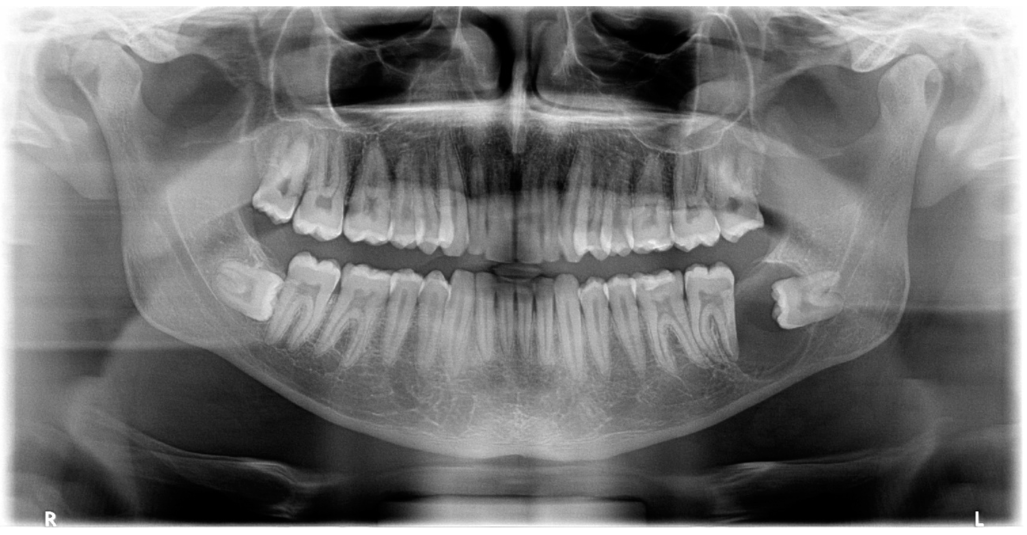
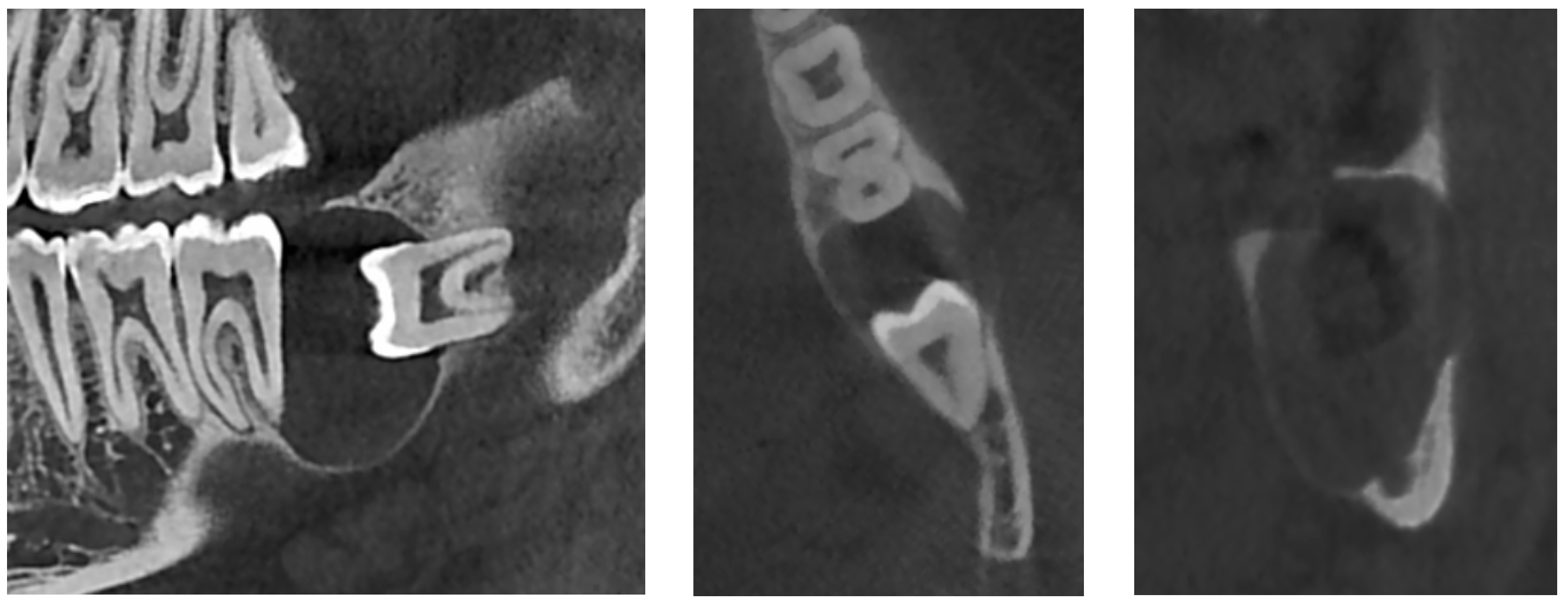
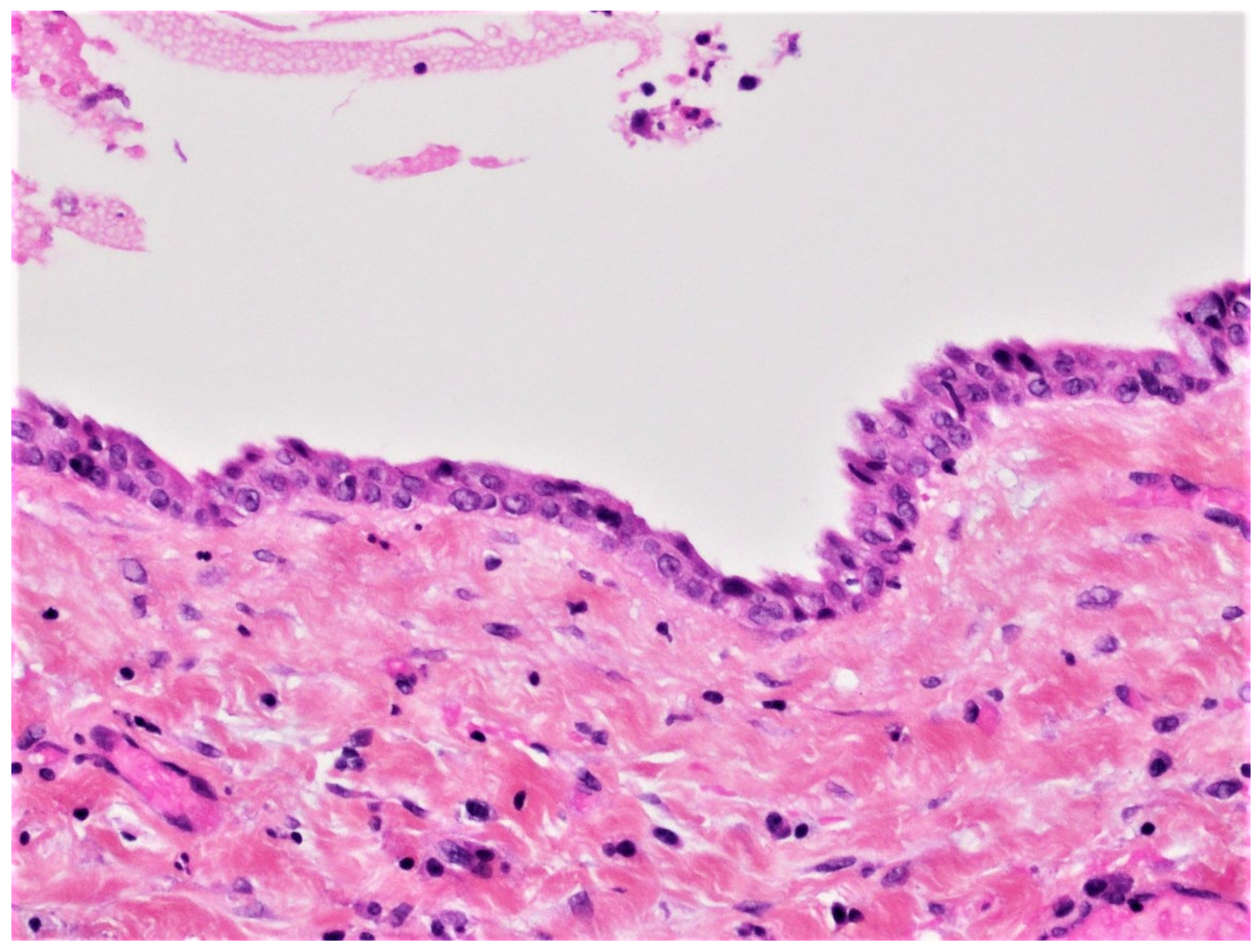
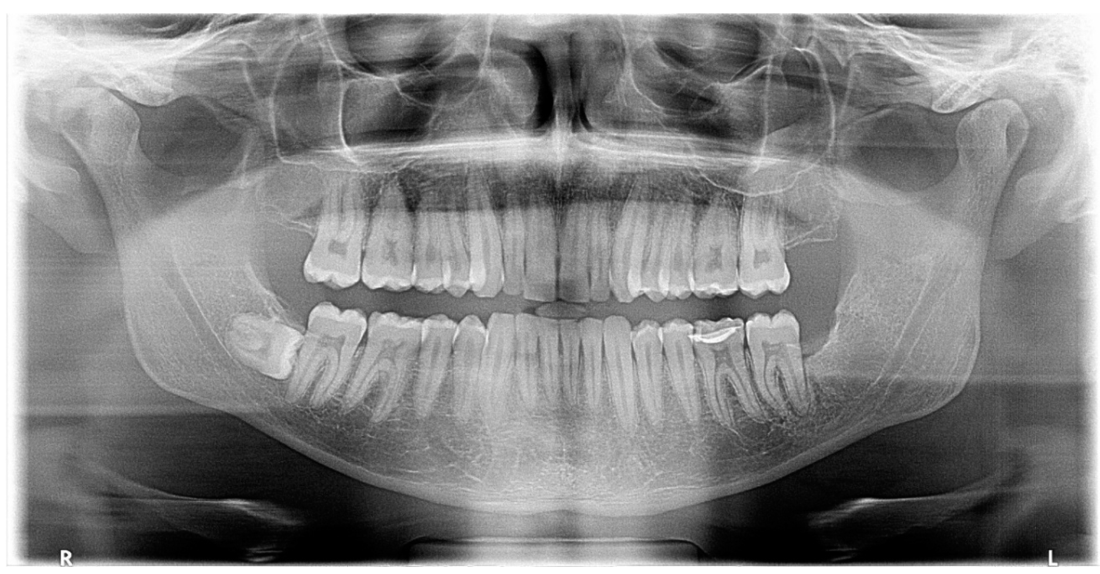
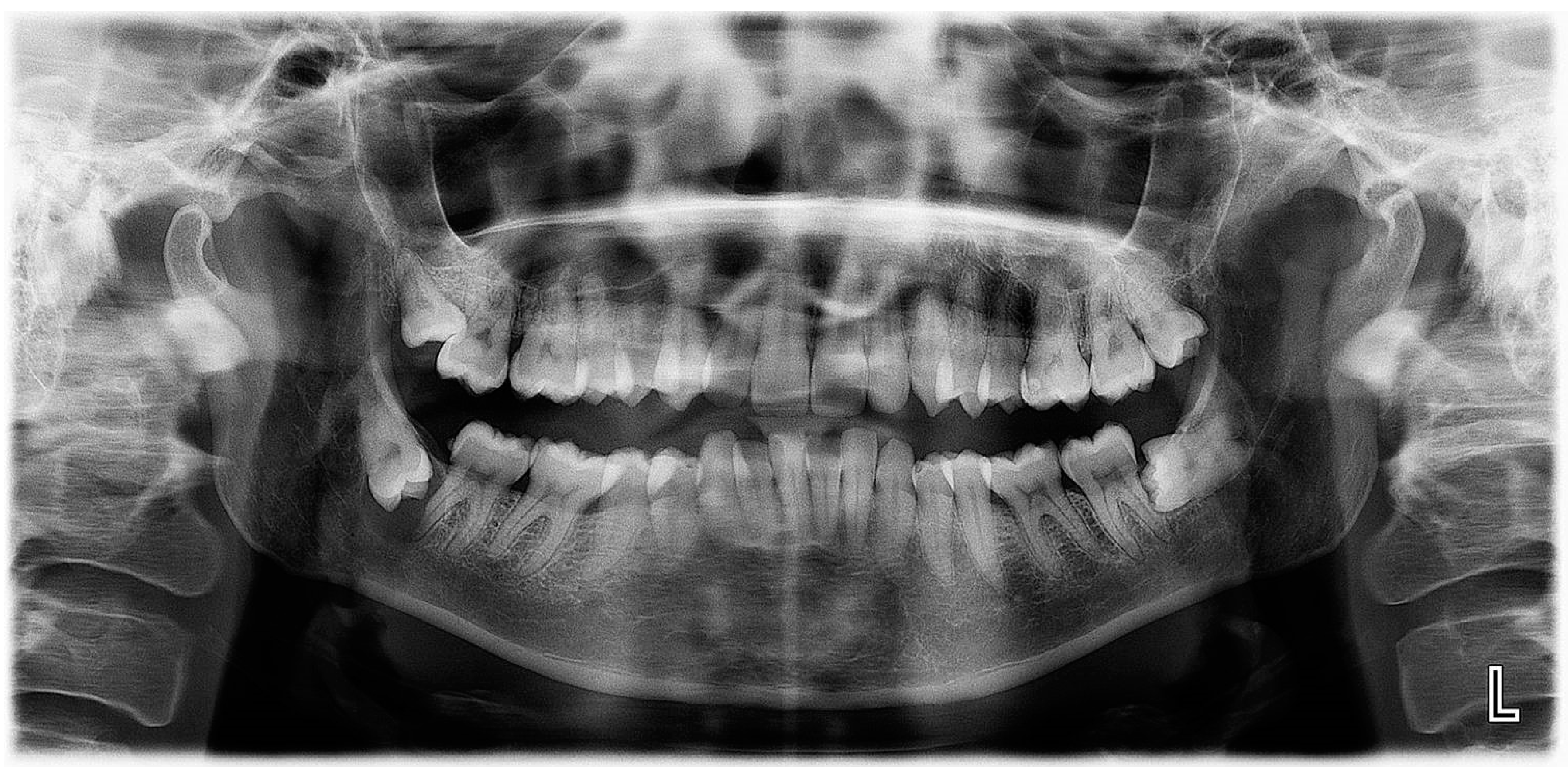
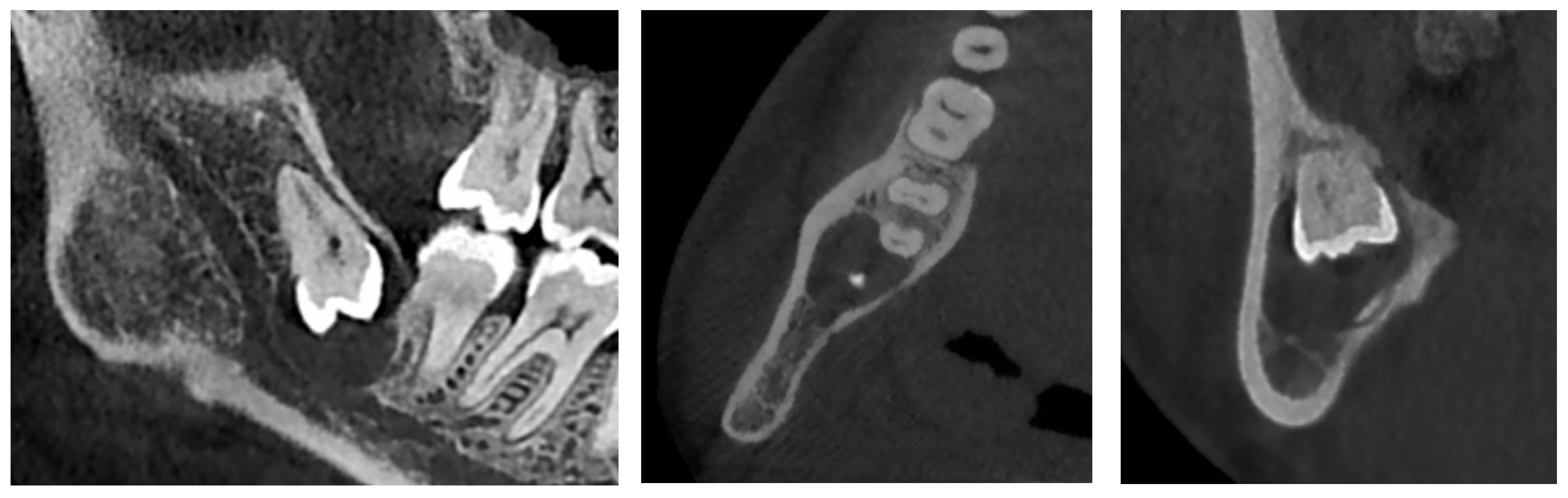
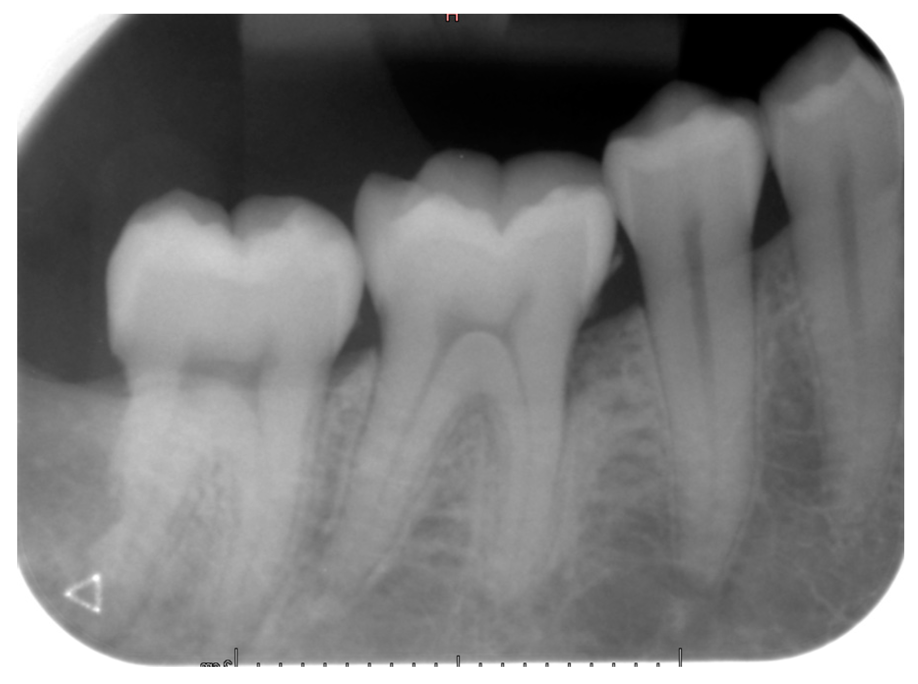
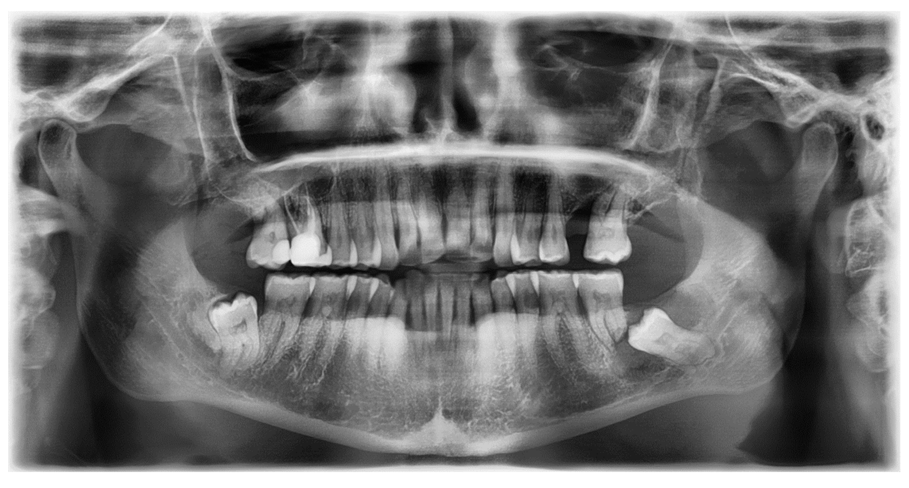
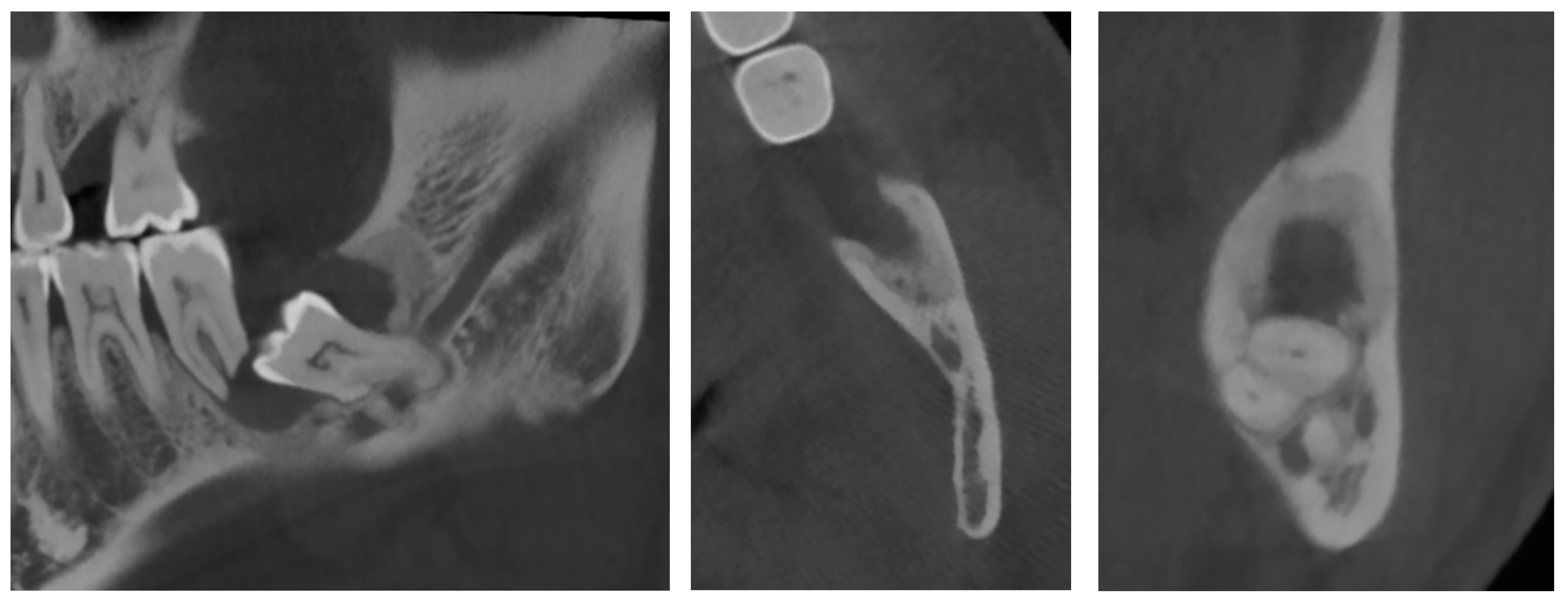
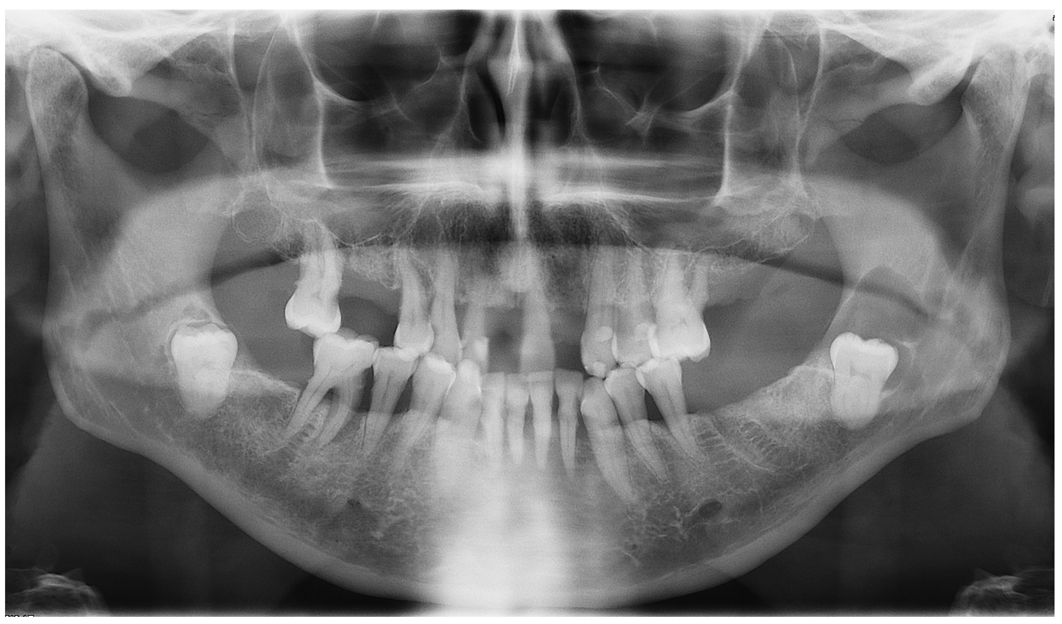
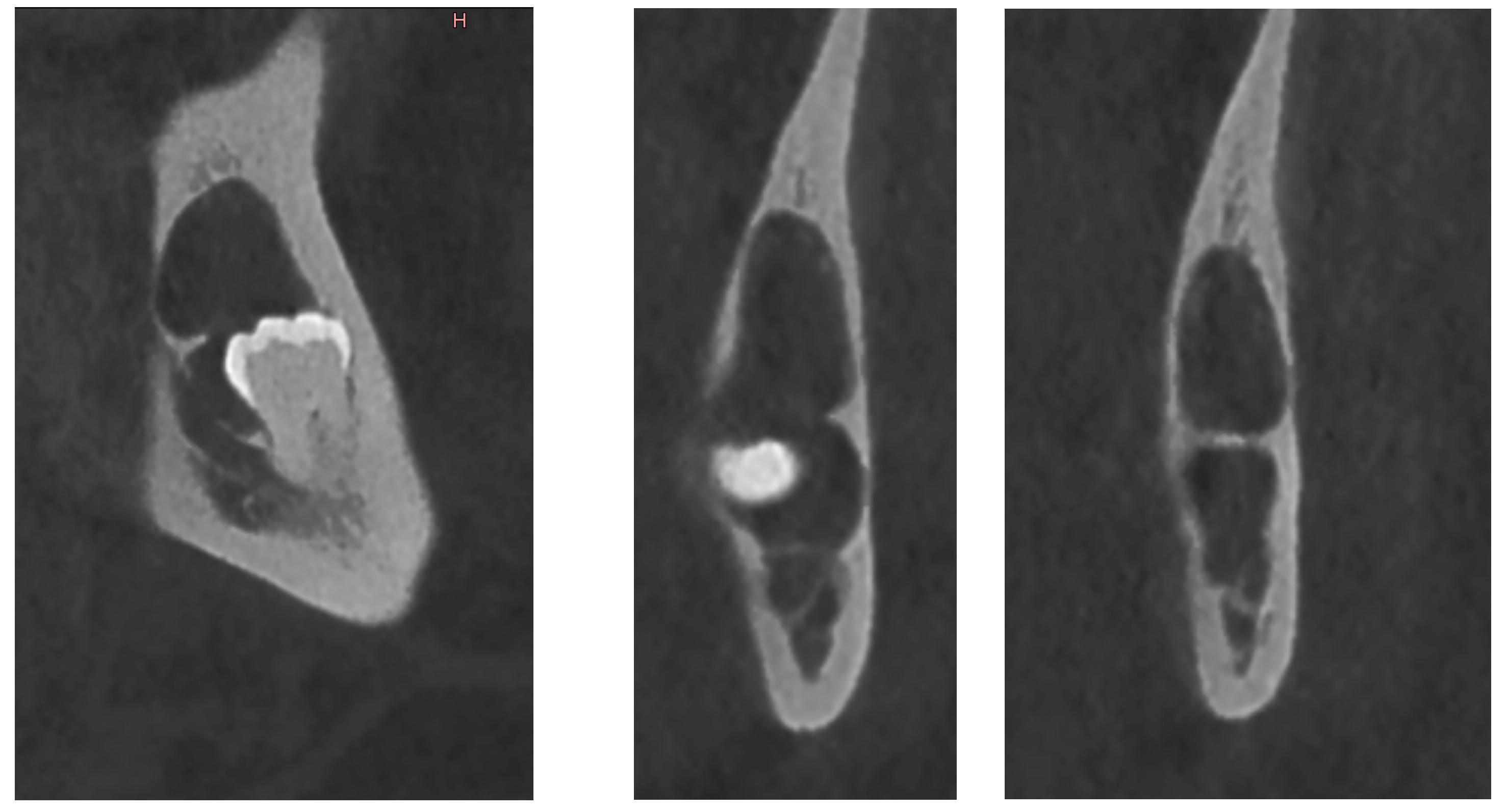
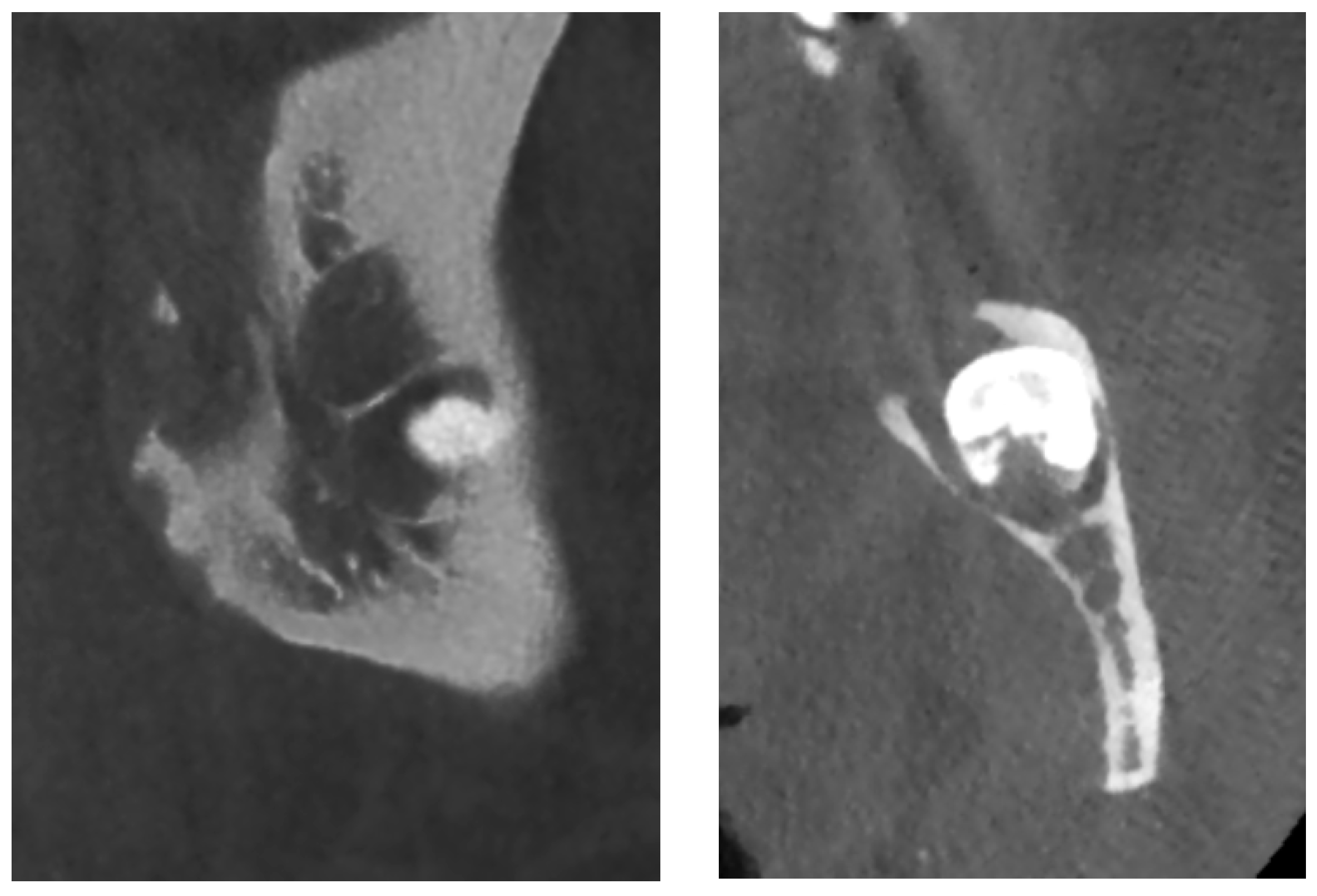
Dentigerous cyst is an odontogenic developmental cyst arising from the pericoronal tissue of an impacted tooth, and that may exhibit various radiological aspects. The aim of this article is to present four cases of histologically confirmed mandibular dentigerous cysts to highlight diverse radiological presentations: one of classical appearance (well-limited unilocular radiolucent lesion surrounding the crown) and three which have shown radiological peculiarities (one cyst displacing the adjacent tooth, with bone but no root resorption, one cyst presenting hallmarks of infection and one multilocular cyst with thin septa). Such radiologic diversity may, on occasion, suggest a clinical aggressive lesion such as an odontogenic keratocyst or ameloblastoma. The diagnosis of dentigerous cyst requires a thorough evaluation of the clinical presentation and accurate radiological studies.
Keywords: odontogenic cysts; dentigerous cyst; jaws; oral surgery; oral pathology; imaging; OPG; CBCT odontogenic cysts; dentigerous cyst; jaws; oral surgery; oral pathology; imaging; OPG; CBCT