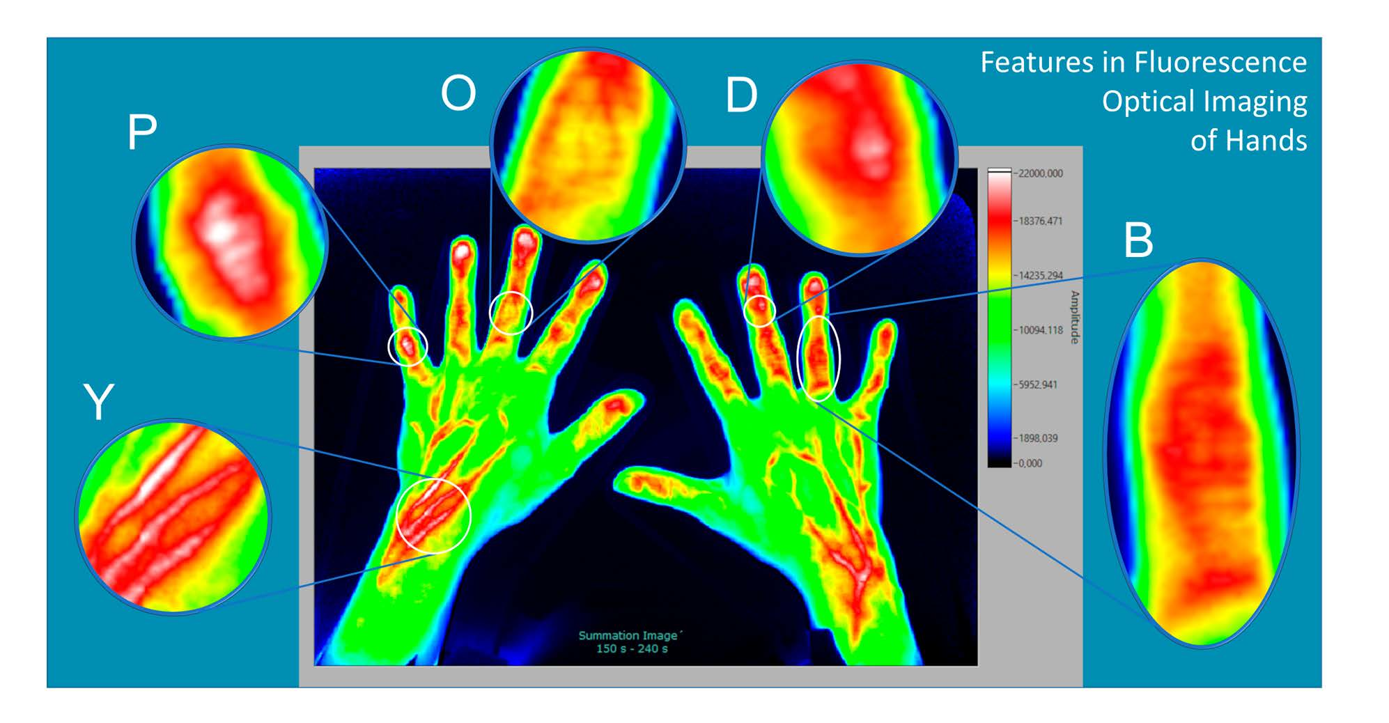
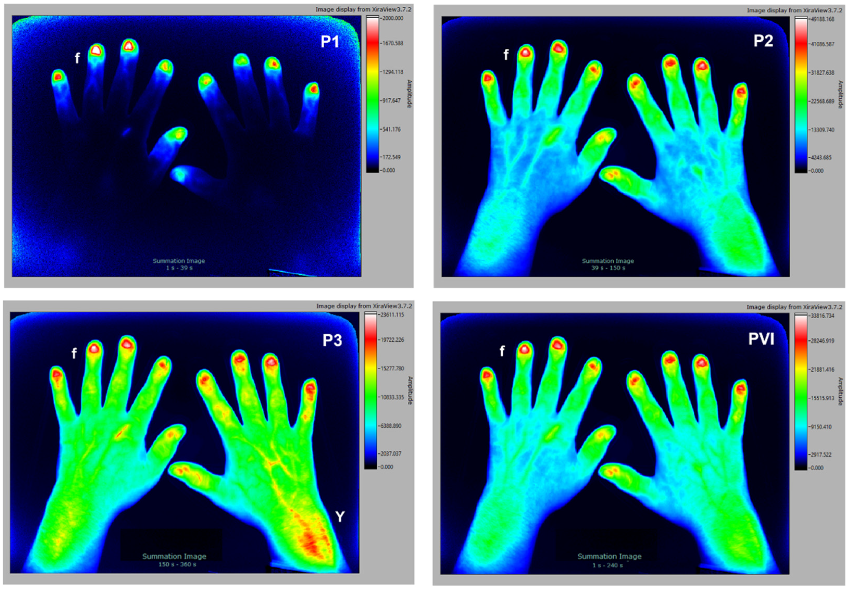
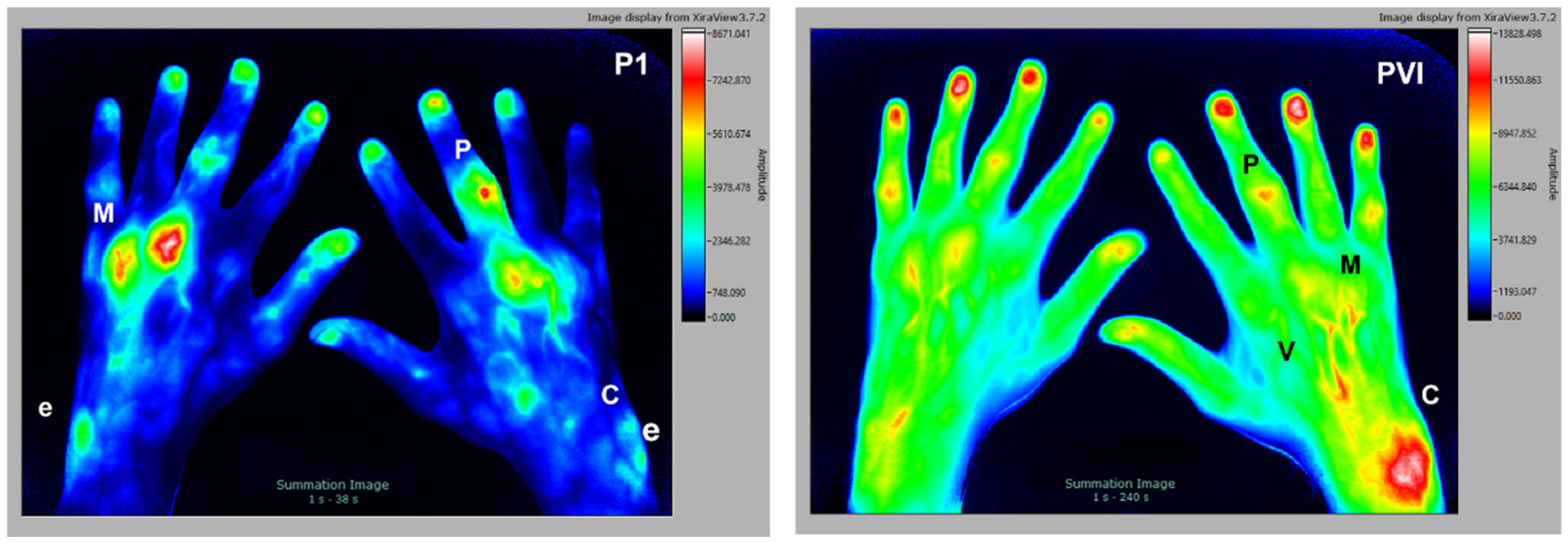
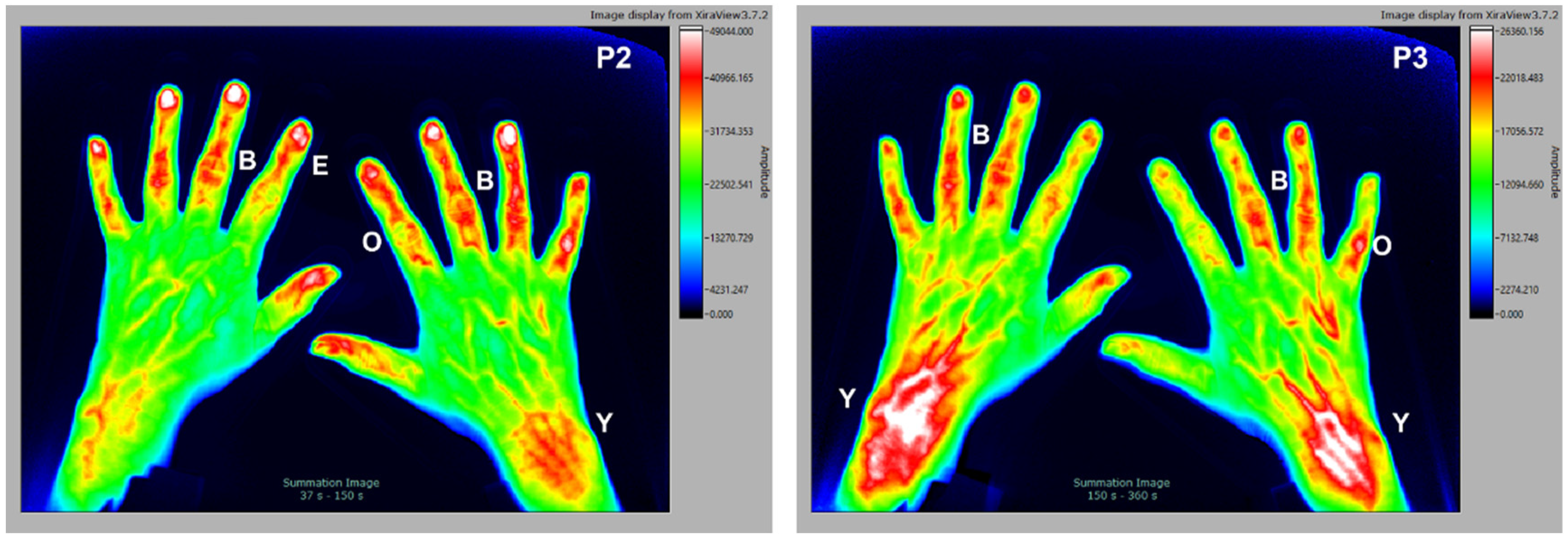
Features Found in Indocyanine Green-Based Fluorescence Optical Imaging of Inflammatory Diseases of the Hands
 ,
, Abstract
Share and Cite
Gedat, E.; Berger, J.; Kiesel, D.; Failli, V.; Briel, A.; Welker, P. Features Found in Indocyanine Green-Based Fluorescence Optical Imaging of Inflammatory Diseases of the Hands. Diagnostics 2022, 12, 1775. https://doi.org/10.3390/diagnostics12081775
Gedat E, Berger J, Kiesel D, Failli V, Briel A, Welker P. Features Found in Indocyanine Green-Based Fluorescence Optical Imaging of Inflammatory Diseases of the Hands. Diagnostics. 2022; 12(8):1775. https://doi.org/10.3390/diagnostics12081775
Chicago/Turabian StyleGedat, Egbert, Jörn Berger, Denise Kiesel, Vieri Failli, Andreas Briel, and Pia Welker. 2022. "Features Found in Indocyanine Green-Based Fluorescence Optical Imaging of Inflammatory Diseases of the Hands" Diagnostics 12, no. 8: 1775. https://doi.org/10.3390/diagnostics12081775
APA StyleGedat, E., Berger, J., Kiesel, D., Failli, V., Briel, A., & Welker, P. (2022). Features Found in Indocyanine Green-Based Fluorescence Optical Imaging of Inflammatory Diseases of the Hands. Diagnostics, 12(8), 1775. https://doi.org/10.3390/diagnostics12081775