


Cytological Diagnosis of Pancreatic Solid-Pseudopapillary Neoplasm: A Single-Institution Community Practice Experience

Abstract
1. Introduction
2. Materials and Methods
3. Results
4. Discussions
5. Conclusions
Author Contributions
Funding
Institutional Review Board Statement
Informed Consent Statement
Data Availability Statement
Conflicts of Interest
References
- Klöppel, G.; Basturk, O.; Klimstra, D.S.; Lam, A.K.; Notohara, K. Solid pseudopapillary neoplasm of the pancreas. In WHO Classification of Tumors: Digestive System Tumours, 5th ed.; WHO Classification of Tumours Editorial Board; IARC Press: Lyon, France, 2019; Volume 1, pp. 340–342. [Google Scholar]
- Hruban, R.H.; Pitman, M.B.; Klimstra, D.S. Solid-pseudopapillary neoplasms. In Atlas of Tumor Pathology, Series 4: Tumors of the Pancreas; Hruban, R.H., Pitman, M.B., Klimstra, D.S., Eds.; Armed Forces Institute of Pathology: Washington, DC, USA, 2007; pp. 231–250. [Google Scholar]
- Klimstra, D.S.; Wenig, B.M.; Heffess, C.S. Solid-pseudopapillary tumor of the pancreas: A typically cystic carcinoma of low malignant potential. Semin. Diagn. Pathol. 2000, 17, 66–80. [Google Scholar] [PubMed]
- Dinarvand, P.; Lai, J. Solid pseudopapillary neoplasm of the pancreas: A rare entity with unique features. Arch. Pathol. Lab. Med. 2017, 141, 990–995. [Google Scholar] [CrossRef] [Scilit]
- La Rosa, S.; Bongiovanni, M. Pancreatic solid pseudopapillary neoplasm: Key pathologic and genetic features. Arch. Pathol. Lab. Med. 2020, 144, 829–837. [Google Scholar] [CrossRef] [Scilit]
- Estrella, J.S.; Li, L.; Rashid, A.; Wang, H.; Katz, M.H.; Fleming, J.B.; Abbruzzese, J.L.; Wang, H. Solid pseudopapillary neoplasm of the pancreas: Clinicopathologic and survival analyses of 64 cases from a single institution. Am. J. Surg. Pathol. 2014, 38, 147–157. [Google Scholar] [CrossRef] [Scilit]
- Pelosi, G.; Iannucci, A.; Zamboni, G.; Bresaola, E.; Iacono, C.; Serio, G. Solid and cystic papillary neoplasm of the pancreas: A clinico-cytopathologic and immunocytochemical study of five new cases diagnosed by fine-needle aspiration cytology and a review of the literature. Diagn. Cytopathol. 1995, 13, 233–246. [Google Scholar] [CrossRef] [Scilit] [PubMed]
- Pettinato, G.; Di Vizio, D.; Manivel, J.C.; Pambuccian, S.E.; Somma, P.; Insabato, L. Solid-pseudopapillary tumor of the pancreas: A neoplasm with distinct and highly characteristic cytological features. Diagn. Cytopathol. 2002, 27, 325–334. [Google Scholar] [CrossRef] [Scilit]
- Bardales, R.H.; Centeno, B.; Mallery, J.S.; Lai, R.; Pochapin, M.; Guiter, G.; Stanley, M.W. Endoscopic ultrasound-guided fine-needle aspiration cytology diagnosis of solid-pseudopapillary tumor of the pancreas: A rare neoplasm of elusive origin but characteristic cytomorphologic features. Am. J. Clin. Pathol. 2004, 121, 654–662. [Google Scholar] [CrossRef]
- Jhala, N.; Siegal, G.P.; Jhala, D. Large, clear cytoplasmic vacuolation: An under-recognized cytologic clue to distinguish solid pseudopapillary neoplasms of the pancreas from pancreatic endocrine neoplasms on fine-needle aspiration. Cancer 2008, 114, 249–254. [Google Scholar] [CrossRef] [Scilit] [PubMed]
- Samad, A.; Shah, A.A.; Stelow, E.B.; Alsharif, M.; Cameron, S.E.H.; Pambuccian, S.E. Cercariform cells: Another cytologic feature distinguishing solid pseudopapillary neoplasms from pancreatic endocrine neoplasms and acinar cell carcinomas in endoscopic ultrasound-guided fine-needle aspirates. Cancer Cytopathol. 2012, 121, 298–310. [Google Scholar] [CrossRef] [Scilit]
- Burford, H.; Baloch, Z.; Liu, X.; Jhala, D.; Siegal, G.P.; Jhala, N. E-cadherin/β-catenin and CD10: A limited immunohistochemical panel to distinguish pancreatic endocrine neoplasm from solid pseudopapillary neoplasm of the pancreas on endoscopic ultrasound-guided fine-needle aspirates of the pancreas. Am. J. Clin. Pathol. 2009, 132, 831–839. [Google Scholar] [CrossRef] [Scilit]
- Bal, M.M.; Deodhar, K.; Shrikhande, S.; Shukla, P.; Arya, S.; Ramadwar, M. Solid pseudopapillary tumor of the pancreas: “Experiences” and “Lessons” at a tertiary-care oncology center. Diagn. Cytopathol. 2012, 41, 599–606. [Google Scholar] [CrossRef] [Scilit]
- Misra, S.; Saran, R.K.; Dm, S.S.; Barman, S.; Dahale, A. Utility of cytomorphology in distinguishing solid pseudopapillary neoplasm of pancreas from pancreatic neuroendocrine tumor with emphasis on nuclear folds and nuclear grooves. Diagn. Cytopathol. 2019, 47, 531–540. [Google Scholar] [CrossRef] [Scilit] [PubMed]
- Singh, P.; Kumar, P.; Rohilla, M.; Gupta, P.; Gupta, N.; Dey, P.; Srinivasan, R.; Rajwanshi, A.; Nada, R. Fine needle aspiration cytology with the aid of immunocytochemistry on cell-block confirms the diagnosis of solid pseudopapillary neoplasm of the pancreas. Cytopathology 2020, 32, 57–64. [Google Scholar] [CrossRef] [Scilit]
- Wu, J.; Mao, Y.; Jiang, Y.; Song, Y.; Yu, P.; Sun, S.; Li, S. Sex differences in solid pseudopapillary neoplasm of the pancreas: A population-based study. Cancer Med. 2020, 9, 6030–6041. [Google Scholar] [CrossRef] [Scilit] [PubMed]
- Lanke, G.; Ali, F.S.; Lee, J.H. Clinical update on the management of pseudopapillary tumor of pancreas. World J. Gastrointest. Endosc. 2018, 10, 145–155. [Google Scholar] [CrossRef] [Scilit]
- Ardengh, J.C.; Lopes, C.V.; Venco, F.E.; Machado, M.A. Diagnosis of pancreatic solid pseudopapillary neoplasms using cell-blocks and immunohistochemical evaluation of endoscopic ultrasound-guided fine needle aspiration biopsy specimens. Cytopathology 2020, 32, 50–56. [Google Scholar] [CrossRef] [Scilit]
- Albores-Saavedra, J.; Simpson, K.W.; Bilello, S.J. The clear cell variant of solid pseudopapillary tumor of the pancreas: A previously unrecognized pancreatic neoplasm. Am. J. Surg. Pathol. 2006, 30, 1237–1242. [Google Scholar] [CrossRef] [Scilit]
- Zhao, P.; Debrito, P.; Ozdemirli, M.; Sidawy, M.K. Solid-pseudopapillary neoplasm of the pancreas: Awareness of unusual clinical presentations and morphology of the clear cell variant can prevent diagnostic errors. Diagn. Cytopathol. 2013, 41, 889–895. [Google Scholar] [CrossRef] [Scilit] [PubMed]
- Tanaka, Y.; Kato, K.; Notohara, K.; Hojo, H.; Ijiri, R.; Miyake, T.; Nagahara, N.; Sasaki, F.; Kitagawa, N.; Nakatani, Y.; et al. Frequent beta-catenin mutation and cytoplasmic/nuclear accumulation in pancreatic solid-pseudopapillary neoplasm. Cancer Res. 2001, 61, 8401–8404. [Google Scholar]
- Kim, M.-J.; Jang, S.-J.; Yu, E. Loss of E-cadherin and cytoplasmic-nuclear expression of β-catenin are the most useful immunoprofiles in the diagnosis of solid-pseudopapillary neoplasm of the pancreas. Hum. Pathol. 2008, 39, 251–258. [Google Scholar] [CrossRef] [Scilit]
- Li, L.; Othman, M.; Rashid, A.; Wang, H.; Li, Z.; Katz, M.H.; Lee, J.E.; Pisters, P.W.; Abbruzzese, J.L.; Fleming, J.B.; et al. Solid pseudopapillary neoplasm of the pancreas with prominent atypical multinucleated giant tumour cells. Histopathology 2012, 62, 465–471. [Google Scholar] [CrossRef] [Scilit] [PubMed]
- Kim, S.A.; Kim, M.-S.; Kim, S.C.; Choi, J.; Yu, E.; Hong, S.-M. Pleomorphic solid pseudopapillary neoplasm of the pancreas: Degenerative change rather than high-grade malignant potential. Hum. Pathol. 2014, 45, 166–174. [Google Scholar] [CrossRef] [Scilit] [PubMed]
- Policarpio-Nicolas, M.L.C.; McHugh, K.E.; Sae-Ow, W.; Brainard, J.A. Pleomorphic and atypical multinucleated giant cells in solid pseudopapillary neoplasm of pancreas: A diagnostic pitfall in cytology and a review of the literature. Diagn. Cytopathol. 2018, 47, 488–493. [Google Scholar] [CrossRef] [Scilit] [PubMed]
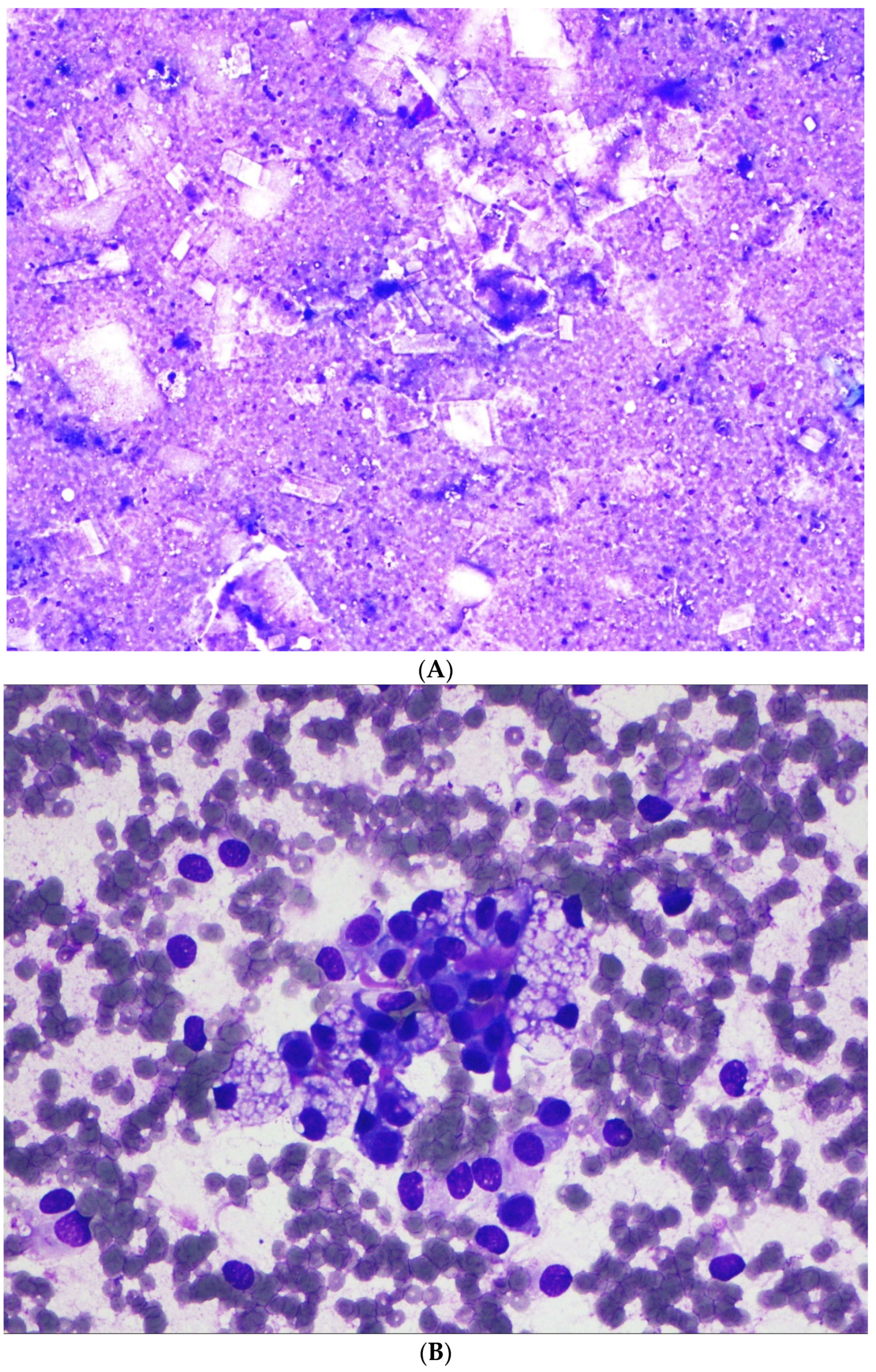
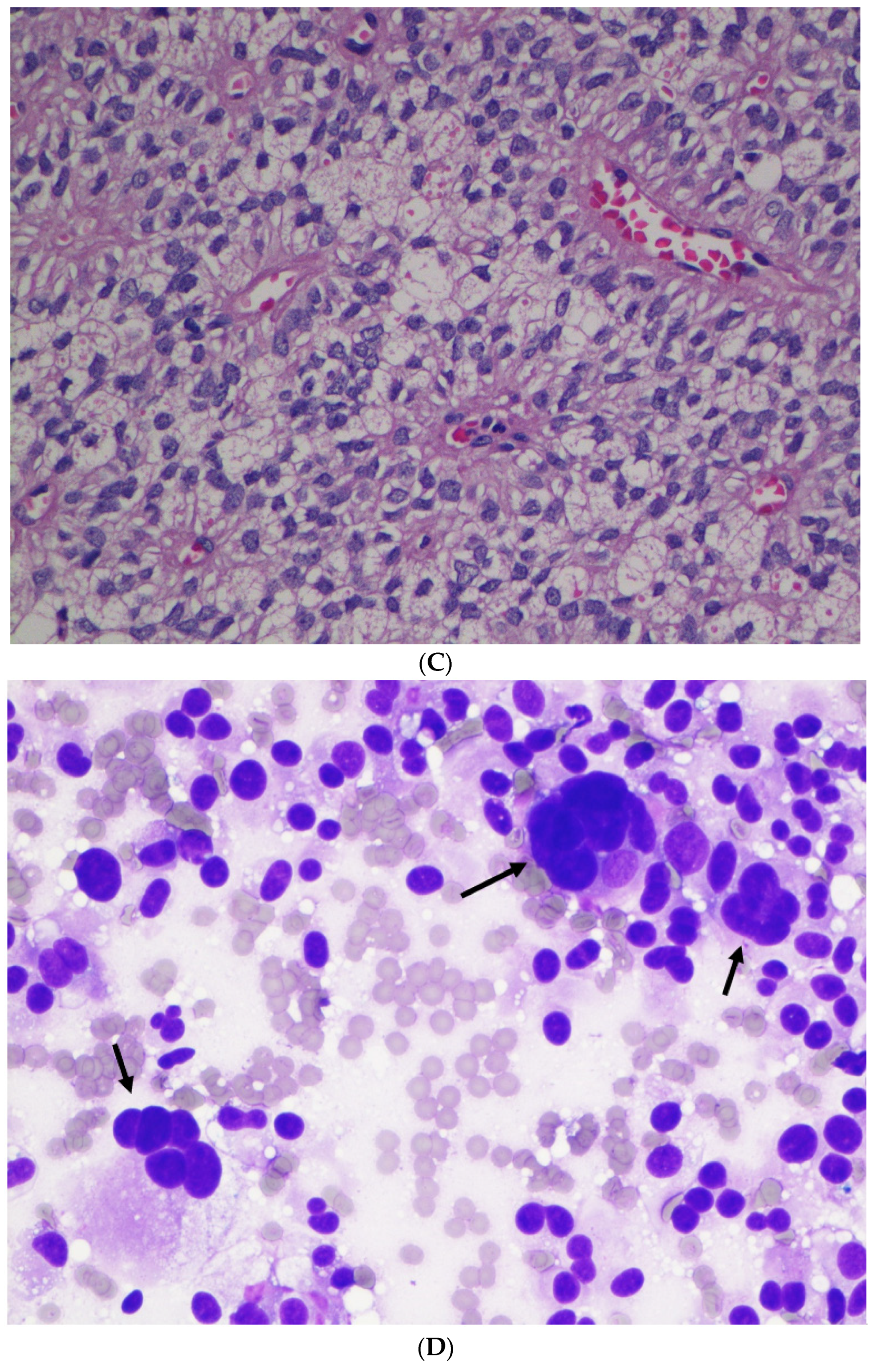
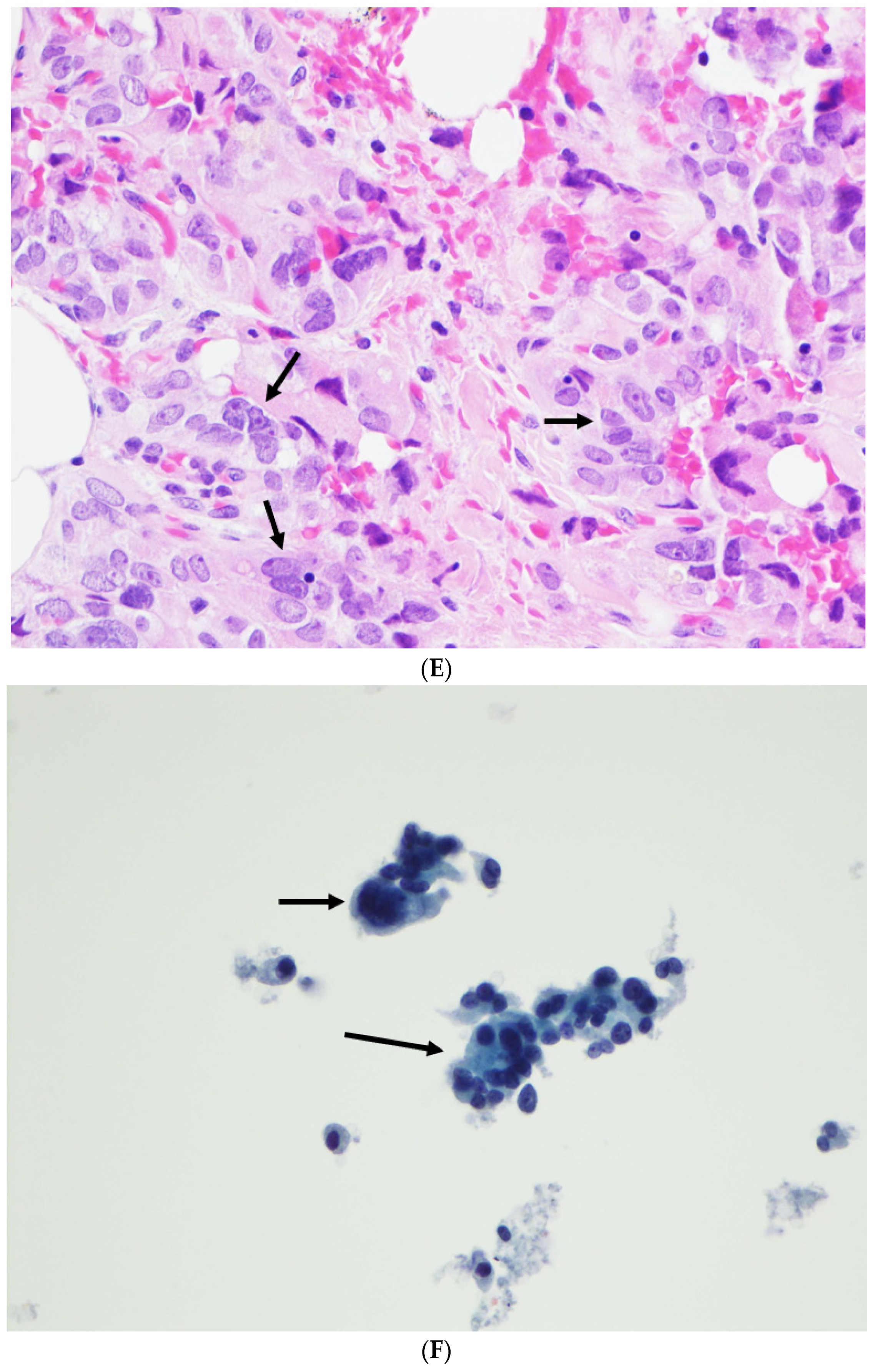
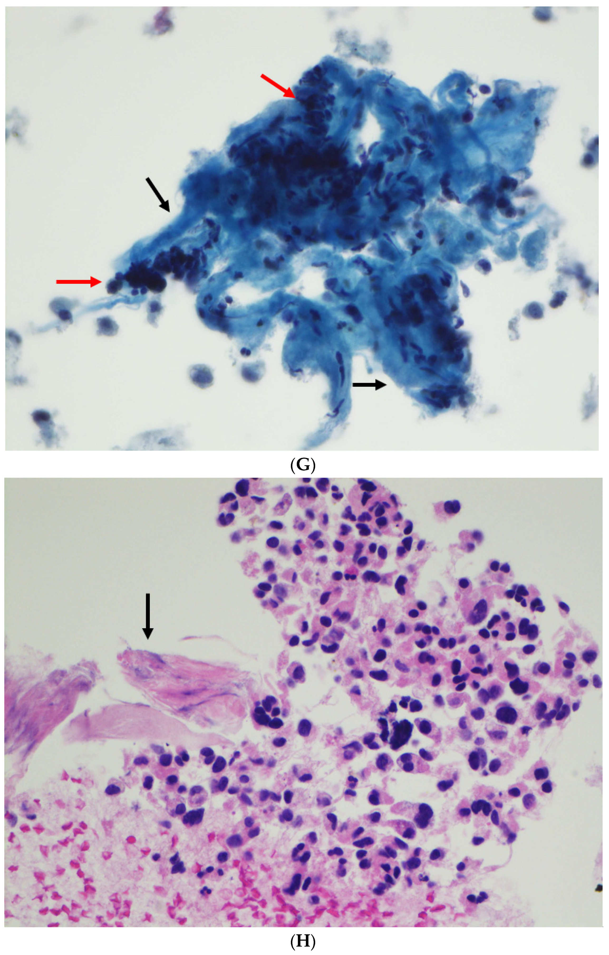
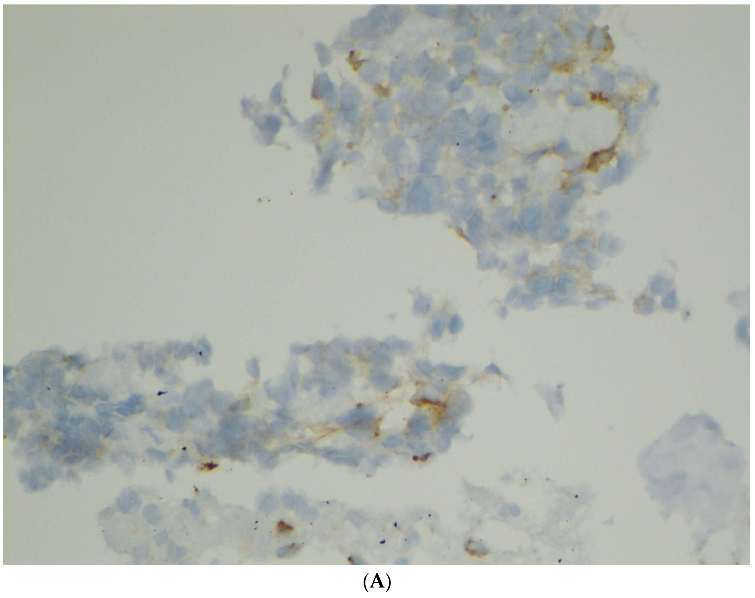
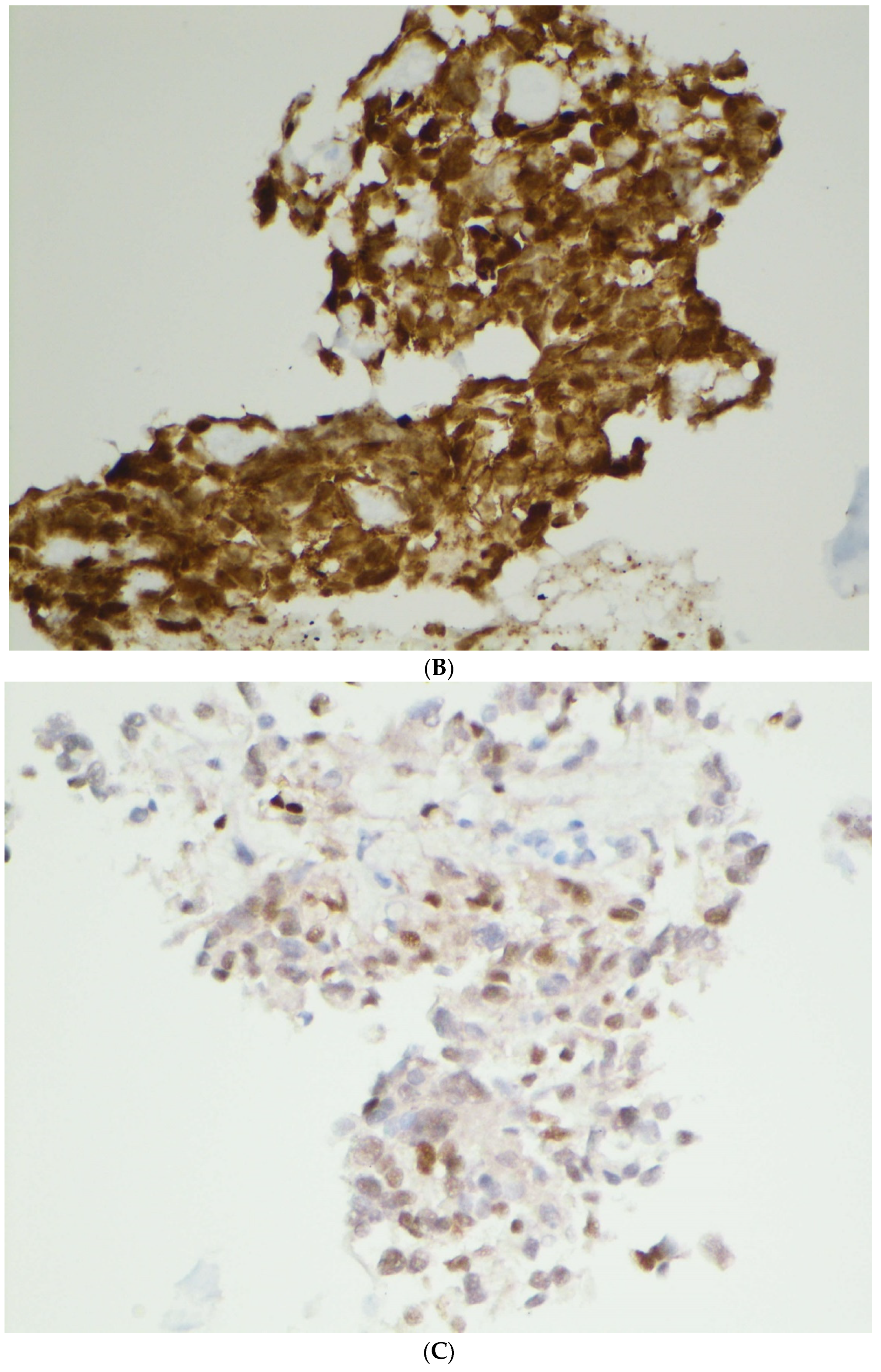
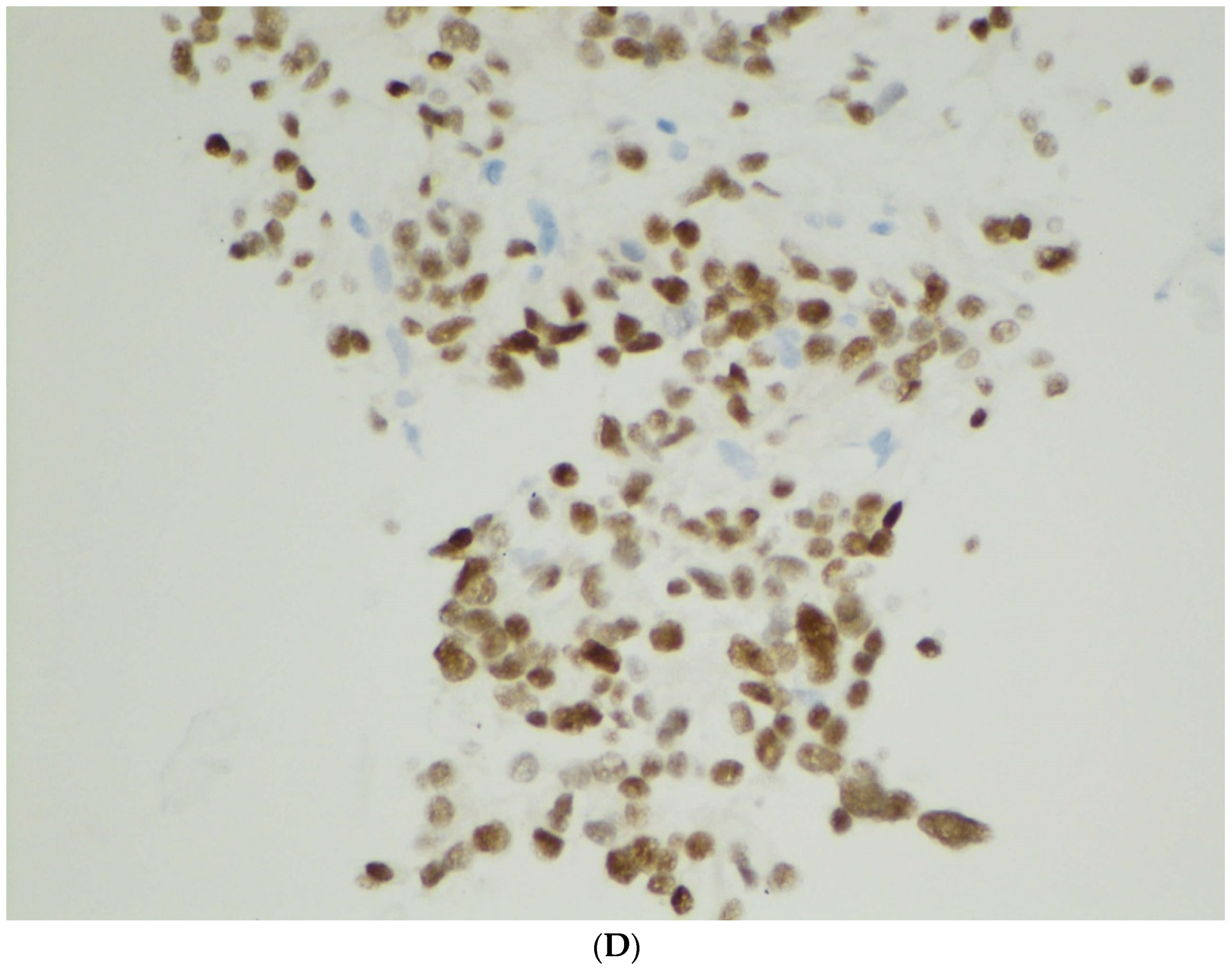

| Case | Age (Years) | Sex | Site | Radiology/Gross Features | Size (cm) | ROSE Diagnosis | FNA/Biopsy Diagnosis |
|---|---|---|---|---|---|---|---|
| 1 | 65 | F | Body | Solid and cystic | 4.0 | Atypical cells with mucinous features | SPN |
| 2 | 39 | F | Tail | Cystic | 3.0 | Serous papillary lesion | SPN |
| 3 | 28 | F | Body | Solid and cystic | 4.0 | NA | SPN |
| 4 | 26 | F | Tail | Solid and cystic | 4.0 | NA | SPN |
| 5 | 33 | F | Body | Solid and cystic | 6.5 | Lesional tissue | SPN |
| 6 | 26 | F | Tail | Solid and cystic | 1.5 | Suspicious for SPN vs. PanNET | SPN |
| 7 | 24 | F | Tail | Solid and cystic | 4.0 | SPN | SPN |
| 8 | 26 | M | Tail | Solid and cystic | 3.5 | NA | SPN |
| 9 | 25 | M | Body | Solid | 2.2 | Favor PanNET | SPN |
| 10 | 29 | F | Body | Solid | 2.9 | Lesional tissue | SPN |
| 11 | 23 | M | Body | Solid and cystic | 4.8 | NA | SPN |
| 12 | 41 | F | Tail | Solid and cystic | 2.2 | NA | SPN |
| 13 | 20 | F | Head | Solid | 6.0 | NA | SPN |
| 14 | 73 | F | Body | Solid and cystic | 1.2 | NA | SPN |
| 15 | 21 | F | Tail | Solid and cystic | 15 | Round cells | SPN (biopsy) |
| 16 | 21 | F | Tail | Solid and cystic | 12 | Lesional cells | SPN (biopsy) |
| 17 | 47 | F | Body | Solid and cystic | 4.7 | Tumor | SPN (biopsy) |
| 18 | 46 | F | Tail | Solid and cystic | 4.5 | NA | NA |
| 19 | 14 | F | Tail | Solid and cystic | 3.7 | NA | NA |
| 20 | 71 | F | Body | Solid and cystic | 15 | NA | NA |
| 21 | 12 | F | Tail | Solid and cystic | 3.5 | NA | NA |
| 22 | 16 | F | Tail | Solid and cystic | 3.5 | NA | NA |
| Case | Papillation | Monomorphic Cells | Central Capillaries | Nuclear Groove | Myxoid Fibrovascular Stroma | Cytoplasmic Hyaline Globules | Atypical Multinucleated Cells |
|---|---|---|---|---|---|---|---|
| 1 | + | + | + | + | + | + | - |
| 2 | + | + | + | + | + | + | - |
| 3 | + | + | + | + | + | + | - |
| 4 | + | + | + | + | + | + | - |
| 5 | + | + | + | + | + | + | - |
| 6 | + | + | + | + | + | + | - |
| 7 | + | + | + | + | + | + | - |
| 8 | + | + | + | + | - | + | - |
| 9 | + | + | + | + | + | + | - |
| 10 | + | + | + | + | + | + | - |
| 11 | + | + | + | + | + | + | - |
| 12 | + | + | + | + | + | + | + |
| 13 | + | + | + | + | + | + | - |
| 14 | + | - | - | + | + | + | + |
| 15 | + | + | + | + | + | + | - |
| 16 | + | + | + | + | + | + | - |
| 17 | + | + | + | + | + | + | - |
| Case | β-Cat | CyD1 | PR | CD56 | SYN | CHR | CD10 | CK | VIM | AAT | Ki-67 |
|---|---|---|---|---|---|---|---|---|---|---|---|
| 1 | + | NA | NA | + | Focal | NA | + | - | + | + | NA |
| 2 | + | NA | NA | + | NA | NA | + | NA | + | + | NA |
| 3 | NA | NA | NA | + | - | - | + | NA | NA | NA | NA |
| 4 | + | NA | NA | + | Focal | - | + | - | + | + | NA |
| 5 | + | NA | NA | + | Focal | - | + | - | + | + | NA |
| 6 | + | NA | NA | NA | - | - | NA | NA | NA | + | NA |
| 7 | NA | NA | NA | NA | NA | NA | NA | NA | NA | + | NA |
| 8 | + | NA | NA | + | NA | NA | + | NA | NA | + | NA |
| 9 | + | NA | NA | NA | Focal | - | + | + | NA | + | <5% |
| 10 | + | NA | + | + | - | - | + | - | NA | NA | <1% |
| 11 | + | + | + | NA | - | - | + | - | NA | NA | <5% |
| 12 | + | NA | NA | NA | - | - | NA | - | NA | NA | <5% |
| 13 | + | + | + | NA | Focal | - | NA | - | NA | NA | 5% |
| 14 | + | + | + | NA | Focal | - | NA | - | NA | NA | 1% |
| 15 | + | + | + | + | - | - | + | - | NA | + | NA |
| 16 | + | + | + | NA | - | - | + | NA | + | NA | NA |
| 17 | + | + | NA | NA | NA | NA | + | - | NA | NA | NA |
| Total | 15/15 100% | 6/6 100% | 6/6 100% | 8/8 100% | 6/13 46% | 0/12 0% | 12/12 100% | 1/11 9% | 5/5 100% | 9/9 100% |
Publisher’s Note: MDPI stays neutral with regard to jurisdictional claims in published maps and institutional affiliations. |
© 2022 by the authors. Licensee MDPI, Basel, Switzerland. This article is an open access article distributed under the terms and conditions of the Creative Commons Attribution (CC BY) license (https://creativecommons.org/licenses/by/4.0/).
Share and Cite
Wang, B.G.; Mani, H.; Wang, Z.Q.; Li, W. Cytological Diagnosis of Pancreatic Solid-Pseudopapillary Neoplasm: A Single-Institution Community Practice Experience. Diagnostics 2022, 12, 449. https://doi.org/10.3390/diagnostics12020449
Wang BG, Mani H, Wang ZQ, Li W. Cytological Diagnosis of Pancreatic Solid-Pseudopapillary Neoplasm: A Single-Institution Community Practice Experience. Diagnostics. 2022; 12(2):449. https://doi.org/10.3390/diagnostics12020449
Chicago/Turabian StyleWang, Brant G., Haresh Mani, Zoe Q. Wang, and Wenping Li. 2022. "Cytological Diagnosis of Pancreatic Solid-Pseudopapillary Neoplasm: A Single-Institution Community Practice Experience" Diagnostics 12, no. 2: 449. https://doi.org/10.3390/diagnostics12020449
APA StyleWang, B. G., Mani, H., Wang, Z. Q., & Li, W. (2022). Cytological Diagnosis of Pancreatic Solid-Pseudopapillary Neoplasm: A Single-Institution Community Practice Experience. Diagnostics, 12(2), 449. https://doi.org/10.3390/diagnostics12020449

