
Mesonephric-like Adenocarcinoma of the Ovary: Clinicopathological and Molecular Characteristics


Abstract
:1. Introduction
2. Materials and Methods
2.1. Case Selection and Clinicopathological Data Collection
2.2. Immunohistochemical Staining
2.3. Immunohistochemical Interpretation
2.4. DNA Extraction, Library Preparation, and Targeted Sequencing
2.5. Bioinformatics and Data Analysis Pipeline
3. Results
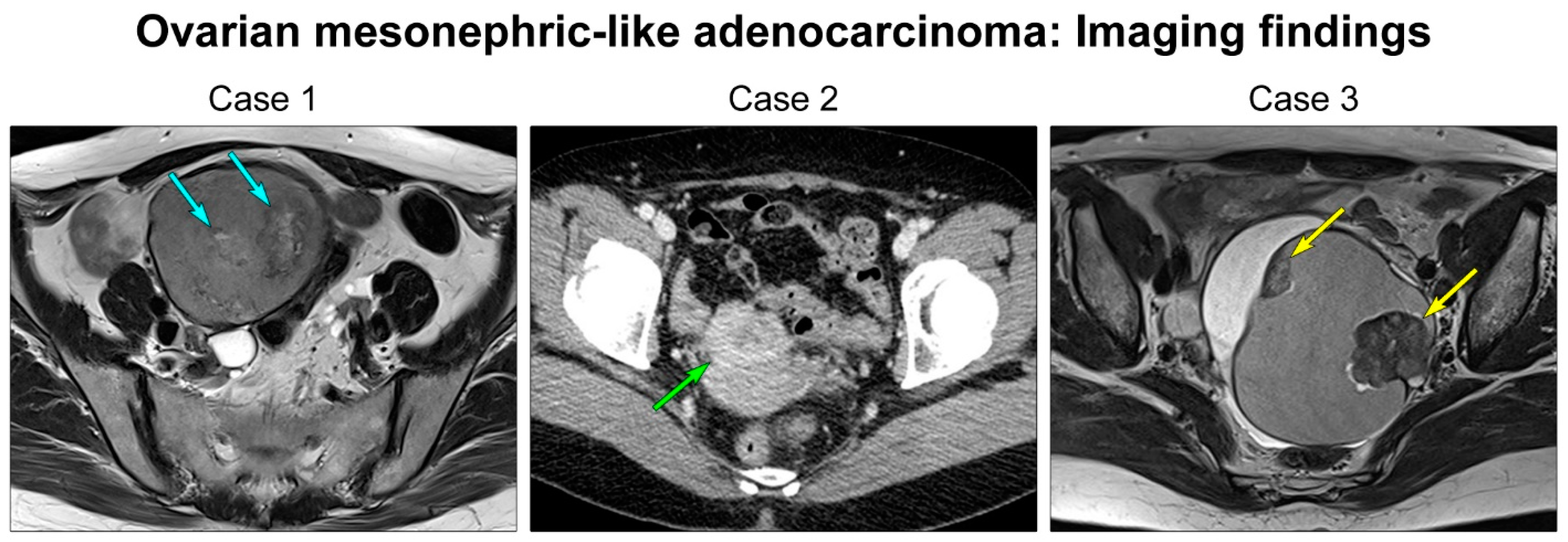
3.1. Clinical Characteristics
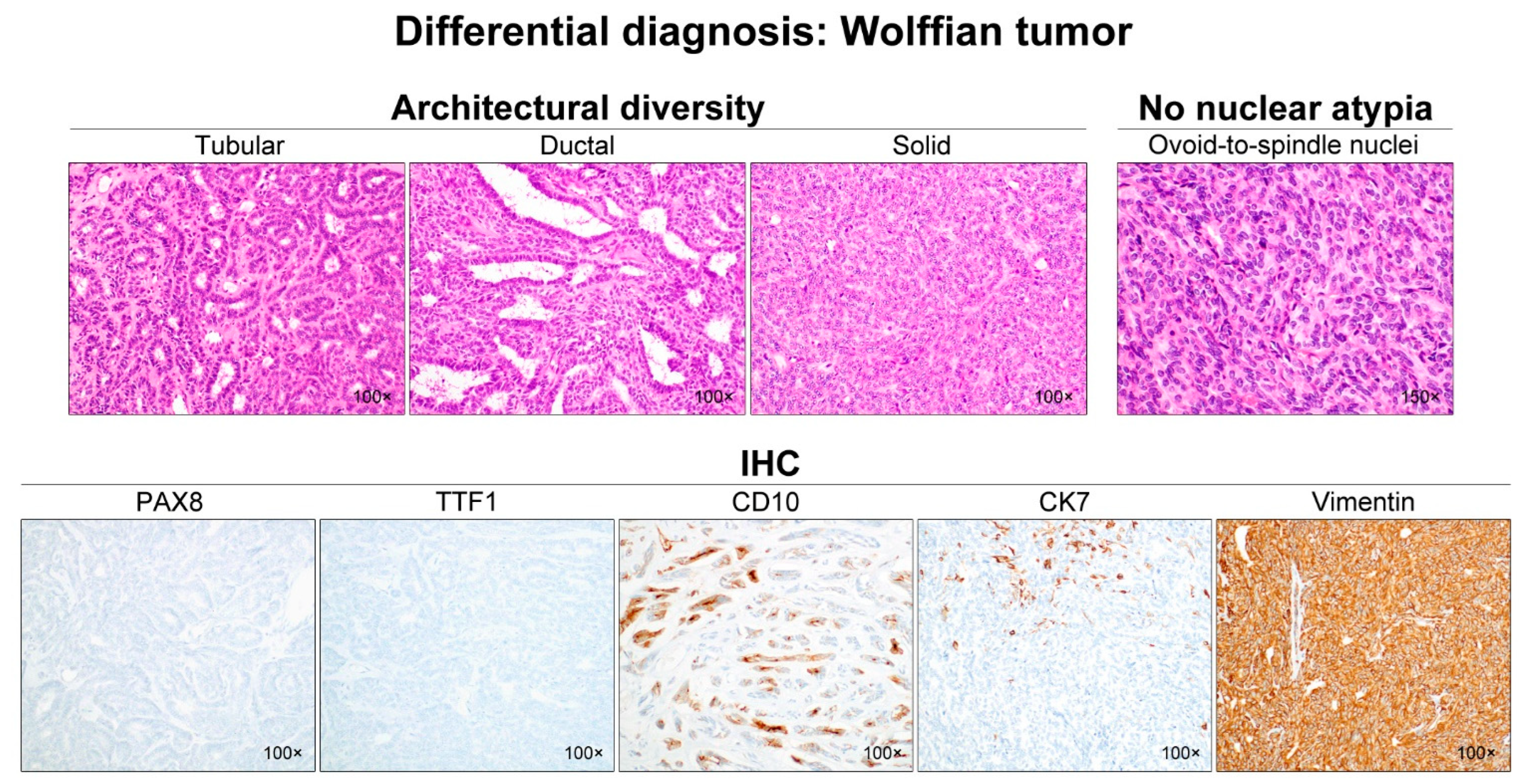
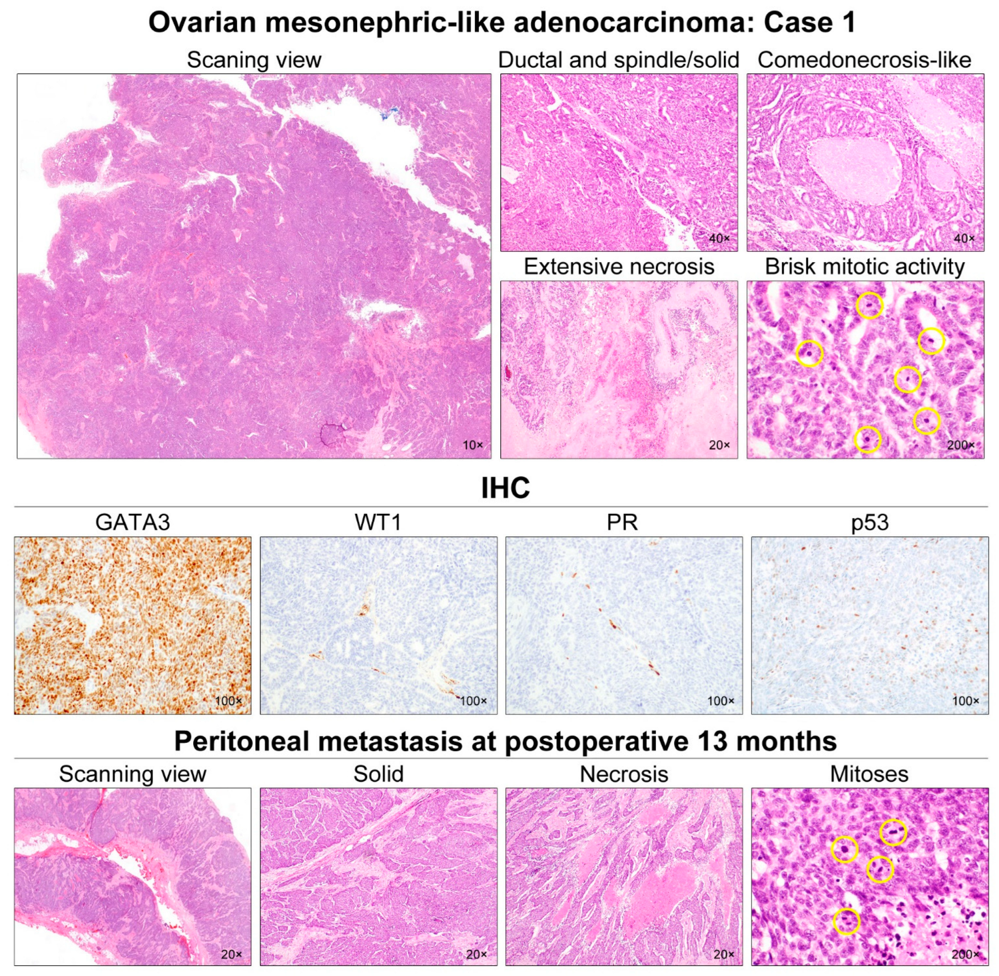
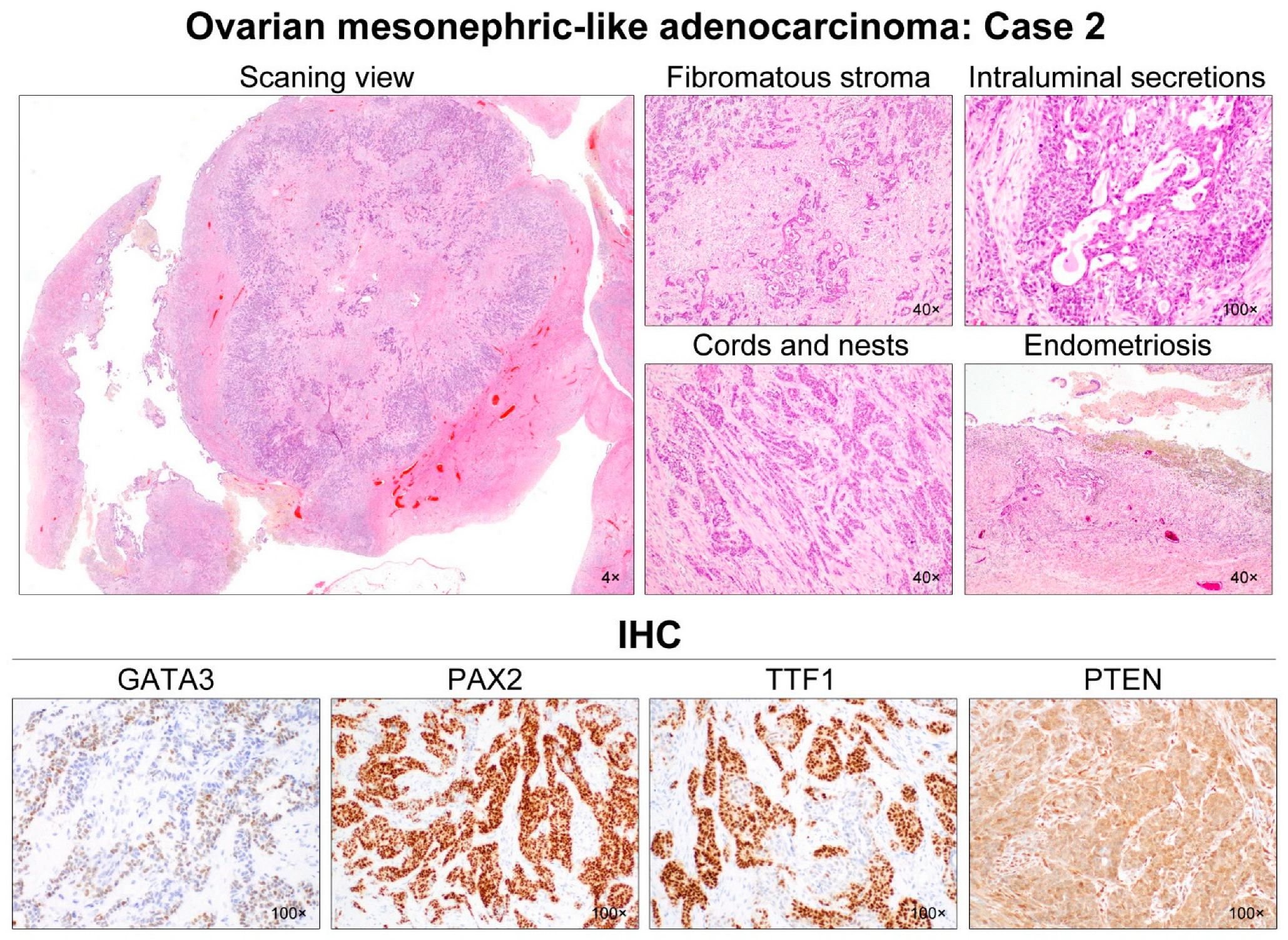
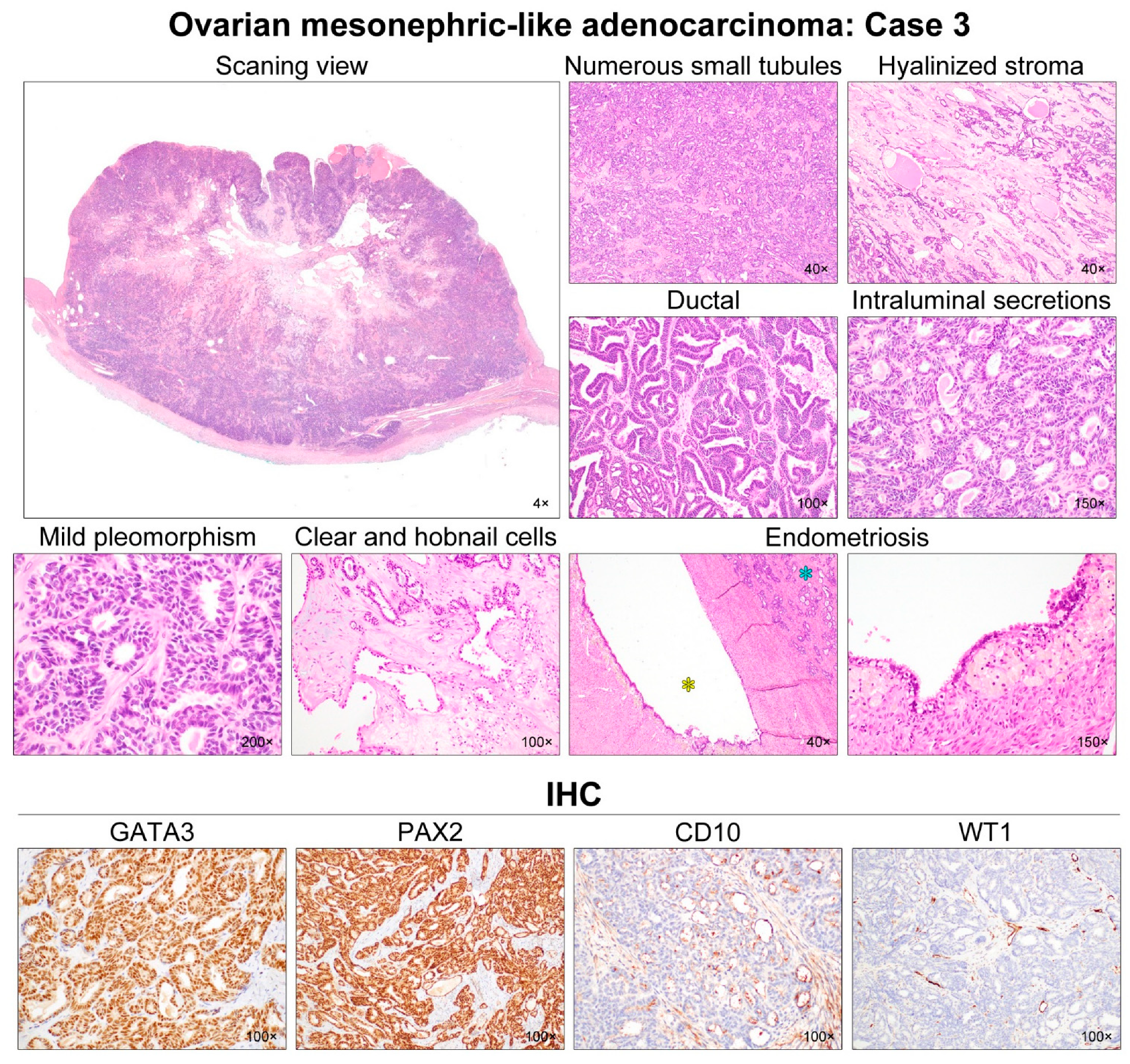
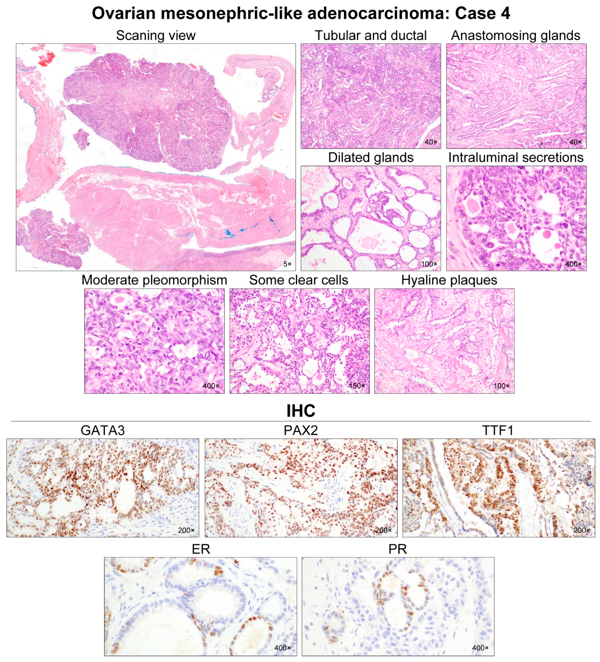
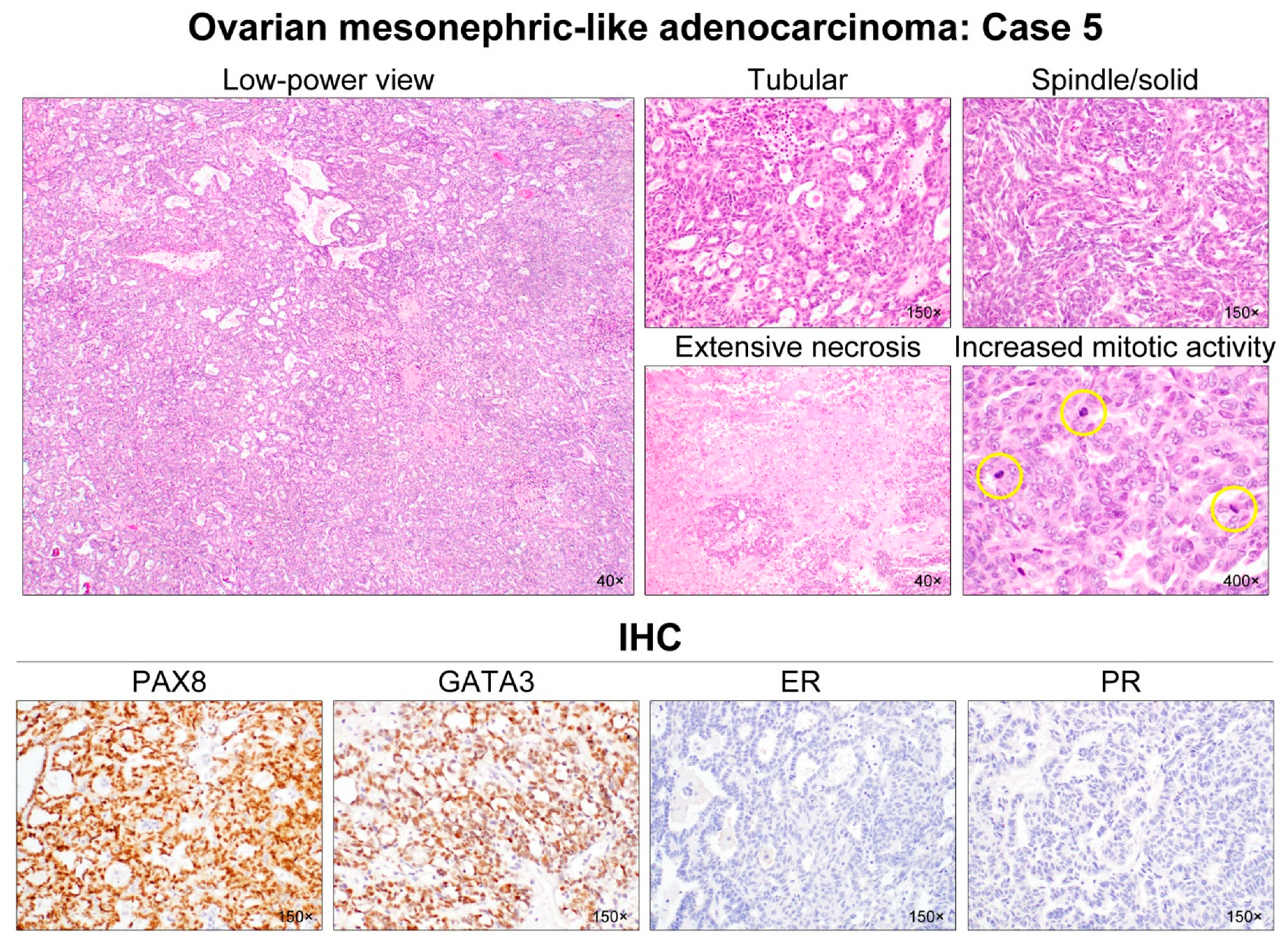
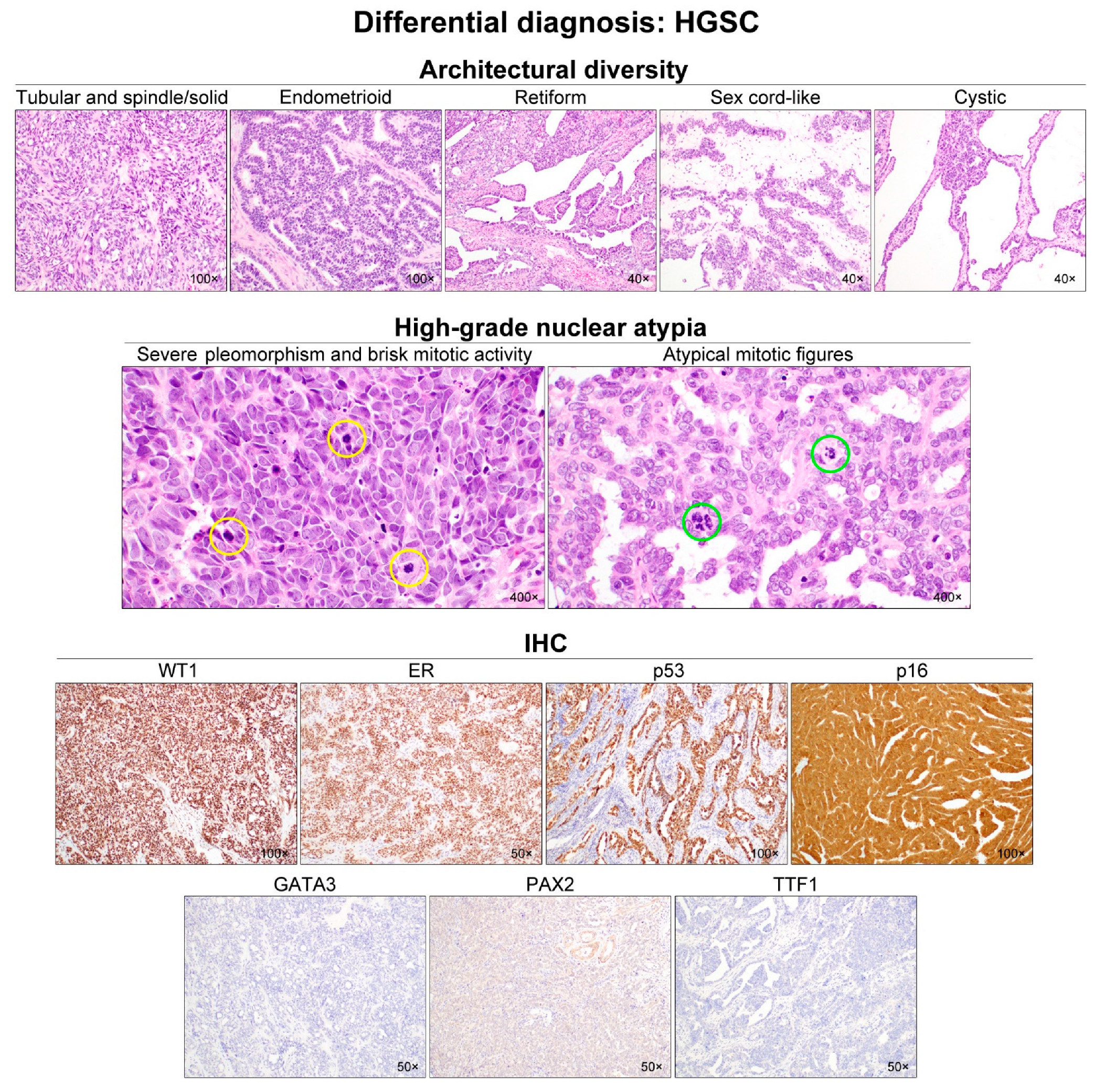
3.2. Pathological Characteristics
3.3. Immunostaining Results
3.4. Targeted Sequencing Results
4. Discussion
5. Conclusions
Author Contributions
Funding
Institutional Review Board Statement
Informed Consent Statement
Data Availability Statement
Conflicts of Interest
References
- Goyal, A.; Yang, B. Differential patterns of PAX8, p16, and ER immunostains in mesonephric lesions and adenocarcinomas of the cervix. Int. J. Gynecol. Pathol. 2014, 33, 613–619. [Google Scholar] [CrossRef] [PubMed]
- Ferry, J.A.; Scully, R.E. Mesonephric remnants, hyperplasia, and neoplasia in the uterine cervix. A study of 49 cases. Am. J. Surg. Pathol. 1990, 14, 1100–1111. [Google Scholar] [CrossRef] [PubMed]
- Kezlarian, B.; Muller, S.; Werneck Krauss Silva, V.; Gonzalez, C.; Fix, D.J.; Park, K.J.; Murali, R. Cytologic features of upper gynecologic tract adenocarcinomas exhibiting mesonephric-like differentiation. Cancer Cytopathol. 2019, 127, 521–528. [Google Scholar] [CrossRef]
- McFarland, M.; Quick, C.M.; McCluggage, W.G. Hormone receptor-negative, thyroid transcription factor 1-positive uterine and ovarian adenocarcinomas: Report of a series of mesonephric-like adenocarcinomas. Histopathology 2016, 68, 1013–1020. [Google Scholar] [CrossRef] [PubMed]
- Mirkovic, J.; McFarland, M.; Garcia, E.; Sholl, L.M.; Lindeman, N.; MacConaill, L.; Dong, F.; Hirsch, M.; Nucci, M.R.; Quick, C.M.; et al. Targeted genomic profiling reveals recurrent KRAS mutations in mesonephric-like adenocarcinomas of the female genital tract. Am. J. Surg. Pathol. 2018, 42, 227–233. [Google Scholar] [CrossRef]
- Pors, J.; Cheng, A.; Leo, J.M.; Kinloch, M.A.; Gilks, B.; Hoang, L. A comparison of GATA3, TTF1, CD10, and calretinin in identifying mesonephric and mesonephric-like carcinomas of the gynecologic tract. Am. J. Surg. Pathol. 2018, 42, 1596–1606. [Google Scholar] [CrossRef] [PubMed]
- Chapel, D.B.; Joseph, N.M.; Krausz, T.; Lastra, R.R. An ovarian adenocarcinoma with combined low-grade serous and mesonephric morphologies suggests a Mullerian origin for some mesonephric carcinomas. Int. J. Gynecol. Pathol. 2018, 37, 448–459. [Google Scholar] [CrossRef]
- McCluggage, W.G.; Vosmikova, H.; Laco, J. Ovarian combined low-grade serous and mesonephric-like adenocarcinoma: Further evidence for a Mullerian origin of mesonephric-like adenocarcinoma. Int. J. Gynecol. Pathol. 2020, 39, 84–92. [Google Scholar] [CrossRef]
- Pors, J.; Segura, S.; Chiu, D.S.; Almadani, N.; Ren, H.; Fix, D.J.; Howitt, B.E.; Kolin, D.; McCluggage, W.G.; Mirkovic, J.; et al. Clinicopathologic characteristics of mesonephric adenocarcinomas and mesonephric-like adenocarcinomas in the gynecologic tract: A multi-institutional study. Am. J. Surg. Pathol. 2021, 45, 498–506. [Google Scholar] [CrossRef]
- Dundr, P.; Gregova, M.; Nemejcova, K.; Bartu, M.; Hajkova, N.; Hojny, J.; Struzinska, I.; Fischerova, D. Ovarian mesonephric-like adenocarcinoma arising in serous borderline tumor: A case report with complex morphological and molecular analysis. Diagn. Pathol. 2020, 15, 91. [Google Scholar] [CrossRef]
- Seay, K.; Akanbi, T.; Bustamante, B.; Chaudhary, S.; Goldberg, G.L. Mesonephric-like adenocarcinoma of the ovary with co-existent endometriosis: A case report and review of the literature. Gynecol. Oncol. Rep. 2020, 34, 100657. [Google Scholar] [CrossRef] [PubMed]
- Chen, Q.; Shen, Y.; Xie, C. Mesonephric-like adenocarcinoma of the ovary: A case report and a review of the literature. Medicine 2020, 99, e23450. [Google Scholar] [CrossRef] [PubMed]
- da Silva, E.M.; Fix, D.J.; Sebastiao, A.P.M.; Selenica, P.; Ferrando, L.; Kim, S.H.; Stylianou, A.; Da Cruz Paula, A.; Pareja, F.; Smith, E.S.; et al. Mesonephric and mesonephric-like carcinomas of the female genital tract: Molecular characterization including cases with mixed histology and matched metastases. Mod. Pathol. 2021, 34, 1570–1587. [Google Scholar] [CrossRef] [PubMed]
- Deolet, E.; Arora, I.; Van Dorpe, J.; Van der Meulen, J.; Desai, S.; Van Roy, N.; Kaur, B.; Van de Vijver, K.; McCluggage, W.G. Extrauterine mesonephric-like neoplasms: Expanding the morphologic spectrum. Am. J. Surg. Pathol. 2022, 46, 124–133. [Google Scholar] [CrossRef] [PubMed]
- Na, K.; Kim, H.S. Clinicopathologic and molecular characteristics of mesonephric adenocarcinoma arising from the uterine body. Am. J. Surg. Pathol. 2019, 43, 12–25. [Google Scholar] [CrossRef] [PubMed]
- Chang, S.; Hur, J.Y.; Choi, Y.L.; Lee, C.H.; Kim, W.S. Current status and future perspectives of liquid biopsy in non-small cell lung cancer. J. Pathol. Transl. Med. 2020, 54, 204–212. [Google Scholar] [CrossRef]
- Chang, S.; Shim, H.S.; Kim, T.J.; Choi, Y.L.; Kim, W.S.; Shin, D.H.; Kim, L.; Park, H.S.; Lee, G.K.; Lee, C.H.; et al. Molecular biomarker testing for non-small cell lung cancer: Consensus statement of the Korean Cardiopulmonary Pathology Study Group. J. Pathol. Transl. Med. 2021, 55, 181–191. [Google Scholar] [CrossRef]
- Choi, S.; Cho, J.; Lee, S.E.; Baek, C.H.; Kim, Y.K.; Kim, H.J.; Ko, Y.H. Adenocarcinoma of the minor salivary gland with concurrent MAML2 and EWSR1 alterations. J. Pathol. Transl. Med. 2021, 55, 132–138. [Google Scholar] [CrossRef]
- Jang, Y.; Jung, H.; Kim, H.N.; Seo, Y.; Alsharif, E.; Nam, S.J.; Kim, S.W.; Lee, J.E.; Park, Y.H.; Cho, E.Y.; et al. Clinicopathologic characteristics of HER2-positive pure mucinous carcinoma of the breast. J. Pathol. Transl. Med. 2020, 54, 95–102. [Google Scholar] [CrossRef]
- Lee, J.; Cho, Y.; Choi, K.H.; Hwang, I.; Oh, Y.L. Metastatic leiomyosarcoma of the thyroid gland: Cytologic findings and differential diagnosis. J. Pathol. Transl. Med. 2021, 55, 360–365. [Google Scholar] [CrossRef]
- Choi, S.; Joo, J.W.; Do, S.I.; Kim, H.S. Endometrium-limited metastasis of extragenital malignancies: A challenge in the diagnosis of endometrial curettage specimens. Diagnostics 2020, 10, 150. [Google Scholar] [CrossRef] [PubMed] [Green Version]
- Choi, S.; Jung, Y.Y.; Kim, H.S. Serous carcinoma of the endometrium with mesonephric-like differentiation initially misdiagnosed as uterine mesonephric-like adenocarcinoma: A case report with emphasis on the immunostaining and the identification of splice site TP53 mutation. Diagnostics 2021, 11, 717. [Google Scholar] [CrossRef] [PubMed]
- Hwang, S.; Kim, B.G.; Song, S.Y.; Kim, H.S. Ovarian gynandroblastoma with a juvenile granulosa cell tumor component in a postmenopausal woman. Diagnostics 2020, 10, 537. [Google Scholar] [CrossRef] [PubMed]
- Kim, H.; Choi, S.; Do, S.I.; Lee, S.H.; Yoon, N.; Kim, H.S. Clinicopathological characteristics of pleomorphic high-grade squamous intraepithelial lesion of the uterine cervix: A single-institutional series of 31 cases. Diagnostics 2020, 10, 595. [Google Scholar] [CrossRef]
- Park, S.; Bae, G.E.; Kim, J.; Kim, H.S. Mesonephric-like differentiation of endometrial endometrioid carcinoma: Clinicopathological and molecular characteristics distinct from those of uterine mesonephric-like adenocarcinoma. Diagnostics 2021, 11, 1450. [Google Scholar] [CrossRef]
- Park, S.; Kim, H.S. Primary retroperitoneal mucinous carcinoma with carcinosarcomatous mural nodules: A case report with emphasis on its histological features and immunophenotype. Diagnostics 2020, 10, 580. [Google Scholar] [CrossRef]
- Kobel, M.; Ronnett, B.M.; Singh, N.; Soslow, R.A.; Gilks, C.B.; McCluggage, W.G. Interpretation of p53 immunohistochemistry in endometrial carcinomas: Toward increased reproducibility. Int. J. Gynecol. Pathol. 2019, 38, S123–S131. [Google Scholar] [CrossRef]
- Köbel, M.; Piskorz, A.M.; Lee, S.; Lui, S.; LePage, C.; Marass, F.; Rosenfeld, N.; Mes Masson, A.M.; Brenton, J.D. Optimized p53 immunohistochemistry is an accurate predictor of TP53 mutation in ovarian carcinoma. J. Pathol. Clin. Res. 2016, 2, 247–258. [Google Scholar] [CrossRef]
- Na, K.; Kim, H.S. Clinicopathological characteristics of fallopian tube metastases from primary endometrial, cervical, and nongynecological malignancies: A single institutional experience. Virchows Arch. 2017, 471, 363–373. [Google Scholar] [CrossRef]
- Chung, T.; Do, S.I.; Na, K.; Kim, G.; Jeong, Y.I.; Kim, Y.W.; Kim, H.S. Stromal p16 overexpression in gastric-type mucinous carcinoma of the uterine cervix. Anticancer Res. 2018, 38, 3551–3558. [Google Scholar] [CrossRef] [Green Version]
- Jung, Y.Y.; Nahm, J.H.; Kim, H.S. Cytomorphological characteristics of glassy cell carcinoma of the uterine cervix: Histopathological correlation and human papillomavirus genotyping. Oncotarget 2016, 7, 74152–74161. [Google Scholar] [CrossRef] [PubMed] [Green Version]
- Stelloo, E.; Jansen, A.M.L.; Osse, E.M.; Nout, R.A.; Creutzberg, C.L.; Ruano, D.; Church, D.N.; Morreau, H.; Smit, V.; van Wezel, T.; et al. Practical guidance for mismatch repair-deficiency testing in endometrial cancer. Ann. Oncol. 2017, 28, 96–102. [Google Scholar] [CrossRef]
- Watkins, J.C.; Nucci, M.R.; Ritterhouse, L.L.; Howitt, B.E.; Sholl, L.M. Unusual mismatch repair immunohistochemical patterns in endometrial carcinoma. Am. J. Surg. Pathol. 2016, 40, 909–916. [Google Scholar] [CrossRef] [PubMed]
- Hovelson, D.H.; McDaniel, A.S.; Cani, A.K.; Johnson, B.; Rhodes, K.; Williams, P.D.; Bandla, S.; Bien, G.; Choppa, P.; Hyland, F.; et al. Development and validation of a scalable next-generation sequencing system for assessing relevant somatic variants in solid tumors. Neoplasia 2015, 17, 385–399. [Google Scholar] [CrossRef] [PubMed] [Green Version]
- Jeon, J.; Maeng, L.S.; Bae, Y.J.; Lee, E.J.; Yoon, Y.C.; Yoon, N. Comparing clonality between components of combined hepatocellular carcinoma and cholangiocarcinoma by targeted sequencing. Cancer Genom. Proteom. 2018, 15, 291–298. [Google Scholar] [CrossRef] [PubMed] [Green Version]
- Yang, H.; Wang, K. Genomic variant annotation and prioritization with ANNOVAR and wANNOVAR. Nat. Protoc. 2015, 10, 1556–1566. [Google Scholar] [CrossRef]
- Kim, H.; Bae, G.E.; Jung, Y.Y.; Kim, H.S. Ovarian Mesonephric-like Adenocarcinoma With Multifocal Microscopic Involvement of the Fimbrial Surface: Potential for Misdiagnosis of Tubal Intraepithelial Metastasis as Serous Tubal Intraepithelial Carcinoma Associated With Ovarian High-grade Serous Carcinoma. In Vivo 2021, 35, 3613–3622. [Google Scholar] [CrossRef]
- Euscher, E.D.; Bassett, R.; Duose, D.Y.; Lan, C.; Wistuba, I.; Ramondetta, L.; Ramalingam, P.; Malpica, A. Mesonephric-like carcinoma of the endometrium: A subset of endometrial carcinoma with an aggressive behavior. Am. J. Surg. Pathol. 2020, 44, 429–443. [Google Scholar] [CrossRef]
- Kim, H.; Na, K.; Bae, G.E.; Kim, H.S. Mesonephric-like adenocarcinoma of the uterine corpus: Comprehensive immunohistochemical analyses using markers for mesonephric, endometrioid and serous tumors. Diagnostics 2021, 11, 2042. [Google Scholar] [CrossRef]
- Patel, V.; Kipp, B.; Schoolmeester, J.K. Corded and hyalinized mesonephric-like adenocarcinoma of the uterine corpus: Report of a case mimicking endometrioid carcinoma. Hum. Pathol. 2019, 86, 243–248. [Google Scholar] [CrossRef]
- Yamamoto, S.; Sakai, Y. Pulmonary metastasis of mesonephric-like adenocarcinoma arising from the uterine body: A striking mimic of follicular thyroid carcinoma. Histopathology 2019, 74, 651–653. [Google Scholar] [CrossRef] [PubMed]
- Kolin, D.L.; Costigan, D.C.; Dong, F.; Nucci, M.R.; Howitt, B.E. A combined morphologic and molecular approach to retrospectively identify KRAS-mutated mesonephric-like adenocarcinomas of the endometrium. Am. J. Surg. Pathol. 2019, 43, 389–398. [Google Scholar] [CrossRef] [PubMed]
- Mirkovic, J.; Sholl, L.M.; Garcia, E.; Lindeman, N.; MacConaill, L.; Hirsch, M.; Dal Cin, P.; Gorman, M.; Barletta, J.A.; Nucci, M.R.; et al. Targeted genomic profiling reveals recurrent KRAS mutations and gain of chromosome 1q in mesonephric carcinomas of the female genital tract. Mod. Pathol. 2015, 28, 1504–1514. [Google Scholar] [CrossRef] [PubMed] [Green Version]
- Dillon, M.; Lopez, A.; Lin, E.; Sales, D.; Perets, R.; Jain, P. Progress on Ras/MAPK signaling research and targeting in blood and solid cancers. Cancers 2021, 13, 5059. [Google Scholar] [CrossRef] [PubMed]
- Nakayama, N.; Nakayama, K.; Yeasmin, S.; Ishibashi, M.; Katagiri, A.; Iida, K.; Fukumoto, M.; Miyazaki, K. KRAS or BRAF mutation status is a useful predictor of sensitivity to MEK inhibition in ovarian cancer. Br. J. Cancer 2008, 99, 2020–2028. [Google Scholar] [CrossRef] [PubMed] [Green Version]
- Choi, K.H.; Kim, H.; Bae, G.E.; Lee, S.H.; Woo, H.Y.; Kim, H.S. Mesonephric-like differentiation of ovarian endometrioid and high-grade serous carcinomas: Clinicopathological and molecular characteristics distinct from those of mesonephric-like adenocarcinoma. Anticancer Res. 2021, 41, 4587–4601. [Google Scholar] [CrossRef]
- Pors, J.; Segura, S.; Cheng, A.; Ji, J.X.; Tessier-Cloutier, B.; Cochrane, D.; Fix, D.J.; Park, K.; Gilks, B.; Hoang, L. Napsin-A and AMACR are superior to HNF-1beta in distinguishing between mesonephric carcinomas and clear cell carcinomas of the gynecologic tract. Appl. Immunohistochem. Mol. Morphol. 2020, 28, 593–601. [Google Scholar] [CrossRef]
- Shalaby, A.; Shenoy, V. Female adnexal tumor of probable Wolffian origin: A review. Arch. Pathol. Lab. Med. 2020, 144, 24–28. [Google Scholar] [CrossRef] [Green Version]
- Sinha, R.; Bustamante, B.; Tahmasebi, F.; Goldberg, G.L. Malignant female adnexal tumor of probable Wolffian origin (FATWO): A case report and review for the literature. Gynecol. Oncol. Rep. 2021, 36, 100726. [Google Scholar] [CrossRef]
- Qazi, M.; Movahedi-Lankarani, S.; Wang, B.G. Cytohistopathologic correlation of ovarian mesonephric-like carcinoma and female adnexal tumor of probable Wolffian origin. Diagn. Cytopathol. 2021, 49, E207–E213. [Google Scholar] [CrossRef]
- Mirkovic, J.; Dong, F.; Sholl, L.M.; Garcia, E.; Lindeman, N.; MacConaill, L.; Crum, C.P.; Nucci, M.R.; McCluggage, W.G.; Howitt, B.E. Targeted genomic profiling of female adnexal tumors of probable Wolffian origin (FATWO). Int. J. Gynecol. Pathol. 2019, 38, 543–551. [Google Scholar] [CrossRef] [PubMed]
- Howitt, B.E.; Emori, M.M.; Drapkin, R.; Gaspar, C.; Barletta, J.A.; Nucci, M.R.; McCluggage, W.G.; Oliva, E.; Hirsch, M.S. GATA3 Is a Sensitive and Specific Marker of Benign and Malignant Mesonephric Lesions in the Lower Female Genital Tract. Am. J. Surg. Pathol. 2015, 39, 1411–1419. [Google Scholar] [CrossRef] [PubMed]
- Roma, A.A.; Goyal, A.; Yang, B. Differential Expression Patterns of GATA3 in Uterine Mesonephric and Nonmesonephric Lesions. Int. J. Gynecol. Pathol. 2015, 34, 480–486. [Google Scholar] [CrossRef] [PubMed]


| Antibody | Clone | Company | Dilution |
|---|---|---|---|
| PAX8 | Polyclonal | Cell Marque (Rocklin, CA, USA) | 1:50 |
| WT1 | 6F-H2 | Cell Marque (Rocklin, CA, USA) | 1:800 |
| GATA3 | L50-823 | Cell Marque (Rocklin, CA, USA) | 1:400 |
| TTF1 | 8G7G3/1 | Dako (Agilent Technologies, Santa Clara, CA, USA) | 1:100 |
| PAX2 | Polyclonal | Invitrogen (Thermo Fisher Scientific, Waltham, MA, USA) | 1:100 |
| CD10 | 56C6 | Novocastra (Leica Biosystems, Buffalo Grove, IL, USA) | 1:100 |
| ER | 6F11 | Novocastra (Leica Biosystems, Buffalo Grove, IL, USA) | 1:150 |
| PR | 16 | Novocastra (Leica Biosystems, Buffalo Grove, IL, USA) | 1:100 |
| p53 | DO-7 | Novocastra (Leica Biosystems, Buffalo Grove, IL, USA) | 1:200 |
| p16 | E6H4 | Ventana Medical Systems (Roche, Oro Valley, AZ, USA) | Prediluted |
| PTEN | SP218 | Ventana Medical Systems (Roche, Oro Valley, AZ, USA) | Prediluted |
| MLH1 | M1 | Ventana Medical Systems (Roche, Oro Valley, AZ, USA) | Prediluted |
| PMS2 | MRQ-28 | Cell Marque (Rocklin, CA, USA) | 1:20 |
| MSH2 | G219-1129 | Cell Marque (Rocklin, CA, USA) | 1:500 |
| MSH6 | 44/MSH6 | BD Biosciences (Franklin Lakes, NJ, USA) | 1:500 |
| Case No. | Age (yrs) | Presenting Symptom | Imaging Finding | CA 125 (U/mL) | CA 19-9 (U/mL) | Clinical Impression | Surgical Treatment | Adjuvant Treatment | Recurrence | Treatment for Recurrence | DFS (mos) | Status | OS (mos) |
|---|---|---|---|---|---|---|---|---|---|---|---|---|---|
| 1 | 42 | Pelvic mass | 7.4-cm cystic right ovarian mass with internal solid nodules | 72.5 | 80.1 | Ovarian clear cell carcinoma | TH with BSO, PLND, PALND, peritonectomy, and omentectomy (PDS) | – | Lung, liver, and peritoneum | PLND, PALND, peritonectomy, omentectomy, appendectomy, and hepatectomy (SDS), and chemotherapy | 13 | Dead | 39 |
| 2 | 53 | – | 4.8-cm cystic left ovarian mass with small enhancing nodules | 26.8 | NA | Ovarian borderline tumor or carcinoma | TH with BSO, PLND, peritonectomy, and omentectomy (PDS) | Chemotherapy | – | – | 21 | Alive (NED) | 21 |
| 3 | 57 | Abdominal distension | 10.9-cm cystic left ovarian mass with solid component | 12.2 | 25.4 | Ovarian borderline tumor or carcinoma | BSO with PLNS, peritonectomy, and omentectomy (PDS) | Chemotherapy | – | – | 11 | Alive (NED) | 11 |
| 4 | 61 | Pelvic mass | Solid and cystic left ovarian mass showing intense hypermetabolic activity | NA | NA | Ovarian carcinoma | TH with BSO, PLND, PALND, peritoneal biopsy, and omentectomy (PDS) | Chemotherapy | NA (follow-up loss) | NA | NA | NA | NA |
| 5 | 52 | Pelvic mass | 5.8-cm solid mass involving the right lateral uterine wall | 108.8 | 9.3 | Uterine leiomyoma or LMS | TH with BSO and peritonectomy (PDS) | Chemotherapy | – | – | 53 | Alive (NED) | 53 |
| Case no. | Tumor Location/Size (cm) | Ovarian Surface Extension | Peritoneal Washing Cytology | Uterine Extension | Pelvic Peritoneal Metastasis | Extrapelvic Peritoneal Metastasis | FIGO Stage | Associated Histology | Dominant Growth Pattern | Severe Nuclear Pleomorphism | Mitotic Count (per 10 HPFs) | TCN |
|---|---|---|---|---|---|---|---|---|---|---|---|---|
| 1 | RO/7.5 | – | – | – | – (p); + (r) | – (p); + (r) | IA (p); IVB (r) | – | Ductal, spindle/solid, and tubular | Focal (p); Diffuse (r) | 27 (p); 25 (r) | + |
| 2 | LO/4.7 | + | + | – | – | – | IC | Endometriotic cyst | Tubular, ductal, and sex cord-like | – | 6 | – |
| 3 | LO/11.0 | + | – | – | – | – | IC | Endometriotic cyst | Tubular and ductal | – | 3 | – |
| 4 | LO/6.0 | – | + | – | – | – | IC | Endometriotic cyst | Ductal, tubular, papillary, and clear | – | 5 | – |
| 5 | RO/6.0 | – | NA | + | + | – | IIB | Endometriosis | Tubular andspindle/solid | Focal | 17 | + |
| Case No. | PAX8 | GATA3 (Nuclear) | PAX2 (Nuclear) | TTF1 (Nuclear) | WT1 | ER | PR | p16 | p53 | CD10 (Luminal) | PTEN | MMR Proteins | KRAS Mutation | Other Mutation |
|---|---|---|---|---|---|---|---|---|---|---|---|---|---|---|
| 1 | +D, S | +F, S | +D, S | +F, S | – | +F, W | – | – | WT | +F, S | No loss | No loss | p.G12V | – |
| 2 | +D, S | +F, M | +D, S | +D, S | – | – | – | +P | WT | +F, S | No loss | No loss | p.G12V | – |
| 3 | +D, S | +D, S | +D, S | – | – | +F, W | – | +P | WT | +F, M | No loss | No loss | p.G12D | – |
| 4 | +D, S | +F, S | +F, S | +F, S | – | +F, M | +F, M | +P | WT | NA | NA | NA | NA | NA |
| 5 | +D, S | +D, S | +D, M | +D, M | – | – | – | – | WT | – | No loss | No loss | p.G12C | – |
| Author | No. of Cases | Age (yrs) | Presenting Symptom and Sign | Imaging Finding | Surgical Treatment | FIGO Stage | Recurrence | Status | Survival | OS (mos) |
|---|---|---|---|---|---|---|---|---|---|---|
| McFarland et al. [4]; Mirkovic et al. [5] | 5 | 42–62 (4/5); NA (1/5) | NA | NA | TH + BSO (2/5); BSO (1/5); NA (2/5) | IA (1/5); IC (1/5); IIB (1/5); IIIC (1/5); NA (1/5) | +(1/5); –(4/5) | PD (1/5); NED (4/5) | Alive (5/5) | 7–37 (4/5); NA (1/5) |
| Pors et al. [6] | 1 | 67 | NA | NA | NA | IC | NA | NA | NA | NA |
| Chapel et al. [7] | 1 | 80 | Abdominal pain | 12.5-cm pelvic mass, omental cake, mesenteric LNE, multiple hepatic nodules | NAC + TH + BSO + O + P | IIIC | – | NED | Alive | 3 |
| McCluggage et al. [8] | 5 | 50–77 | Pelvic pain, vaginal discharge (1/5); NA (4/5) | 6-cm solid LO mass (1/5); NA (4/5) | TH + BSO + PLND + O + P (1/5); NA (4/5) | IIIA (1/5); NA (4/5) | NA | NA | NA | NA |
| Pors et al. [9] | 25 | 36–81 | Pelvic pain (10/25); uterine bleeding (4/25); abdominal distension or bloating (4/25); incidental (5/25); NA (2/25) | NA | NA | I (11/25); II–IV (7/25); NA (7/25) | +(10/25); –(14/25); NA (1/25) | NA | 68% (PFS), 71% (OS) (23/25); NA (2/25) | 1–1346 |
| Dundr et al. [10] | 1 | 61 | NA | Non-resectable LO mass, hepatic metastases, peritoneal carcinomatosis | NAC + TH + BSO + O + P + A | IVB | – | NED | Alive | NA |
| Seay et al. [11] | 1 | 67 | Pelvic heaviness, polyuria | 11-cm complex RA mass | TH + RSO + PLND + O | IA | + | SD | Alive | NA; 18 (RFS) |
| Chen et al. [12] | 1 | 29 | Abdominal discomfort | 10-cm solid and cystic RA and 5-cm cystic LA masses | TH + BSO + PLND + PALND + O | IC | – | NED | Alive | 13 |
| da Silva et al. [13] | 15 | 36–76 | NA | NA | NA | IA (2/15); IC (3/15); IIB (2/15); IIIA (1/15); IIIC (2/15); IV (3/15); NA (2/15) | +(10/15); –(5/15) | NA | NA | NA |
| Deolet et al. [14] | 4 | 33–75 | Acute abdominal pain, hydronephrosis, bilateral pleural effusions (1/4); NA (3/4) | 7-cm LO mass (1/4); 15-cm solid and cystic LO mass (1/4); ovarian mass (1/4); 11-cm RO mass (1/4) | LSO (1/4); TH + BSO + O (1/4); BSO (1/4); right partialoophorectomy (1/4) | IA (1/4); IC (1/4); IIIC (1/4); IVB (1/4) | +(1/4); –(3/4) | PR (1/4); NED (3/4) | Alive (4/4) | 8–46 |
| Kim et al. [37] | 1 | 47 | Pelvic mass | 4.4-cm solid and cystic LO mass | PLND + PALND + O+P | IIIC | – | NED | Alive | 11 |
| Author | No. of Cases | GATA3 (Nuclear) | WT1 | ER | PR | p16 | p53 | PTEN | CD10 (Luminal) | TTF1 (Nuclear) |
|---|---|---|---|---|---|---|---|---|---|---|
| McFarland et al. [4]; Mirkovic et al. [5] | 5 | +D (1/5); +F (1/5); –(2/5); NA (1/5) | –(5/5) | –(5/5) | –(5/5) | +F (2/5); NA (3/5) | WT (3/5); NA (2/5) | NA (5/5) | +F (2/5); –(1/5); NA (2/5) | +D (4/5); +F (1/5) |
| Pors et al. [6] | 1 | +D, M-S | NA | – | NA | NA | NA | NA | +FS | +FM-S |
| Chapel et al. [7] | 1 | + | – | – | – | +F | WT | NA | +F | + |
| McCluggage et al. [8] | 5 | +F (1/5); NA (4/5) | –(1/5); NA (4/5) | –(1/5); NA (4/5) | –(1/5); NA (4/5) | NA | WT (1/5); NA (4/5) | NA | +D (1/5); NA (4/5) | +F (1/5); NA (4/5) |
| Pors et al. [9] | 25 | NA | NA | NA | NA | NA | NA | NA | NA | NA |
| Dundr et al. [10] | 1 | +D | – | – | – | NA | WT | NA | +F | +D |
| Seay et al. [11] | 1 | +F | – | – | – | +F | WT | No loss | +F | +F |
| Chen et al. [12] | 1 | + | NA | – | – | – | WT | NA | + | + |
| da Silva et al. [13] | 15 | +(14/15); NA (1/15) | –(12/15); NA (3/15) | +F (2/15); –(13/15) | –(11/15); NA (4/5) | NA | WT (7/15); NA (8/15) | NA | +(2/15); –(3/15); NA (10/15) | +(11/15); –(4/15) |
| Deolet et al. [14] | 4 | +D (3/4); +F (1/4) | –(3/4); NA (1/4) | –(4/4) | –(3/4); NA (1/4) | NA | WT (3/4); NA (1/4) | NA | +D (2/4); –(1/4); NA (1/4) | +D (1/4); +F (3/4) |
| Kim et al. [37] | 1 | +D | – | – | – | +P | WT | No loss | NA | NA |
| Total [4,5,6,7,8,9,10,11,12,13,14,37] | 60 | +(27/29); –(2/29) | –(25/25) | –(29/31); +F (2/31) | – (25/25) | +F (5/6); –(1/6) | WT (19/19) | No loss (2/2) | +(12/17); –(5/17) | +(26/30); –(4/30) |
| Author | No. of Cases | KRAS Mutation | Other Recurrent Mutation | Recurrent Chromosomal Gain | Recurrent Chromosomal Loss |
|---|---|---|---|---|---|
| McFarland et al. [4]; Mirkovic et al. [5] | 5 | p.G12D (4/5); NA (1/5) | PIK3CA | 1q | 1p |
| Pors et al. [6] | 1 | NA | NA | NA | NA |
| Chapel et al. [7] | 1 | – | NRAS, BCOR | 1q, 4, 7, 13, 18p, 21 | 1p, 11, 18q, 19, 22, X |
| McCluggage et al. [8] | 5 | p.G12D (1/5); NA (4/5) | – | NA | NA |
| Pors et al. [9] | 25 | NA | NA | NA | NA |
| Dundr et al. [10] | 1 | p.G12C | PIK3CA, CHEK2 | – | – |
| Seay et al. [11] | 1 | – | – | – | – |
| Chen et al. [12] | 1 | NA | NA | NA | NA |
| da Silva et al. [13] | 15 | p.G12D (5/15); p.G12V (8/15); –(2/15) | PIK3CA, SPOP, NRAS, SETD8, CTNNB1, CREBBP, NOTCH3, ARID1A, FBXW7, FANCA, AKT1, ASXL1, RAD54L | 1q, 2, 6, 7, 8, 9, 10, 12, 16, 17, 20, 21, X | 1p, 4, 6, 15, 18, 22, X |
| Deolet et al. [14] | 4 | p.G12A (2/4); p.G12V (1/4); –(1/4) | PIK3CA, PTEN amplification, 12p isochromosome | 1q, 12, 17, 21 | – |
| Kim et al. [37] | 1 | p.G12V | – | – | – |
| Total [4,5,6,7,8,9,10,11,12,13,14,37] | 60 | p.G12D (10/28); p.G12V (10/28); p.G12A (2/28); p.G12C (1/28); –(5/28) |
Publisher’s Note: MDPI stays neutral with regard to jurisdictional claims in published maps and institutional affiliations. |
© 2022 by the authors. Licensee MDPI, Basel, Switzerland. This article is an open access article distributed under the terms and conditions of the Creative Commons Attribution (CC BY) license (https://creativecommons.org/licenses/by/4.0/).
Share and Cite
Koh, H.H.; Park, E.; Kim, H.-S. Mesonephric-like Adenocarcinoma of the Ovary: Clinicopathological and Molecular Characteristics. Diagnostics 2022, 12, 326. https://doi.org/10.3390/diagnostics12020326
Koh HH, Park E, Kim H-S. Mesonephric-like Adenocarcinoma of the Ovary: Clinicopathological and Molecular Characteristics. Diagnostics. 2022; 12(2):326. https://doi.org/10.3390/diagnostics12020326
Chicago/Turabian StyleKoh, Hyun Hee, Eunhyang Park, and Hyun-Soo Kim. 2022. "Mesonephric-like Adenocarcinoma of the Ovary: Clinicopathological and Molecular Characteristics" Diagnostics 12, no. 2: 326. https://doi.org/10.3390/diagnostics12020326
APA StyleKoh, H. H., Park, E., & Kim, H.-S. (2022). Mesonephric-like Adenocarcinoma of the Ovary: Clinicopathological and Molecular Characteristics. Diagnostics, 12(2), 326. https://doi.org/10.3390/diagnostics12020326