
Rapid Evolution of an Aortic Endocarditis


Abstract
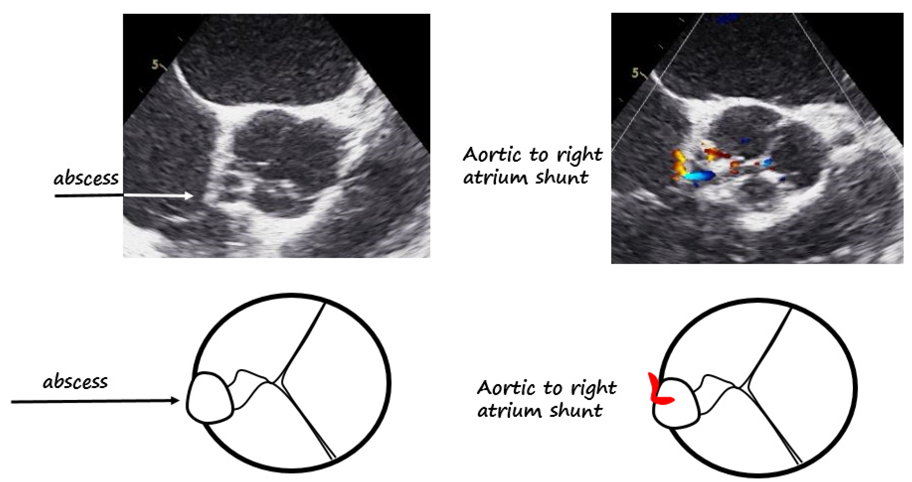
:

Author Contributions
Funding
Institutional Review Board Statement
Informed Consent Statement
Data Availability Statement
Conflicts of Interest
References
- Durack, D.T.; Lukes, A.S.; Bright, D.K. New criteria for diagnosis of infective endocarditis: Utilization of specific echocardiographic findings. Duke Endocarditis Serv. Am. J. Med. 1994, 96, 200–209. [Google Scholar] [CrossRef]
- Otto, C.M.; Nishimura, R.A.; Bonow, R.O.; Carabello, B.A.; Erwin, J.P., 3rd; Gentile, F.; Jneid, H.; Krieger, E.V.; Mack, M.; McLeod, C.; et al. 2020 ACC/AHA Guideline for the Management of Patients With Valvular Heart Disease: Executive Summary: A Report of the American College of Cardiology/American Heart Association Joint Committee on Clinical Practice Guidelines. J. Am. Coll. Cardiol. 2021, 77, 450–500. [Google Scholar] [CrossRef] [PubMed]
- Writing Committee; Pettersson, G.B.; Coselli, J.S.; Hussain, S.T.; Griffin, B.; Blackstone, E.H.; Gordon, S.M.; LeMaire, S.A.; Woc-Colburn, L.E. 2016 The American Association for Thoracic Surgery (AATS) consensus guidelines: Surgical treatment of infective endocarditis: Executive summary. J. Thorac. Cardiovasc. Surg. 2017, 153, 1241–1258. [Google Scholar] [CrossRef] [PubMed] [Green Version]
Publisher’s Note: MDPI stays neutral with regard to jurisdictional claims in published maps and institutional affiliations. |
© 2022 by the authors. Licensee MDPI, Basel, Switzerland. This article is an open access article distributed under the terms and conditions of the Creative Commons Attribution (CC BY) license (https://creativecommons.org/licenses/by/4.0/).
Share and Cite
Todde, G.; Gargiulo, P.; Canciello, G.; Borrelli, F.; Pilato, E.; Esposito, G.; Losi, M.A. Rapid Evolution of an Aortic Endocarditis. Diagnostics 2022, 12, 327. https://doi.org/10.3390/diagnostics12020327
Todde G, Gargiulo P, Canciello G, Borrelli F, Pilato E, Esposito G, Losi MA. Rapid Evolution of an Aortic Endocarditis. Diagnostics. 2022; 12(2):327. https://doi.org/10.3390/diagnostics12020327
Chicago/Turabian StyleTodde, Gaetano, Paola Gargiulo, Grazia Canciello, Felice Borrelli, Emanuele Pilato, Giovanni Esposito, and Maria Angela Losi. 2022. "Rapid Evolution of an Aortic Endocarditis" Diagnostics 12, no. 2: 327. https://doi.org/10.3390/diagnostics12020327
APA StyleTodde, G., Gargiulo, P., Canciello, G., Borrelli, F., Pilato, E., Esposito, G., & Losi, M. A. (2022). Rapid Evolution of an Aortic Endocarditis. Diagnostics, 12(2), 327. https://doi.org/10.3390/diagnostics12020327