Article

Interleaving Automatic Segmentation and Expert Opinion for Retinal Conditions
1
Department of Computer Science, Technical University of Cluj-Napoca, 400114 Cluj-Napoca, Romania
2
Department of Ophthalmology, “Iuliu Hatieganu” University of Medicine and Pharmacy, Emergency County Hospital, 400337 Cluj-Napoca, Romania
*
Author to whom correspondence should be addressed.
Diagnostics 2022, 12(1), 22; https://doi.org/10.3390/diagnostics12010022

Submission received: 17 November 2021 / Revised: 14 December 2021 / Accepted: 15 December 2021 / Published: 23 December 2021
(This article belongs to the Special Issue Artificial Intelligence in Eye Disease)

Abstract

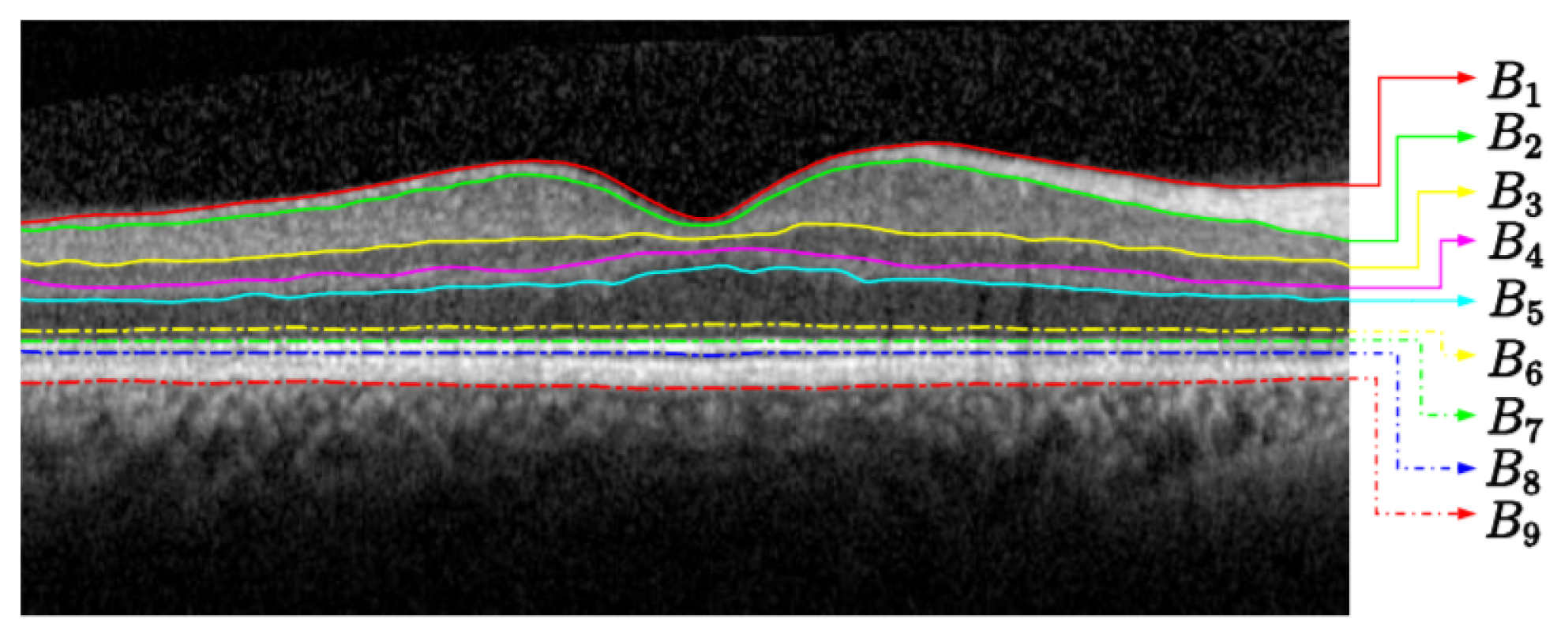
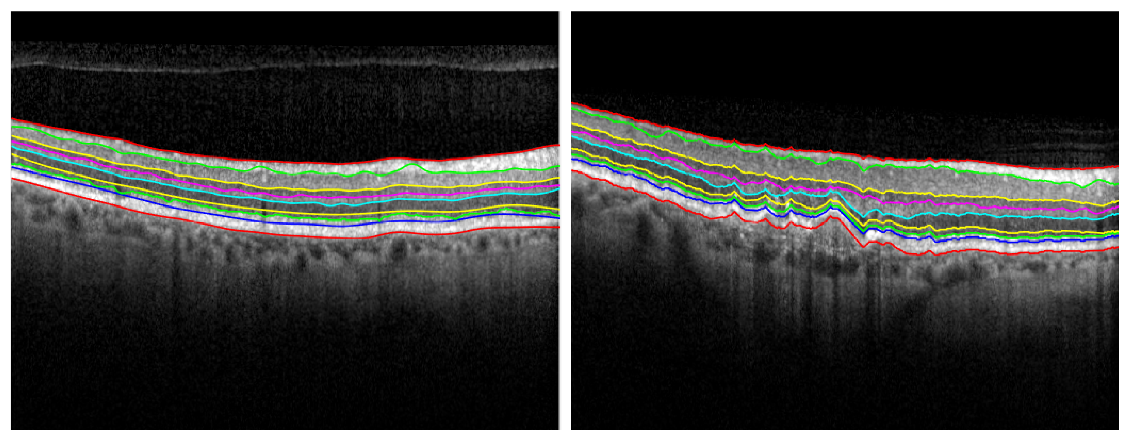
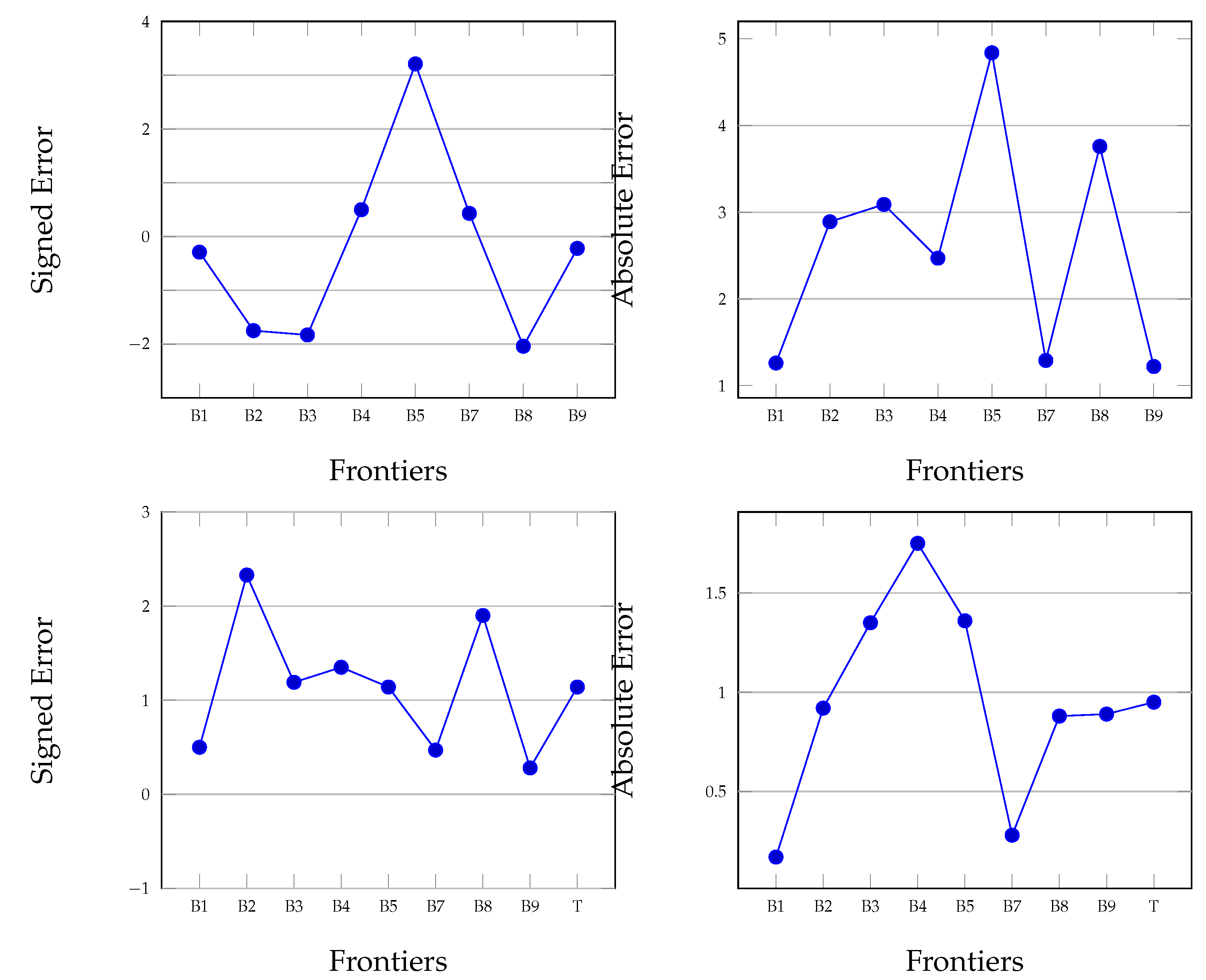
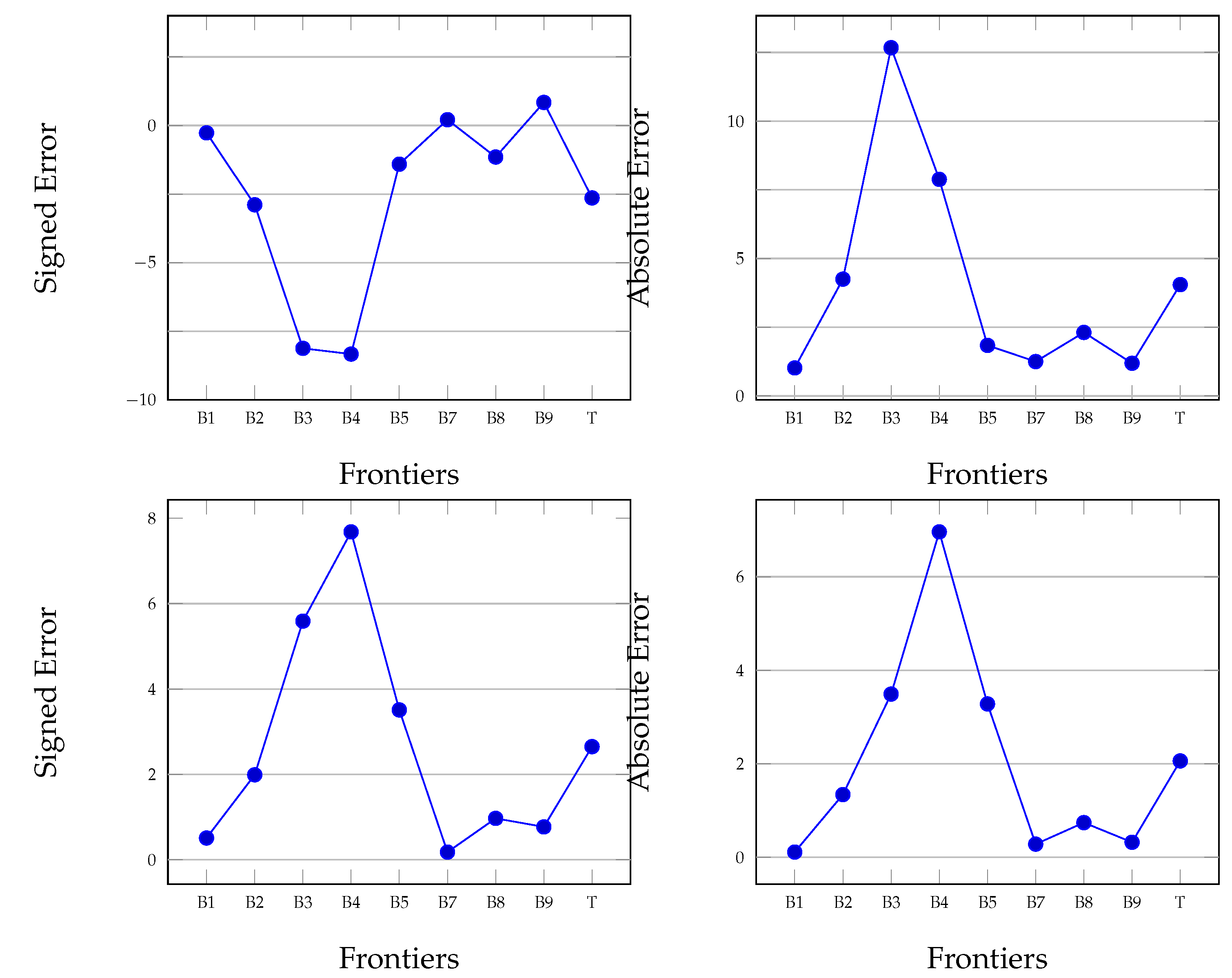
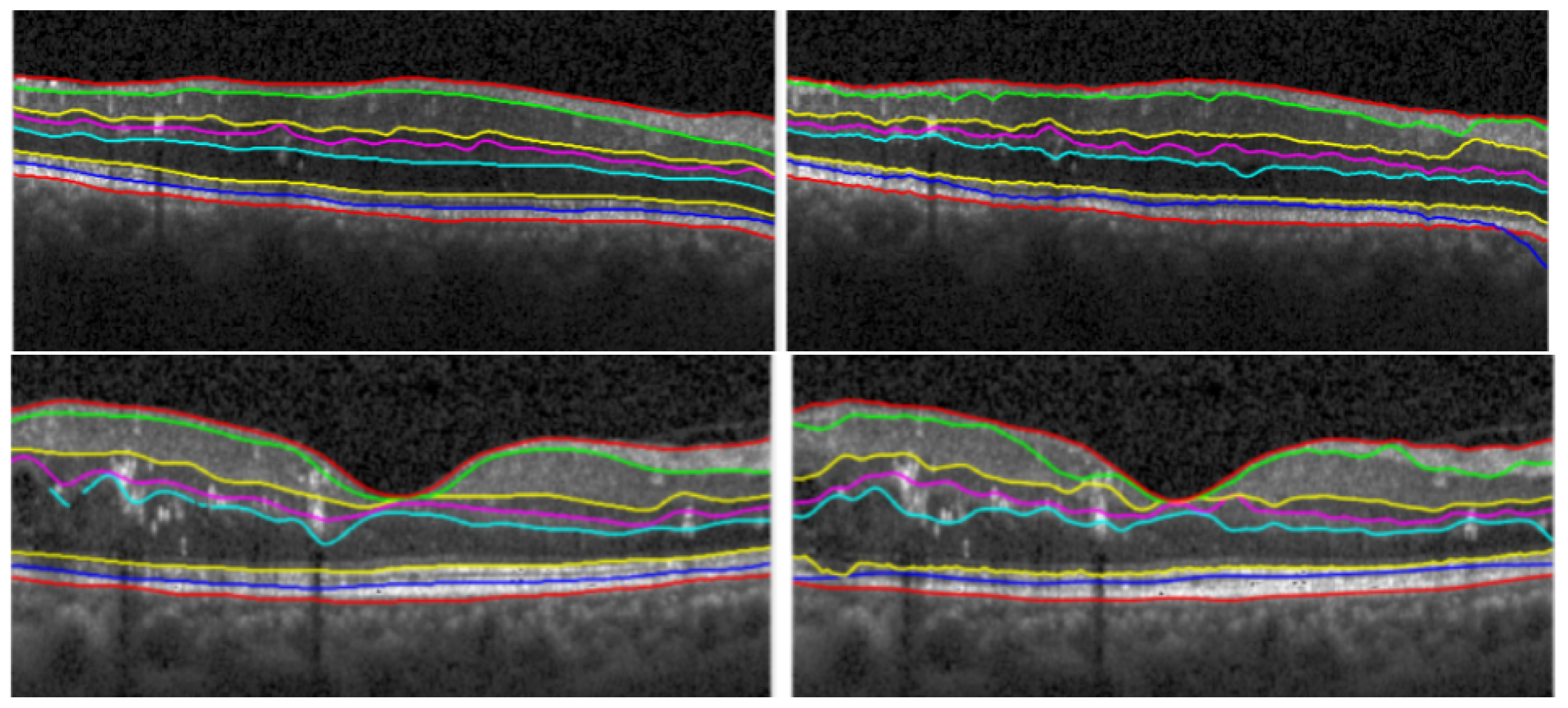
Optical coherence tomography (OCT) has become the leading diagnostic tool in modern ophthalmology. We are interested here in developing a support tool for the segmentation of retina layers. The proposed method relies on graph theory and geodesic distance. As each retina layer is characterised by different features, the proposed method interleaves various gradients during detection, such as horizontal and vertical gradients or open-closed gradients. The method was tested on a dataset of 750 OCT B-Scan Spectralis provided by the Ophthalmology Department of the County Emergency Hospital Cluj-Napoca. The method has smaller signed error on layers B1, B7 and B8, with the highest value of 0.43 pixels. The average value of signed error on all layers is −1.99 ± 1.14 px. The average value for mean absolute error is 2.60 ± 0.95 px. Since the target is a support tool for the human agent, the ophthalmologist can intervene after each automatic step. Human intervention includes validation or fine tuning of the automatic segmentation. In line with design criteria advocated by explainable artificial intelligence (XAI) and human-centered AI, this approach gives more control and transparency as well as more of a global perspective on the segmentation process.
Keywords: optical coherence tomography; retina layer segmentation; geodesic distance; vertical and horizontal gradients; human-centered AI optical coherence tomography; retina layer segmentation; geodesic distance; vertical and horizontal gradients; human-centered AI