Interesting Images

Cell Proliferation PET Imaging with 4DST PET/CT in Colorectal Adenocarcinoma and Adenoma
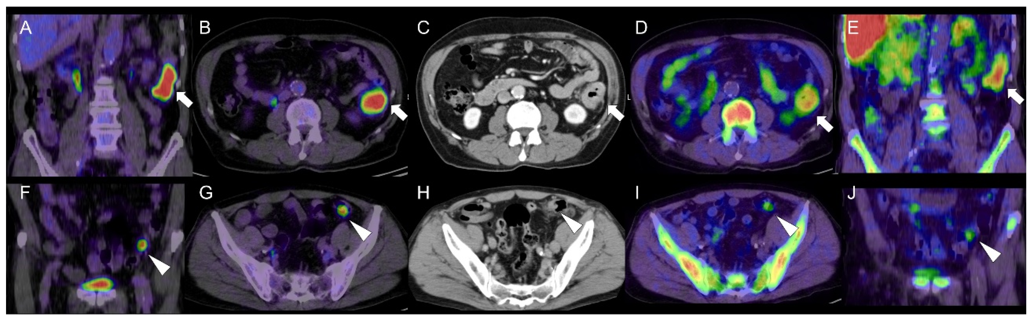
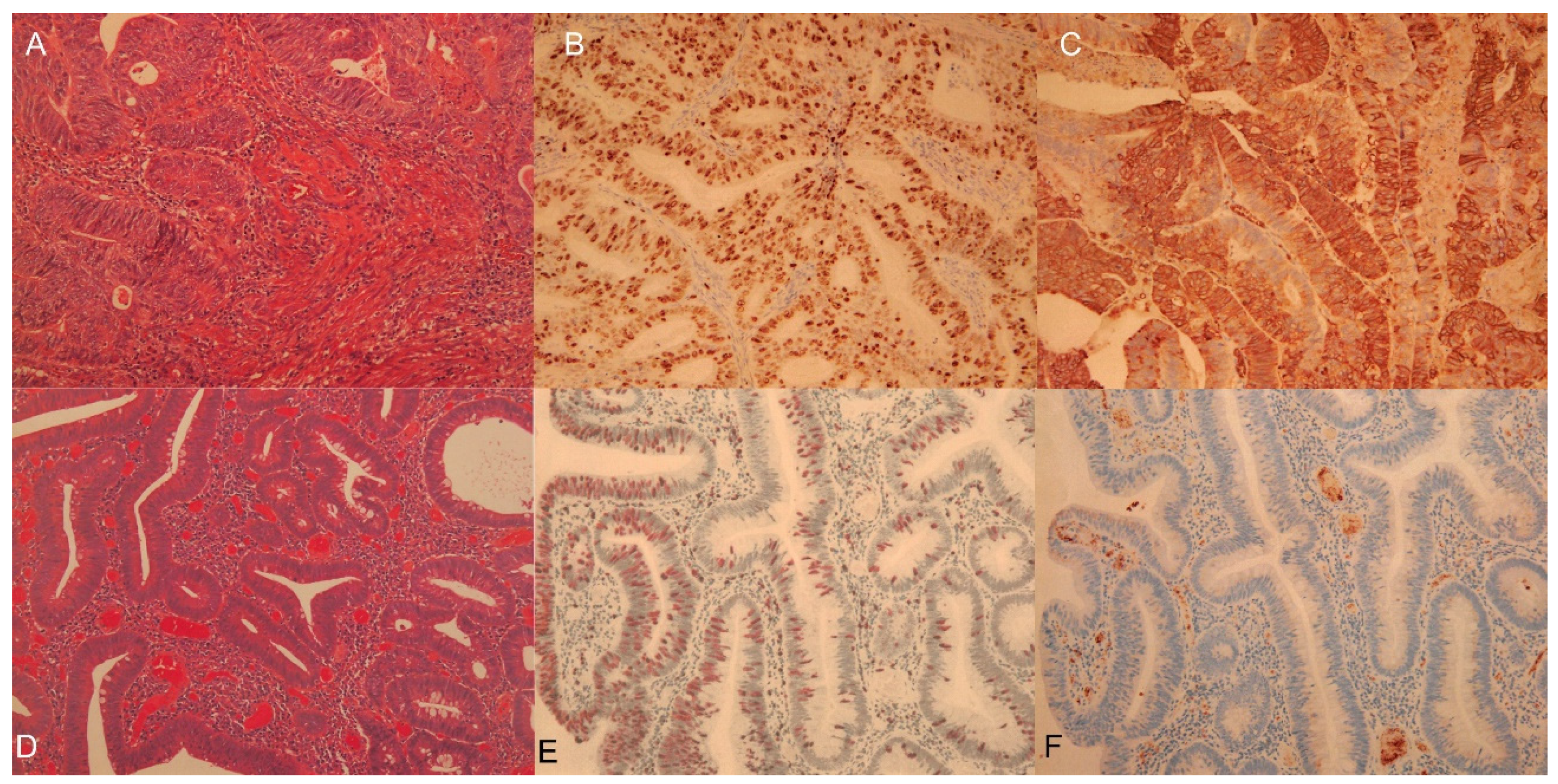
1
Division of Nuclear Medicine, Department of Radiology, National Center for Global Health and Medicine, Tokyo 1628655, Japan
2
Department of Clinical Pathology, Edogawa Hospital, Tokyo 1330052, Japan
*
Author to whom correspondence should be addressed.
Diagnostics 2021, 11(9), 1658; https://doi.org/10.3390/diagnostics11091658
Submission received: 25 July 2021 / Revised: 6 September 2021 / Accepted: 8 September 2021 / Published: 10 September 2021
(This article belongs to the Collection Interesting Images)

Abstract

:
An age of 70-year-old man was incidentally found two focal high 2-[18F]-fluoro-2-deoxy-d-glucose (FDG) uptake in the descending colon and in the sigmoid colon. We observed the feature of these two areas in the preplanned 4′-[methyl-11C]-thiothymidine (4DST) positron emission tomography (PET)/computed Tomography (CT)providing cell proliferation imaging. A mass forming high 4DST uptake in the descending colon and focal moderate 4DST uptake in the sigmoid colon was confirmed, and that were proven pathologically as adenocarcinoma and moderate to severe type tubular adenoma, respectively. This is the first report to present that colorectal adenoma can be visualized by proliferation PET imaging and the degree of uptake may enable discrimination of colorectal adenoma from adenocarcinoma, based on pathological considerations.

Colon adenomas are regarded as precursors of colorectal cancer (CRC) [1], and the adenoma–carcinoma sequence is a widely accepted theory [2]. Detecting and removing advanced colon adenomas are significant aspects of prevention of CRC [3]. CRC can develop over several years from precancerous adenomatous polyps; however, as most colon adenomas will not progress to carcinoma, the growth and the transition rates of adenomas are unpredictable. Therefore, definitive noninvasive imaging enables differentiation of lesion status between CRC and colon adenoma.
The usefulness of FDG PET and PET/CT for colon adenoma detection has been previously reported [4,5,6]; the sensitivity of FDG-PET depends on size and histologic grade of the colon adenoma [4]. Although FDG-PET has a high potential as an examination to detect colon adenomas that should be removed, the cut-off value of FDG uptake for differentiating malignant from benign lesions tends to be a high maximum standardized uptake value (SUVmax) [7]. Moreover, the FDG uptake pattern of cancer has much in common with that of colon adenoma, resulting in difficulty differentiating between CRC and colon adenoma [8].
A PET tracer for cell proliferation imaging, known as 4DST, is incorporated into DNA [9]. A previous study has reported a higher correlation of proliferating lung cancers and renal cell cancers with 4DST than FDG uptake [10,11]. The potential of 4DST PET/CT for identification of advanced CRC has been reported, with uptake values lower than those for FDG, which shows a similar tendency to the present case [12]. We found that tubular adenoma could be visualized by 4DST-PET/CT (SUVmax 2.5) as well as by FDG (SUVmax 7.7) (Figure 1). In addition, we demonstrated the potential of proliferation PET for discriminating between CRC and colon adenoma. To clarify the significance of 4DST uptake in the colon adenoma, we compared the 4DST and FDG findings with respect to the pathological results (Figure 2).
Güreşci et al. reported a significant correlation of Ki-67 index with SUVmax of FDG uptake in colorectal lesions including CRC and colon adenoma [13]. Moreover, higher proliferation is related to poorer outcomes in CRC [14]. In contrast, among the various types of cancer, the correlation between GLUT-1 expression and SUVmax is lowest in colorectal cancer (r = 0.21) [15], which indicates that several factors, other than GLUT-1 expression, may influence FDG uptake in CRC. The expression of Ki-67 shows clear differences between advanced and nonadvanced adenomas [16]. The strong reactions of Ki-67 in adenomas with severe dysplasia show a close association with colorectal carcinoma [17]. GLUT-1 immunostaining was absent in normal colorectal epithelium and tubular adenomas, and absent or only weakly apparent in tubulovillous adenomas [18]. In contrast, GLUT-1 expression in colon adenoma has been reported in several studies. Greijer et al. concluded that HIF-1α expression led to the expression of downstream target genes, such as GLUT-1 [19]. de Wit et al. found that that plasma membrane staining of GLUT-1 was completely absent in low-risk colon adenomas and present in high-risk colon adenomas (21.4%), which is significantly lower than that in CRCs (42.5%) [20]. In the present case, Ki-67 appeared to relate with 4DST and FDG uptake, whereas GLUT-1 expression was low in colon adenoma specimens even in the case of high FDG uptake. This finding suggests that proliferation imaging may have potential for predicting the growth of colon adenomas and their transition to CRC.

Author Contributions

R.M.: involved in drafting of manuscript and in review of the images. H.E.: involved in review of the pathological images. Both authors have read and agreed to the published version of the manuscript.

Funding

This report received no external funding.

Institutional Review Board Statement

This study was conducted according to the guidelines of the Declaration of Helsinki, and approval by the institutional review board of our facility (NCGM-G-000807, 25 June 2010).

Informed Consent Statement

Written informed consent was obtained from the parents of the patient involved in the study.

Data Availability Statement

Not applicable.

Conflicts of Interest

The authors declare no conflict of interest.

References

  1. Bond, J.H. Polyp guideline: Diagnosis, treatment, and surveillance for patients with colorectal polyps. Practice Parameters Committee of the American College of Gastroenterology. Am. J. Gastroenterol. 2000, 95, 3053–3063. [Google Scholar] [CrossRef] [PubMed]
  2. Chen, C.D.; Yen, M.F.; Wang, W.M.; Wong, J.M.; Chen, T.H. A case cohort study for the disease natural history of adenoma-carcinoma and de novo carcinoma and surveillance of colon and rectum after polypectomy: Implication for efficacy of colonoscopy. Br. J. Cancer 2003, 88, 1866–1873. [Google Scholar] [CrossRef] [Green Version]
  3. Winawer, S.J.; Zauber, A.G.; Ho, M.N.; O’Brien, M.J.; Gottlieb, L.S.; Sternberg, S.S.; Waye, J.D.; Schapiro, M.; Bond, J.H.; Panish, J.F.; et al. Prevention of colorectal cancer by colonoscopic polypectomy. The National Polyp Study Workgroup. N. Engl. J. Med. 1993, 329, 1977–1981. [Google Scholar] [CrossRef] [PubMed]
  4. Drenth, J.P.; Nagengast, F.M.; Oyen, W.J. Evaluation of (pre-)malignant colonic abnormalities: Endoscopic validation of FDG-PET findings. Eur. J. Nucl. Med. Mol. Imaging 2001, 28, 1766–1769. [Google Scholar] [CrossRef]
  5. Agress, H., Jr.; Cooper, B.Z. Detection of clinically unexpected malignant and premalignant tumors with whole-body FDG PET: Histopathologic comparison. Radiology 2004, 230, 417–422. [Google Scholar] [CrossRef] [PubMed]
  6. Minamimoto, R.; Senda, M.; Jinnouchi, S.; Terauchi, T.; Yoshida, T.; Inoue, T. Detection of colorectal cancer and adenomas by FDG-PET cancer screening program: Results based on a nationwide Japanese survey. Ann. Nucl. Med. 2013, 28, 212–219. [Google Scholar] [CrossRef] [PubMed]
  7. Oh, J.R.; Min, J.J.; Song, H.C.; Chong, A.; Kim, G.E.; Choi, C.; Seo, J.H.; Bom, H.S. A stepwise approach using metabolic volume and SUVmax to differentiate malignancy and dysplasia from benign colonic uptakes on 18F-FDG PET/CT. Clin. Nuc. Med. 2012, 37, 134–140. [Google Scholar] [CrossRef] [PubMed]
  8. Minamimoto, R.; Terauchi, T.; Jinnouchi, S.; Yoshida, T.; Tsukamoto, E.; Shimbo, T.; Ito, K.; Uno, K.; Ohno, H.; Oguchi, K.; et al. Observer variation study of the assessment and diagnosis of incidental colonic FDG uptake. Ann. Nucl. Med. 2013, 27, 468–477. [Google Scholar] [CrossRef] [PubMed]
  9. Toyohara, J.; Kumata, K.; Fukushi, K.; Irie, T.; Suzuki, K. Evaluation of 4’-[methyl-14C] thiothymidine for in vivo DNA synthesis imaging. J. Nucl. Med. 2006, 47, 1717–1722. [Google Scholar] [PubMed]
  10. Minamimoto, R.; Toyohara, J.; Seike, A.; Ito, H.; Endo, H.; Morooka, M.; Nakajima, K.; Mitsumoto, T.; Ito, K.; Okasaki, M.; et al. 4’-[Methyl-11C]-Thiothymidine PET/CT for Proliferation Imaging in Non-Small Cell Lung Cancer. J. Nucl. Med. 2011, 53, 199–206. [Google Scholar] [CrossRef] [PubMed] [Green Version]
  11. Minamimoto, R.; Nakaigawa, N.; Nagashima, Y.; Toyohara, J.; Ueno, D.; Namura, K.; Nakajima, K.; Yao, M.; Kubota, K. Comparison of 11C-4DST and 18F-FDG PET/CT imaging for advanced renal cell carcinoma: Preliminary study. Abdom. Radiol. 2016, 41, 521–530. [Google Scholar] [CrossRef]
  12. Fukuda, Y.; Yamamoto, Y.; Mitamura, K.; Ishikawa, R.; Asano, E.; Toyohara, J.; Norikane, T.; Nishiyama, Y. 4′-[methyl-11C]-thiothymidine as a proliferation imaging tracer for detection of colorectal cancer: Comparison with 18F-FDG. Ann. Nucl. Med. 2019, 33, 822–827. [Google Scholar] [CrossRef]
  13. Güreşci, S.; Özmen, Ö.; Uzman, M.; Şimşek, G.; Tatci, E.; Gökçek, A.; Yeniova, A.Ö. Correlation of Ki-67 proliferation index and 18-fluorodeoxyglucose uptake in colorectal incidental lesions detected by positron emission tomography-computed tomography. Turk. J. Med. Sci. 2016, 46, 1182–1187. [Google Scholar] [CrossRef] [PubMed]
  14. Luo, Z.-W.; Zhu, M.-G.; Zhang, Z.-Q.; Ye, F.-J.; Huang, W.-H.; Luo, X.-Z. Increased expression of Ki-67 is a poor prognostic marker for colorectal cancer patients: A meta analysis. BMC Cancer 2019, 19, 1–13. [Google Scholar] [CrossRef] [PubMed] [Green Version]
  15. Meyer, H.-J.; Wienke, A.; Surov, A. Associations between GLUT expression and SUV values derived from FDG-PET in different tumors—A systematic review and meta analysis. PLoS ONE 2019, 14, e0217781. [Google Scholar] [CrossRef] [PubMed] [Green Version]
  16. Radovanovic-Dinic, B.; Nagorni, A.; Katic, V.; Katic, V.; Stamenkovic, I.; Zlatic, A. An immunohistochemical study of Ki-67 in colorectal adenoma. Med. Arh. 2009, 63, 16–18. [Google Scholar] [PubMed]
  17. Barone, M.; Scavo, M.P.; Papagni, S.; Piscitelli, D.; Guido, R.; Di Lena, M.; Comelli, M.C.; Di Leo, A. ERβ expression in normal, adenomatous and carcinomatous tissues of patients with familial adenomatous polyposis. Scand. J. Gastroenterol. 2010, 45, 1320–1328. [Google Scholar] [CrossRef] [PubMed]
  18. Haber, R.S.; Rathan, A.; Weiser, K.R.; Pritsker, A.; Itzkowitz, S.H.; Bodian, C.; Slater, G.; Weiss, A.; Burstein, D.E. GLUT1 glucose transporter expression in colorectal carcinoma: A marker for poor prognosis. Cancer 1998, 83, 34–40. [Google Scholar] [CrossRef]
  19. Greijer, A.E.; Diemen, P.M.D.-V.; Fijneman, R.; Giles, R.H.; Voest, E.E.; Van Hinsbergh, V.W.M.; Meijer, G.A. Presence of HIF-1 and related genes in normal mucosa, adenomas and carcinomas of the colorectum. Virchows Arch. 2008, 452, 535–544. [Google Scholar] [CrossRef] [PubMed] [Green Version]
  20. De Wit, M.; Jimenez, C.R.; Carvalho, B.; Belien, J.A.; Delis-van Diemen, P.M.; Mongera, S.; Piersma, S.R.; Vikas, M.; Navani, S.; Pontén, F.; et al. Cell surface proteomics identifies glucose transporter type 1 and prion protein as candidate biomarkers for colorectal adenoma-to-carcinoma progression. Gut 2012, 61, 855–864. [Google Scholar] [CrossRef] [PubMed] [Green Version]
Figure 1. A 70-year-old man with pathologically proven adenocarcinoma in the descending colon and moderate to severe type tubular adenoma in the sigmoid colon. An adenocarcinoma is seen in the descending colon on (A) coronal- and (B) axial-fused FDG PET/CT imaging (arrows); (C) contrast-enhanced CT (arrow); and (D) axial- and (E) coronal-fused 4DST PET/CT imaging (arrows). The colon adenoma is seen on (F) coronal- and (G) axial-fused FDG PET/CT (arrowheads); (H) contrast-enhanced CT (arrowhead); and (J) axial- and (I) coronal-fused 4DST PET/CT imaging (arrowheads). FDG PET/CT revealed high FDG uptake (SUVmax 15.2) in the advanced CRC at the descending colon, and the lesion was also confirmed with high 4DST uptake (SUVmax 4.8). We found that tubular adenoma could be visualized by 4DST-PET/CT (SUVmax 2.5) as well as by FDG (SUVmax 7.7).
Diagnostics 11 01658 g001
Figure 2. Pathological features of colorectal cancer and adenoma. (A) Hematoxylin and Eosin (HE) staining in CRC (moderately differentiated tubular adenocarcinoma), (B) Ki-67 staining in CRC (Ki-67 index: 84%), (C) GLUT-1 staining in CRC, (D) HE staining in colon adenoma (tubular adenoma with moderate to severe atypia), (E) Ki-67 staining in colon adenoma (Ki-67 index: 23%), and (F) GLUT-1 staining in colon adenoma. Immunohistochemical expression of GLUT-1 and Ki-67 were clearly confirmed in CRC. In colon adenoma, Ki-67 was less intense but GLUT-1 expression was quite low despite the high FDG uptake.
Diagnostics 11 01658 g002
Publisher’s Note: MDPI stays neutral with regard to jurisdictional claims in published maps and institutional affiliations.

