A Novel Quantitative Method for Tooth Grinding Surface Assessment Using 3D Scanning
 , , ,
, , ,
Abstract
1. Introduction
2. Materials and Methods
3. Results
4. Discussion
5. Conclusions
Author Contributions
Funding
Institutional Review Board Statement
Data Availability Statement
Acknowledgments
Conflicts of Interest
Appendix A.—Software Workflow
References
- Ferro, K.J.; Morgano, S.M.; Driscoll, C.F.; Freilich, M.A.; Guckes, A.D.; Knoernschild, K.L.; Mc Garry, T.J. The Glossary of Prosthodontic Terms: Ninth Edition. J. Prosthet. Dent. 2017, 117, e1–e105. [Google Scholar] [CrossRef] [Scilit]
- Carra, M.C.; Huynh, N.; Lavigne, G. Sleep Bruxism: A Comprehensive Overview for the Dental Clinician Interested in Sleep Medicine. Dent. Clin. N. Am. 2012, 56, 387–413. [Google Scholar] [CrossRef] [Scilit] [PubMed]
- Bader, G.; Lavigne, G. Sleep bruxism; an overview of an oromandibular sleep movement disorder: Review Article. Sleep Med. Rev. 2000, 4, 27–43. [Google Scholar] [CrossRef] [Scilit]
- Lavigne, G.J.; Montplaisir, J.Y. Restless Legs Syndrome and Sleep Bruxism: Prevalence and Association Among Canadians. Sleep 1994, 17. [Google Scholar] [CrossRef] [Scilit]
- Sateia, M.J. International Classification of Sleep Disorders-Third Edition. Chest 2014, 146, 1387–1394. [Google Scholar] [CrossRef] [Scilit] [PubMed]
- Manfredini, D.; Winocur, E.; Guarda-Nardini, L.; Paesani, D.; Lobbezoo, F. Epidemiology of Bruxism in Adults: A Systematic Review of the Literature. J. Orofac. Pain 2013, 27, 99–110. [Google Scholar] [CrossRef] [Scilit] [PubMed]
- Lavigne, G.; Rompre, P.; Montplaisir, J. Sleep Bruxism: Validity of Clinical Research Diagnostic Criteria in a Controlled Polysomnographic Study. J. Dent. Res. 1996, 75, 546–552. [Google Scholar] [CrossRef] [Scilit]
- Thymi, M.; Lobbezoo, F.; Aarab, G.; Ahlberg, J.; Baba, K.; Carra, M.C.; Gallo, L.M.; de Laat, A.; Manfredini, D.; Lavigne, G.; et al. Signal acquisition and analysis of ambulatory electromyographic recordings for the assessment of sleep bruxism: A scoping review. J. Oral Rehabilitation 2021, 48, 846–871. [Google Scholar] [CrossRef] [Scilit]
- Lavigne, G.; Rompré, P.; Poirier, G.; Huard, H.; Kato, T.; Montplaisir, J. Rhythmic Masticatory Muscle Activity during Sleep in Humans. J. Dent. Res. 2001, 80, 443–448. [Google Scholar] [CrossRef] [Scilit]
- Mayer, P.; Heinzer, R.; Lavigne, G. Sleep Bruxism in Respiratory Medicine Practice. Chest 2016, 149, 262–271. [Google Scholar] [CrossRef] [Scilit] [PubMed]
- Robin, O.; Claude, A.; Gehin, C.; Massot, B.; McAdams, E. Recording of bruxism events in sleeping humans at home with a smart instrumented splint. Cranio 2020, 1–9. [Google Scholar] [CrossRef] [Scilit] [PubMed]
- Forgione, A.G. A Simple but Effective Method of Quantifying Bruxing Behavior. J. Dent. Res. 1974, 53, 127. [Google Scholar]
- Heller, R.F.; Forgione, A.G. An Evaluation of Bruxism Control: Massed Negative Practice and Automated Relaxation Training. J. Dent. Res. 1975, 54, 1120–1123. [Google Scholar] [CrossRef] [Scilit]
- Ommerborn, M.; Giraki, M.; Schneider, C.; Schaefer, R.; Gotter, A.; Franz, M.; Raab, W.H.M. A new analyzing method for quantification of abrasion on the Bruxcore device for sleep bruxism diagnosis. J. Orofac. Pain 2005, 19, 232–238. [Google Scholar]
- Isacsson, G.; Bodin, L.; Seldén, A.; Barregård, L. Variability in the quantification of abrasion on the Bruxcore device. J. Orofac. Pain 1996, 10, 362–368. [Google Scholar]
- Onodera, K.; Kawagoe, T.; Sasaguri, K.; Protacio-Quismundo, C.; Sato, S. The Use of a BruxChecker in the Evaluation of Different Grinding Patterns During Sleep Bruxism. Cranio 2006, 24, 292–299. [Google Scholar] [CrossRef] [Scilit]
- Tao, J.; Liu, W.; Wu, J.; Zhang, X.; Zhang, Y. The study of grinding patterns and factors influencing the grinding areas during sleep bruxism. Arch. Oral Biol. 2015, 60, 1595–1600. [Google Scholar] [CrossRef] [Scilit]
- Padmaja Satheeswarakumar, L.; Elenjickal, T.J.; Ram, S.K.M.; Thangasamy, K. Assessment of Mandibular Surface Area Changes in Bruxers Versus Controls on Panoramic Radiographic Images: A Case Control Study. Open Dent J. 2018, 12, 753–761. [Google Scholar] [CrossRef] [Scilit] [PubMed]
- Sugimoto, K.; Yoshimi, H.; Sasaguri, K.; Sato, S. Occlusion Factors Influencing the Magnitude of Sleep Bruxism Activity. Cranio 2011, 29, 127–137. [Google Scholar] [CrossRef] [Scilit] [PubMed]
- Hokama, H.; Masaki, C.; Mukaibo, T.; Tsuka, S.; Kondo, Y.; Hosokawa, R. The effectiveness of an occlusal disclosure sheet to diagnose sleep bruxism: A pilot study. Cranio 2017, 37, 5–11. [Google Scholar] [CrossRef] [Scilit]
- Tokiwa, O.; Park, B.-K.; Takezawa, Y.; Takahashi, Y.; Sasaguri, K.; Sato, S. Relationship of Tooth Grinding Pattern During Sleep Bruxism and Dental Status. Cranio 2008, 26, 287–293. [Google Scholar] [CrossRef] [Scilit] [PubMed]
- Schneider, C.A.; Rasband, W.S.; Eliceiri, K.W. NIH Image to ImageJ: 25 years of image analysis. Nat. Methods 2012, 9, 671–675. [Google Scholar] [CrossRef] [Scilit] [PubMed]
- Sagl, B.; Schmid-Schwap, M.; Piehslinger, E.; Kundi, M.; Stavness, I. Effect of facet inclination and location on TMJ loading during bruxism: An in-silico study. J. Adv. Res. 2021. [Google Scholar] [CrossRef] [Scilit]
- Pierce, C.; Gale, E. Methodological Considerations Concerning the Use of Bruxcore Plates to Evaluate Nocturnal Bruxism. J. Dent. Res. 1989, 68, 1110–1114. [Google Scholar] [CrossRef] [Scilit] [PubMed]
- Persson, A.S.; Odén, A.; Andersson, M.; Englund, G.S. Digitization of simulated clinical dental impressions: Virtual three-dimensional analysis of exactness. Dent. Mater. 2009, 25, 929–936. [Google Scholar] [CrossRef] [Scilit]
- Medina-Sotomayor, P.; Pascual-Moscardo, A.; Camps, I. Accuracy of 4 digital scanning systems on prepared teeth digitally isolated from a complete dental arch. J. Prosthet. Dent. 2019, 121, 811–820. [Google Scholar] [CrossRef] [Scilit]
- Bosniac, P.; Rehmann, P.; Wöstmann, B. Comparison of an indirect impression scanning system and two direct intraoral scanning systems in vivo. Clin. Oral Investig. 2018, 23, 2421–2427. [Google Scholar] [CrossRef] [Scilit]
- Akhlaghian, M.; Khaledi, A.-A.; Farzin, M.; Pardis, S. Vertical marginal fit of zirconia copings fabricated with one direct and three indirect digital scanning techniques. J. Prosthet. Dent. 2020. [Google Scholar] [CrossRef] [Scilit]
- Mizumoto, R.M.; Yilmaz, B. Intraoral scan bodies in implant dentistry: A systematic review. J. Prosthet. Dent. 2018, 120, 343–352. [Google Scholar] [CrossRef] [Scilit]
- Schmidt, A.; Klussmann, L.; Wöstmann, B.; Schlenz, M.A. Accuracy of Digital and Conventional Full-Arch Impressions in Patients: An Update. J. Clin. Med. 2020, 9, 688. [Google Scholar] [CrossRef] [Scilit]
- Dutton, E.; Ludlow, M.; Mennito, A.; Kelly, A.; Evans, Z.; Culp, A.; Kessler, R.; Renne, W. The effect different substrates have on the trueness and precision of eight different intraoral scanners. J. Esthet. Restor. Dent. 2020, 32, 204–218. [Google Scholar] [CrossRef] [Scilit] [PubMed]
- Ender, A.; Zimmermann, M.; Mehl, A. Accuracy of complete- and partial-arch impressions of actual intraoral scanning systems in vitro. Int. J. Comput. Dent. 2019, 22, 11–19. [Google Scholar] [PubMed]


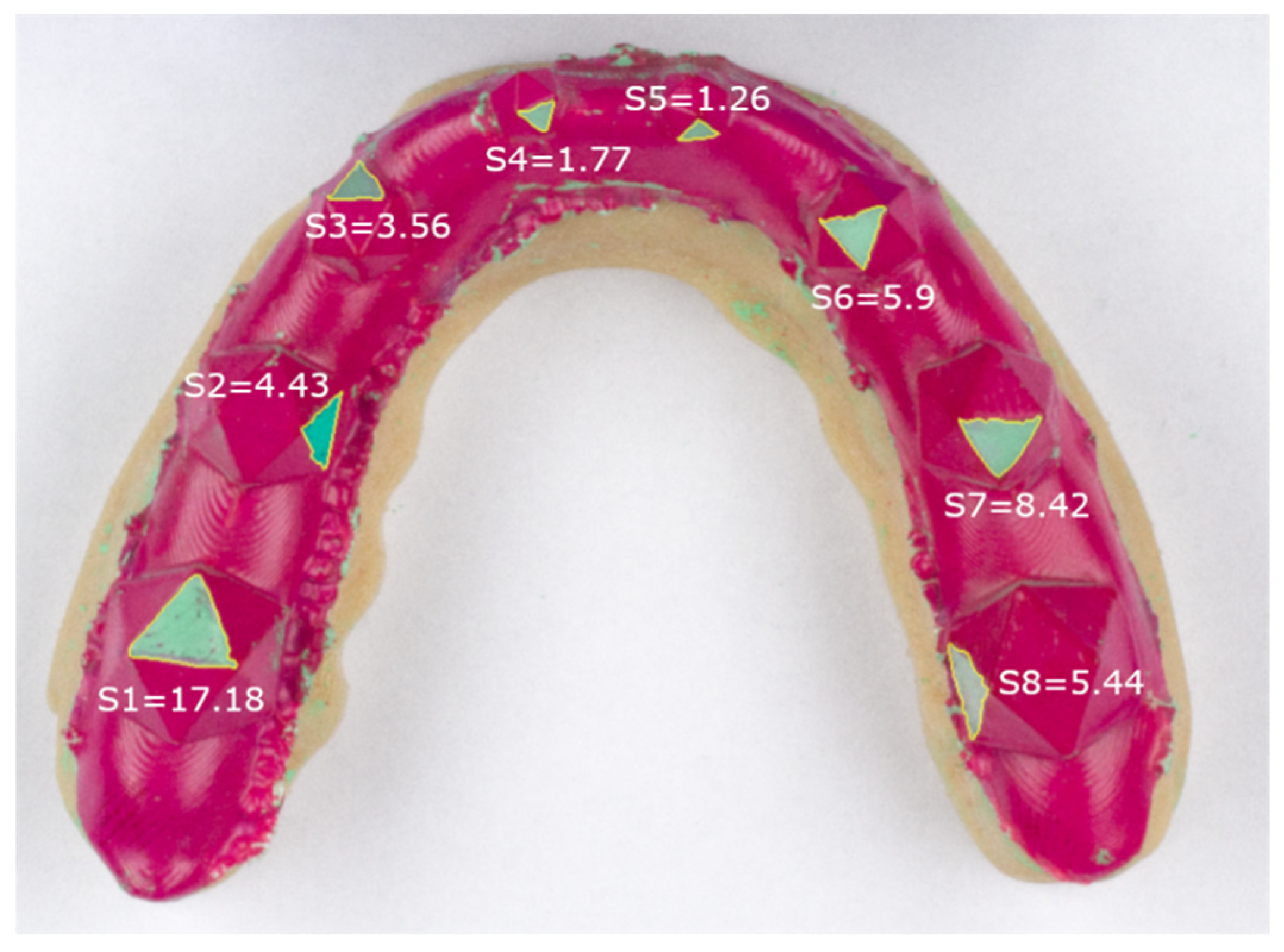

| Surface Area in 2D [mm2] | Surface Area in 3D [mm2] | p-Value | |
|---|---|---|---|
| Surface 1 | 13.2 ± 2.85 | 17.41 ± 1.2 | <0.001 |
| Surface 2 | 7.61 ± 2.27 | 11.20 ± 0.61 | <0.001 |
| Surface 3 | 4.90 ± 1.47 | 6.46 ± 0.61 | <0.001 |
| Surface 4 | 1.98 ± 0.37 | 2.78 ± 0.29 | <0.001 |
| Surface 5 | 2.01 ± 0.57 | 2.97 ± 0.32 | <0.001 |
| Surface 6 | 4.80 ± 1.66 | 6.45 ± 0.71 | <0.001 |
| Surface 7 | 8.80 ± 1.12 | 10.65 ± 0.44 | <0.001 |
| Surface 8 | 13.56 ± 3.16 | 16.93 ± 0.86 | <0.001 |
| Maximum [mm2] | Minimum [mm2] | Mean [mm2] | Percentage of Mean Surface | |
|---|---|---|---|---|
| Surface 1 | 0.41 | 0.11 | 0.25 | 2.35 |
| Surface 2 | 0.59 | 0.14 | 0.28 | 5.28 |
| Surface 3 | 0.48 | 0.09 | 0.19 | 7.43 |
| Surface 4 | 0.29 | 0.03 | 0.12 | 10.36 |
| Surface 5 | 0.26 | 0.08 | 0.15 | 8.91 |
| Surface 6 | 0.35 | 0.04 | 0.17 | 5.38 |
| Surface 7 | 0.59 | 0.15 | 0.28 | 5.5 |
| Surface 8 | 0.63 | 0.15 | 0.32 | 3.73 |
Publisher’s Note: MDPI stays neutral with regard to jurisdictional claims in published maps and institutional affiliations. |
© 2021 by the authors. Licensee MDPI, Basel, Switzerland. This article is an open access article distributed under the terms and conditions of the Creative Commons Attribution (CC BY) license (https://creativecommons.org/licenses/by/4.0/).
Share and Cite
Sagl, B.; Besirevic-Bulic, F.; Schmid-Schwap, M.; Laky, B.; Janjić, K.; Piehslinger, E.; Rausch-Fan, X. A Novel Quantitative Method for Tooth Grinding Surface Assessment Using 3D Scanning. Diagnostics 2021, 11, 1483. https://doi.org/10.3390/diagnostics11081483
Sagl B, Besirevic-Bulic F, Schmid-Schwap M, Laky B, Janjić K, Piehslinger E, Rausch-Fan X. A Novel Quantitative Method for Tooth Grinding Surface Assessment Using 3D Scanning. Diagnostics. 2021; 11(8):1483. https://doi.org/10.3390/diagnostics11081483
Chicago/Turabian StyleSagl, Benedikt, Ferida Besirevic-Bulic, Martina Schmid-Schwap, Brenda Laky, Klara Janjić, Eva Piehslinger, and Xiaohui Rausch-Fan. 2021. "A Novel Quantitative Method for Tooth Grinding Surface Assessment Using 3D Scanning" Diagnostics 11, no. 8: 1483. https://doi.org/10.3390/diagnostics11081483
APA StyleSagl, B., Besirevic-Bulic, F., Schmid-Schwap, M., Laky, B., Janjić, K., Piehslinger, E., & Rausch-Fan, X. (2021). A Novel Quantitative Method for Tooth Grinding Surface Assessment Using 3D Scanning. Diagnostics, 11(8), 1483. https://doi.org/10.3390/diagnostics11081483