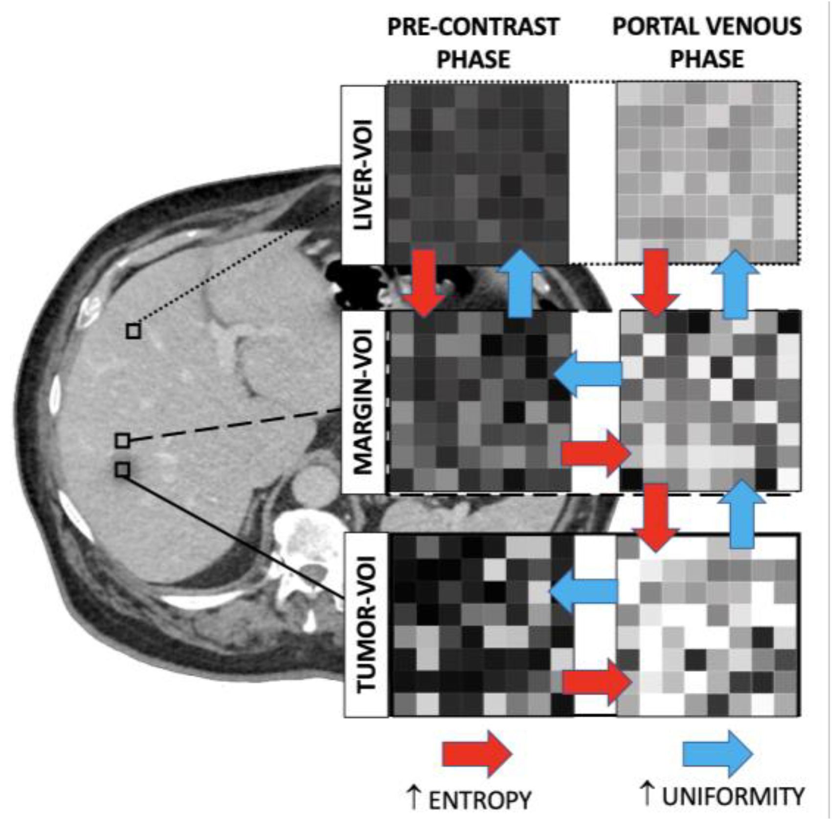
Contrast Administration Impacts CT-Based Radiomics of Colorectal Liver Metastases and Non-Tumoral Liver Parenchyma Revealing the “Radiological” Tumour Microenvironment

 , , ,
, , ,  ,
, 
Abstract
Share and Cite
Fiz, F.; Costa, G.; Gennaro, N.; la Bella, L.; Boichuk, A.; Sollini, M.; Politi, L.S.; Balzarini, L.; Torzilli, G.; Chiti, A.; et al. Contrast Administration Impacts CT-Based Radiomics of Colorectal Liver Metastases and Non-Tumoral Liver Parenchyma Revealing the “Radiological” Tumour Microenvironment. Diagnostics 2021, 11, 1162. https://doi.org/10.3390/diagnostics11071162
Fiz F, Costa G, Gennaro N, la Bella L, Boichuk A, Sollini M, Politi LS, Balzarini L, Torzilli G, Chiti A, et al. Contrast Administration Impacts CT-Based Radiomics of Colorectal Liver Metastases and Non-Tumoral Liver Parenchyma Revealing the “Radiological” Tumour Microenvironment. Diagnostics. 2021; 11(7):1162. https://doi.org/10.3390/diagnostics11071162
Chicago/Turabian StyleFiz, Francesco, Guido Costa, Nicolò Gennaro, Ludovico la Bella, Alexandra Boichuk, Martina Sollini, Letterio S. Politi, Luca Balzarini, Guido Torzilli, Arturo Chiti, and et al. 2021. "Contrast Administration Impacts CT-Based Radiomics of Colorectal Liver Metastases and Non-Tumoral Liver Parenchyma Revealing the “Radiological” Tumour Microenvironment" Diagnostics 11, no. 7: 1162. https://doi.org/10.3390/diagnostics11071162
APA StyleFiz, F., Costa, G., Gennaro, N., la Bella, L., Boichuk, A., Sollini, M., Politi, L. S., Balzarini, L., Torzilli, G., Chiti, A., & Viganò, L. (2021). Contrast Administration Impacts CT-Based Radiomics of Colorectal Liver Metastases and Non-Tumoral Liver Parenchyma Revealing the “Radiological” Tumour Microenvironment. Diagnostics, 11(7), 1162. https://doi.org/10.3390/diagnostics11071162