Case Report

A Case Report of Tongue Lymphoepithelial Carcinoma with a Histological Diagnostic Dilemma

1
Division of Oral and Maxillofacial Surgery, Department of Surgery Related, Kobe University Graduate School of Medicine, 7-5-2 Kusunoki-cho, Chuo-ku, Kobe 650-0017, Japan
2
Division of Pathology, Department of Pathology, Kobe University Graduate School of Medicine, 7-5-1 Kusunoki-cho, Chuo-ku, Kobe 650-0017, Japan
3
Division of Diagnostic Pathology, Department of Pathology, Kobe University Graduate School of Medicine, 7-5-2 Kusunoki-cho, Chuo-ku, Kobe 650-0017, Japan
*
Author to whom correspondence should be addressed.
Diagnostics 2021, 11(6), 1039; https://doi.org/10.3390/diagnostics11061039
Submission received: 21 April 2021 / Revised: 31 May 2021 / Accepted: 1 June 2021 / Published: 4 June 2021
(This article belongs to the Special Issue Biomarkers of Oral Cancer 2.0)

Abstract

:
Most head and neck lymphoepithelial carcinomas (LECs) arise in the nasopharynx and harbor Epstein–Barr virus (EBV). LEC is also a rare subtype of the oral squamous cell carcinoma (SCC). Morphologically, LEC is defined as resembling non-keratinizing nasopharyngeal carcinoma, undifferentiated subtype. The histological features and pathogenesis of oral LEC are not established. We describe a case of tongue LEC with histopathological diagnostic difficulties. A 72-year-old Japanese female presented with a whitish change on her left-side tongue. The diagnosis was atypical epithelium; neoplastic change could not be ruled out by a biopsy. Although the lesion was monitored at our hospital per her request, invasive carcinoma was detected 11 months later. Microscopically, conventional SCC was observed with the characteristic features as LEC confined to the deep part of the lesion. We briefly discuss this unusual histological finding and make a novel proposal for distinguishing oral LEC from LECs in other regions based on these histological findings.

1. Introduction

Oral squamous cell carcinoma (SCC) is a carcinoma with squamous differentiation arising from the mucosal epithelium [1]. There are architectural and cytological epithelial changes associated with an increased risk of progression to SCC. Unlike mucosal SCC with a high mutational burden due to chronic alcohol or tobacco exposure, virus-associated carcinomas have a distinct pathogenesis [2].
Lymphoepithelial carcinoma (LEC) is a ‘subtype’ of oral SCC [1]. In the World Health Organization (WHO) classification, LEC is defined as an “SCC morphologically similar to non-keratinizing nasopharyngeal carcinoma (NK-NPC), undifferentiated subtype” [1]. LEC is mostly located in the nasopharynx, and NK-NPC has a strong association with Epstein–Barr virus (EBV) [1]. EBV infection is consistently associated with LEC epidemiology and pathogenesis [3], but differences are noted depending on geographic regions and affected sites [4,5,6]. Once infected with EBV, the virus lies latent in the epithelial cells of the oropharynx and salivary glands and in B-lymphocytes and persists as a low-grade active infection throughout life [3].
Oral LEC is extremely rare, and not all LECs are EBV-positive [1]. The histological features of oral LECs and the biologic significance of EBV detection in LECs are not established. Different pathogenesis may be present in oral LEC without EBV detection. We report a case of tongue LEC with a histological diagnostic dilemma.

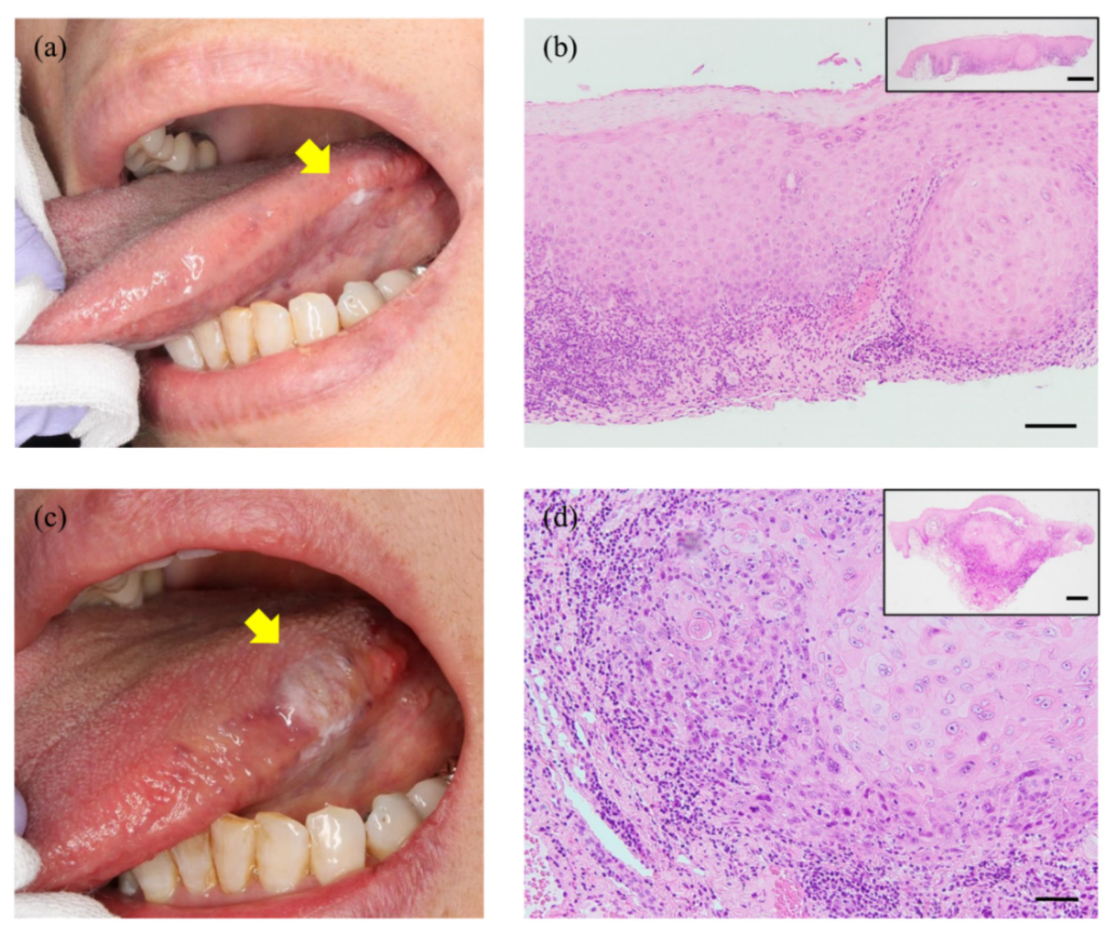
2. Case Presentation

A 72-year-old Japanese female was referred to our hospital for the examination of a whitish lesion on her left tongue. She became aware of its existence with discomfort 5 months prior to this presentation. She had a history of appendicitis and clear cell renal cell carcinoma, but the EBV infection had not been pointed out. Intraoral examination revealed a unilateral white patch with non-detachable at the left lingual edge (Figure 1a). We performed a local biopsy, and the histological diagnosis ‘atypical epithelium that does not rule out neoplastic change’ was obtained (Figure 1b). As she did not desire surgical treatment, we monitored the lesion, and 11 months later, a mass with induration was observed.
The exophytic lesion was ~12 × 6 mm in maximum diameter, and a pale whitish lesion was present in the anterior part (Figure 1c). We performed a second biopsy, and the histological diagnosis of SCC was confirmed (Figure 1d). Contrast-enhanced CT and FDG-PET showed no cervical lymph node metastasis. The clinical stage was thus classified as cT2N0M0 based on the TNM classification (8th edition) of the Union for International Cancer Control [7].
We performed a partial glossectomy with 10-mm tumor-free margins. A microscopic examination was performed using whole-tissue sections. The hematoxylin–eosin (HE)-stained specimens revealed a 4.6-mm-deep moderately differentiated conventional SCC characterized by invasion into the lamina propria with little keratin pearl formation, and cellular and nuclear pleomorphism was observed in whole tissue (Figure 2a). Solid nests with non-keratinization and lymphoplasmacytic cell infiltration at a deep stromal area were observed; the desmoplastic stromal reaction was not remarkable (Figure 2b). These findings are unusual patterns as conventional SCC. At higher magnification, the tumor cells showed large round-to-oval nuclei with hyperchromasia, an increased nuclear-to-cytoplasmic ratio, and prominent nucleoli; the indistinct cell border showed a syncytial appearance (Figure 2c). These findings were thought to be morphologically similar to NK-NPC. The pathologists discussed this histological diagnostic dilemma, LEC was considered in histological differential diagnosis.
In situ hybridization (ISH) was negative for EBV-encoded RNA (EBER) in the tumor cells (Figure 2d). These tumor cells were highlighted by a pan-cytokeratin marker, CK AE1/AE3 (Figure 2e), and tumor-infiltrating lymphocytes (TILs) did not show monoclonal reactivity for a B-cell marker (CD20) or a T-cell marker (CD3) (Figure 2f,g). The diagnosis of LEC (pT1cN0M0, stage I) was confirmed. She showed no evidence of disease at the 1-year follow-up. Written informed consent was obtained from the patient for the publication of this case report.

3. Discussion

The microscopic features of oral LEC are not established, due to the limited number of this rare tumor. We have identified only 16 cases of tongue LEC in the English literature [8,9,10], and all 16 cases were at the base of the tongue, and their association with EBV was not described. We have presented a case of LEC occurring at the lingual edge, which is extremely uncommon.
The most peculiar feature of the present case is the overlap of two histological features: conventional SCC and LEC. Although the whole cancer tissue exhibited features of conventional SCC, poorly differentiated malignant epithelial cells accompanied by TILs were observed in the deep area. The desmoplastic reaction has been highlighted as an important stromal reaction in various solid tumors, including oral SCC [11,12,13,14]. Unusually, the desmoplastic reaction was unremarkable at the invasive front in our patient. The lesion thus was finally diagnosed as LEC.
Oral SCCs are commonly well or moderately differentiated, and poorly differentiated cases are much less [1]. It was recently proposed that the characteristics of oral carcinogenesis differ from those of carcinogenesis of the uterine cervix or esophagus because oral SCC retains the maturation and differentiation characteristics of the stratified squamous epithelium [15,16,17]. It is thus reasonable to consider that the characteristics of oral LEC should be distinguished from LECs occurring at other sites. The specific findings of LEC as a ‘subtype’ of oral SCC characterized by superficial maturation and differentiation might be likely to appear in the deep area of the tumor tissue. Although we should recognize that only limited conclusions can be drawn from this single case, this histological finding might provide a clue to the differential diagnosis.
Most cases of head and neck LEC harbor EBV [1], but several reports [18,19,20,21] described cases of EBV-negative LEC in oral mucosa and adjacent structures [22]. Consistent with these reports, the ISH results was negative for EBV in our patient. These findings support our hypothesis that the characteristics of LEC in oral mucosa differ from those at other head and neck sites. To clarify what causes these differences, the pathogenesis of EBV-negative LEC should be determined.
Head and neck LECs tend to metastasize to regional lymph nodes, which affects the prognosis [1,9,23,24], and these tumors have high sensitivity to radiotherapy [1,9,23,25]. Our patients tumor was completely resected, and thus we did not conduct additional radiotherapy. In addition, no cervical lymph nodes suspected of metastasis were present preoperatively, we considered additional prophylactic neck dissection to be overtreatment. Despite the tendency of LECs in the prior reports to metastasize or spread locally, the prognosis remains rather favorable compared to other poorly differentiated epithelial tumors [24]. It was also suggested that patients with EBV-positive malignancies have better survival than those with EBV-negative malignancies, as is the case for oncogenic human papillomavirus-positive oropharyngeal cancer [21]. We plan to longitudinally monitor our patient.
In conclusion, this case report provides three take-home messages for readers. (1) While the tongue LECs in all of the published reports arose at the base of the tongue, this tongue LEC occurred at the lingual edge. (2) The most peculiar feature of the present case is the overlap of histological features of conventional SCC and LEC. The whole cancer tissue indicated conventional SCC, but poorly differentiated malignant epithelial cells and TILs were observed in the deep area. Although we should recognize the weakness of the single case, we would like to stress this histological feature as a promising clue to distinguish oral LEC from LECs at other sites. (3) Consistent with previous reports, in situ hybridization (ISH) was negative for EBV in this patient with oral LEC. These findings support our hypothesis that the characteristics of LEC in oral mucosa differ from those at other head and neck sites. To clarify what caused these differences, the pathogenesis of EBV-negative LEC should be determined in the future.

Author Contributions

D.T. was responsible for the clinical analysis and manuscript drafting. M.S. was responsible for the morphological and immunohistochemical evaluation and manuscript drafting. T.S. participated in the morphological and immunohistochemical evaluation. N.Y. and T.H. contributed to the collection of clinical record. M.A. edited the manuscript and supervised the study. All authors have read and agreed to the published version of the manuscript.

Funding

This work was supported by a Grant-in-Aid for Scientific Research (19K19157) from the Japan Society for Promotion of Science.

Institutional Review Board Statement

This report was conducted in compliance with the Declaration of Helsinki. Based on the Japanese “Ethical Guidelines for Medical and Health Research Involving Human Subjects”, case reports are not recognized as research. Therefore, Kobe University Hospital does not require ethical approval for reporting individual cases.

Informed Consent Statement

Written informed consent was obtained from the patient to publish this paper.

Acknowledgments

We thank Tomonori Tanaka for the valuable discussions and histopathological suggestions and all of the technicians of the Division of Diagnostic Pathology, Department of Pathology, Kobe University for the excellent technical assistance.

Conflicts of Interest

The authors declare no conflict of interest.

References

  1. El-Naggar, A.K.; Centre International de Recherche sur le cancer. WHO Classification of Head and Neck Tumours; IARC Publications: Lyon, France, 2017. [Google Scholar]
  2. Holmes, B.J.; Wenig, B.M. Virus-associated carcinomas of the head & neck: Update from the 2017 WHO classification. Ann. Diagn. Pathol. 2019, 38, 29–42. [Google Scholar] [CrossRef] [PubMed]
  3. Iezzoni, J.C.; Gaffey, M.J.; Weiss, L.M. The role of epstein-barr virus in lymphoepithelioma-like carcinomas. Am. J. Clin. Pathol. 1995, 103, 308–315. [Google Scholar] [CrossRef]
  4. Lanier, A.P.; Bornkamm, G.W.; Henle, W.; Henle, G.; Bender, T.R.; Talbot, M.L.; Dohan, P.H. Association of Epstein-Barr virus with nasopharyngeal carcinoma in alaskan native patients: Serum antibodies and tissue EBNA and DNA. Int. J. Cancer 1981, 28, 301–305. [Google Scholar] [CrossRef]
  5. Zong, Y.; Liu, K.; Zhong, B.; Chen, G.; Wu, W. Epstein-Barr virus infection of sinonasal lymphoepithelial carcinoma in Guangzhou. Chin. Med. J. 2001, 114, 132–136. [Google Scholar] [PubMed]
  6. Sckolnick, J.; Murphy, J.; Hunt, J.L. Microsatellite instability in nasopharyngeal and lymphoepithelial carcinomas of the head and neck. Am. J. Surg. Pathol. 2006, 30, 1250–1253. [Google Scholar] [CrossRef]
  7. Brierley, J.D.; Gospodarowicz, M.K.; Wittekind, C. TNM Classification of Malignant Tumours; John Wiley & Sons: New York, NY, USA, 2016; ISBN 978-1-119-26354-8. [Google Scholar]
  8. Bansberg, S.F.; Olsen, K.D.; Gaffey, T.A. Lymphoepithelioma of the Oropharynx. Otolaryngol. Head Neck Surg. 1989, 100, 303–307. [Google Scholar] [CrossRef] [PubMed]
  9. Dubey, P.; Ha, C.S.; Ang, K.K.; El-Naggar, A.K.; Knapp, C.; Byers, R.M.; Morrison, W.H. Nonnasopharyngeal lymphoepithelioma of the head and neck. Cancer 1998, 82, 1556–1562. [Google Scholar] [CrossRef]
  10. Merz, H.; Marnitz, S.; Erbersdobler, A.; Goektas, O. Schmincke’s Tumor, Carcinoma of the Base of the Tongue c T1-2, cN2c M0–A Case Report. Case Rep. Oncol. 2010, 3, 77–82. [Google Scholar] [CrossRef]
  11. Wang, L.M.; Silva, M.A.; D’Costa, Z.; Bockelmann, R.; Soonawalla, Z.; Liu, S.; O’Neill, E.; Mukherjee, S.; McKenna, W.G.; Muschel, R.; et al. The prognostic role of desmoplastic stroma in pancreatic ductal adenocarcinoma. Oncotarget 2016, 7, 4183–4194. [Google Scholar] [CrossRef] [Green Version]
  12. Ueno, H.; Kanemitsu, Y.; Sekine, S.; Ishiguro, M.; Ito, E.; Hashiguchi, Y.; Kondo, F.; Shimazaki, H.; Mochizuki, S.; Kajiwara, Y.; et al. Desmoplastic Pattern at the Tumor Front Defines Poor-prognosis Subtypes of Colorectal Cancer. Am. J. Surg. Pathol. 2017, 41, 1506–1512. [Google Scholar] [CrossRef]
  13. Ao, T.; Kajiwara, Y.; Yonemura, K.; Shinto, E.; Mochizuki, S.; Okamoto, K.; Aosasa, S.; Ueno, H. Prognostic significance of histological categorization of desmoplastic reaction in colorectal liver metastases. Virchows Archiv 2019, 475, 341–348. [Google Scholar] [CrossRef]
  14. Almangush, A.; Bello, I.O.; Heikkinen, I.; Hagström, J.; Haglund, C.; Kowalski, L.P.; Nieminen, P.; Coletta, R.D.; Mäkitie, A.A.; Salo, T.; et al. Stromal categorization in early oral tongue cancer. Virchows Archiv 2021, 478, 925–932. [Google Scholar] [CrossRef]
  15. Izumo, T. Oral premalignant lesions: From the pathological viewpoint. Int. J. Clin. Oncol. 2011, 16, 15–26. [Google Scholar] [CrossRef]
  16. Arsenic, R.; Kurrer, M.O. Differentiated dysplasia is a frequent precursor or associated lesion in invasive squamous cell carcinoma of the oral cavity and pharynx. Virchows Archiv 2013, 462, 609–617. [Google Scholar] [CrossRef] [PubMed] [Green Version]
  17. Ranganathan, K.; Kavitha, L. Oral epithelial dysplasia: Classifications and clinical relevance in risk assessment of oral potentially malignant disorders. J. Oral Maxillofac. Pathol. 2019, 23, 19–27. [Google Scholar] [CrossRef] [PubMed]
  18. Mahomed, F.; Grayson, W. A rare case of lymphoepithelial carcinoma of the lip. Oral Surgery, Oral Med. Oral Pathol. Oral Radiol. Endodontol. 2008, 105, e49–e52. [Google Scholar] [CrossRef] [PubMed]
  19. Gultekin, M.; Omer, G.; Sefik, H.; Mustaf, C.; Murat, G. Lymphoepithelial Carcinoma of the Lower Lip: Report of a Case. Int. J. Hematol. Oncol. 2014, 24, 70–72. [Google Scholar] [CrossRef]
  20. Almeida, L.Y.; Silveira, H.A.; Silva, E.V.; Barbeiro, C.D.O.; De Paula, J.A.; Bufalino, A.; Ribeiro-Silva, A.; León, J.E. EBV-negative lymphoepithelial-like carcinoma of the lower lip. Autops. Case Rep. 2019, 10, e2020138. [Google Scholar] [CrossRef] [Green Version]
  21. Shimizu, S.; Miyazaki, A.; Nakamori, K.; Nakai, H.; Ogi, K.; Hasegawa, T.; Hiratsuka, H. Immunophenotypic analysis of tumor infiltrating lymphocytes in Epstein-Barr virus-negative lymphoepithelial carcinoma of the oral cavity: Report of a case. J. Oral Maxillofac. Surg. Med. Pathol. 2017, 29, 581–586. [Google Scholar] [CrossRef]
  22. Izumo, T.; Kirita, T.; Ariji, E.; Ozeki, S.; Okada, N.; Okabe, S.; Okazaki, Y.; Omura, K.; Kusama, M.; Sato, T.; et al. General rules for clinical and pathological studies on oral cancer: A synopsis. Jpn. J. Clin. Oncol. 2012, 42, 1099–1109. [Google Scholar] [CrossRef] [PubMed] [Green Version]
  23. Chow, T.; Lui, Y.; Sze, W.; Yuen, N.; Kwok, S.; Chow, T. Lymphoepithelioma-like carcinoma of oral cavity: Report of three cases and literature review. Int. J. Oral Maxillofac. Surg. 2002, 31, 212–218. [Google Scholar] [CrossRef] [PubMed]
  24. Rytkönen, A.E.; Hirvikoski, P.P.; Salo, T.A. Lymphoepithelial carcinoma: Two case reports and a systematic review of oral and sinonasal cases. Head Neck Pathol. 2011, 5, 327–334. [Google Scholar] [CrossRef] [PubMed] [Green Version]
  25. Wenig, B.M. Lymphoepithelial-like carcinomas of the head and neck. Semin. Diagn. Pathol. 2015, 32, 74–86. [Google Scholar] [CrossRef] [PubMed]
Figure 1. Clinical appearance and histological findings of the patient’s biopsy. (a) A whitish lesion was found on the left lingual edge at her first visit (arrow). (b) This lesion was histologically diagnosed as ‘atypical epithelium that does not rule out neoplastic change’ based on this first biopsy. (c) After 11 months of monitoring, a mass with induration was observed (arrow). (d) A morphological evaluation by second biopsy confirmed invasive SCC with dyskeratosis. Scale bars: 100 µm (b), 50 µm (d). Insets in (b,d) are lower magnifications of the images (scale bars: 500 µm).
Diagnostics 11 01039 g001
Figure 2. The morphological and immunohistochemical images. (a) Low-power image (HE staining). Invasive cancer was detected in resected specimens. The depth of invasion was 4.6 mm. The lesion was classified as pT1cN0M0. Dotted line indicates a feature of conventional SCC. (b) In the deep area (square A), the observation of the cancer nests was difficult due to lymphoplasmacytic infiltration. Both keratinization and desmoplastic stromal reaction were absent. (c) High-power view of the invasive front (square B). The tumor cells have enlarged vesicular nuclei with prominent nucleoli. Nuclear pleomorphism is also increased. (d) The tumor cells were negative for EBER-ISH. (e) The tumor cells were positive for CK AE1/AE3. (f,g) The monoclonal reactivities for both a B-cell (CD20) and a T-cell (CD3) marker in infiltrating lymphocytes could not be detected. Scale bars: 1 mm (a), 100 µm (b,e), 20 µm (c,d,f,g).
Diagnostics 11 01039 g002
Publisher’s Note: MDPI stays neutral with regard to jurisdictional claims in published maps and institutional affiliations.

