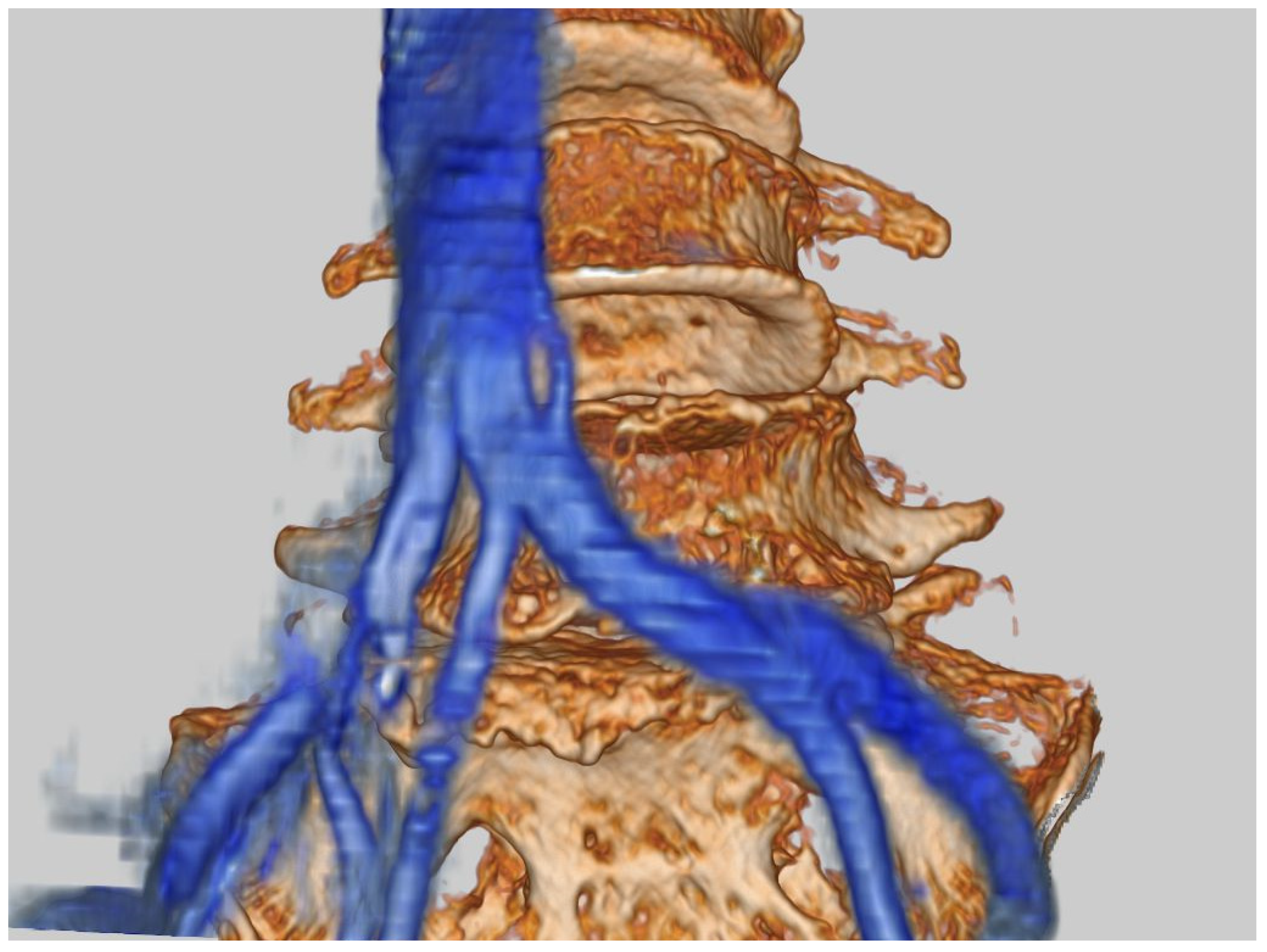
Assessment of 3D Lumbosacral Vascular Anatomy for OLIF51 by Non-Enhanced MRI and CT Medical Image Fusion Technique

 , ,
, , Abstract
Share and Cite
Nagamatsu, M.; Ruparel, S.; Tanaka, M.; Fujiwara, Y.; Uotani, K.; Arataki, S.; Yamauchi, T.; Takeshita, Y.; Takamoto, R.; Tanaka, M.; et al. Assessment of 3D Lumbosacral Vascular Anatomy for OLIF51 by Non-Enhanced MRI and CT Medical Image Fusion Technique. Diagnostics 2021, 11, 1744. https://doi.org/10.3390/diagnostics11101744
Nagamatsu M, Ruparel S, Tanaka M, Fujiwara Y, Uotani K, Arataki S, Yamauchi T, Takeshita Y, Takamoto R, Tanaka M, et al. Assessment of 3D Lumbosacral Vascular Anatomy for OLIF51 by Non-Enhanced MRI and CT Medical Image Fusion Technique. Diagnostics. 2021; 11(10):1744. https://doi.org/10.3390/diagnostics11101744
Chicago/Turabian StyleNagamatsu, Masakazu, Sameer Ruparel, Masato Tanaka, Yoshihiro Fujiwara, Koji Uotani, Shinya Arataki, Taro Yamauchi, Yoshiyuki Takeshita, Rika Takamoto, Masato Tanaka, and et al. 2021. "Assessment of 3D Lumbosacral Vascular Anatomy for OLIF51 by Non-Enhanced MRI and CT Medical Image Fusion Technique" Diagnostics 11, no. 10: 1744. https://doi.org/10.3390/diagnostics11101744
APA StyleNagamatsu, M., Ruparel, S., Tanaka, M., Fujiwara, Y., Uotani, K., Arataki, S., Yamauchi, T., Takeshita, Y., Takamoto, R., Tanaka, M., & Moriue, S. (2021). Assessment of 3D Lumbosacral Vascular Anatomy for OLIF51 by Non-Enhanced MRI and CT Medical Image Fusion Technique. Diagnostics, 11(10), 1744. https://doi.org/10.3390/diagnostics11101744