Hydrophilic Biocompatible Poly(Acrylic Acid-co-Maleic Acid) Polymer as a Surface-Coating Ligand of Ultrasmall Gd2O3 Nanoparticles to Obtain a High r1 Value and T1 MR Images


 ,
,  , ,
, , 
Abstract
1. Introduction
2. Materials and Methods
2.1. Chemicals
2.2. Synthesis of PAAMA-Coated Ultrasmall Gd2O3 Nanoparticles
2.3. Characterizations
2.4. In Vitro Cellular Toxicity Measurements
2.5. Relaxometric Property Measurements
2.6. In Vivo T1 MR Image Measurements
3. Results
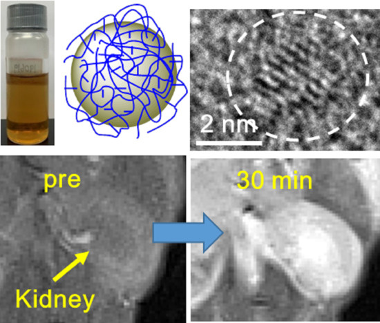
3.1. Particle Diameter
3.2. Colloidal Stability
3.3. Crystal Structure
3.4. Surface-Coating Results
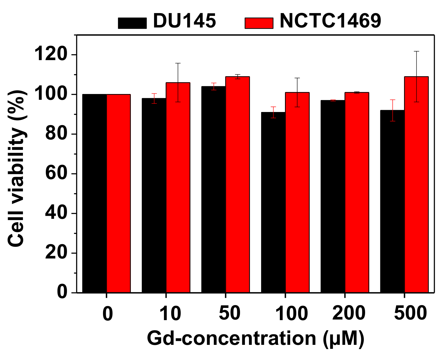
3.5. Cell Viability
3.6. Magnetic Properties
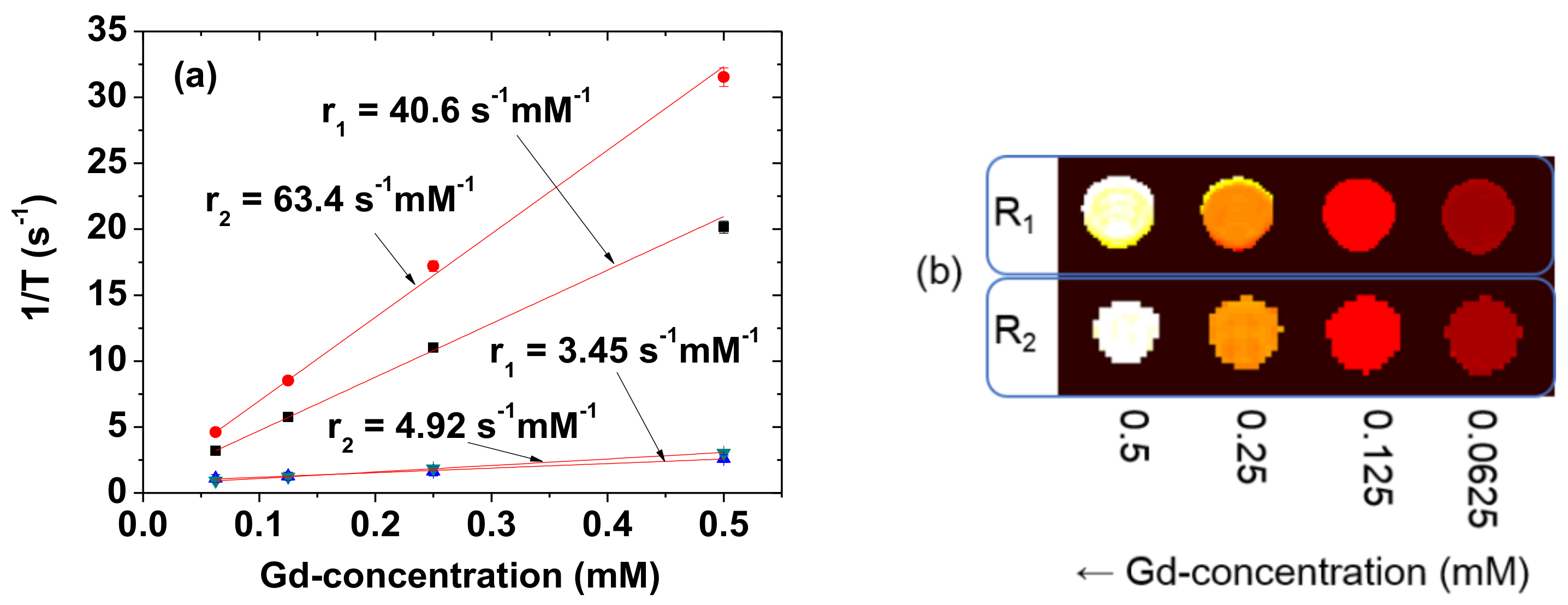
3.7. r1 and r2 Values
3.8. In Vivo T1 MR Images at the 3.0 T MR Field
4. Discussion
5. Conclusions
- (1)
- The abundant carboxylic groups in PAAMA allowed its strong bonding to the nanoparticle surface and the attraction of numerous water molecules around the nanoparticle surface, thereby achieving a large aavg of 9.0 nm. This led to an extremely high r1 value of 40.6 s−1mM−1 (r2/r1 = 1.56) and good colloidal stability, as confirmed by the high zeta potential and absence of precipitation of the nanoparticles in the aqueous solution;
- (2)
- The biocompatible PAAMA coating resulted in extremely low cellular toxicity;
- (3)
- The observed r1 value was ~10 times higher than those of commercial molecular contrast agents. Thus, strong positive-contrast enhancements in the in vivo T1 MR images were observed. However, more studies, including the pharmacokinetic study, are required to further demonstrate the possibility of using the synthesized nanoparticles as a powerful T1 MRI contrast agent.
Author Contributions
Funding
Acknowledgments
Conflicts of Interest
References
- Hashemi, R.H.; Bradley, W.G.; Lisanti, C.J. MRI the Basics; Lippincott Williams & Wilkins: New York, NY, USA, 2004. [Google Scholar]
- Lauffer, R.B. Paramagnetic metal complexes as water proton relaxation agents for NMR imaging: Theory and design. Chem. Rev. 1987, 87, 901–927. [Google Scholar] [CrossRef]
- Caravan, P.; Ellison, J.J.; McMurry, T.J.; Lauffer, R.B. Gadolinium(III) chelates as MRI contrast agents: Structure, dynamics, and applications. Chem. Rev. 1999, 99, 2293–2352. [Google Scholar] [CrossRef]
- Xiao, Y.-D.; Paudel, R.; Liu, J.; Ma, C.; Zhang, Z.-S.; Zhou, S.-K. MRI contrast agents: Classification and application (Review). Int. J. Mol. Med. 2016, 38, 1319–1326. [Google Scholar] [CrossRef]
- Greenwood, N.N.; Earnshaw, A. Chemistry of the Elements; Butterworth-Heinemann: Oxford, UK, 1997; p. 1243. [Google Scholar]
- Guay-Bégin, A.-A.; Chevallier, P.; Faucher, L.; Turgeon, S.; Fortin, A.-A. Surface modification of gadolinium oxide thin films and nanoparticles using poly(ethylene glycol)-phosphate. Langmuir 2012, 28, 774–782. [Google Scholar] [CrossRef]
- Engström, M.; Klasson, A.; Pedersen, H.; Vahlberg, C.; Käll, P.-O.; Uvdal, K. High proton relaxivity for gadolinium oxide nanoparticles. Magn. Reson. Mater. Phys. Biol. Med. 2006, 19, 180–186. [Google Scholar] [CrossRef]
- Mekuria, S.L.; Debele, T.A.; Tsai, H.-C. Encapsulation of gadolinium oxide nanoparticle (Gd2O3) contrasting agents in PAMAM dendrimer templates for enhanced magnetic resonance imaging in vivo. ACS Appl. Mater. Interfaces 2017, 9, 6782–6795. [Google Scholar] [CrossRef] [PubMed]
- Cho, M.; Sethi, R.; Ananta narayanan, J.S.; Lee, S.S.; Benoit, D.N.; Taheri, N.; Decuzzi, P.; Colvin, V.L. Gadolinium oxide nanoplates with high longitudinal relaxivity for magnetic resonance imaging. Nanoscale 2014, 6, 13637–13645. [Google Scholar] [CrossRef] [PubMed]
- Choi, H.S.; Liu, W.; Misra, P.; Tanaka, E.; Zimmer, J.P.; Ipe, B.I.; Bawendi, M.G.; Frangioni, J.V. Renal clearance of quantum dots. Nat. Biotechnol. 2007, 25, 1165–1170. [Google Scholar] [CrossRef] [PubMed]
- Longmire, M.; Choyke, P.L.; Kobayashi, H. Clearance properties of nano-sized particles and molecules as imaging agents: Considerations and caveats. Nanomedicine 2008, 3, 703–717. [Google Scholar] [CrossRef] [PubMed]
- Hainfeld, J.F.; Slatkin, D.N.; Focella, T.M.; Smilowitz, H.M. Gold nanoparticles: A new X-ray contrast agent. Br. J. Radiol. 2006, 79, 248–253. [Google Scholar] [CrossRef] [PubMed]
- Singh, R.; Singh, S. Surface Modification of Nanomaterials for Biomedical Applications: Strategies and Recent Advances. In Nanobiotechnology; Dhawan, A., Singh, S., Kumar, A., Shanker, R., Eds.; CRC Press: Boca Raton, FL, USA, 2018. [Google Scholar]
- Zeng, J.; Jing, L.; Hou, Y.; Jiao, M.; Qiao, R.; Jia, Q.; Liu, C.; Fang, F.; Lei, H.; Gao, M. Anchoring group effects of surface ligands on magnetic properties of Fe3O4 nanoparticles: Towards high performance MRI contrast agents. Adv. Mater. 2014, 26, 2694–2698. [Google Scholar] [CrossRef] [PubMed]
- Tegafaw, T.; Xu, W.; Lee, S.-H.; Chae, K.S.; Cha, H.; Chang, Y.; Lee, G.H. Ligand-size and ligand-chain hydrophilicity effects on the relaxometric properties of ultrasmall Gd2O3 nanoparticles. AIP Adv. 2016, 6, 065114. [Google Scholar] [CrossRef]
- Duan, H.; Kuang, M.; Wang, X.; Wang, Y.A.; Mao, H.; Nie, S. Reexamining the effects of particle size and surface chemistry on the magnetic properties of iron oxide nanocrystals: New insights into spin disorder and proton relaxivity. J. Phys. Chem. C 2008, 112, 8127–8131. [Google Scholar] [CrossRef]
- Thomsen, H.S. Nephrogenic systemic fibrosis: A serious late adverse reaction to gadodiamide. Eur. Radiol. 2006, 16, 2619–2621. [Google Scholar] [CrossRef]
- Gao, S.; Chen, M.-L.; Zhou, Z.-H. Substitution of gadolinium ethylenediaminetetraacetate with phosphites: Towards gadolinium deposit in nephrogenic systemic fibrosis. Dalton Trans. 2014, 43, 639–645. [Google Scholar] [CrossRef]
- Penfield, J.G.; Reilly, R.F., Jr. What nephrologists need to know about gadolinium. Nat. Clin. Pract. Nephrol. 2007, 3, 654–668. [Google Scholar] [CrossRef]
- Hifumi, H.; Yamaoka, S.; Tanimoto, A.; Citterio, D.; Suzuki, K. Gadolinium-based hybrid nanoparticles as a positive MR contrast agent. J. Am. Chem. Soc. 2006, 128, 15090–15091. [Google Scholar] [CrossRef]
- Bridot, J.-L.; Faure, A.-C.; Laurent, S.; Rivière, C.; Billotey, C.; Hiba, B.; Janier, M.; Josserand, V.; Coll, J.-L.; Elst, L.V.; et al. Hybrid gadolinium oxide nanoparticles: Multimodal contrast agents for in vivo imaging. J. Am. Chem. Soc. 2007, 129, 5076–5084. [Google Scholar] [CrossRef]
- Ottenbrite, R.M. Antitumor activity of polycarboxylic acid polymers. J. Macromol. Sci. A 1985, 22, 819–832. [Google Scholar] [CrossRef]
- Tóth, I.Y.; Illés, E.; Bauer, R.A.; Nesztor, D.; Szekeres, M.; Zupkó, I.; Tombácz, E. Designed polyelectrolyte shell on magnetite nanocore for dilution-resistant biocompatible magnetic fluids. Langmuir 2012, 28, 16638–16646. [Google Scholar] [CrossRef]
- International Center for Diffraction Data. Card No. 43-1014, JCPDS-International Centre for Diffraction Data, PCPDFWIN, Software Version 1.30; International Center for Diffraction Data: Newtown Square, PA, USA, 1997. [Google Scholar]
- Söderlind, F.; Pedersen, H.; Petoral, R.M., Jr.; Käll, P.-O.; Uvdal, K. Synthesis and characterisation of Gd2O3 nanocrystals functionalised by organic acids. J. Colloid Interface Sci. 2005, 288, 140–148. [Google Scholar] [CrossRef] [PubMed]
- Kruis, F.E.; Nielsch, K.; Fissan, H.; Rellinghaus, B.; Wassermann, E.F. Preparation of size-classified PbS nanoparticles in the gas phase. Appl. Phys. Lett. 1998, 73, 547–549. [Google Scholar] [CrossRef]
- Duckworth, O.W.; Martin, S.T. Surface complexation and dissolution of hematite by C1-C6 dicarboxylic acids at pH = 5.0. Geochim. Cosmochim. Acta 2001, 65, 4289–4301. [Google Scholar] [CrossRef]
- Hug, S.J.; Bahnemann, D. Infrared spectra of oxalate, malonate and succinate adsorbed on the aqueous surface of rutile, anatase and lepidocrocite measured with in situ ATR-FTIR. J. Electron Spectrosc. Related Phenomena 2006, 150, 208–219. [Google Scholar] [CrossRef]
- Hug, S.J.; Sulzberger, B. In situ Fourier transform infrared spectroscopic evidence for the formation of several different surface complexes of oxalate on TiO2 in the aqueous phase. Langmuir 1994, 10, 3587–3597. [Google Scholar] [CrossRef]
- Mendive, C.B.; Bredow, T.; Blesa, M.A.; Bahnemann, D.W. ATR-FTIR measurements and quantum chemical calculations concerning the adsorption and photoreaction of oxalic acid on TiO2. Phys. Chem. Chem. Phys. 2006, 8, 3232–3247. [Google Scholar] [CrossRef]
- Pearson, R.G. Hard and soft acids and bases, HSAB, part 1: Fundamental principles. J. Chem. Edu. 1968, 45, 581–587. [Google Scholar] [CrossRef]
- Pearson, R.G. Hard and soft acids and bases, HSAB, part II: Underlying theories. J. Chem. Edu. 1968, 45, 643–648. [Google Scholar] [CrossRef]
- Haynes, W.M.; Lide, D.R.; Bruno, T.J. CRC Handbook of Chemistry and Physcis; CRC Press: Boca Raton, FL, USA, 2015–2016; pp. 4–64. [Google Scholar]
- Corbierre, M.K.; Cameron, N.S.; Lennox, R.B. Polymer-stabilized gold nanoparticles with high grafting densities. Langmuir 2004, 20, 2867–2873. [Google Scholar] [CrossRef]
- Benoit, D.N.; Zhu, H.; Lilierose, M.H.; Verm, R.A.; Ali, N.; Morrison, A.N.; Fortner, J.D.; Avendano, C.; Colvin, V.L. Measuring the grafting density of nanoparticles in solution by analytical ultracentrifugation and total organic carbon analysis. Anal. Chem. 2012, 84, 9238–9245. [Google Scholar] [CrossRef]
- Moon, R.M.; Koehler, W.C. Magnetic properties of Gd2O3. Phys. Rev. B 1975, 11, 1609–1622. [Google Scholar] [CrossRef]
- Schinkel, C.J.; van Amstel, W.D. Reduced magnetic moment of gadolinium in the oxyde and the sulphate. Phys. Lett. 1973, 44, 467–468. [Google Scholar] [CrossRef]
- Wolf, W.P.; Meissner, H.; Catanese, C.A. Magnetic properties of rare earth hydroxides. J. Appl. Phys. 1968, 39, 1134–1136. [Google Scholar] [CrossRef]
- Park, J.Y.; Baek, M.J.; Choi, E.S.; Woo, S.; Kim, J.H.; Kim, T.J.; Jung, J.C.; Chae, K.S.; Chang, Y.; Lee, G.H. Paramagnetic ultrasmall gadolinium oxide nanoparticles as advanced T1 MRI contrast agent: Account for large longitudinal relaxivity, optimal particle diameter, and in vivo T1 MR images. ACS Nano 2009, 3, 3663–3669. [Google Scholar] [CrossRef]
- Wyss, P.P.; Lamichhane, S.P.; Abed, A.; Vonwil, D.; Kretz, O.; Huber, T.B.; Sarem, M.; Shastri, V.P. Renal clearance of polymeric nanoparticles by mimicry of glycan surface of viruses. Biomaterials 2020, 230, 119643. [Google Scholar] [CrossRef]
- Huang, H.; Hernandez, R.; Geng, J.; Sun, H.; Song, W.; Chen, F.; Graves, S.A.; Nickles, R.J.; Cheng, C.; Cai, W.; et al. A porphyrin-PEG polymer with rapid renal clearance. Biomaterials 2016, 76, 25–32. [Google Scholar] [CrossRef]
- Miao, X.; Ho, S.L.; Tegafaw, T.; Cha, H.; Chang, Y.; Oh, I.T.; Yaseen, A.M.; Marasini, S.; Ghazanfari, A.; Yue, H.; et al. Stable and non-toxic ultrasmall gadolinium oxide nanoparticle colloids (coating material = polyacrylic acid) as high-performance T1 magnetic resonance imaging contrast agents. RSC Adv. 2018, 8, 3189–3197. [Google Scholar] [CrossRef]
- Ahmad, M.Y.; Ahmad, M.W.; Yue, H.; Ho, S.L.; Park, J.A.; Jung, K.-H.; Cha, H.; Marasini, S.; Ghazanfari, A.; Liu, S.; et al. In vivo positive magnetic resonance imaging applications of poly(methyl vinyl ether-alt-maleic acid)-coated ultra-small paramagnetic gadolinium oxide nanoparticles. Molecules 2020, 25, 1159. [Google Scholar] [CrossRef]


| davg (nm) | aavg (nm) | ξavg (mV) | Average Surface-Coating Amount | Net M at 2.0 T and 300 K (emu/g) 4 | Water Proton Spin Relaxivity at 3.0 T and 22 °C (s−1 mM−1) | |||
|---|---|---|---|---|---|---|---|---|
| P 1 (wt.%) | σ 2 (nm−2) | N 3 | r1 | r2 | ||||
| 1.8 ± 0.1 | 9.0 ± 0.2 | −43.9 ± 0.2 | 40.3 ± 0.2 | 0.48 ± 0.05 | 6 ± 1 | 1.71 ± 0.05 | 40.6 ± 0.1 | 63.4 ± 0.1 |
| Chemical | Polymer | MW (amu) | davg (nm) | aavg (nm) | Water Proton Spin Relaxivity at 3.0 T and 22 °C (s−1mM−1) | Ref. | |
|---|---|---|---|---|---|---|---|
| r1 | r2 | ||||||
| Gd2O3 | PAAMA | 3000 | 1.8 | 9.0 | 40.6 | 63.4 | This work |
| Gd2O3 | PAA 1 | 5000 | 2.0 | 6.3 | 31.0 | 37.4 | [42] |
| Gd2O3 | PMVEMA 2 | 80,000 | 1.9 | 19.8 | 36.2 | 74.0 | [43] |
| Gd-DOTA 3 | - | - | - | - | 3.45 | 4.92 | This work |
Publisher’s Note: MDPI stays neutral with regard to jurisdictional claims in published maps and institutional affiliations. |
© 2020 by the authors. Licensee MDPI, Basel, Switzerland. This article is an open access article distributed under the terms and conditions of the Creative Commons Attribution (CC BY) license (http://creativecommons.org/licenses/by/4.0/).
Share and Cite
Jang, Y.-J.; Liu, S.; Yue, H.; Park, J.A.; Cha, H.; Ho, S.L.; Marasini, S.; Ghazanfari, A.; Ahmad, M.Y.; Miao, X.; et al. Hydrophilic Biocompatible Poly(Acrylic Acid-co-Maleic Acid) Polymer as a Surface-Coating Ligand of Ultrasmall Gd2O3 Nanoparticles to Obtain a High r1 Value and T1 MR Images. Diagnostics 2021, 11, 2. https://doi.org/10.3390/diagnostics11010002
Jang Y-J, Liu S, Yue H, Park JA, Cha H, Ho SL, Marasini S, Ghazanfari A, Ahmad MY, Miao X, et al. Hydrophilic Biocompatible Poly(Acrylic Acid-co-Maleic Acid) Polymer as a Surface-Coating Ligand of Ultrasmall Gd2O3 Nanoparticles to Obtain a High r1 Value and T1 MR Images. Diagnostics. 2021; 11(1):2. https://doi.org/10.3390/diagnostics11010002
Chicago/Turabian StyleJang, Yeong-Ji, Shuwen Liu, Huan Yue, Ji Ae Park, Hyunsil Cha, Son Long Ho, Shanti Marasini, Adibehalsadat Ghazanfari, Mohammad Yaseen Ahmad, Xu Miao, and et al. 2021. "Hydrophilic Biocompatible Poly(Acrylic Acid-co-Maleic Acid) Polymer as a Surface-Coating Ligand of Ultrasmall Gd2O3 Nanoparticles to Obtain a High r1 Value and T1 MR Images" Diagnostics 11, no. 1: 2. https://doi.org/10.3390/diagnostics11010002
APA StyleJang, Y.-J., Liu, S., Yue, H., Park, J. A., Cha, H., Ho, S. L., Marasini, S., Ghazanfari, A., Ahmad, M. Y., Miao, X., Tegafaw, T., Chae, K.-S., Chang, Y., & Lee, G. H. (2021). Hydrophilic Biocompatible Poly(Acrylic Acid-co-Maleic Acid) Polymer as a Surface-Coating Ligand of Ultrasmall Gd2O3 Nanoparticles to Obtain a High r1 Value and T1 MR Images. Diagnostics, 11(1), 2. https://doi.org/10.3390/diagnostics11010002