Review

Metals in Cyanobacteria: Analysis of the Copper, Nickel, Cobalt and Arsenic Homeostasis Mechanisms

by
María José Huertas
1,†,
Luis López-Maury
1,†,
Joaquín Giner-Lamia
2,
Ana María Sánchez-Riego
1 and
Francisco Javier Florencio
1,*
1
Instituto de Bioquímica Vegetal y Fotosíntesis, Universidad de Sevilla-CSIC, Américo Vespucio 49, E-41092 Sevilla, Spain
2
Systems Biology and Bioinformatics Laboratory, IBB-CBME, University of Algarve, Campus de Gambelas, 8005-139 Faro, Portugal
*
Author to whom correspondence should be addressed.
These authors contributed equally to this work.
Life 2014, 4(4), 865-886; https://doi.org/10.3390/life4040865
Submission received: 30 October 2014 / Revised: 27 November 2014 / Accepted: 4 December 2014 / Published: 9 December 2014
(This article belongs to the Special Issue Cyanobacteria: Ecology, Physiology and Genetics)

Abstract

:
Traces of metal are required for fundamental biochemical processes, such as photosynthesis and respiration. Cyanobacteria metal homeostasis acquires an important role because the photosynthetic machinery imposes a high demand for metals, making them a limiting factor for cyanobacteria, especially in the open oceans. On the other hand, in the last two centuries, the metal concentrations in marine environments and lake sediments have increased as a result of several industrial activities. In all cases, cells have to tightly regulate uptake to maintain their intracellular concentrations below toxic levels. Mechanisms to obtain metal under limiting conditions and to protect cells from an excess of metals are present in cyanobacteria. Understanding metal homeostasis in cyanobacteria and the proteins involved will help to evaluate the use of these microorganisms in metal bioremediation. Furthermore, it will also help to understand how metal availability impacts primary production in the oceans. In this review, we will focus on copper, nickel, cobalt and arsenic (a toxic metalloid) metabolism, which has been mainly analyzed in model cyanobacterium Synechocystis sp. PCC 6803.

Graphical Abstract

1. Introduction

Metals are basic micronutrients for all living organisms and play essential roles in fundamental biochemical processes that sustain life on Earth, like respiration and photosynthesis. They are critical for correct folding and functioning of many proteins, but many metal-containing proteins remain unknown, at least in their function [1]. Recently, it has been shown that up to a third of the total proteome of microbes contains a metal cofactor [2], but there are limitations in metallo-protein prediction that make it difficult to predict if a protein will contain metals or not. In vitro, the majority of metalloproteins will bind metals with an order of affinity following the Irving-Williams series for divalent cations (Mg2+ < Ca2+ < Mn2+ < Fe2+ < Co2+ < Ni2+ < Cu2+ > Zn2+), and therefore, cells have a great challenge delivering the right metal to the right proteins [1]. Furthermore, when the concentrations of metals increase in the cell, they undergo undesirable redox reactions or bind inappropriately to the metal binding sites of several enzymes, leading to toxic effects [3,4]. Thus, the acquisition of metals has to be strictly balanced in the cells, and hence, all organisms have evolved to respond to the bioavailability of metals in order to sustain metal homeostasis.
The occurrence of heavy metals in the biosphere is a natural phenomenon, as they are elements that cannot be destroyed or degraded. In the last century, heavy metals have accumulated in the environment as a result of their enhanced utilization in several industrial activities, including their extraction in mining processes. Their management is a major global concern, because of their toxicity, and a threat to both human life and biosphere conservation. Heavy metal accumulation has been especially intense in marine environments [5] and lake sediments [6]. In metal-polluted environments, metal homeostasis is important to protect cells from metal overload, which will cause oxidative stress, lipid peroxidation, damage of nucleic acids and the inhibition of essential processes, like respiration, carbon metabolism and photosynthesis [7,8]. One of the approaches to control the presence of heavy metals involves the use of microorganisms to remediate these contaminants [8,9].
Cyanobacteria are the only group of prokaryotes capable of performing oxygenic photosynthesis and spread out in almost any ecological niche from fresh and salt water to terrestrial and extreme environments, including metal-contaminated areas [10]. Metal homeostasis is especially important in cyanobacteria, because the photosynthetic machinery imposes a high demand for metals, acting as cofactors of several proteins [11]. Metals play essential roles in cyanobacterial metabolism, for example: iron is required as a cofactor for all three photosynthetic electron transfer chain complexes [7]; Mn is specifically required for PSII (Photosystem II) function [12]; Mg is coordinated at the center of the chlorophyll ring [13]; and zinc is required for carbonic anhydrase [14]. In fact, the distribution and abundance of marine cyanobacteria and, therefore, phytoplankton’s productivity in natural habitats is often determined by the metal bioavailability, especially by iron, but also by copper, cobalt, zinc or nickel [15,16,17]. This availability can limit cyanobacterial growth, not only because of scarcity, but also due to toxic effects, as it has been shown that different clades and species have different metal susceptibilities [15,16].
Adaptation to high metal concentration in cyanobacteria includes mechanisms, such as their ability to excrete to the media heavy metal ligands, like siderophores [18,19] or extracellular polymeric substances (EPS) [9,20,21,22], the production of metallothioneins [23,24,25,26,27] or induction of metal transporters [26,28,29,30,31,32,33,34]. Understanding the mechanisms of how metals interact with cyanobacteria in natural systems will allow the use of these organisms as promising detector systems for metal bioremediation. Furthermore, knowing the influence of metal availability in the cyanobacterial distribution in the oceans, where they have an important role in global primary production, therefore, could have a great impact on our understanding of the global carbon cycle. It will also help to identify in which conditions cyanobacteria growth is favored, allowing the design of optimized processes to use them as biofactories [35]. In this review, we will focus on the more recent advances in cyanobacterial homeostasis of the metals, copper, nickel and cobalt, and the metalloid, arsenic. Other metals, like iron, zinc or manganese, have been recently reviewed and will not be included in this study [7,15,36].

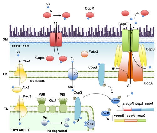
2. Copper, an Essential Element for Photosynthesis and Respiration

Copper is an essential micro-nutrient that is required as a cofactor for a number of cuproenzymes, including amine oxidases, cytochrome c oxidases, laccases, methane monooxygenases, multicopper oxidases, nitrite oxidases, plastocyanin, superoxide dismutases and tyrosinases. These proteins are involved in diverse cellular processes, such as energy transduction, iron mobilization and oxidative stress response [37,38,39]. The ability of copper to alternate between its cuprous Cu(I) and cupric Cu(II) oxidation states makes it an ideal biological cofactor, especially for processes in which the electron transfer is a key factor, such as photosynthesis and respiration. However, the two oxidation states of copper not only allow its participation in essential redox reactions, but also allow it to catalyze the production of reactive oxygen species (ROS) through the Fenton and Haber–Weiss reactions, which leads to severe damage to lipids, proteins, DNA and other cytoplasmic molecules [38]. Furthermore, an excess of copper competes with other metals for their binding sites in proteins following the Irving-Williams series [1], resulting in a perturbation of protein function and, in some cases, protein degradation. Recently, it has been proposed that the main target for copper toxicity in Escherichia coli, Bacillus subtilis and Synechocystis sp. PCC 6803 (or simply Synechocystis) is the replacement of iron by copper in the Fe-S clusters of essential enzymes [40,41,42,43] and not the DNA damage mediated by copper-catalyzed production of ROS [42,44].
In bacteria, the first barrier of protection against copper is to localize all cuproproteins in the periplasm and the plasma membrane, avoiding the presence of this metal in the cytosol. Cyanobacteria are unusual among bacteria, as they require copper to enter into the cytosol to reach the thylakoid, their internal membrane system, which is discrete from the periplasm. Two essential proteins are present in the thylakoid: the blue-copper protein, plastocyanin, and the caa3-type cytochrome oxidase, which are involved in the photosynthetic and respiratory electron transport processes, respectively (Figure 1). This enzymatic demand for copper in the thylakoids sets cyanobacteria as an attractive model system to study copper trafficking to an internal compartment.
Figure 1. Schematic representation of the copper homeostasis mechanism in Synechocystis sp. PCC 6803. The proteins (and genes) mentioned in the figure are CopM (sll0788 and slr6039), CopR (sll0789 and slr6040), CopS (sll0790 and slr6041), PacS (sll1920), CtaA (slr1950), Atx1 (ssr2857), FutA2 (slr0513), CopB (slr6042), CopA (slr6043), CopC (slr6044), Cox (cytochrome oxidase complex), Pc (sll0199), PSI (Photosystem I), PSII (Photosystem II) and Cb6f (cytochrome b6f). OM, outer membrane; PM, plasma membrane; TM, thylakoid membrane.
Life 04 00865 g001
As for most of the metals, copper metabolism has been mainly analyzed in model cyanobacterium Synechocystis. Copper is imported in Synechocystis mediated by two PI-type ATPases, CtaA and PacS, which are located in the plasma and thylakoidal membranes, respectively. These two proteins are assisted by a small cytosolic soluble copper metallochaperone, Atx1 [42,45,46]. Copper import inside the cell is mediated by CtaA, which delivers it to Atx1 [42]; then, it is transferred to PacS, which finally transports it inside the thylakoid lumen (Figure 1). This model is supported by the data that shows that mutants in either ctaA or pacS render cells with reduced cytochrome caa3 oxidase and plastocyanin activities [45] and the lower internal copper quota of ctaA mutants. These phenotypes, along with the proposed location of PacS at the thylakoid membrane [47] and its requirement for copper tolerance, are in agreement with this proposed model. Although this pathway was widely accepted in the past, it is currently under discussion. Structural and biochemical analyses show that PI-type ATPases cannot import copper, and hence, CtaA and PacS can only efflux copper from the cell [48,49]. In fact, copper can reach plastocyanin in pacS and atx1 mutant strains, but not in ctaA mutants, when cells are grown in standard copper-replete medium, suggesting that only CtaA is involved in the delivery to plastocyanin. This is in agreement with its low transport rate, as has been shown for other PI-ATPases involved in cuproprotein assembly [42,48,49]. Finally, both PI-type ATPases are required for CucA (a periplasmic copper-containing protein) to be loaded with copper, suggesting that they could also affect periplasmic copper metabolism [3]. This suggests that an additional copper import system should be present in Synechocystis (which will allow copper to enter the cytoplasm) and that both ATPases export copper to either the periplasm (CtaA) or both the periplasm and the thylakoid lumen (PacS). Because of this, the pathway for copper assembly in plastocyanin and caa3-type cytochrome oxidase could be similar to that of manganese loading in PSII, which seems to happen through the periplasm [50,51,52]. Another important protein related to copper import in Synechocystis is the periplasmic iron-binding protein, FutA2 [53]. Deletion of futA2 leads to lower copper-dependent cytochrome caa3 oxidase activity and hyperaccumulation of copper in the periplasm. It is thought that FutA2 influences copper uptake into the cytosol by chelation of Fe(III), which, in the absence of FutA2, interferes with copper transport [53]. Transport through the outer membrane is also essential for metal homeostasis, and this is usually mediated by porins [54,55]. Although this has not been studied in Synechocystis, in Anabaena sp. PCC 7120 mutants, a TonB-dependent transporter (iacT, all4026), located at the outer membrane, affected the rate of copper transport. Furthermore, overexpression of this gene confers hypersensitivity to both copper and iron, suggesting that this protein is needed for copper and iron transport [56]. In marine environments, free copper is scarce, and most copper is complexed to an organic ligand that has been denoted, L1, a compound that has been shown to be produced by Synechococcus in response to copper [57]. In addition, Anabaena sp. PCC 7120 mutants affected in the production and transport of hydroxamate-based siderophore are also affected in copper transport [16,56,58].
Finally, one of the best known responses to copper availability in cyanobacteria is the replacement of cytochrome c6 by plastocyanin when copper is available [43,59,60,61,62]. Although this regulation has been known for a long time and the genes for both electron carriers are present in most cyanobacterial genomes (and most probably, its regulation is also conserved), nothing is known about the mechanisms of regulation. The appearance of plastocyanin probably conferred a selective advantage in iron-limited ecosystems, such as more oxidizing environments that were being generated by cyanobacteria through the released O2 by oxygenic photosynthesis [62].
Copper resistance has been mainly studied in Synechocystis and comprises a two-component system, CopRS, CopM, a periplasmic and extracellular copper binding protein [63], and an Heavy Metal Efflux-Resistance-Nodulation-Division (HME-RND) family export system, CopBAC (Figure 1, [29]). These genes are regulated by the presence of copper in the media through the CopRS two-component system. CopS is able to detect copper directly and probably activates CopR that is able to directly bind to the promoters of the copMRS and copBAC operons [29]. However, CopRS does not control the expression of any of the copper metabolism genes described above, such as ctaA, pacS and atx1 [29,43]. Strains lacking any of these genes are more sensitive to copper, with copRS mutants being more sensitive than copM or copBAC mutants, suggesting that both CopM and CopBAC contribute to copper resistance [29,63]. These mutants also accumulate higher amounts of intracellular copper than the wild type. Moreover, CopS, the histidine kinase that detects copper and that belongs to the membrane-attached histidine kinases, is localized not only in the plasma membrane, but also in the thylakoid membrane. Therefore, CopS could be involved in copper detection in both the periplasm and the thylakoid lumen [29]. This could allow CopS not only to respond to conditions that alter extracellular copper supply, but also to those that alter the internal copper requirements, as almost (if not all) intracellular copper is localized to the thylakoids in plastocyanin and cytochrome cca3 oxidase. In fact, the copMRS operon is expressed in conditions that alter the photosynthetic electron transport and induced plastocyanin degradation (Figure 1, [29,64,65]). In Anabaena sp. PCC 7120 and Synechococcus sp. CC9311, operons coding for a putative RND transport system has been shown to be expressed in response to copper [34,56], although their contributions to copper resistance have not been analyzed. In contrast, two genes, coding for a membrane thioredoxin and an S-layer protein (and probably, the whole operon in which they are located), which were identified as copper induced in Synechococcus sp. CC9311, contribute to copper resistance and oxidative stress [34,66].

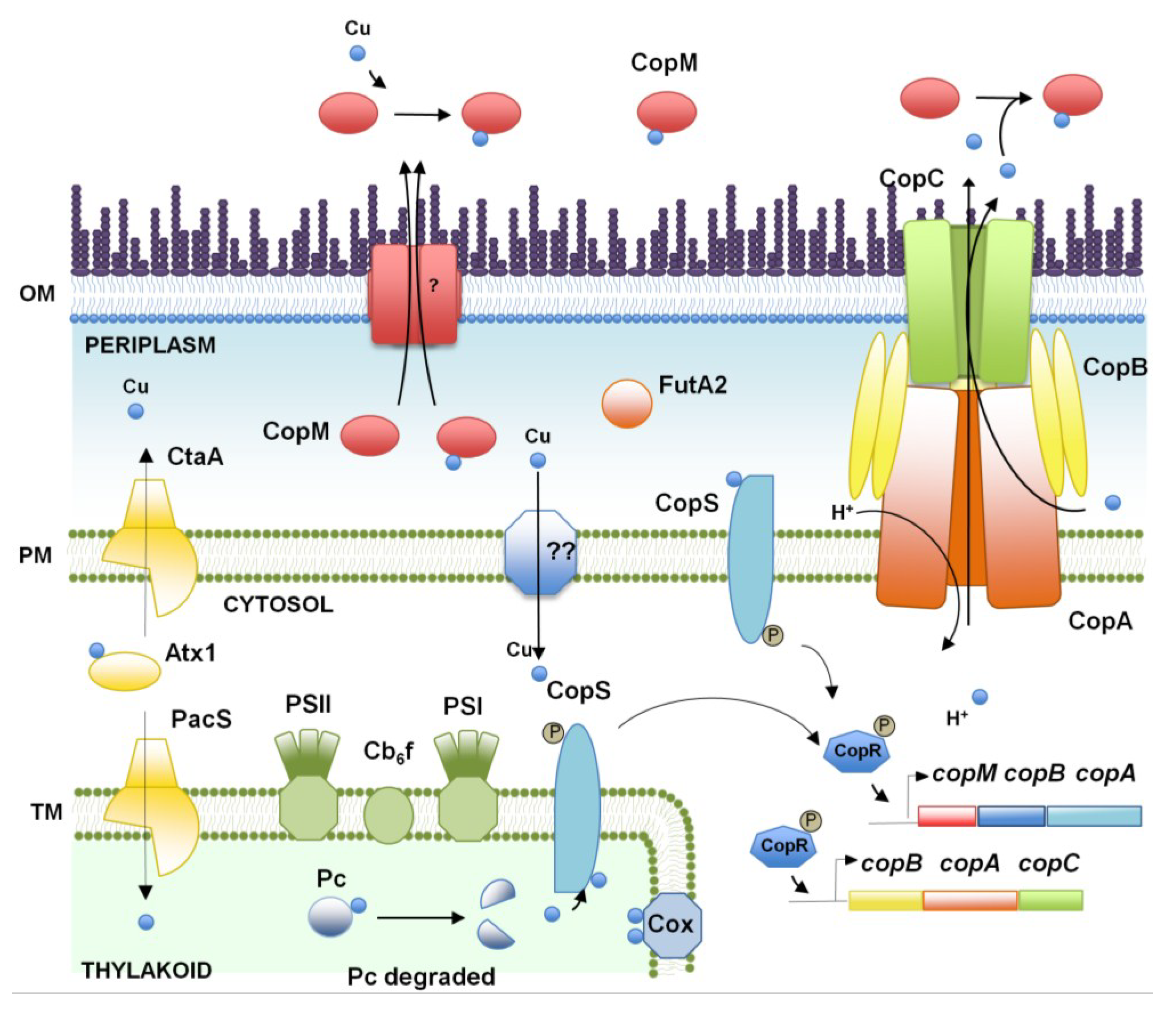
3. Nickel and Cobalt, Two Essential Metals for Cyanobacteria?

Nickel and cobalt are essential transition metals that are present at low levels, but play important roles in the cellular physiology of cyanobacteria [15,67]. Ni(II) is directly coordinated by proteins [68] or through the tetrapyrrole ring of coenzyme F430, which coordinates a nickel atom in methyl-coenzyme M reductase, while Co(II) is mainly used as a component in vitamin B12 [69]. In cyanobacteria, the major Ni-binding enzymes are urease and hydrogenases. Urease is needed to use urea as a nitrogen source [70] and probably also for arginine catabolism [71]. Urea appears to be a major nitrogen resource in the sea, and it has been suggested that both marine and freshwater cyanobacteria use it as a nitrogen source [72,73]; in some cases, the urease operon has been shown to be controlled by the nitrogen global regulator, NtcA [70,73,74]. The second Ni-containing enzymes are hydrogen-metabolizing/producing enzymes, named hydrogenases. Two distinct types are found in different cyanobacterial species: the bidirectional hydrogenases and the uptake hydrogenases. The bidirectional Ni-Fe hydrogenase (encoded by the hox genes, hoxEFUYH) can either take up or produce hydrogen and is found in both nitrogen fixing and non-fixing cyanobacterial genomes. This enzyme belongs to the NAD(P)-reducing class [75,76,77], although in Synechocystis, it has been recently described that it can be reduced by flavodoxin and ferredoxin [78]. This enzyme has great interest for the development of solar hydrogen production technologies using cyanobacteria. The second one is the uptake hydrogenase, which has the ability to recycle hydrogen (consuming O2) that is evolved as a side reaction during nitrogen fixation, since it is found in all nitrogen fixing species [79]. It is composed of small and large subunits encoded by the hupS and hupL genes, respectively. HupS mediates electron transport from the active site to the electron transport chain, and HupL contains two putative nickel-binding sites necessary for the coordination of the nickel in the active site [80,81]. Since this enzyme is an obstacle for hydrogen production, mutations disrupting the hupSL genes have been constructed in different cyanobacteria, and these mutant strains produce more hydrogen than the wild-type [82,83]. Other cyanobacterial Ni-enzymes described to date are Ni-superoxide dismutase [84] and glyoxalase I, a glutathione-dependent enzyme that requires Ni(II) and/or Co(II) for its activity [85,86]. Cobalt is mainly found in the corrin ring of coenzyme B12, a cofactor involved in both methyl group transfer and rearrangements reactions, and also in some non-corrin cobalt-containing enzymes, such as nitrile hydratase [69]. Although some cyanobacteria can synthesize vitamin B12-related compounds, others, like Synechococcus sp. PCC 7002, require B12 for growth. Little is known about how these compounds are used as cofactors in cobalamin-dependent enzymes beyond the essential enzyme, cobalamin-dependent methionine synthase [87].
Synthesis of Ni(II)/Co(II) enzymes and coenzyme B12 requires high-affinity uptake of the metal ions from natural environments, where they are available only in trace amounts most of the time. Nickel/cobalt transport is mediated by specific systems that belong to both secondary and primary transporters [15,88]. In general, the secondary systems found in cyanobacteria are related to nickel/cobalt transporters (NiCoTs) found in bacteria, which include the UreH transporters and HupE/UreJ permeases and which have been shown to mediate nickel transport. Most of the UreH proteins are predicted to function as high-affinity nickel transporters based on co-localization of these genes with those coding for NiFe hydrogenase, urease or nickel superoxide dismutase. UreH proteins are predicted to contain six transmembrane helices (TMH) and a histidine-rich segment in the cytoplasmic loop between TMH III and TMH IV. A member of the UreH family (called SodT) is found in some Synechococcus and all Prochlorococcus genomes and is predicted to contain six transmembrane domains [15,89]. sodT co-localizes with sodN, which code for a Ni-superoxide dismutase (SodN), and sodX, coding for a putative maturation peptidase, in all cyanobacterial genomes where they are present. SodT has been proposed to supply nickel for SodN, which is an essential protein in these organisms [15]. The HupE/UreJ proteins represent a secondary transporter that is widespread among bacteria, and they are encoded within certain NiFe hydrogenase and urease gene clusters. HupE/UreJ permeases are produced as a precursor and a mature protein that contain six transmembrane domains. Based on this genomic localization, a role in nickel uptake has been proposed. In the genome of marine cyanobacteria, a gene coding for a HupE transporter appears to be under the control of a B12-responsive riboswitch and probably involved in cobalt transport [90]. In the same way, a function in cobalt transport has been attributed to HupE in Synechocystis, because the hupE mutant growth defects were suppressed by adding cobalt or methionine [91]. Primary active nickel importers contain subunits with an ATP binding cassette (ABC) and are driven by ATP hydrolysis. Bioinformatics studies using 19 cyanobacterial genomes only found the CbiMNQO/NikMNQO systems as this primary cobalt/nickel transporters [92,93]. These genes code for a member of the energy-coupling factor (ECF) family of micronutrient importers and consist of a conserved transmembrane protein (Nik/CbiQ), an ABC ATPases (Nik/CbiO) [93] and a component that is responsible for substrate specificity (Nik/CbiMN) [92]. CbiMNQO systems have been annotated as cobalt uptake systems based on their genomic co-localization with B12 biosynthesis genes or by the presence of the regulatory B12 riboswitch.
The systems described above imply routes that are used by bacteria to import nickel and cobalt across the inner membrane to the cytosol, but metals need to also enter the periplasm through the outer membrane. As has been discussed for copper, this transport is usually carried out by porins [54,55] and a TonB-dependent transporter. In Proteobacteria, nickel-regulated genes for outer-membrane transporters, ExbB-EsbD-TonB clusters, have been identified [93]. Putative TonB-dependent transporters, implicated in the transport of cobalamin and metals, such as nickel, are predicted in bioinformatics studies based on the sequence similarity of these systems in cyanobacteria [87]. In Anabaena sp. PCC 7120, several TonB-dependent receptors genes have been shown to change its expression levels in response to metal and nitrogen source availability, which suggest that they could be involved in transporting different metals across the outer membrane [56,58,94,95].
These import system must be tightly regulated in response to metal availability and cellular requirements/demands. The known nickel regulatory systems used in bacteria to regulate nickel import, such as NikR and Nur [96,97], are absent from cyanobacterial genomes, implying that a novel form of nickel transport regulation should exist. The cobalt regulatory system is also unknown, but as has been indicated above, several transporters are associated with a putative B12 riboswitch, suggesting a role in cobalt uptake and coordination with the synthesis of coenzyme B12 [89].
Finally, an excess of nickel/cobalt that could cause a perturbation of protein function is counterbalanced by the presence of efflux pumps, which expel these metal ions out of the cell. In addition, there could be other proteins involved in metal tolerance. In this regard, a global proteomic approach has revealed that the chaperones, HspA and GroES, are specifically upregulated in the presence of high concentrations of nickel and cobalt, respectively [8]. These data suggest that these chaperones may be directly involved in the tolerance for these metals. Furthermore, it also has been shown that FutA2, a periplasmic iron-binding protein that seems to be essential for metal homeostasis [53], is downregulated in the presence of both nickel and cobalt [8], pointing to cross-talk between different metal metabolisms.
As for the other metals, nickel and cobalt resistance systems have been mainly studied in Synechocystis (Figure 2). The genes involved in the resistance to these two metals are part of a metal-regulated gene cluster involved in resistance against zinc, copper, cobalt and nickel [28,29,31,32]. This 14-kb region consists of 14 ORFs organized into seven putative transcriptional units: the nrsBACD operon involved in nickel tolerance and regulated by the upstream nrsRS operon [28,98], corT (coaA), encoding a putative co-translocating PI-type ATPase under the regulation of the upstream corR (coaR) product [28,31], ziaA, encoding a putative Zn-efflux PI-type ATPase and regulated by the product of ziaR [32], and the copMRS operon, which is involved in copper resistance (see above, [29]).
Figure 2. Schematic representation of the cobalt and nickel homeostasis mechanisms in Synechocystis sp. PCC 6803. The proteins (and genes) mentioned in the figure are CorT or CoaT (slr0797), CorR or CoaR (sll0794), NrsB (slr0793), NrsA (slr0794), NrsC (slr0795), NrsD (slr0796), InrS (sll0176), HupE (slr2135), NrsR (sll0797), NrsS (sll0798), CbiO (sll0384), CbiQ (sll0385) and CbiM (sll0383). OM, outer membrane; PM, plasma membrane.
Life 04 00865 g002
The cobalt resistance system is composed of CorR (or CoaR), a MerR homolog that has been identified as activator of the corT (coaT) gene, which codes for a co-dependent PI-type ATPase [28,31]. This transcription factor contains a precorrin isomerase domain and responds to vitamin B12 intermediaries [31,99], suggesting that, in addition, to coaT (corT) activation in response to cobalt excess, it could be also involved in coordinating cobalt transport and vitamin B12 synthesis. Mutation of any of these genes confers sensitivity to cobalt excess in the media, although its effects on vitamin B12 metabolism were not analyzed [28,31].
The nrsBACD operon encodes two putative nickel efflux systems: an RND efflux system (encoded nrsBAC genes) and a major facilitator superfamily (MFS) permease (encoded by nrsD) that contains a metal binding domain (histidine-rich region) in its C-terminus [28]. A two-component signal transduction system (encoded by the nrsRS genes, also known as Hik30-Rre33) controls the Ni-dependent induction of the nrsBACD operon and its own expression [98]. NrsS is a histidine kinase sensor with an N-terminal periplasmic domain that shares sequence similarity with the alpha subunit of the methyl-coenzyme M reductase (MCR) from Methanobacteria (an enzyme able to bind nickel through a F430 cofactor, suggesting that this domain could bind Ni(II) using a related structure). This histidine kinase senses nickel and modulates the Ni(II) efflux operon via the DNA-binding transcriptional regulator, NrsR, a response regulator of the PhoB/OmpR family [98]. Mutants in any of these genes are also more sensitive than the wild-type to nickel excess [28,98]. nrsD, the last gene of the nrsBACD operon, has its own promoter between the final two ORFs in the nrsBACD operon, and it has been recently shown to be also regulated by InrS (for internal nickel-responsive sensor), a CsoR/RcnR-like transcriptional repressor [100]. This family of transcription factors were identified as copper responsive [97,101] and InrS contains conserved copper binding residues. In vitro, InrS is able to bind Cu(I) more tightly than Ni(II), although it was proposed that this protein does not have access to copper in vivo under steady-state conditions, because copper is completely buffered in Synechocystis’ cytoplasm [42]. Later, it was shown that the nrsD gene is also induced by copper (and Zn) after a short challenge [102], although neither nrsBAC nor nrsRS genes were induced by copper [43]. InrS mutants grow poorly in the absence of added nickel [43,100], probably because they contain lower internal nickel quota [100], suggesting that this metal is essential for Synechocystis growth. This phenotype is suppressed in a double-mutant lacking also nrsD (InrS NrsD), but surprisingly was shown extremely sensitive to different metals, suggesting that InrS could have a central role in metal homeostasis, probably controlling other elements [43].

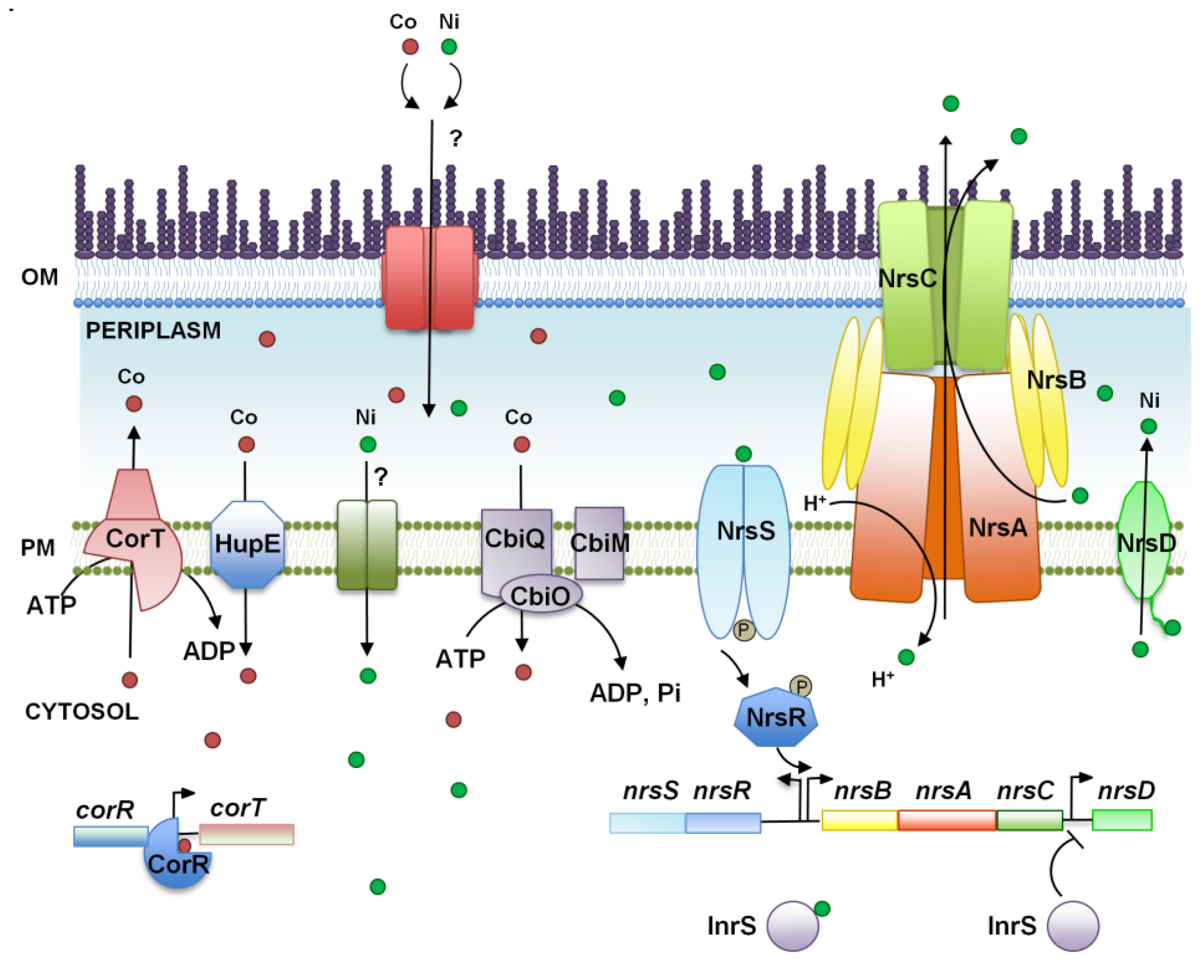
4. Arsenic, a World Wide Spread Phosphate Competitor

Arsenic is an ubiquitous toxic metalloid that causes serious health problems in many places of the world where the arsenic content of drinking water is well over the recommended limits [103]. Arsenic is present in two biologically active forms, arsenate [AsV] and arsenite [AsIII], depending on the redox potential of the environment. Arsenate is a phosphate analogue and, therefore, enters the cells through phosphate transporters. Its toxicity is mediated by replacing phosphate in essential biochemical reactions, such as oxidative phosphorylation and glycolysis. The resulting arseno-compounds are extremely labile and hydrolyze spontaneously at millisecond rates, making them unable to be used by living organisms [104,105,106,107]. On the other hand, arsenite enters the cell through aquaglyceroporins [108,109,110] and exerts its toxicity through binding to dithiols, forming arsenothiols that disrupt protein function and that ultimately generate reactive oxygen species (ROS) [108,109,110,111]. Because of the high affinity for sulfur, arsenite is able to bind to the main redox buffer in the cells, glutathione (GSH), forming AsIII-GSH2, and depletes its pool, thus contributing to ROS generation. Despite being toxic, arsenic is also used by some microorganisms as an electron acceptor in the anaerobic respiratory chain, as an electron donor to grow chemo-lithotrophically and even for anoxygenic photosynthesis by some photosynthetic bacteria and cyanobacteria [112,113]. It has been postulated that arsenic compounds played an important role during early life on Earth before the appearance of molecular oxygen as an electron acceptor and donor [113,114].
Because of the extensive use and distribution of arsenic compounds, arsenic resistance is widespread among living organisms. Many resistance systems consist of the reduction of arsenate to arsenite, followed by export of the latter outside the cell or its transport to the vacuole. Another detoxification system that is present from bacteria to animals is arsenic methylation, which conjugates arsenic to methyl groups and can lead to the formation of arsenic volatile species [115,116].
In cyanobacteria, arsenic metabolism and resistance is best understood in Synechocystis (Figure 3). The main arsenic resistance mechanism is mediated by an operon of three genes (arsBHC) that is regulated by an unlinked arsR [30,117]. The operon includes an Acr3-arsenite transporter gene, arsB, arsH, which codes for a NADPH-FMN reductase, able to reduce several compounds without a clear function in arsenic resistance [30,118,119], and an arsenate reductase gene, arsC. At least two other resistance determinants have been described in Synechocystis: an additional arsenate reductase from the E. coli family (encoded by two nearly identical genes, arsI1 and arsI2 [120]) and an arsenite methylase gene, arsM [121]. ArsI is only essential for arsenic resistance in the absence of ArsC, probably due to its low level of expression [117,120]. Furthermore, global studies have shown that arsenic induces a strong oxidative stress response, disrupts metal homeostasis and depletes the GSH pool [117,122,123], which is in agreement with what has been observed in other organisms [111].
Arsenate reduction to arsenite is catalyzed by arsenate reductase, an enzymatic activity carried out by at least three non-related families of enzymes that use thioredoxin, glutaredoxin or mycoredoxin as electron donors [120,124,125,126]. Two types of arsenate reductases appear in cyanobacteria encoded by arsC and arsI genes in Synechocystis. arsC codes for a new type of hybrid arsenate reductase exclusively found in cyanobacteria, that although related to thioredoxin-dependent ones, uses the glutathione/glutaredoxin system for reduction [30,120,124], while arsI genes code for E. coli-like glutaredoxin-dependent arsenate reductases [30,120]. Although the ArsCSyn is exclusively found in cyanobacteria, many genomes also contain genes coding for E. coli-like arsenate reductases [120,127]. The fact that all arsenate reductases present in cyanobacteria are glutaredoxin dependent could be related to the essentiality of the thioredoxin system in these organisms [128,129], which has forced them to find alternative reductants for these enzymes.
Arsenite export is mediated by two families of proteins: ArsB proteins, which are present only in bacteria [130], and Acr3 proteins, found in different organisms, including bacteria, fungi and plants [131,132]. Many cyanobacterial genomes contain arsenic resistance gene clusters with the ACR3 family of transporters and arsenate reductase genes (of either ArsCSyn or the E. coli-type) [120,127]. In contrast, no homologs of the E. coli ArsB protein can be easily identified in them.
Figure 3. Schematic representation of the arsenic resistance mechanism in Synechocystis sp. PCC 6803. The proteins (and genes) mentioned in the figure are ArsR (sll1957), ArsB (slr0944), ArsH (slr0945), ArsC (slr0946), ArsI (sll5104 and slr6037), Grx (GrxA or GrxB; ssr2061 and slr1562), ArsM (slr0303), AqpZ (aquaporin; slr2057), PstA (sll0682 and slr1242), PstB (sll0683-84 and slr1250), PstC (sll0681 and slr1248) and PstS (sll0680, sll0679, slr1247 and slr0540). OM, outer membrane; PM, plasma membrane.
Life 04 00865 g003
ArsM from several cyanobacteria is able to methylate arsenite to the volatile trimethylarsine [TMA(III)] using S-adenosyl methionine and glutathione as methyl donors in vitro [121], and the arsM gene is present in many cyanobacterial genomes. Although the role of ArsM in arsenic resistance has not been tested in vivo in cyanobacteria, E. coli strains carrying arsM genes from different cyanobacteria are more resistant to arsenite [121]. Furthermore, methylated arsenic forms are abundant in the oceans [133], and methylated arsenic species are detected when several cyanobacterial strains are treated with arsenic [121,134,135], suggesting that arsenic methylation could play a role in resistance. On the other hand, arsM expression has been reported to be both upregulated by arsenic after two weeks of exposure [121] or not regulated after one hour of both arsenate and arsenite treatments [117]. Although the conditions were completely different, what seems to be clear is that the arsM gene is not regulated by the transcriptional repressor, ArsR, at least in Synechocystis [117].
Bioinformatics analyses have shown that arsenic resistance genes are widespread in most sequenced cyanobacterial genomes [120,127]. Inspection of the genomic context have allowed us to identify four additional genes associated with arsenic resistance genes: a periplasmic phosphate binding protein (PstS), a glyceraldehyde-3-phosphate dehydrogenase (Gap3), a major facilitator superfamily transporter (MFS) and a putative glyoxalase, which are probably organized forming an operon with arsB (ACR3) and arsC (ArsCSyn) genes (Figure 4). In some cases, an ArsR coding gene is also localized upstream of the PstS coding gene, strongly suggesting that these genes are co-regulated in response to arsenic. The function of these proteins in arsenic resistance remains to be determined, but it is possible that they could influence phosphate transport (PstS and MFS transporter), be arsenate insensitive isoforms (PstS or Gap3) or constitute a new arsenic resistance mechanism transporting arsenic outside the cell (MFS or PtsS or the glyoxalase).
Figure 4. Genomic organization of arsenic resistance genes in several cyanobacteria.
Life 04 00865 g004
Phosphate has a great influence on arsenic resistance in cyanobacteria, because the phosphate concentration interferes with arsenate transport. The phosphate transport system (which is the main arsenate transport system) is induced under phosphate deprivation [136,137], therefore increasing arsenate transport [134,135,138,139,140,141]. Furthermore, phosphate starvation also induced arsB expression in Crocosphaera watsonii [137], and arsenic represses phosphate transport gene expression in Synechocystis [117]. In addition, the specificity and selectivity of the phosphate transport system greatly influence arsenate transport [142]. Phosphate also influences the redox state of the arsenic that accumulated in the media after it is metabolized by cyanobacteria. In Synechocystis sp. PCC 6803, arsenate is the main form accumulated in the media after long-term exposure to both arsenite and arsenate in phosphate-rich conditions, but arsenite accumulates in phosphate-depleted media [134]. This is a consequence of the active arsenate reduction in the cytoplasm, which efficiently reduces all arsenate to arsenite, which is extruded outside the cells. Once outside, this arsenite is oxidized to arsenate, which, under phosphate-replete conditions, is not transported inside the cells, while under phosphate-deficient conditions, any arsenate that is produced is transported into the cells and reduced. The mechanism for extracellular arsenite oxidation is not clear and remains to be elucidated [134].
Finally, phytochelatins, which are GSH polymers (n = 2–16), are well known to contribute to arsenite resistance [143]. Although genes with homology to phytochelatin synthase are present in some cyanobacteria [144] and phytochelatin contents increase in response to arsenic [122], it is not known whether phytochelatin is able to confer arsenic resistance.

5. Conclusions

This review highlights our understanding of metal homeostasis systems in cyanobacteria and how metal transporters, metal sensing and regulators are selective for one (or two) metal ion(s). This cellular-level approach is necessary to establish accurately the mechanism involved in the interaction between cyanobacteria and metals under deficiency or toxic concentration conditions. As discussed above, this information may be used for the design of promising detector systems for metal bioremediation or for the determination of the optimal growth conditions in order to use them as biofactories. Nevertheless, it is becoming clear that metal metabolism, as occurs with many cellular process, is interconnected. In addition, natural ecosystems are usually limited by several nutritional factors, including the availability of essential metals, like iron (this limitation is a key point for ocean productivity). The excess of metals happens frequently in nature, in many cases due to contamination produced by human activity. This type of contamination includes more than one metal, as they are in general more soluble in acid environments, for example mine drainage or the chemical industry. Furthermore, the nutrition status for one metal can greatly determine the responses to other metal excesses or deficiencies, as has been shown for Zn starved Chlamydomonas reinhardtii, which show symptoms of copper deficiency and accumulate higher intracellular amounts of Mn and Fe [145]. This highlights the importance of studying the metal responses to different combinations of metal nutrition statuses in the lab to be able to understand what happens in natural systems.

Acknowledgments

Part of the work described here was supported by Ministerio de Economia y Competividad (MINECO) Grants BFU2010-15708 and BFU2013-41712, and by Junta de Andalucía (Group BIO-284 and Grant P12-BIO-1119), co-financed by European Regional Funds (FEDER) to Francisco Javier Florencio, Luis López-Maury was supported by a postdoctoral contract from Universidad de Sevilla. We thank Sandra Díaz-Troya for critical reading of the manuscript.

Author Contributions

All authors analyzed the literature and wrote the paper. All authors have read and approved the final manuscript.

Conflicts of Interest

The authors declare no conflict of interest.

References

  1. Waldron, K.J.; Robinson, N.J. How do bacterial cells ensure that metalloproteins get the correct metal? Nat. Rev. Microbiol. 2009, 7, 25–35. [Google Scholar] [CrossRef] [PubMed]
  2. Cvetkovic, A.; Menon, A.L.; Thorgersen, M.P.; Scott, J.W.; Poole, F.L., II; Jenney, F.E., Jr.; Lancaster, W.A.; Praissman, J.L.; Shanmukh, S.; Vaccaro, B.J.; et al. Microbial metalloproteomes are largely uncharacterized. Nature 2010, 466, 779–782. [Google Scholar] [CrossRef]
  3. Waldron, K.J.; Firbank, S.J.; Dainty, S.J.; Perez-Rama, M.; Tottey, S.; Robinson, N.J. Structure and metal loading of a soluble periplasm cuproprotein. J. Biol. Chem. 2010, 285, 32504–32511. [Google Scholar] [CrossRef] [PubMed]
  4. Waldron, K.J.; Rutherford, J.C.; Ford, D.; Robinson, N.J. Metalloproteins and metal sensing. Nature 2009, 460, 823–830. [Google Scholar] [CrossRef] [PubMed]
  5. Guerra-García, J.M.; García-Gómez, J.C. Assessing pollution levels in sediments of a harbour with two opposing entrances. Environmental implications. J. Environ. Manag. 2005, 77, 1–11. [Google Scholar] [CrossRef]
  6. Peng, J.-F.; Song, Y.-H.; Yuan, P.; Cui, X.-Y.; Qiu, G.-L. The remediation of heavy metals contaminated sediment. J. Hazard. Mater. 2009, 161, 633–640. [Google Scholar] [CrossRef] [PubMed]
  7. Shcolnick, S.; Keren, N. Metal Homeostasis in Cyanobacteria and Chloroplasts. Balancing Benefits and Risks to the Photosynthetic Apparatus. Plant Physiol. 2006, 141, 805–810. [Google Scholar] [CrossRef] [PubMed]
  8. Mehta, A.; López-Maury, L.; Florencio, F.J. Proteomic pattern alterations of the cyanobacterium Synechocystis sp. PCC 6803 in response to cadmium, nickel and cobalt. J. Proteomics 2014, 102, 98–112. [Google Scholar] [CrossRef] [PubMed]
  9. Pereira, S.; Micheletti, E.; Zille, A.; Santos, A.; Moradas-Ferreira, P.; Tamagnini, P.; de Philippis, R. Using extracellular polymeric substances (EPS)-producing cyanobacteria for the bioremediation of heavy metals: Do cations compete for the EPS functional groups and also accumulate inside the cell? Microbiology 2011, 157, 451–458. [Google Scholar] [CrossRef] [PubMed]
  10. Burnat, M.; Diestra, E.; Esteve, I.; Solé, A. In Situ Determination of the Effects of Lead and Copper on Cyanobacterial Populations in Microcosms. PLoS One 2009, 4. [Google Scholar] [CrossRef] [PubMed]
  11. Merchant, S.S.; Helmann, J.D.; Robert, K.P. Chapter 2—Elemental Economy: Microbial Strategies for Optimizing Growth in the Face of Nutrient Limitation. In Advances in Microbial Physiology; Academic Press: Waltham, MA, USA, 2012; Volume 60, pp. 91–210. [Google Scholar]
  12. Hillier, W.; Babcock, G.T. Photosynthetic Reaction Centers. Plant Physiol. 2001, 125, 33–37. [Google Scholar] [CrossRef] [PubMed]
  13. Jordan, P.; Fromme, P.; Witt, H.T.; Klukas, O.; Saenger, W.; Krausz, N. Three-dimensional structure of cyanobacterial photosystem I at 2.5 Å resolution. Nature 2001, 411, 909–917. [Google Scholar] [CrossRef] [PubMed]
  14. Barnett, J.P.; Millard, A.; Ksibe, A.; Scanlan, D.J.; Schmid, R.; Blindauer, C.A. Mining genomes of cyanobacteria for elements of zinc homeostasis. Front. Microbiol. 2012, 3. [Google Scholar] [CrossRef]
  15. Dupont, C.L.; Johnson, D.A.; Phillippy, K.; Paulsen, I.T.; Brahamsha, B.; Palenik, B. Genetic Identification of a High-Affinity Ni Transporter and the Transcriptional Response to Ni Deprivation in Synechococcus sp. Strain WH8102. Appl. Environ. Microbiol. 2012, 78, 7822–7832. [Google Scholar] [CrossRef] [PubMed]
  16. Mann, E.L.; Ahlgren, N.; Moffett, J.W.; Chisholm, S.W. Copper toxicity and cyanobacteria ecology in the Sargasso Sea. Limnol. Oceanogr. 2002, 47, 976–988. [Google Scholar] [CrossRef]
  17. Twining, B.S.; Baines, S.B. The trace metal composition of marine phytoplankton. Ann. Rev. Mar. Sci. 2013, 5, 191–215. [Google Scholar] [CrossRef] [PubMed]
  18. Trick, C.; Kerry, A. Isolation and purification of siderophores produced by cyanobacteria, Synechococcus sp. PCC 7942 and Anabaena variabilis ATCC 29413. Curr. Microbiol. 1992, 24, 241–245. [Google Scholar] [CrossRef]
  19. Itou, Y.; Okada, S.; Murakami, M. Two structural isomeric siderophores from the freshwater cyanobacterium Anabaena cylindrica (NIES-19). Tetrahedron 2001, 57, 9093–9099. [Google Scholar] [CrossRef]
  20. De Philippis, R.; Sili, C.; Paperi, R.; Vincenzini, M. Exopolysaccharide-producing cyanobacteria and their possible exploitation: A review. J. Appl. Phycol. 2001, 13, 293–299. [Google Scholar] [CrossRef]
  21. Jittawuttipoka, T.; Planchon, M.; Spalla, O.; Benzerara, K.; Guyot, F.; Cassier-Chauvat, C.; Chauvat, F. Multidisciplinary evidences that Synechocystis PCC 6803 exopolysaccharides operate in cell sedimentation and protection against salt and metal stresses. PLoS One 2013, 8. [Google Scholar] [CrossRef] [PubMed]
  22. Micheletti, E.; Pereira, S.; Mannelli, F.; Moradas-Ferreira, P.; Tamagnini, P.; de Philippis, R. Sheathless mutant of Cyanobacterium Gloeothece sp. strain PCC 6909 with increased capacity to remove copper ions from aqueous solutions. Appl. Environ. Microbiol. 2008, 74, 2797–2804. [Google Scholar] [CrossRef] [PubMed]
  23. Huckle, J.W.; Morby, A.P.; Turner, J.S.; Robinson, N.J. Isolation of a prokaryotic metallothionein locus and analysis of transcriptional control by trace metal ions. Mol. Microbiol. 1993, 7, 177–187. [Google Scholar] [CrossRef] [PubMed]
  24. Robinson, N.J.; Whitehall, S.K.; Cavet, J.S. Microbial metallothioneins. Adv. Microb. Physiol. 2001, 44, 183–213. [Google Scholar] [PubMed]
  25. Turner, J.S.; Morby, A.P.; Whitton, B.A.; Gupta, A.; Robinson, N.J. Construction of Zn2+/Cd2+ hypersensitive cyanobacterial mutants lacking a functional metallothionein locus. J. Biol. Chem. 1993, 268, 4494–4498. [Google Scholar] [PubMed]
  26. Liu, T.; Nakashima, S.; Hirose, K.; Shibasaka, M.; Katsuhara, M.; Ezaki, B.; Giedroc, D.P.; Kasamo, K. A novel cyanobacterial SmtB/ArsR family repressor regulates the expression of a CPx-ATPase and a metallothionein in response to both Cu(I)/Ag(I) and Zn(II)/Cd(II). J. Biol. Chem. 2004, 279, 17810–17818. [Google Scholar] [CrossRef] [PubMed]
  27. Liu, T.; Nakashima, S.; Hirose, K.; Uemura, Y.; Shibasaka, M.; Katsuhara, M.; Kasamo, K. A metallothionein and CPx-ATPase handle heavy-metal tolerance in the filamentous cyanobacterium Oscillatoria brevis. FEBS Lett. 2003, 542, 159–163. [Google Scholar] [CrossRef] [PubMed]
  28. García-Domínguez, M.; Lopez-Maury, L.; Florencio, F.J.; Reyes, J.C. A Gene Cluster Involved in Metal Homeostasis in the Cyanobacterium Synechocystis sp. Strain PCC 6803. J. Bacteriol. 2000, 182, 1507–1514. [Google Scholar] [CrossRef] [PubMed]
  29. Giner-Lamia, J.; López-Maury, L.; Reyes, J.C.; Florencio, F.J. The CopRS two-component system is responsible for resistance to copper in the cyanobacterium Synechocystis sp. PCC 6803. Plant Physiol. 2012, 159, 1806–1818. [Google Scholar] [CrossRef] [PubMed]
  30. López-Maury, L.; Florencio, F.J.; Reyes, J.C. Arsenic sensing and resistance system in the cyanobacterium Synechocystis sp. strain PCC 6803. J. Bacteriol. 2003, 185, 5363–5371. [Google Scholar] [CrossRef] [PubMed]
  31. Rutherford, J.C.; Cavet, J.S.; Robinson, N.J. Cobalt-dependent transcriptional switching by a dual-effector MerR-like protein regulates a cobalt-exporting variant CPx-type ATPase. J. Biol. Chem. 1999, 274, 25827–25832. [Google Scholar] [CrossRef] [PubMed]
  32. Thelwell, C.; Robinson, N.J.; Turner-Cavet, J.S. An SmtB-like repressor from Synechocystis PCC 6803 regulates a zinc exporter. Proc. Natl. Acad. Sci. USA 1998, 95, 10728–10733. [Google Scholar] [CrossRef] [PubMed]
  33. Liu, T.; Golden, J.W.; Giedroc, D.P. A Zinc(II)/Lead(II)/Cadmium(II)-Inducible Operon from the Cyanobacterium Anabaena Is Regulated by AztR, an α3N ArsR/SmtB Metalloregulator. Biochemistry 2005, 44, 8673–8683. [Google Scholar] [CrossRef] [PubMed]
  34. Stuart, R.K.; Dupont, C.L.; Johnson, D.A.; Paulsen, I.T.; Palenik, B. Coastal strains of marine Synechococcus species exhibit increased tolerance to copper shock and a distinctive transcriptional response relative to those of open-ocean strains. Appl. Environ. Microbiol. 2009, 75, 5047–5057. [Google Scholar] [CrossRef] [PubMed]
  35. Yu, Y.; You, L.; Liu, D.; Hollinshead, W.; Tang, Y.; Zhang, F. Development of Synechocystis sp. PCC 6803 as a Phototrophic Cell Factory. Mar. Drugs 2013, 11, 2894–2916. [Google Scholar] [CrossRef] [PubMed]
  36. Blindauer, C.A. Zinc-Handling in Cyanobacteria: An Update. Chem. Biodivers. 2008, 5, 1990–2013. [Google Scholar] [CrossRef] [PubMed]
  37. Rensing, C.; Grass, G. Escherichia coli mechanisms of copper homeostasis in a changing environment. FEMS Microbiol. Rev. 2003, 27, 197–213. [Google Scholar] [CrossRef] [PubMed]
  38. Osman, D.; Cavet, J.S. Copper homeostasis in bacteria. Adv. Appl. Microbiol. 2008, 65, 217–247. [Google Scholar] [PubMed]
  39. Rademacher, C.; Masepohl, B. Copper-responsive gene regulation in bacteria. Microbiology 2012, 158, 2451–2464. [Google Scholar] [CrossRef] [PubMed]
  40. Macomber, L.; Imlay, J.A. The iron-sulfur clusters of dehydratases are primary intracellular targets of copper toxicity. Proc. Natl. Acad. Sci. USA 2009, 106, 8344–8349. [Google Scholar] [CrossRef] [PubMed]
  41. Chillappagari, S.; Seubert, A.; Trip, H.; Kuipers, O.P.; Marahiel, M.A.; Miethke, M. Copper stress affects iron homeostasis by destabilizing iron-sulfur cluster formation in Bacillus subtilis. J. Bacteriol. 2010, 192, 2512–2524. [Google Scholar] [CrossRef] [PubMed]
  42. Tottey, S.; Patterson, C.J.; Banci, L.; Bertini, I.; Felli, I.C.; Pavelkova, A.; Dainty, S.J.; Pernil, R.; Waldron, K.J.; Foster, A.W.; et al. Cyanobacterial metallochaperone inhibits deleterious side reactions of copper. Proc. Natl. Acad. Sci. USA 2012, 109, 95–100. [Google Scholar] [CrossRef] [PubMed]
  43. Giner-Lamia, J.; López-Maury, L.; Florencio, F.J. Global Transcriptional Profiles of the Copper Responses in the Cyanobacterium Synechocystis sp. PCC 6803. PLoS One 2014, 9. [Google Scholar] [CrossRef] [PubMed]
  44. Macomber, L.; Rensing, C.; Imlay, J.A. Intracellular copper does not catalyze the formation of oxidative DNA damage in Escherichia coli. J. Bacteriol. 2007, 189, 1616–1626. [Google Scholar] [CrossRef] [PubMed]
  45. Tottey, S.; Rich, P.R.; Rondet, S.A.; Robinson, N.J. Two Menkes-type atpases supply copper for photosynthesis in Synechocystis. PCC 6803. J. Biol. Chem. 2001, 276, 19999–20004. [Google Scholar] [CrossRef] [PubMed]
  46. Tottey, S.; Rondet, S.A.; Borrelly, G.P.; Robinson, P.J.; Rich, P.R.; Robinson, N.J. A copper metallochaperone for photosynthesis and respiration reveals metal-specific targets, interaction with an importer, and alternative sites for copper acquisition. J. Biol. Chem. 2002, 277, 5490–5497. [Google Scholar] [CrossRef] [PubMed]
  47. Kanamaru, K.; Kashiwagi, S.; Mizuno, T. A copper-transporting P-type ATPase found in the thylakoid membrane of the cyanobacterium Synechococcus species PCC7942. Mol. Microbiol. 1994, 13, 369–377. [Google Scholar] [CrossRef] [PubMed]
  48. González-Guerrero, M.; Raimunda, D.; Cheng, X.; Argüello, J.M. Distinct functional roles of homologous Cu+ efflux ATPases in Pseudomonas aeruginosa. Mol. Microbiol. 2010, 78, 1246–1258. [Google Scholar] [CrossRef] [PubMed]
  49. Raimunda, D.; González-Guerrero, M.; Leeber, B., III; Argüello, J. The transport mechanism of bacterial Cu+-ATPases: Distinct efflux rates adapted to different function. BioMetals 2011, 24, 467–475. [Google Scholar] [CrossRef] [PubMed]
  50. Stengel, A.; Gugel, I.L.; Hilger, D.; Rengstl, B.; Jung, H.; Nickelsen, J. Initial steps of photosystem II de novo assembly and preloading with manganese take place in biogenesis centers in Synechocystis. Plant Cell 2012, 24, 660–675. [Google Scholar] [CrossRef] [PubMed]
  51. Schottkowski, M.; Gkalympoudis, S.; Tzekova, N.; Stelljes, C.; Schunemann, D.; Ankele, E.; Nickelsen, J. Interaction of the periplasmic PratA factor and the PsbA (D1) protein during biogenesis of photosystem II in Synechocystis sp. PCC 6803. J. Biol. Chem. 2009, 284, 1813–1819. [Google Scholar] [CrossRef] [PubMed]
  52. Rengstl, B.; Oster, U.; Stengel, A.; Nickelsen, J. An intermediate membrane subfraction in cyanobacteria is involved in an assembly network for Photosystem II biogenesis. J. Biol. Chem. 2011, 286, 21944–21951. [Google Scholar] [CrossRef] [PubMed]
  53. Waldron, K.J.; Tottey, S.; Yanagisawa, S.; Dennison, C.; Robinson, N.J. A periplasmic iron-binding protein contributes toward inward copper supply. J. Biol. Chem. 2007, 282, 3837–3846. [Google Scholar] [CrossRef] [PubMed]
  54. Lutkenhaus, J. Role of a major outer membrane protein in Escherichia coli. J. Bacteriol. 1977, 131, 631–637. [Google Scholar] [PubMed]
  55. Speer, A.; Rowland, J.L.; Haeili, M.; Niederweis, M.; Wolschendorf, F. Porins increase copper susceptibility of Mycobacterium tuberculosis. J. Bacteriol. 2013, 195, 5133–5140. [Google Scholar] [CrossRef] [PubMed]
  56. Nicolaisen, K.; Hahn, A.; Valdebenito, M.; Moslavac, S.; Samborski, A.; Maldener, I.; Wilken, C.; Valladares, A.; Flores, E.; Hantke, K.; et al. The interplay between siderophore secretion and coupled iron and copper transport in the heterocyst-forming cyanobacterium Anabaena sp. PCC 7120. Biochim. Biophys. Acta 2010, 1798, 2131–2140. [Google Scholar] [CrossRef] [PubMed]
  57. Moffett, J.W.; Brand, L.E. Production of strong, extracellular Cu chelators by marine cyanobacteria in response to Cu stress. Limnol. Oceanogr. 1996, 41, 388–395. [Google Scholar] [CrossRef]
  58. Stevanovic, M.; Lehmann, C.; Schleiff, E. The response of the TonB-dependent transport network in Anabaena sp. PCC 7120 to cell density and metal availability. BioMetals 2013, 26, 549–560. [Google Scholar] [CrossRef] [PubMed]
  59. Duran, R.V.; Hervas, M.; de la Rosa, M.A.; Navarro, J.A. The efficient functioning of photosynthesis and respiration in Synechocystis sp. PCC 6803 strictly requires the presence of either cytochrome c6 or plastocyanin. J. Biol. Chem. 2004, 279, 7229–7233. [Google Scholar] [CrossRef] [PubMed]
  60. Zhang, L.; McSpadden, B.; Pakrasi, H.B.; Whitmarsh, J. Copper-mediated regulation of cytochrome c553 and plastocyanin in the cyanobacterium Synechocystis 6803. J. Biol. Chem. 1992, 267, 19054–19059. [Google Scholar] [PubMed]
  61. Briggs, L.M.; Pecoraro, V.L.; McIntosh, L. Copper-induced expression, cloning, and regulatory studies of the plastocyanin gene from the cyanobacterium Synechocystis sp. PCC 6803. Plant Mol. Biol. 1990, 15, 633–642. [Google Scholar] [CrossRef] [PubMed]
  62. De la Rosa, M.A.; Navarro, J.A.; Dı́az-Quintana, A.; de la Cerda, B.; Molina-Heredia, F.P.; Balme, A.; Murdoch, P.D.S.; Dı́az-Moreno, I.; Durán, R.V.; Hervás, M.; et al. An evolutionary analysis of the reaction mechanisms of photosystem I reduction by cytochrome c6 and plastocyanin. Bioelectrochemistry 2002, 55, 41–45. [Google Scholar] [CrossRef] [PubMed]
  63. Giner-Lamia, J.; López-Maury, L.; Florencio, F.J. CopM is a novel copper binding protein involved in copper resistance in Synechocystis sp. PCC 6803. MicrobiologyOpen 2014, in press. [Google Scholar]
  64. López-Maury, L.; Giner-Lamia, J.; Florencio, F.J. Redox control of copper homeostasis in cyanobacteria. Plant Signal. Behav. 2012, 7, 1712–1714. [Google Scholar] [CrossRef] [PubMed]
  65. Osanai, T.; Imamura, S.; Asayama, M.; Shirai, M.; Suzuki, I.; Murata, N.; Tanaka, K. Nitrogen induction of sugar catabolic gene expression in Synechocystis sp. PCC 6803. DNA Res. 2006, 13, 185–195. [Google Scholar] [CrossRef] [PubMed]
  66. Stuart, R.K.; Brahamsha, B.; Busby, K.; Palenik, B. Genomic island genes in a coastal marine Synechococcus strain confer enhanced tolerance to copper and oxidative stress. ISME J. 2012, 7, 1139–1149. [Google Scholar] [CrossRef]
  67. Rodriguez, I.B.; Ho, T.-Y. Diel nitrogen fixation pattern of Trichodesmium: The interactive control of light and Ni. Sci. Rep. 2014, 4. [Google Scholar] [CrossRef] [PubMed]
  68. Ragsdale, S.W. Nickel-based Enzyme Systems. J. Biol. Chem. 2009, 284, 18571–18575. [Google Scholar] [CrossRef] [PubMed]
  69. Kobayashi, M.; Shimizu, S. Cobalt proteins. Eur. J. Biochem. 1999, 261, 1–9. [Google Scholar] [CrossRef] [PubMed]
  70. Collier, J.L.; Brahamsha, B.; Palenik, B. The marine cyanobacterium Synechococcus sp. WH7805 requires urease (urea amiohydrolase, EC 3.5.1.5) to utilize urea as a nitrogen source: Molecular-Genetic and biochemical analysis of the enzyme. Microbiology 1999, 145, 447–459. [Google Scholar] [CrossRef] [PubMed]
  71. Quintero, M.J.; Muro-Pastor, A.M.; Herrero, A.; Flores, E. Arginine catabolism in the cyanobacterium Synechocystis sp. Strain PCC 6803 involves the urea cycle and arginase pathway. J. Bacteriol. 2000, 182, 1008–1015. [Google Scholar] [CrossRef] [PubMed]
  72. Mitamura, O.; Kawashima, M.; Maeda, H. Urea degradation by picophytoplankton in the euphotic zone of Lake Biwa. Limnology 2000, 1, 19–26. [Google Scholar] [CrossRef]
  73. Saito, M.A.; McIlvin, M.R.; Moran, D.M.; Goepfert, T.J.; DiTullio, G.R.; Post, A.F.; Lamborg, C.H. Multiple nutrient stresses at intersecting Pacific Ocean biomes detected by protein biomarkers. Science 2014, 345, 1173–1177. [Google Scholar] [CrossRef] [PubMed]
  74. Valladares, A.; Montesinos, M.L.; Herrero, A.; Flores, E. An ABC-type, high-affinity urea permease identified in cyanobacteria. Mol. Microbiol. 2002, 43, 703–715. [Google Scholar] [CrossRef] [PubMed]
  75. McIntosh, C.L.; Germer, F.; Schulz, R.; Appel, J.; Jones, A.K. The [NiFe]-Hydrogenase of the Cyanobacterium Synechocystis sp. PCC 6803 Works Bidirectionally with a Bias to H2 Production. J. Am. Chem. Soc. 2011, 133, 11308–11319. [Google Scholar] [CrossRef] [PubMed]
  76. Carrieri, D.; Wawrousek, K.; Eckert, C.; Yu, J.; Maness, P.-C. The role of the bidirectional hydrogenase in cyanobacteria. Bioresour. Technol. 2011, 102, 8368–8377. [Google Scholar] [CrossRef] [PubMed]
  77. Eckert, C.; Boehm, M.; Carrieri, D.; Yu, J.; Dubini, A.; Nixon, P.J.; Maness, P.-C. Genetic Analysis of the Hox Hydrogenase in the Cyanobacterium Synechocystis sp. PCC 6803 Reveals Subunit Roles in Association, Assembly, Maturation, and Function. J. Biol. Chem. 2012, 287, 43502–43515. [Google Scholar] [CrossRef] [PubMed]
  78. Gutekunst, K.; Chen, X.; Schreiber, K.; Kaspar, U.; Makam, S.; Appel, J. The bidirectional NiFe-hydrogenase in Synechocystis sp. PCC 6803 is reduced by flavodoxin and ferredoxin and is essential under mixotrophic, nitrate-limiting conditions. J. Biol. Chem. 2014, 289, 1930–1937. [Google Scholar] [CrossRef] [PubMed]
  79. Leitão, E.; Oxelfelt, F.; Oliveira, P.; Moradas-Ferreira, P.; Tamagnini, P. Analysis of the hupSL Operon of the Nonheterocystous Cyanobacterium Lyngbya majuscula CCAP 1446/4: Regulation of Transcription and Expression under a Light-Dark Regimen. Appl. Environ. Microbiol. 2005, 71, 4567–4576. [Google Scholar] [CrossRef] [PubMed]
  80. Wunschiers, R.; Batur, M.; Lindblad, P. Presence and expression of hydrogenase specific C-terminal endopeptidases in cyanobacteria. BMC Microbiol. 2003, 3. [Google Scholar] [CrossRef] [Green Version]
  81. Zhang, X.; Sherman, D.M.; Sherman, L.A. The Uptake Hydrogenase in the Unicellular Diazotrophic Cyanobacterium Cyanothece sp. Strain PCC 7822 Protects Nitrogenase from Oxygen Toxicity. J. Bacteriol. 2014, 196, 840–849. [Google Scholar] [CrossRef] [PubMed]
  82. Khetkorn, W.; Lindblad, P.; Incharoensakdi, A. Inactivation of uptake hydrogenase leads to enhanced and sustained hydrogen production with high nitrogenase activity under high light exposure in the cyanobacterium Anabaena siamensis TISTR 8012. J. Biol. Eng. 2012, 6. [Google Scholar] [CrossRef]
  83. Happe, T.; Schütz, K.; Böhme, H. Transcriptional and Mutational Analysis of the Uptake Hydrogenase of the Filamentous Cyanobacterium Anabaena Variabilis ATCC 29413. J. Bacteriol. 2000, 182, 1624–1631. [Google Scholar] [CrossRef] [PubMed]
  84. Dupont, C.L.; Neupane, K.; Shearer, J.; Palenik, B. Diversity, function and evolution of genes coding for putative Ni-containing superoxide dismutases. Environ. Microbiol. 2008, 10, 1831–1843. [Google Scholar] [CrossRef] [PubMed]
  85. Suttisansanee, U.; Lau, K.; Lagishetty, S.; Rao, K.N.; Swaminathan, S.; Sauder, J.M.; Burley, S.K.; Honek, J.F. Structural Variation in Bacterial Glyoxalase I Enzymes: Investigation of the metalloenzyme glyoxalase I from clostridium acetobutylicum. J. Biol. Chem. 2011, 286, 38367–38374. [Google Scholar] [CrossRef] [PubMed]
  86. Kaur, C.; Vishnoi, A.; Ariyadasa, T.U.; Bhattacharya, A.; Singla-Pareek, S.L.; Sopory, S.K. Episodes of horizontal gene-transfer and gene-fusion led to co-existence of different metal-ion specific glyoxalase I. Sci. Rep. 2013, 3. [Google Scholar] [CrossRef] [PubMed]
  87. Tanioka, Y.; Yabuta, Y.; Yamaji, R.; Shigeoka, S.; Nakano, Y.; Watanabe, F.; Inui, H. Occurrence of Pseudovitamin B12 and Its Possible Function as the Cofactor of Cobalamin-Dependent Methionine Synthase in a Cyanobacterium Synechocystis sp. PCC6803. J. Nutr. Sci. Vitaminol. 2009, 55, 518–521. [Google Scholar] [CrossRef] [PubMed]
  88. Dupont, C.L.; Buck, K.N.; Palenik, B.; Barbeau, K. Nickel utilization in phytoplankton assemblages from contrasting oceanic regimes. Deep Sea Res. Part I Oceanogr. Res. Pap. 2010, 57, 553–566. [Google Scholar] [CrossRef]
  89. Zhang, Y.; Rodionov, D.; Gelfand, M.; Gladyshev, V. Comparative genomic analyses of nickel, cobalt and vitamin B12 utilization. BMC Genomics 2009, 10. [Google Scholar] [CrossRef] [PubMed]
  90. Rodionov, D.A.; Vitreschak, A.G.; Mironov, A.A.; Gelfand, M.S. Comparative Genomics of the Vitamin B12 Metabolism and Regulation in Prokaryotes. J. Biol. Chem. 2003, 278, 41148–41159. [Google Scholar] [CrossRef] [PubMed]
  91. Hoffmann, D.; Gutekunst, K.; Klissenbauer, M.; Schulz-Friedrich, R.; Appel, J. Mutagenesis of hydrogenase accessory genes of Synechocystis sp. PCC 6803. FEBS J. 2006, 273, 4516–4527. [Google Scholar] [CrossRef] [PubMed]
  92. Eitinger, T.; Rodionov, D.A.; Grote, M.; Schneider, E. Canonical and ECF-type ATP-binding cassette importers in prokaryotes: Diversity in modular organization and cellular functions. FEMS Microbiol. Rev. 2011, 35, 3–67. [Google Scholar] [CrossRef] [PubMed]
  93. Rodionov, D.A.; Hebbeln, P.; Gelfand, M.S.; Eitinger, T. Comparative and Functional Genomic Analysis of Prokaryotic Nickel and Cobalt Uptake Transporters: Evidence for a Novel Group of ATP-Binding Cassette Transporters. J. Bacteriol. 2006, 188, 317–327. [Google Scholar] [CrossRef] [PubMed]
  94. Napolitano, M.; Rubio, M.A.; Santamaria-Gomez, J.; Olmedo-Verd, E.; Robinson, N.J.; Luque, I. Characterization of the response to zinc deficiency in the cyanobacterium Anabaena sp. strain PCC 7120. J. Bacteriol. 2012, 194, 2426–2436. [Google Scholar] [CrossRef] [PubMed]
  95. Mirus, O.; Strauss, S.; Nicolaisen, K.; von Haeseler, A.; Schleiff, E. TonB-dependent transporters and their occurrence in cyanobacteria. BMC Biol. 2009, 7. [Google Scholar] [CrossRef] [PubMed]
  96. An, Y.J.; Ahn, B.E.; Han, A.R.; Kim, H.M.; Chung, K.M.; Shin, J.H.; Cho, Y.B.; Roe, J.H.; Cha, S.S. Structural basis for the specialization of Nur, a nickel-specific Fur homolog, in metal sensing and DNA recognition. Nucleic Acids Res. 2009, 37, 3442–3451. [Google Scholar] [CrossRef] [PubMed]
  97. Iwig, J.S.; Chivers, P.T. Coordinating intracellular nickel-metal-site structure-function relationships and the NikR and RcnR repressors. Nat. Prod. Rep. 2010, 27, 658–667. [Google Scholar] [CrossRef] [PubMed]
  98. López-Maury, L.; García-Domínguez, M.; Florencio, F.J.; Reyes, J.C. A two-component signal transduction system involved in nickel sensing in the cyanobacterium Synechocystis sp. PCC 6803. Mol. Microbiol. 2002, 43, 247–256. [Google Scholar] [CrossRef] [PubMed]
  99. Patterson, C.J.; Pernil, R.; Dainty, S.J.; Chakrabarti, B.; Henry, C.E.; Money, V.A.; Foster, A.W.; Robinson, N.J. Co(ll)-detection does not follow Kco(ll) gradient: Channelling in Co(ll)-sensing. Metallomics 2013, 5, 352–362. [Google Scholar] [CrossRef] [PubMed] [Green Version]
  100. Foster, A.W.; Patterson, C.J.; Pernil, R.; Hess, C.R.; Robinson, N.J. Cytosolic Ni(II) Sensor in Cyanobacterium: Nickel Detection Follows Nickel Affinity Across Four Families of Metal Sensors. J. Biol. Chem. 2012, 287, 12142–12151. [Google Scholar] [CrossRef] [PubMed] [Green Version]
  101. Liu, T.; Ramesh, A.; Ma, Z.; Ward, S.K.; Zhang, L.; George, G.N.; Talaat, A.M.; Sacchettini, J.C.; Giedroc, D.P. CsoR is a novel Mycobacterium tuberculosis copper-sensing transcriptional regulator. Nat. Chem. Biol. 2007, 3, 60–68. [Google Scholar] [CrossRef] [PubMed]
  102. Foster, A.W.; Pernil, R.; Patterson, C.J.; Robinson, N.J. Metal-specificity of cyanobacterial nickel-responsive repressor InrS: Cells maintain zinc and copper below the detection-threshold for InrS. Mol. Microbiol. 2014, 92, 797–812. [Google Scholar] [CrossRef] [PubMed] [Green Version]
  103. Nordstrom, D.K. Public health. Worldwide occurrences of arsenic in ground water. Science 2002, 296, 2143–2145. [Google Scholar] [CrossRef] [PubMed]
  104. Tawfik, D.S.; Viola, R.E. Arsenate replacing phosphate: Alternative life chemistries and ion promiscuity. Biochemistry 2011, 50, 1128–1134. [Google Scholar] [CrossRef] [PubMed]
  105. Fekry, M.I.; Tipton, P.A.; Gates, K.S. Kinetic Consequences of Replacing the Internucleotide Phosphorus Atoms in DNA with Arsenic. ACS Chem. Biol. 2011, 6, 127–130. [Google Scholar] [CrossRef] [PubMed]
  106. Kamerlin, S.C.; Sharma, P.K.; Prasad, R.B.; Warshel, A. Why nature really chose phosphate. Q. Rev. Biophys. 2013, 46, 1–132. [Google Scholar] [CrossRef] [PubMed]
  107. Westheimer, F.H. Why Nature Chose Phosphates. Science 1987, 235, 1173–1178. [Google Scholar] [CrossRef] [PubMed]
  108. Liu, Z.; Shen, J.; Carbrey, J.M.; Mukhopadhyay, R.; Agre, P.; Rosen, B.P. Arsenite transport by mammalian aquaglyceroporins AQP7 and AQP9. Proc. Natl. Acad. Sci. USA 2002, 99, 6053–6058. [Google Scholar] [CrossRef] [PubMed]
  109. Meng, Y.L.; Liu, Z.; Rosen, B.P. As(III) and Sb(III) uptake by GlpF and efflux by ArsB in Escherichia coli. J. Biol. Chem. 2004, 279, 18334–18341. [Google Scholar] [CrossRef] [PubMed]
  110. Wysocki, R.; Chery, C.C.; Wawrzycka, D.; van Hulle, M.; Cornelis, R.; Thevelein, J.M.; Tamas, M.J. The glycerol channel Fps1p mediates the uptake of arsenite and antimonite in Saccharomyces cerevisiae. Mol. Microbiol. 2001, 40, 1391–1401. [Google Scholar] [CrossRef] [PubMed]
  111. Bernstam, L.; Nriagu, J. Molecular aspects of arsenic stress. J. Toxicol. Environ. Health B Crit. Rev. 2000, 3, 293–322. [Google Scholar] [CrossRef] [PubMed]
  112. Oremland, R.S.; Kulp, T.R.; Blum, J.S.; Hoeft, S.E.; Baesman, S.; Miller, L.G.; Stolz, J.F. A Microbial Arsenic Cycle in a Salt-Saturated, Extreme Environment. Science 2005, 308, 1305–1308. [Google Scholar] [CrossRef] [PubMed]
  113. Kulp, T.R.; Hoeft, S.E.; Asao, M.; Madigan, M.T.; Hollibaugh, J.T.; Fisher, J.C.; Stolz, J.F.; Culbertson, C.W.; Miller, L.G.; Oremland, R.S.; et al. Arsenic(III) Fuels Anoxygenic Photosynthesis in Hot Spring Biofilms from Mono Lake, California. Science 2008, 321, 967–970. [Google Scholar] [CrossRef] [PubMed]
  114. Van Lis, R.; Nitschke, W.; Duval, S.; Schoepp-Cothenet, B. Arsenics as bioenergetic substrates. Biochim. Biophys. Acta 2013, 1827, 176–188. [Google Scholar] [CrossRef] [PubMed]
  115. Ye, J.; Rensing, C.; Rosen, B.P.; Zhu, Y.G. Arsenic biomethylation by photosynthetic organisms. Trends Plant Sci. 2012, 17, 155–162. [Google Scholar] [CrossRef] [PubMed]
  116. Thomas, D.J.; Li, J.; Waters, S.B.; Xing, W.; Adair, B.M.; Drobna, Z.; Devesa, V.; Styblo, M. Arsenic (+3 oxidation state) methyltransferase and the methylation of arsenicals. Exp. Biol. Med. 2007, 232, 3–13. [Google Scholar]
  117. Sánchez-Riego, A.M.; López-Maury, L.; Florencio, F.J. Genomic Responses to Arsenic in the Cyanobacterium Synechocystis sp. PCC 6803. PLoS One 2014, 9. [Google Scholar] [CrossRef] [PubMed]
  118. Hervas, M.; Lopez-Maury, L.; Leon, P.; Sanchez-Riego, A.M.; Florencio, F.J.; Navarro, J.A. ArsH from the cyanobacterium Synechocystis sp. PCC 6803 is an efficient NADPH-dependent quinone reductase. Biochemistry 2012, 51, 1178–1187. [Google Scholar] [CrossRef] [PubMed]
  119. Xue, X.M.; Yan, Y.; Xu, H.J.; Wang, N.; Zhang, X.; Ye, J. ArsH from Synechocystis sp. PCC 6803 reduces chromate and ferric iron. FEMS Microbiol. Lett. 2014, 356, 105–112. [Google Scholar] [CrossRef] [PubMed]
  120. López-Maury, L.; Sanchez-Riego, A.M.; Reyes, J.C.; Florencio, F.J. The glutathione/glutaredoxin system is essential for arsenate reduction in Synechocystis sp. strain PCC 6803. J. Bacteriol. 2009, 191, 3534–3543. [Google Scholar] [CrossRef] [PubMed]
  121. Yin, X.X.; Chen, J.; Qin, J.; Sun, G.X.; Rosen, B.P.; Zhu, Y.G. Biotransformation and volatilization of arsenic by three photosynthetic cyanobacteria. Plant Physiol. 2011, 156, 1631–1638. [Google Scholar] [CrossRef] [PubMed]
  122. Pandey, S.; Rai, R.; Rai, L.C. Proteomics combines morphological, physiological and biochemical attributes to unravel the survival strategy of Anabaena sp. PCC7120 under arsenic stress. J. Proteomics 2012, 75, 921–937. [Google Scholar] [CrossRef] [PubMed]
  123. Bhattacharya, P.; Pal, R. Response of cyanobacteria to arsenic toxicity. J. Appl. Phycol. 2011, 23, 293–299. [Google Scholar] [CrossRef]
  124. Li, R.; Haile, J.D.; Kennelly, P.J. An arsenate reductase from Synechocystis sp. strain PCC 6803 exhibits a novel combination of catalytic characteristics. J. Bacteriol. 2003, 185, 6780–6789. [Google Scholar] [CrossRef] [PubMed]
  125. Ordoñez, E.; van Belle, K.; Roos, G.; de Galan, S.; Letek, M.; Gil, J.A.; Wyns, L.; Mateos, L.M.; Messens, J. Arsenate reductase, mycothiol, and mycoredoxin concert thiol/disulfide exchange. J. Biol. Chem. 2009, 284, 15107–15116. [Google Scholar] [CrossRef] [PubMed]
  126. Messens, J.; Silver, S. Arsenate reduction: Thiol cascade chemistry with convergent evolution. J. Mol. Biol. 2006, 362, 1–17. [Google Scholar] [CrossRef] [PubMed]
  127. Scanlan, D.J.; Ostrowski, M.; Mazard, S.; Dufresne, A.; Garczarek, L.; Hess, W.R.; Post, A.F.; Hagemann, M.; Paulsen, I.; Partensky, F.; et al. Ecological genomics of marine picocyanobacteria. Microbiol. Mol. Biol. Rev. 2009, 73, 249–299. [Google Scholar] [CrossRef] [PubMed]
  128. Navarro, F.; Florencio, F.J. The cyanobacterial thioredoxin gene is required for both photoautotrophic and heterotrophic growth. Plant Physiol. 1996, 111, 1067–1075. [Google Scholar] [CrossRef] [PubMed]
  129. Muller, E.G.; Buchanan, B.B. Thioredoxin is essential for photosynthetic growth. The thioredoxin m gene of Anacystis nidulans. J. Biol. Chem. 1989, 264, 4008–4014. [Google Scholar] [PubMed]
  130. Rosen, B.P. Families of arsenic transporters. Trends Microbiol. 1999, 7, 207–212. [Google Scholar] [CrossRef] [PubMed]
  131. Bobrowicz, P.; Wysocki, R.; Owsianik, G.; Goffeau, A.; Ułaszewski, S. Isolation of Three Contiguous Genes, ACR1, ACR2 and ACR3, Involved in Resistance to Arsenic Compounds in the Yeast Saccharomyces cerevisiae. Yeast 1997, 13, 819–828. [Google Scholar] [CrossRef] [PubMed]
  132. Indriolo, E.; Na, G.; Ellis, D.; Salt, D.E.; Banks, J.A. A Vacuolar Arsenite Transporter Necessary for Arsenic Tolerance in the Arsenic Hyperaccumulating Fern Pteris vittata is Missing in Flowering Plants. Plant Cell Online 2010, 22, 2045–2057. [Google Scholar] [CrossRef]
  133. Wurl, O.; Zimmer, L.; Cutter, G.A. Arsenic and phosphorus biogeochemistry in the ocean: Arsenic species as proxies for P-limitation. Limnol. Oceanogr. 2013, 58, 729–740. [Google Scholar] [CrossRef]
  134. Zhang, S.; Rensing, C.; Zhu, Y.-G. Cyanobacteria-Mediated Arsenic Redox Dynamics is Regulated by Phosphate in Aquatic Environments. Environ. Sci. Technol. 2014, 48, 994–1000. [Google Scholar] [CrossRef] [PubMed]
  135. Yin, X.-X.; Wang, L.H.; Bai, R.; Huang, H.; Sun, G.-X. Accumulation and Transformation of Arsenic in the Blue-Green Alga Synechocysis sp. PCC 6803. Water Air Soil Pollut. 2012, 223, 1183–1190. [Google Scholar] [CrossRef]
  136. Pitt, F.D.; Mazard, S.; Humphreys, L.; Scanlan, D.J. Functional characterization of Synechocystis sp. strain PCC 6803 pst1 and pst2 gene clusters reveals a novel strategy for phosphate uptake in a freshwater cyanobacterium. J. Bacteriol. 2010, 192, 3512–3523. [Google Scholar] [CrossRef] [PubMed]
  137. Dyhrman, S.T.; Haley, S.T. Arsenate Resistance in the Unicellular Marine Diazotroph Crocosphaera watsonii. Front. Microbiol. 2011, 2. [Google Scholar] [CrossRef] [PubMed]
  138. Takahashi, A.; Kawakami, H.; Bada, A.; Okonogi, Y.; Matsuto, S. Effects of phosphate on arsenate inhibition in a marine cyanobacterium, Phormidium sp. Appl. Organomet. Chem. 1990, 4, 269–279. [Google Scholar] [CrossRef]
  139. Takahashi, A.; Kawakami, H.; Iwakiri, K.; Matsuto, S. Some characteristics of arsenate transport in a marine cyanobacterium, Synechococcus sp. Appl. Organomet. Chem. 2001, 15, 291–298. [Google Scholar] [CrossRef]
  140. Thiel, T. Phosphate transport and arsenate resistance in the cyanobacterium Anabaena variabilis. J. Bacteriol. 1988, 170, 1143–1147. [Google Scholar] [PubMed]
  141. Markley, C.T. Arsenate uptake, sequestration and reduction by a freshwater cyanobacterium: A potential biologic control of arsenic in South Texas. Master’s Thesis, Texas A&M University, College Station, TX, USA, 2004. [Google Scholar]
  142. Elias, M.; Wellner, A.; Goldin-Azulay, K.; Chabriere, E.; Vorholt, J.A.; Erb, T.J.; Tawfik, D.S. The molecular basis of phosphate discrimination in arsenate-rich environments. Nature 2012, 491, 134–137. [Google Scholar] [CrossRef] [PubMed]
  143. Wysocki, R.; Clemens, S.; Augustyniak, D.; Golik, P.; Maciaszczyk, E.; Tamas, M.J.; Dziadkowiec, D. Metalloid tolerance based on phytochelatins is not functionally equivalent to the arsenite transporter Acr3p. Biochem. Biophys. Res. Commun. 2003, 304, 293–300. [Google Scholar] [CrossRef] [PubMed]
  144. Harada, E.; von Roepenack-Lahaye, E.; Clemens, S. A cyanobacterial protein with similarity to phytochelatin synthases catalyzes the conversion of glutathione to gamma-glutamylcysteine and lacks phytochelatin synthase activity. Phytochemistry 2004, 65, 3179–3185. [Google Scholar] [CrossRef] [PubMed]
  145. Malasarn, D.; Kropat, J.; Hsieh, S.I.; Finazzi, G.; Casero, D.; Loo, J.A.; Pellegrini, M.; Wollman, F.A.; Merchant, S.S. Zinc deficiency impacts CO2 assimilation and disrupts copper homeostasis in Chlamydomonas reinhardtii. J. Biol. Chem. 2013, 288, 10672–10683. [Google Scholar] [CrossRef] [PubMed]

