Article

Characterization of Escherichia Phage ND-4 Against Avian Pathogenic Escherichia coli IMT5155 and Its Preliminary Application in Chicks


1
Key Laboratory of China (Guangxi)-ASEAN Transboundary Animal Disease Prevention and Control, Ministry of Agriculture and Rural Affairs, Nanning 530001, China
2
Guangxi Key Laboratory of Veterinary Biotechnology, Guangxi Veterinary Research Institute, Nanning 530001, China
3
School of the Integrated Traditional Chinese and Western Medicine, Hunan University of Chinese Medicine, Changsha 410208, China
4
College of Animal Science and Technology, Guangxi University, Nanning 530004, China
5
College of Animal Science and Technology, Guangxi Agricultural Vocational University, Nanning 530007, China
6
College of Animal Science and Technology, Guangxi Agricultural Engineering Vocational Technical College, Chongzuo 530028, China
7
Nanning University, Nanning 530299, China
*
Authors to whom correspondence should be addressed.
These authors contributed equally to this work.
Life 2026, 16(9), 1549; https://doi.org/10.3390/life16091549
Submission received: 2 July 2026 / Revised: 31 August 2026 / Accepted: 7 September 2026 / Published: 16 September 2026
(This article belongs to the Special Issue Microorganisms as Animal Health Promoters)

Abstract

Colibacillosis caused by avian pathogenic Escherichia coli (APEC) remains an important bacterial disease in poultry production and is increasingly difficult to control because of antimicrobial resistance. In this study, a lytic phage, ND-4, was isolated from poultry-farm sewage using the O2:K1 APEC strain IMT5155 as the host, and its biological characteristics, genome features and preliminary application potential were evaluated. ND-4 formed clear plaques on IMT5155 and showed a tailed morphology, with an elliptical head of 110 ± 5 nm by 82 ± 5 nm and a tail of 97 ± 5 nm by 18 ± 2 nm, giving a total virion length of approximately 204 ± 10 nm. The phage had an optimal multiplicity of infection of 0.01, a latent period of 20 min, a 50 min burst period and a burst size of 231 PFU/cell. ND-4 remained active across pH 4–9 and at temperatures up to 60 °C, and it retained titers above 108 PFU/mL after exposure to chloroform concentrations up to 15%. Whole-genome sequencing revealed a 154,669 bp double-stranded DNA genome with 184 predicted open reading frames and a G + C content of 48.98%. No virulence or antimicrobial resistance genes were detected. ND-4 reduced biofilm biomass in vitro and decreased tissue bacterial loads and alleviated hepatic inflammation and vacuolar degeneration in IMT5155-challenged chicks. These results indicate that ND-4 is a promising candidate for further development as a phage-based control agent against APEC infection, although larger in vivo studies are needed to define its efficacy, dosing window and biosafety profile.

1. Introduction

Escherichia coli (E. coli) is a Gram-negative bacterium in the family Enterobacteriaceae. It mainly colonizes the lower intestine of warm-blooded animals and is released into the environment through fecal shedding [1]. Although many E. coli strains are commensal, pathogenic strains can cause diarrhea and systemic disease in humans and animals. Before molecular virulence typing became widely used, E. coli strains were classified primarily by the O (lipopolysaccharide), H (flagellar), and K (capsular) antigens. Approximately 186 O groups and 53 H types have been described [2,3].
Avian pathogenic Escherichia coli (APEC) is an important cause of economic losses in the poultry industry. Continued studies of APEC have shown that some virulence factors in avian and livestock strains are also associated with extraintestinal infections in mammals [4,5]. APEC and human extraintestinal pathogenic E. coli (ExPEC), including O1, O2, and O18 serotypes, are phylogenetically related and share several genetic and pathogenic features. IMT5155 belongs to serotype O2:K1:H5, sequence type complex ST95, and sequence type ST140. It is closely related to ST95 APEC O1:K1 and human ExPEC O18:K1 strains [6,7]. These genomic similarities, together with the relatively weak host restriction of some E. coli lineages, raise concerns about cross-host transmission and shared pathogenic mechanisms [8].
Antibiotics, especially quinolones and ciprofloxacin, have been widely used to treat E. coli infection [9]. However, extensive antibiotic use has contributed to the emergence and spread of antimicrobial-resistant E. coli, creating an urgent need for alternative control strategies. Lytic bacteriophages are attractive candidates because of their host specificity and their potential to target pathogenic bacteria without broadly disrupting commensal microbiota.
Bacteriophages are abundant viruses that specifically infect bacteria [10]. Unlike conventional antibiotics, lytic phages can adsorb to host cells, replicate, and release progeny phages through bacterial lysis. Their host specificity may help preserve normal microbial communities in humans and animals [11]. Previous studies have shown that phages can be used to control bacterial infections and may be combined with antibiotics to enhance antibacterial activity [12,13,14,15]. Nevertheless, clinical and agricultural applications remain limited, partly because bacterial resistance can also evolve against phages. Continued isolation and characterization of E. coli phages therefore remain important for developing multi-phage preparations.
In this study, we isolated phage ND-4 from farm sewage using the APEC strain IMT5155 as the host. We evaluated its host range, biological characteristics, genome content, biofilm-clearance activity, chloroform tolerance, and preliminary therapeutic effect in chicks. These data provide a basis for further development of phage-based agents against multidrug-resistant E. coli.

2. Materials and Methods

2.1. Materials

2.1.1. Time and Location

The experiments were completed in September 2025 at the Guangxi Zhuang Autonomous Region Veterinary Research Institute.

2.1.2. Bacterial Strains

This study used 40 E. coli strains and 2 Salmonella strains. Twenty-two E. coli strains were provided by the Clinical Diagnosis Laboratory, College of Animal Science and Technology, Guangxi University. Fifteen E. coli strains were isolated from a farm in Guangxi and provided by the Bacteriology Laboratory of the Guangxi Veterinary Research Institute. Two E. coli strains and two Salmonella strains were purchased from the China Veterinary Culture Collection Center. The host strain, E. coli IMT5155 (APEC), was provided by Researcher Shaohui Wang of the Shanghai Veterinary Research Institute.

2.1.3. Main Reagents

The main reagents included 0.22 μm sterile filters (Biosharp, Beijing, China), LB broth and LB agar medium (Beijing Luqiao Technology Co., Ltd., Beijing, China), agar powder (Solarbio Science & Technology Co., Ltd., Beijing, China), a viral nucleic acid extraction kit, DNase, and RNase A (CoWin Biosciences, Taizhou, China).

2.2. Methods

2.2.1. Isolation, Purification, and Morphological Observation

Sewage samples were collected from a poultry farm in Nanning, Guangxi. Samples were centrifuged at 3000 r/min, and the supernatant was added to logarithmic-phase E. coli IMT5155 cultures (OD600 = 0.4–0.5). The mixture was incubated at 37 °C with shaking for 12–18 h. The culture was then filtered through a 0.22 μm sterile filter to obtain the initial phage suspension.
Phage isolation was performed using the double-layer agar method. Briefly, 100 μL of phage suspension and 100 μL of bacterial culture were mixed with LB semi-solid agar at approximately 50 °C, poured onto LB agar plates, and incubated at 28 °C for 12–18 h. Single plaques with regular morphology were picked into SM buffer and resuspended for 30 min. Purification was repeated three to five times until plaque morphology was uniform. The purified phage was negatively stained with phosphotungstic acid and examined by transmission electron microscopy at Chengdu Lilai Biotechnology Co., Ltd. (Chengdu, China)

2.2.2. Host Range Determination

The host range of phage ND-4 was initially screened using a spot assay against 40 E. coli strains and two Salmonella strains. Strains showing clear lysis zones were further evaluated using the double-layer agar plaque assay. Phage titers were determined for each susceptible strain, and the efficiency of plating (EOP) was calculated as the ratio of the phage titer obtained on the test strain to that obtained on the original host strain, E. coli IMT5155. All assays were performed in triplicate.
E O P = P h a g e   t i t e r   o n   t e s t   s t r a i n P h a g e   t i t e r   o n   h o s t   s t r a i n   I M T 5155

2.2.3. Determination of Biological Characteristics

Optimal MOI was determined by mixing E. coli IMT5155 (1 × 10⁶ CFU/mL) with phage at ratios of 100, 10, 1, 0.1, and 0.01. The mixtures were incubated at 37 °C and 150 r/min for 6 h. Phage titers were then measured using the double-layer agar method, and the MOI producing the highest titer was selected. Each assay was performed in triplicate.
For pH stability assays, phage suspension (109 PFU/mL) was added to SM buffer adjusted to pH 1.0–12.0. After incubation at 37 °C for 60 min, phage titers were measured.
For one-step growth analysis, host bacteria and phage were mixed at the optimal MOI of 0.01 and incubated at 37 °C. Samples were collected every 10 min for 180 min to measure phage titers. The latent period, burst period, and burst size were then calculated.
For thermal stability assays, phage suspensions (109 PFU/mL) were incubated at 4, 25, 37, 50, 60, 70, or 80 °C for 30, 60, or 90 min. Samples were cooled in ice water for 2 min before titer determination.

2.2.4. Whole Genome Sequencing and Analysis

Viral nucleic acid was extracted using the CoWin kit and verified by 1.2% agarose gel electrophoresis. Sequencing was performed by Shanghai Personalbio (Personalbio Technology Co., Ltd., Shanghai, China) on the Illumina NovaSeq 6000 platform. Raw reads were quality-controlled using SOAPnuke (v2.1.5), and host-derived sequences were removed using BWA (v0.7.17). Genome assembly was performed using MEGA HIT (v1.1.2), and contigs were evaluated using CheckV (v1.0.3).
Open reading frames were predicted using the RAST online platform (https://rast.nmpdr.org/, accessed on 23 June 2025) and annotated against the COG database. Virulence and antimicrobial resistance genes were screened using RAST and PHASTER. A phylogenetic tree based on the major capsid protein was constructed in MEGA 11 using the neighbor-joining method. Genome synteny was visualized using Easyfig 2.2.3.

2.2.5. Preliminary Application Experiments

For chloroform sensitivity assays, phage suspensions (109 PFU/mL) were treated with chloroform at final concentrations of 1%, 3%, 5%, 10%, and 15%. The mixtures were incubated at 37 °C and 170 r/min for 30 min, centrifuged at 11,000 r/min and 4 °C for 1 min, and the supernatants were collected for titer determination.
For biofilm inhibition assays, bacterial suspension (1 × 10⁹ CFU/mL) was added to 24-well plates at 500 μL per well and incubated at 37 °C for 72 h to allow biofilm formation. Wells were washed with PBS, and phage suspensions were added at MOIs of 1, 0.1, and 0.01. After 12 h of incubation, wells were washed, fixed with methanol for 15 min, stained with crystal violet for 15 min, and solubilized with methanol. Biofilm biomass was assessed by measuring OD590.

2.2.6. Chick Infection and Treatment Assay

Before the animal experiment, the experimental site was fumigated with potassium permanganate and formaldehyde at a 1:2 ratio and then fully ventilated. Commercially sourced fertilized specific-pathogen-free chicken eggs were used. Newly hatched chicks were given a multivitamin and glucose solution before 3 days of age. The chicks were randomly divided into four experimental groups: the negative control group (n = 20), positive control group (n = 20), antibiotic-treatment group (n = 20), and phage-treatment group (n = 20).
On day 4, each chick in the three infection groups was intraperitoneally inoculated with 20 μL of IMT5155 suspension, corresponding to approximately 5 × 10⁷ CFU. The negative control group received no IMT5155 challenge. After infection, the chicks were transferred to cages that met animal welfare requirements and were provided with autoclaved double-distilled water and high-temperature-sterilized starter feed ad libitum.
Treatment was initiated 3 days after challenge, at 7 days of age. The antibiotic-treatment group received gentamicin soluble powder in the drinking water at the recommended dose, whereas the phage-treatment group received ND-4 at approximately 1 × 10⁸ PFU/mL in the drinking water. The average daily water consumption was approximately 20 mL per chick. Both treatments were administered continuously for 3 days. No additional antibiotics or phages were administered after completion of the 3-day treatment period.
Chick health status was evaluated twice daily throughout the experimental period. Body weight was recorded at 10 and 14 days of age. At the end of the experiment, the surviving chicks were euthanized by carotid artery bleeding. Liver and spleen tissues with major lesions were collected and divided into two portions. One portion was placed in 1000 μL sterile PBS and mechanically homogenized with grinding beads for bacterial-load determination. The other portion was fixed in 4% paraformaldehyde for 48 h, dehydrated, embedded, sectioned, and stained with hematoxylin and eosin for histopathological analysis.
The animal experiment was approved by the Animal Ethics Committee of Guangxi Veterinary Research Institute (Approval No. GX-2025-033).

2.2.7. Statistical Analysis

Data are presented as the mean ± standard deviation (SD). Statistical analyses were performed using GraphPad Prism 9.0. Survival curves were generated using the Kaplan–Meier method and compared using the Log-rank (Mantel–Cox) test. Longitudinal body-weight data were analyzed using a mixed-effects model, with treatment group and time as fixed effects, followed by Tukey’s multiple-comparison test. This approach was used because mortality resulted in missing body-weight measurements at later time points. Bacterial loads in the spleen and liver were analyzed separately using one-way ANOVA followed by Tukey’s multiple-comparison test. For other datasets involving comparisons among multiple independent groups, one-way ANOVA followed by Tukey’s multiple-comparison test was used. A value of p < 0.05 was considered statistically significant.

3. Results

3.1. Isolation and Morphology

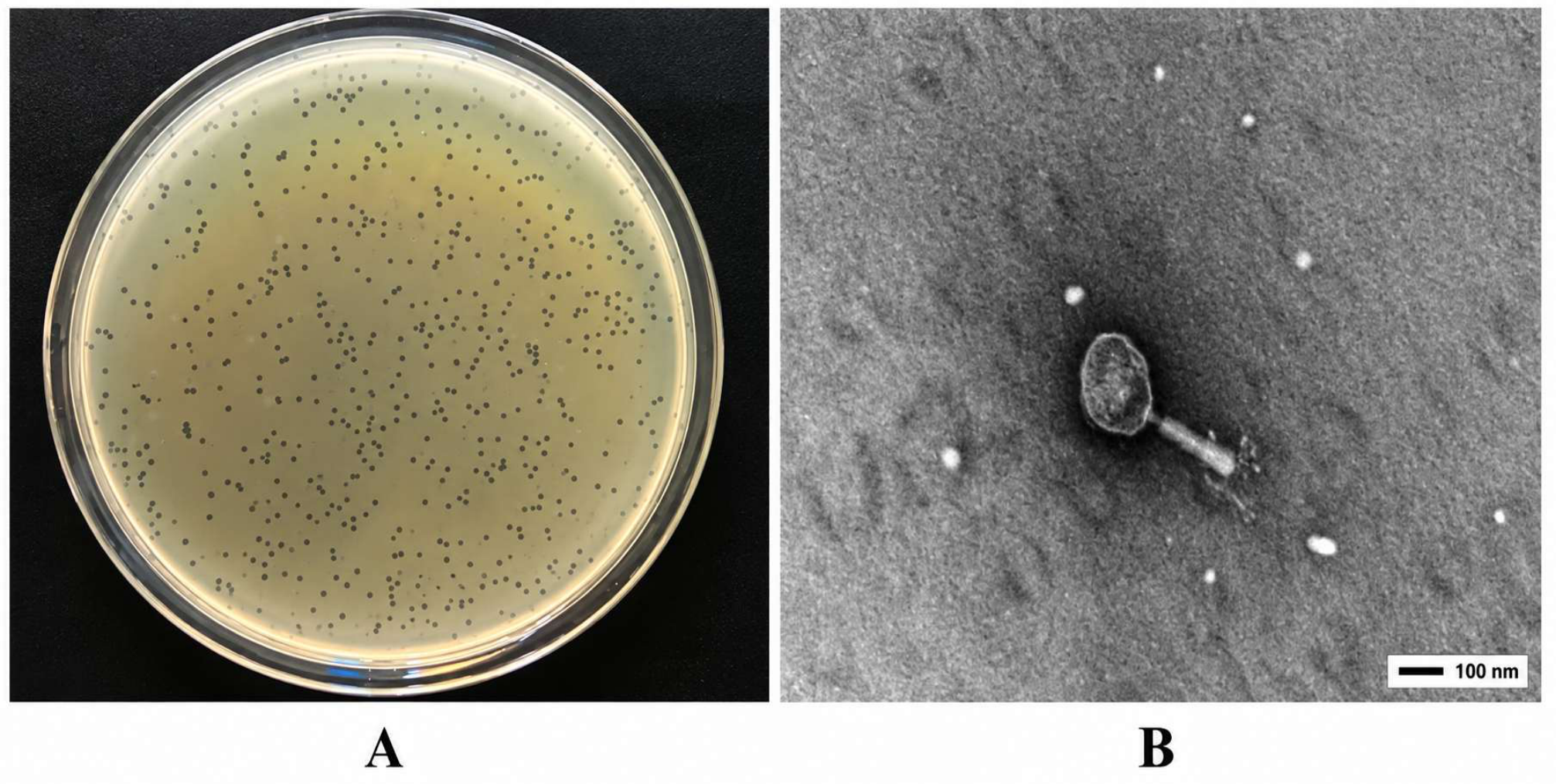
Transmission electron microscopy showed that ND-4 possessed an elliptical head measuring 110 ± 5 nm (long axis) by 82 ± 5 nm (short axis) and a tail measuring approximately 97 ± 5 nm in length and 18 ± 2 nm in width (Figure 1A). Re-examination of the TEM images indicated that the tail morphology is consistent with a contractile tail rather than a non-contractile tail (Figure 1B). Together with the whole-genome and phylogenetic analyses, these morphological features support the assignment of ND-4 to the family Ackermannviridae within the class Caudoviricetes.

3.2. Host Range

ND-4 produced plaques on three of the 40 tested E. coli strains, including the original host IMT5155, CVCC1527, and D-GX3, whereas no plaques were detected on the remaining E. coli strains or the two Salmonella strains. The EOP on IMT5155 was defined as 1.0, while the EOP values on CVCC1527 and D-GX3 were 0.47 and 0.85, respectively (Table 1). These results indicate that ND-4 has a relatively narrow productive host range under the tested conditions.

3.3. Biological Characteristics

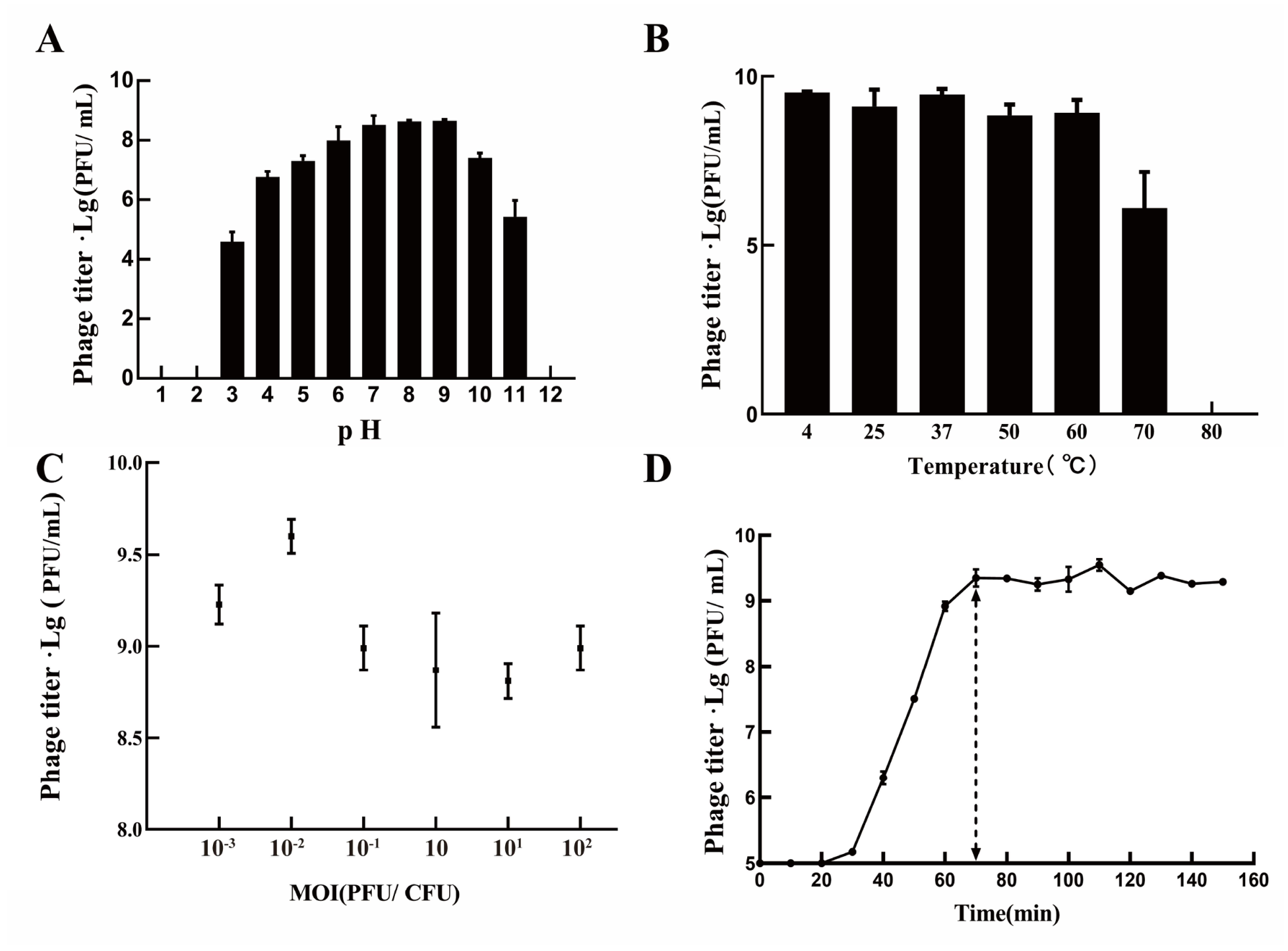
Thermal stability assays showed that ND-4 titers remained stable from 4 °C to 50 °C. At 60 °C, titers began to decline after 30 min. At 80 °C, activity persisted for 30 min but was lost by 60 min (Figure 2A).
ND-4 remained active from pH 3 to pH 11, with the highest titers observed at pH 7–9 (approximately 109 PFU/mL). Activity was lost at pH 2 and pH 12 (Figure 2B).
One-step growth analysis showed a latent period of 20 min, followed by a 50 min burst period. The phage titer reached a plateau at approximately 70 min, with an estimated burst size of approximately 231 PFU/cell (Figure 2C).
The highest phage titer, 3.97 × 10⁹ PFU/mL, was obtained at an MOI of 0.01 after 6 h of incubation (Figure 2D).

3.4. Genomic Analysis

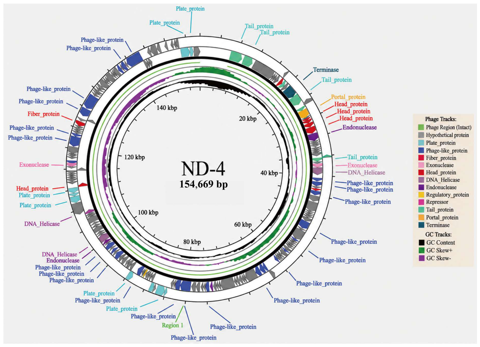
The ND-4 genome is a double-stranded DNA genome of 154,669 bp (GenBank accession: PQ374922), with a G + C content of 48.98%. Genome annotation predicted 184 proteins. Forty-five proteins had assigned functions related mainly to structure, replication, or lysis, whereas the remaining proteins were annotated as hypothetical proteins. No virulence or antimicrobial resistance genes were detected (Figure 3 and Table 2).
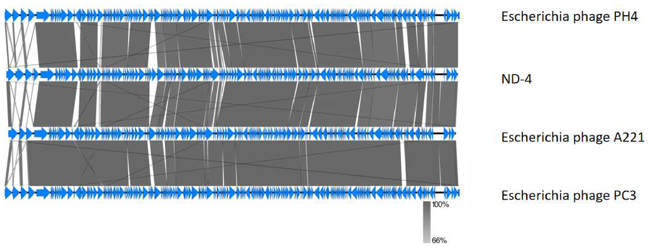
Functional annotation identified terminase, DNA helicase, regulatory proteins, portal protein, tail fiber proteins, capsid proteins, and lysis-related proteins, including lysozyme and endolysin. BLAST (https://blast.ncbi.nlm.nih.gov/, accessed on 23 June 2025) analysis showed that ND-4 was homologous to Escherichia phages A221, PC3, and PH4, with more than 98% nucleotide identity and 86–87% genome coverage (Figure 4).
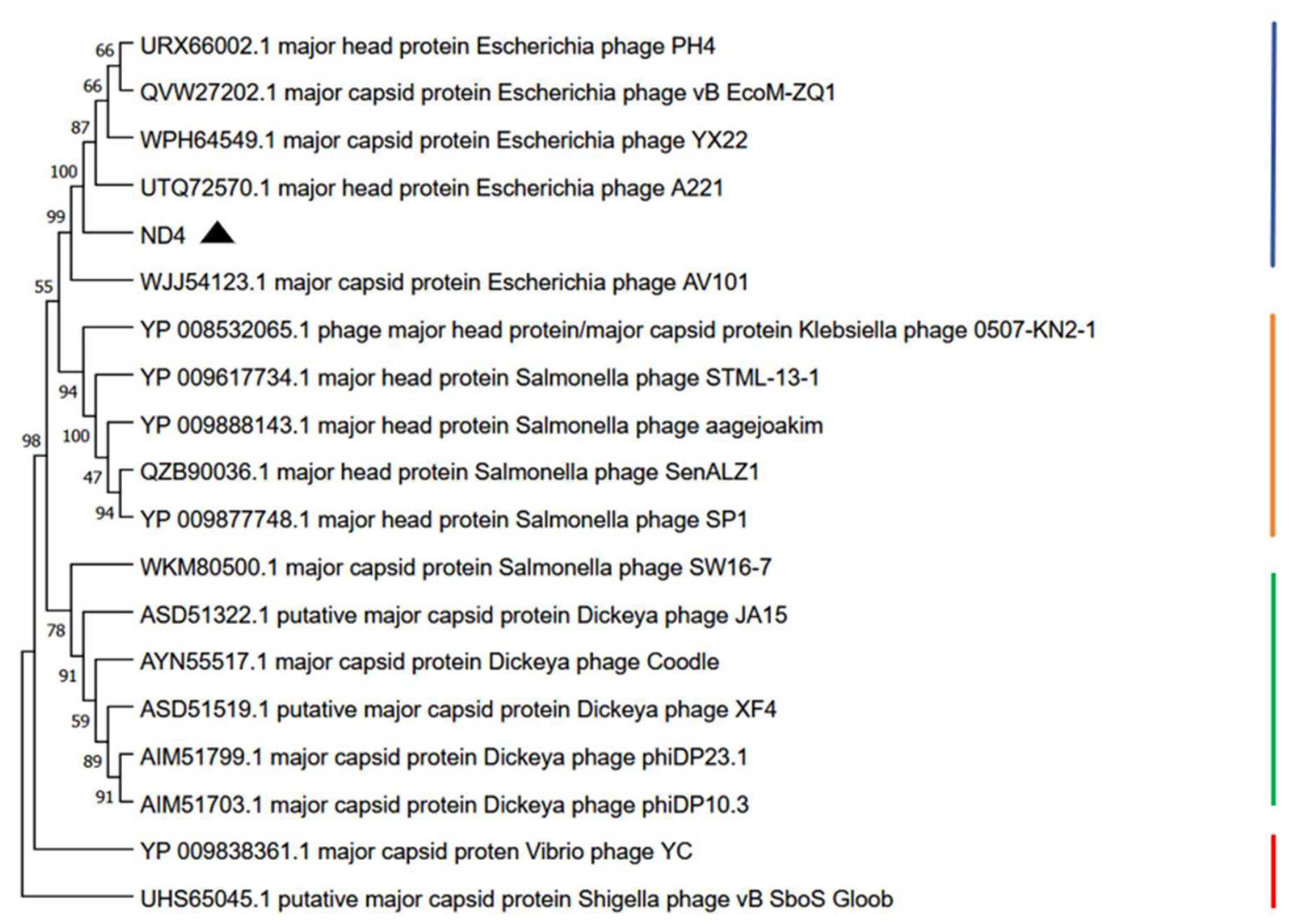
Phylogenetic analysis based on the major capsid protein (ORF22) clustered ND-4 with Escherichia phages A221, YX22, vB_EcoM-ZQ1, and PH44. Together with whole-genome similarity analysis, these phylogenetic relationships supported the assignment of ND-4 to the family Ackermannviridae, subfamily Aglimvirinae, within the class Caudoviricetes, in accordance with the current ICTV genomic classification framework (Figure 5). Re-examination of the TEM images further indicated that the tail morphology of ND-4 is consistent with a contractile-tail morphology, supporting its genomic assignment to the family Ackermannviridae.

3.5. Preliminary Application

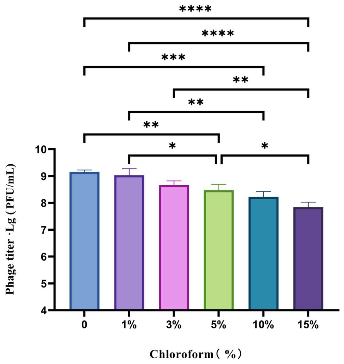
ND-4 maintained titers above 108 PFU/mL after treatment with chloroform concentrations up to 15% (Figure 6). This result is consistent with ND-4 being a non-enveloped phage.
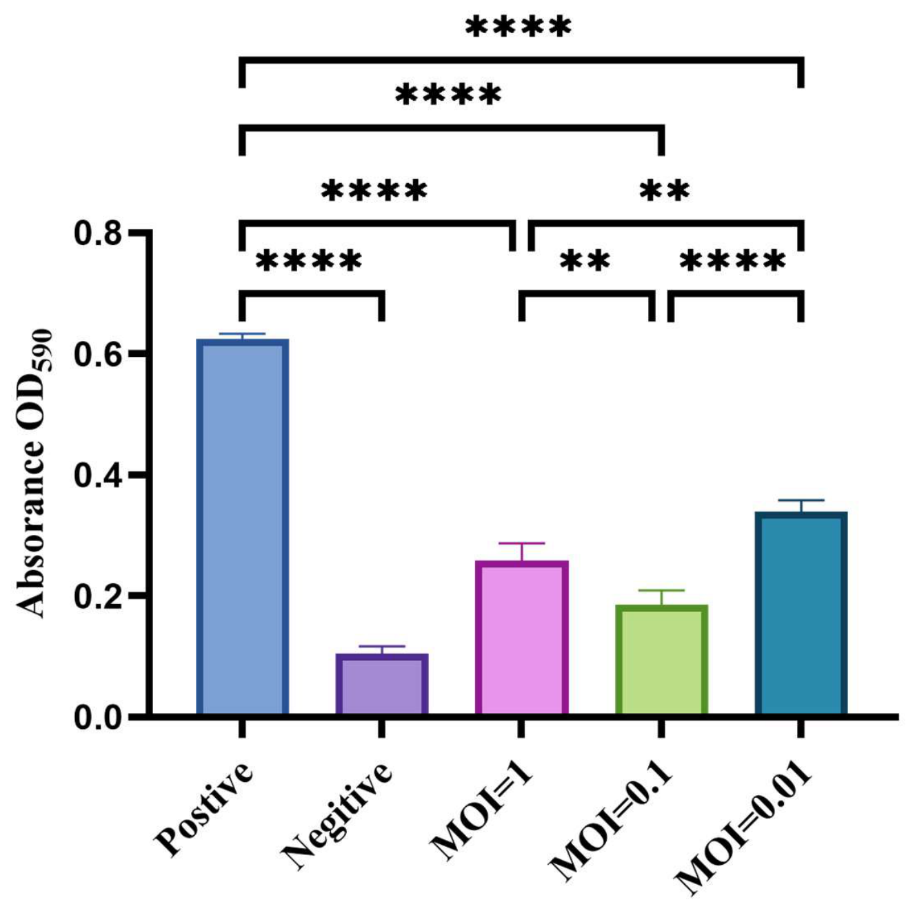
ND-4 significantly reduced biofilm biomass compared with the positive control (OD590 = 0.62) and approached the negative-control level (OD590 = 0.11). The strongest clearance effect was observed at an MOI of 0.1 (Figure 7).

3.6. Preliminary Chick Application Assay

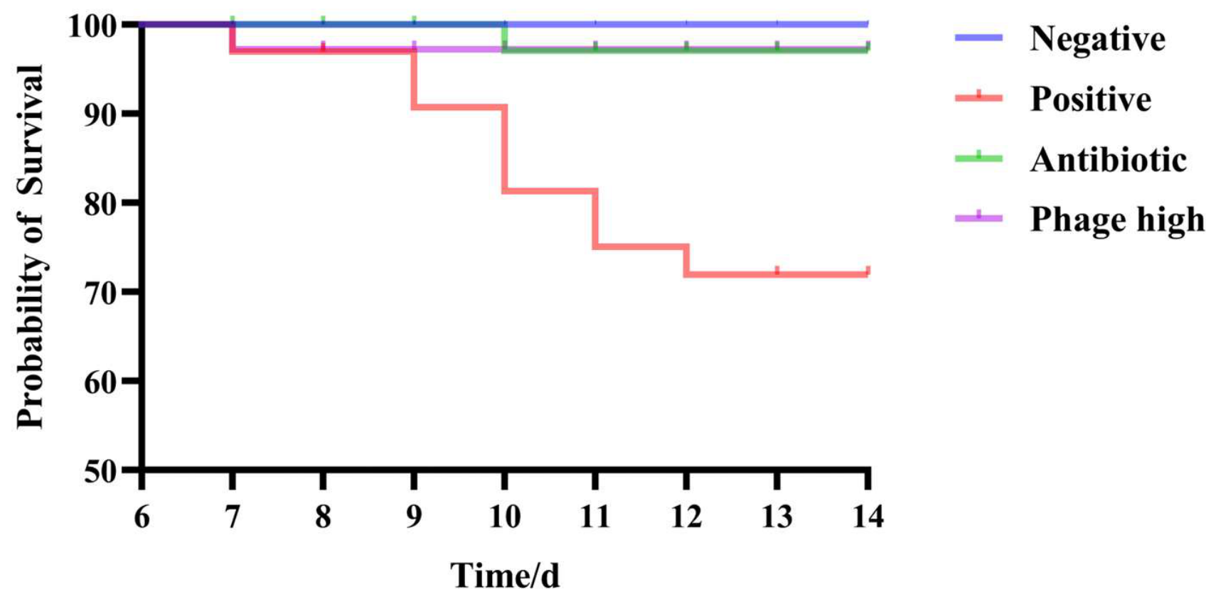
Survival analysis showed no mortality in the negative control group during the rearing period (Figure 8). Before treatment, deaths occurred in all three IMT5155-challenged groups. After treatment, mortality decreased in the antibiotic and phage treatment groups.
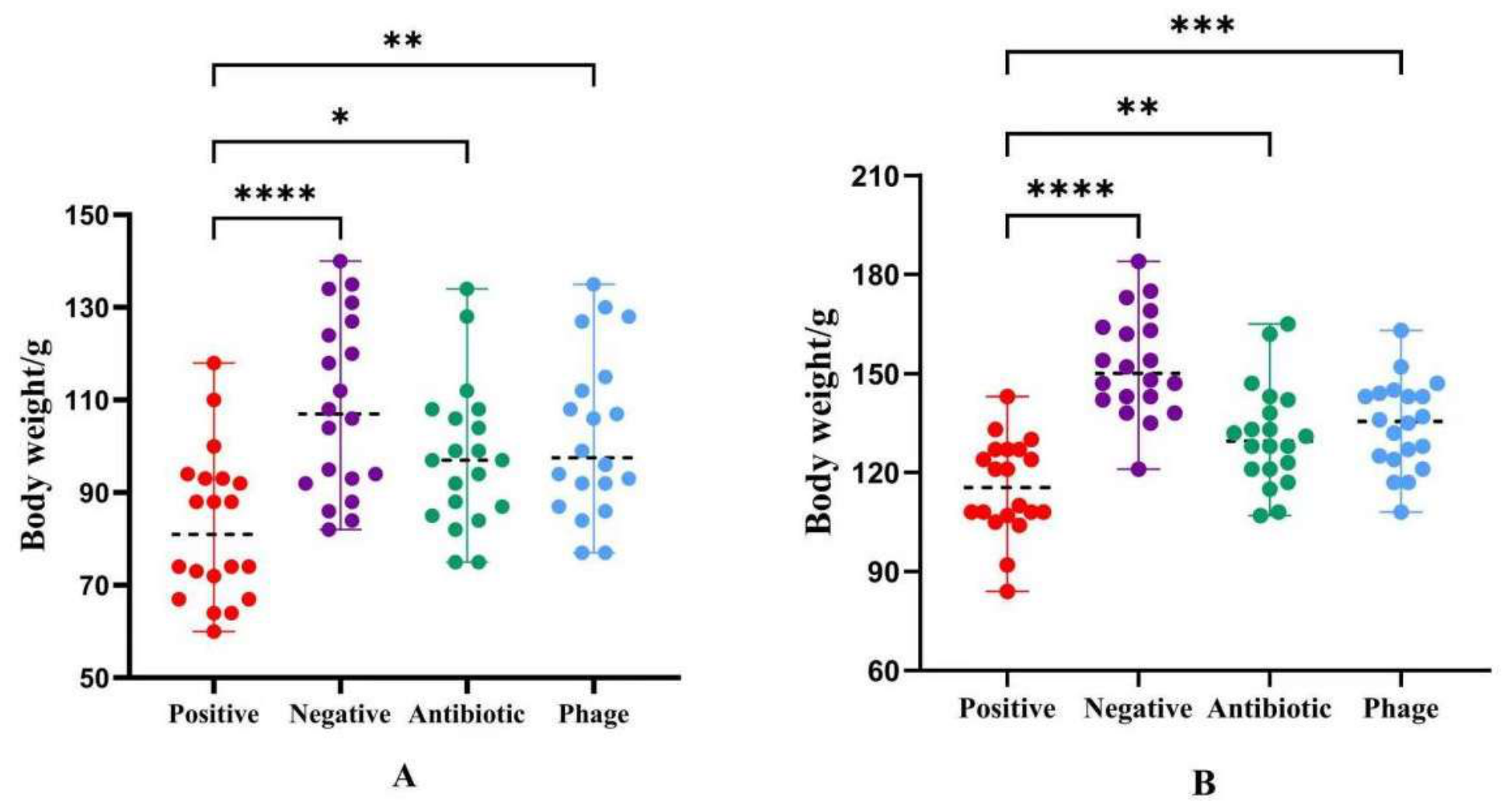
Body-weight analysis showed that IMT5155 infection suppressed weight gain during the monitored periods (Figure 9). Chicks treated with ND-4 or antibiotics showed partial recovery of body weight.
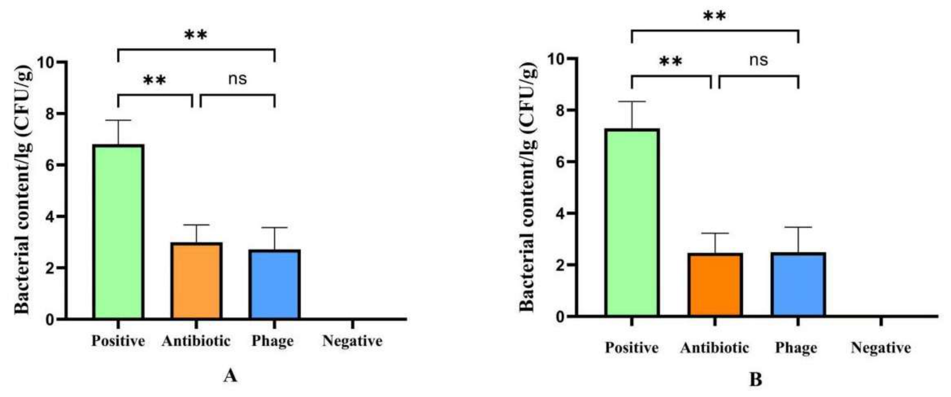
Bacterial-load analysis showed that phage and antibiotic treatment markedly reduced bacterial loads in chick tissues compared with the IMT5155-infected positive control (Figure 10). The reduction was approximately four orders of magnitude.
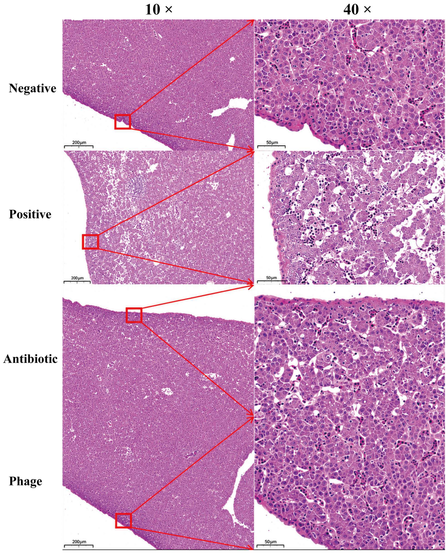
Histopathological analysis showed marked hepatic interstitial congestion and vacuolar degeneration in IMT5155-infected chicks compared with the blank control group (Figure 11). Treatment with ND-4 or antibiotic reduced hepatic inflammation and steatosis. The phage-treated group showed slightly greater histological improvement than the antibiotic-treated group under the conditions tested.

4. Discussion

This study identified ND-4 as a lytic E. coli phage with favorable biological stability, genomic safety features and preliminary application potential against APEC IMT5155. The phage showed a narrow host range, lysing IMT5155, CVCC1527 and D3 but not the other tested E. coli or Salmonella strains. This specificity may be advantageous for targeted control, although it also indicates that ND-4 would probably need to be combined with complementary phages for broader field application.
The replication and stability profiles of ND-4 support its further evaluation as an applied phage candidate. ND-4 had a latent period of 20 min and a burst size of 231 PFU/cell, indicating efficient propagation on IMT5155. Its burst size was higher than that reported for phage A221 [16], and its environmental tolerance was also favorable, with activity across pH 3–11 and short-term retention of activity at 80 °C. These features are relevant for preparation, storage and delivery, although formulation strategies such as microencapsulation may still be needed to improve stability under practical farm conditions [17].
Genome analysis further supported the lytic and safety profile of ND-4. The genome contained 184 predicted open reading frames, including genes associated with structure, replication and lysis. Tail fiber proteins are likely to contribute to the observed host specificity, as receptor-binding proteins are key determinants of phage host range [18,19,20]. No integrase, virulence gene or antimicrobial resistance gene was detected, which is an important prerequisite for therapeutic or preventive development [21,22,23,24,25]. The predicted nicotinamide synthesis-related protein may also be relevant to phage-bacteria interactions because some phages use NAD-related strategies to counter bacterial defense systems [25,26].
ND-4 also showed activity in application-oriented assays. Chloroform tolerance was consistent with the non-enveloped nature of the phage, and biofilm assays showed that ND-4 reduced biofilm biomass at all tested MOIs. The strongest effect occurred at an MOI of 0.1 rather than at the highest tested phage concentration. This pattern suggests that biofilm clearance may depend not only on phage dose but also on phage diffusion, adsorption and movement within the biofilm matrix [27].
The chick infection assay provided preliminary evidence that ND-4 can reduce the pathological impact of IMT5155 challenge. Phage treatment improved survival, body weight, tissue bacterial load and liver histopathology compared with infected controls, and some outcomes were comparable to or slightly better than gentamicin treatment. These effects are consistent with the targeted antibacterial activity expected for lytic phages. Nevertheless, the animal data should be interpreted cautiously because dose–response effects, treatment timing, route of administration, microbiota changes and resistance emergence were not fully resolved.
Although ND-4 was administered orally through drinking water, whereas the IMT5155 challenge was performed intraperitoneally, previous studies suggest that orally administered bacteriophages can cross epithelial barriers and reach extraintestinal sites. Nguyen et al. demonstrated that diverse bacteriophages can undergo rapid apical-to-basolateral transcytosis across epithelial cell layers, providing a potential mechanism for phage entry into the systemic compartment after oral administration [28]. More directly, a recent study in laying hens showed that a phage cocktail administered through drinking water after systemic APEC challenge could be recovered from multiple internal tissues and significantly reduced bacterial-associated morbidity and pathological lesions, supporting the feasibility of oral phage delivery for controlling extraintestinal APEC infection [29]. Once phages reach infected tissues and encounter susceptible bacteria, local replication may further increase the effective phage concentration and enhance antibacterial activity, a phenomenon generally referred to as phage self-amplification [30]. These mechanisms may partly explain the therapeutic effects observed for ND-4 in the present study. However, because phage concentrations in blood and tissues were not directly measured, the systemic bioavailability, tissue distribution, and pharmacokinetics of orally administered ND-4 remain to be determined in future studies.
Overall, ND-4 expands the available phage resources for APEC control and provides a candidate for future cocktail design. Its narrow host range argues for combination with other well-characterized lytic phages, whereas its stability, lack of detectable risk genes, and preliminary in vivo efficacy support further development. Future studies should test ND-4 in larger animal cohorts, define optimal dosing regimens, evaluate resistance dynamics and assess performance in multi-phage preparations under conditions closer to poultry production.

5. Conclusions

ND-4 is a newly characterized lytic E. coli phage isolated against the APEC strain IMT5155. It showed efficient propagation, broad environmental tolerance, biofilm-reduction activity and preliminary protection in challenged chicks. Its genome lacked detectable virulence and antimicrobial resistance genes, supporting further safety assessment. These findings indicate that ND-4 is a promising candidate for phage-based APEC control, especially as part of a rationally designed cocktail, but its efficacy and biosafety require validation in larger and more practical infection models.

Author Contributions

Conceptualization, Y.Z. and H.P.; methodology, L.W., J.Z., Q.D. and H.B.; software, G.L. and D.D.; validation, Y.F. and Y.Y.; formal analysis, C.L. and H.W.; investigation, L.T. and C.M.; resources, X.L.; data curation, R.Q., Y.Y. and C.M.; writing—original draft, L.W.; writing—review and editing, X.L. and Y.Z.; visualization, H.P. and G.L.; supervision, H.P. and X.L.; funding acquisition, H.P. All authors have read and agreed to the published version of the manuscript.

Funding

This research was supported by the Guangxi Key R&D Program (AB241484013. FN2600640636); Guangxi Forestry Science and Technology Program (2026GXZCLK06. 2025GXZCLK09. 2025GXZCLK11. 2025GXZCLK60. 2024GXZCLK37.); Nanning Key R&D Program (NNKJ202408); China Agriculture Resrearch System (CARS-42-55); Open Project of Guangxi Key Laboratory of Veterinary Biological Products (Guike 25-05).

Institutional Review Board Statement

All experiments were conducted in accordance with the guidelines of the Guangxi Animal Ethics Committee and the laboratory animal rules of Guangxi Veterinary Institute (GX-2025-033).

Informed Consent Statement

Not applicable.

Data Availability Statement

The annotated complete genome sequence of the phage ND-4 was deposited in GenBank under accession number PQ374922.

Conflicts of Interest

The authors declare no conflicts of interest.

References

  1. Berthe, T.; Ratajczak, M.; Clermont, O.; Denamur, E.; Petit, F. Evidence for Coexistence of Distinct Escherichia Coli Populations in Various Aquatic Environments and Their Survival in Estuary Water. Appl. Environ. Microb. 2013, 79, 4684. [Google Scholar] [CrossRef] [Scilit]
  2. Fratamico, P.M.; DebRoy, C.; Liu, Y.; Needleman, D.S.; Baranzoni, G.M.; Feng, P. Advances in Molecular Serotyping and Subtyping of Escherichia Coli. Front. Microbiol. 2016, 7, 644. [Google Scholar] [CrossRef] [Scilit]
  3. Kaper, J.B.; Nataro, J.P.; Mobley, H.L. Pathogenic Escherichia Coli. Nat. Rev. Microbiol. 2004, 2, 123. [Google Scholar] [CrossRef] [Scilit]
  4. Dai, J. Suppression Subtractive Hybridization Identifies an Autotransporter Adhesin Gene of E. Coli IMT5155 Specifically Associated with Avian Pathogenic Escherichia Coli (APEC). BMC Microbiol. 2010, 10, 236. [Google Scholar] [CrossRef] [Scilit]
  5. Koonjan, S.; Cooper, C.J.; Nilsson, A.S. Complete Genome Sequence of vB_EcoP_SU7, a Podoviridae Coliphage with the Rare C3 Morphotype. Microorganisms 2021, 9, 1576. [Google Scholar] [CrossRef] [Scilit]
  6. Lagerstrom, K.M.; Scales, N.C.; Hadly, E.A. Impressive Pan-Genomic Diversity of E. Coli from a Wild Animal Community Near Urban Development Reflects Human Impacts. Iscience 2024, 27, 109072. [Google Scholar] [CrossRef] [Scilit]
  7. Ge, X.Z.; Jiang, J.; Pan, Z.; Hu, L.; Wang, S.; Wang, H.; Leung, F.C.; Dai, J.; Fan, H. Comparative Genomic Analysis Shows That Avian Pathogenic Escherichia Coli Isolate IMT5155 (O2:K1:H5; ST Complex 95, ST140) Shares Close Relationship with ST95 APEC O1:K1 and Human ExPEC O18:K1 Strains. PLoS ONE 2014, 9, e112048. [Google Scholar] [CrossRef] [Scilit]
  8. Ali, A.; Kolenda, R.; Khan, M.M.; Weinreich, J.; Li, G.; Wieler, L.H.; Tedin, K.; Roggenbuck, D.; Schierack, P. Novel Avian Pathogenic Escherichia Coli Genes Responsible for Adhesion to Chicken and Human Cell Lines. Appl. Environ. Microb. 2020, 86, e01068-20. [Google Scholar] [CrossRef] [Scilit]
  9. Begum, Y.A.; Talukder, K.A.; Azmi, I.J.; Shahnaij, M.; Sheikh, A.; Sharmin, S.; Svennerholm, A.-M.; Qadri, F. Resistance Pattern and Molecular Characterization of Enterotoxigenic Escherichia Coli (ETEC) Strains Isolated in Bangladesh. PLoS ONE 2016, 11, e157415. [Google Scholar] [CrossRef] [Scilit]
  10. Dion, M.B.; Oechslin, F.; Moineau, S. Phage Diversity, Genomics and Phylogeny. Nat. Rev. Microbiol. 2020, 18, 125. [Google Scholar] [CrossRef] [Scilit]
  11. Łusiak-Szelachowska, M.; Międzybrodzki, R.; Drulis-Kawa, Z.; Cater, K.; Knežević, P.; Winogradow, C.; Amaro, K.; Jończyk-Matysiak, E.; Weber-Dąbrowska, B.; Rękas, J.; et al. Bacteriophages and Antibiotic Interactions in Clinical Practice: What We Have Learned so Far. J. Biomed. Sci. 2022, 29, 23. [Google Scholar] [CrossRef] [Scilit]
  12. Tagliaferri, T.L.; Jansen, M.; Horz, H. Fighting Pathogenic Bacteria on Two Fronts: Phages and Antibiotics as Combined Strategy. Front. Cell. Infect. Microbiol. 2019, 9, 22. [Google Scholar] [CrossRef] [Scilit]
  13. Salmond, G.P.C.; Fineran, P.C. A Century of the Phage: Past, Present and Future. Nat. Rev. Microbiol. 2015, 13, 777. [Google Scholar] [CrossRef] [Scilit]
  14. Duplessis, C.A.; Biswas, B. A Review of Topical Phage Therapy for Chronically Infected Wounds and Preparations for a Randomized Adaptive Clinical Trial Evaluating Topical Phage Therapy in Chronically Infected Diabetic Foot Ulcers. Antibiotics 2020, 9, 377. [Google Scholar] [CrossRef] [Scilit]
  15. Federici, S.; Kredo-Russo, S.; Valdés-Mas, R.; Kviatcovsky, D.; Weinstock, E.; Matiuhin, Y.; Silberberg, Y.; Atarashi, K.; Furuichi, M.; Oka, A.; et al. Targeted Suppression of Human IBD-Associated Gut Microbiota Commensals by Phage Consortia for Treatment of Intestinal Inflammation. Cell 2022, 185, 2879. [Google Scholar] [CrossRef] [Scilit]
  16. Mao, X.; Wu, Y.; Ma, R.; Li, L.; Wang, L.; Tan, Y.; Li, Z.; Liu, H.; Han, K.; Cao, Y.; et al. Oral Phage Therapy with Microencapsulated Phage A221 against Escherichia Coli Infections in Weaned Piglets. BMC Vet. Res. 2023, 19, 165. [Google Scholar] [CrossRef] [Scilit]
  17. Chen, B.; Benavente, L.P.; Chittò, M.; Wychowaniec, J.K.; Post, V.; D’Este, M.; Constant, C.; Zeiter, S.; Feng, W.; Moreno, M.G.; et al. Alginate Microbeads and Hydrogels Delivering Meropenem and Bacteriophages to Treat Pseudomonas Aeruginosa Fracture-Related Infections. J. Control. Release Off. J. Control. Release Soc. 2023, 364, 159. [Google Scholar] [CrossRef] [Scilit]
  18. Nobrega, F.L.; Vlot, M.; de Jonge, P.A.; Dreesens, L.L.; Beaumont, H.J.E.; Lavigne, R.; Dutilh, B.E.; Brouns, S.J.J. Targeting Mechanisms of Tailed Bacteriophages. Nat. Rev. Microbiol. 2018, 16, 760. [Google Scholar] [CrossRef] [Scilit]
  19. Prevelige, P.E.J.; Cortines, J.R. Phage Assembly and the Special Role of the Portal Protein. Curr. Opin. Virol. 2018, 31, 66. [Google Scholar] [CrossRef] [Scilit]
  20. Stone, E.; Campbell, K.; Grant, I.; McAuliffe, O. Understanding and Exploiting Phage-Host Interactions. Viruses 2019, 11, 567. [Google Scholar] [CrossRef] [Scilit]
  21. Selcuk, E.; Dokuz, S.; Ozbek, T. Evaluating the Stability of Lytic and Lysogenic Bacteriophages in Various Protectants. J. Pharm. Sci. 2024, 113, 1488. [Google Scholar] [CrossRef] [Scilit]
  22. Feiner, R.; Argov, T.; Rabinovich, L.; Sigal, N.; Borovok, I.; Herskovits, A.A. A New Perspective on Lysogeny: Prophages as Active Regulatory Switches of Bacteria. Nat. Rev. Microbiol. 2015, 13, 641. [Google Scholar] [CrossRef] [Scilit]
  23. Tang, X.; Zhou, M.; Fan, C.; Zeng, G.; Lu, Y.; Dong, H.; Song, B.; Fu, Q.; Zeng, Y. The Arsenic Chemical Species Proportion and Viral Arsenic Biotransformation Genes Composition Affects Lysogenic Phage Treatment under Arsenic Stress. Sci. Total Environ. 2021, 780, 146628. [Google Scholar] [CrossRef] [Scilit]
  24. Sedhom; J. Solomon, L.A. Lambda CI Binding to Related Phage Operator Sequences Validates Alignment Algorithm and Highlights the Importance of Overlooked Bonds. Genes 2023, 14, 2221. [Google Scholar] [CrossRef] [Scilit]
  25. Millman, A.; Melamed, S.; Leavitt, A.; Doron, S.; Bernheim, A.; Hör, J.; Garb, J.; Bechon, N.; Brandis, A.; Lopatina, A.; et al. An Expanded Arsenal of Immune Systems That Protect Bacteria from Phages. Cell Host Microbe 2022, 30, 1556. [Google Scholar] [CrossRef] [Scilit]
  26. Osterman, I.; Samra, H.; Rousset, F.; Loseva, E.; Itkin, M.; Malitsky, S.; Yirmiya, E.; Millman, A.; Sorek, R. Phages Reconstitute NAD(+) to Counter Bacterial Immunity. Nature 2024, 634, 1160. [Google Scholar] [CrossRef] [Scilit]
  27. Simmons, E.L.; Drescher, K.; Nadell, C.D.; Bucci, V. Phage Mobility Is a Core Determinant of Phage-Bacteria Coexistence in Biofilms. ISME J. 2018, 12, 531. [Google Scholar] [CrossRef] [Scilit]
  28. Nguyen, S.; Baker, K.; Padman, B.S.; Patwa, R.; Dunstan, R.A.; Weston, T.A.; Schlosser, K.; Bailey, B.; Lithgow, T.; Lazarou, M.; et al. Bacteriophage Transcytosis Provides a Mechanism To Cross Epithelial Cell Layers. mBio 2017, 8, e01874-17. [Google Scholar] [CrossRef] [Scilit]
  29. Gohar, M.; Hyun, J.E.; Smith, K.R.; Shaukat, W.; Waldner, M.; Mehina, N.; Moqaddes, S.; Ding, Y.; Abd-Elsalam, R.M.; Fairbrother, J.M.; et al. Bacteriophage Cocktail in Drinking Water Suppresses Systemic Avian Pathogenic Escherichia Coli Infection and Pathology in Laying Hens. Curr. Res. Microb. Sci. 2026, 10, 100569. [Google Scholar] [CrossRef] [Scilit]
  30. Abedon, S.T. Kinetics of Phage-Mediated Biocontrol of Bacteria. Foodborne Pathog. Dis. 2009, 6, 807–815. [Google Scholar] [CrossRef] [Scilit]
Figure 1. Plaque morphology and virion structure of ND-4. (A) Plaque morphology. (B) Transmission electron microscopy image.
Life 16 01549 g001
Figure 2. Biological characteristics of phage ND-4. (A) pH stability. (B) Thermal stability. (C) Optimal MOI determination. (D) One-step growth curve.
Life 16 01549 g002
Figure 3. Genome map of phage ND-4.
Life 16 01549 g003
Figure 4. Genomic synteny analysis of ND-4 and related phages.
Life 16 01549 g004
Figure 5. Phylogenetic tree based on the major capsid protein. Note: The triangle indicates the bacteriophage used in this study.
Life 16 01549 g005
Figure 6. Chloroform sensitivity of phage ND-4. (* p < 0.05, ** p < 0.01, *** p < 0.001, and **** p < 0.0001 (one-way ANOVA followed by Tukey’s multiple-comparison test.)).
Life 16 01549 g006
Figure 7. Biofilm-clearance efficiency of phage ND-4 at different MOIs. (** p < 0.01, and **** p < 0.0001 (one-way ANOVA followed by Tukey’s multiple-comparison test.)).
Life 16 01549 g007
Figure 8. Survival curves of chicks (n = 20 per group).
Life 16 01549 g008
Figure 9. Body weight of chicks at 10 and 14 days of age. (A) Body weight at 10 days. (B) Body weight at 14 days. (n = 20 per group, * p < 0.05, ** p < 0.01, *** p < 0.001, and **** p < 0.0001 (one-way ANOVA followed by Tukey’s multiple-comparison test)).
Life 16 01549 g009
Figure 10. Bacterial load in chick tissues. (A) Bacterial load in spleen tissue. (B) Bacterial load in liver tissue. (** p < 0.01, ns = not significant, (one-way ANOVA followed by Tukey’s multiple-comparison test)).
Life 16 01549 g010
Figure 11. Histological sections of chick liver tissue.
Life 16 01549 g011
Table 1. Host-range analysis of phage ND-4.
StrainsGenusSourceSerotypesLytic
IMT5155E.coliChickenO2:K1+
CVCC1527E.coliPigO8:K88+
CVCC4050E.coliPigO157:H7-
GXEC-01E.coliChickenNA-
GXEC-02E.coliChickenNA-
GXEC-03E.coliChickenO157-
GXEC-04E.coliChickenNA-
GXEC-05E.coliChickenNA-
GXEC-06E.coliChickenO157-
GXEC-07E.coliChickenNA-
GXEC-08E.coliChickenNA-
GXEC-09E.coliChickenNA-
GXEC-10E.coliChickenNA-
GXEC-11E.coliChickenO142:K68-
GXEC-12E.coliChickenNA-
GXEC-13E.coliChickenNA-
GXEC-14E.coliChickenNA-
GXEC-15E.coliChickenNA-
HZDC01E.coliChickenNA-
HZDC02E.coliChickenNA-
HZDC03E.coliChickenO114:K90-
HZDC04E.coliChickenNA-
HZDC05E.coliChickenNA-
HZDC06E.coliChickenNA-
HZDC07E.coliChickenNA-
HZDC08E.coliChickenO26:K60-
HZDC09E.coliChickenNA-
D-GX1E.coliChickenNA-
D-GX2E.coliChickenNA-
D-GX3E.coliChickenNA+
D-GX4E.coliChickenNA-
D-GX5E.coliChickenNA-
D-GX6E.coliChickenNA-
C-D1E.coliChickenO157:H7-
C-D2E.coliChickenNA-
C-D3E.coliChickenNA-
C-D4E.coliChickenNA-
C-D5E.coliChickenO2-
C-D6E.coliChickenNA-
C-D7E.coliChickenNA-
C-D8E.coliChickenNA-
CVCC1806Sal\S. enteritidis-
CVCC3384Sal\S. typhimurium-
Note: +, lytic activity; -, no lytic activity. Strains listed as serotype NA were tested using commercial O2, O26, O142, and O157 detection kits but were not assigned to any of these serotypes.
Table 2. Functional CDS annotation of phage ND-4.
CDSIdentityLength (aa)Functione-ValueAccession Number
296.13%3027Phage tail fibers0WPH64569.1
382.03%2430Tail protein0YP_010659927.1
598.11%1728Tail spike protein0YP_010659925.1
8100.00%753Putative head–tail protein0QZI78659.1
11100.00%696tail stabilization protein2 × 10−173UTQ72558.1
1390.32%343Terminase large subunit0UHHS65537.1
1499.52%1896Tail sheath protein0UHS65542.1
17100.00%1695Portal protein7 × 10−28URX65806.1
2299.77%1323Major capsid protein0QVW27202.1
34100.00%453Recombination mediator protein6 × 10−105WP_242243974.1
3699.80%1509DNA helicase0WPH64536.1
39100.00%990Sliding clamp loader subunit0WPH64532.1
40100.00%423DNA polymerase1 × 10−97WP_276339478.1
41100.00%495Translational repressor4 × 10−111WP_242244075.1
4998.94%855Ribose-phosphate0WKM80470.1
5099.64%1665Nicotinamide0WPH64521.1
65100.00%279DNA-binding protein7 × 10−57WP_276339458.1
6699.48%1719DNA helicase0UTQ72610.1
68100.00%528RnaseH1 × 10−125QVW27164.1
7199.61%2346Endonuclease subunit0UHS65534.1
84100.00%912DNA primase0WPH64489.1
8999.87%2283Ribonucleotide reductase0WPH64482.1
90100.00%1104Ribonucleotide reductase0EJS6255053.1
91100.00%162Glutaredoxin3 × 10−29WP_242244087.1
94100.00%381GPW/gp25 family protein6 × 10−88EJS6255058.1
95100.00%1611Lysozyme0UHS65547.1
10599.41%1023ssDNA binding protein0WPH64466.1
11999.79%1425DNA helicase0UHS65553.1
12599.16%1431DNA ligase0WPH64446.1
12899.54%660DNA helicase loader7 × 10−162QVW27103.1
147100.00%1332DNA topoisomerase II medium subunit0USL85959.1
14899.84%1911DNA topoisomerase0WPH64598.1
15499.63%810tail protein0URX65943.1
17399.50%3000DNA polymerase0USL85796.1
Note: Proteins not labeled in the table were annotated as hypothetical proteins with unknown function.
Disclaimer/Publisher’s Note: The statements, opinions and data contained in all publications are solely those of the individual author(s) and contributor(s) and not of MDPI and/or the editor(s). MDPI and/or the editor(s) disclaim responsibility for any injury to people or property resulting from any ideas, methods, instructions or products referred to in the content.