Challenges in Differential Diagnosis and Management of Lymphoepithelial Sialadenitis (LESA): A Scoping Review


 , and
, and 
Abstract
1. Introduction
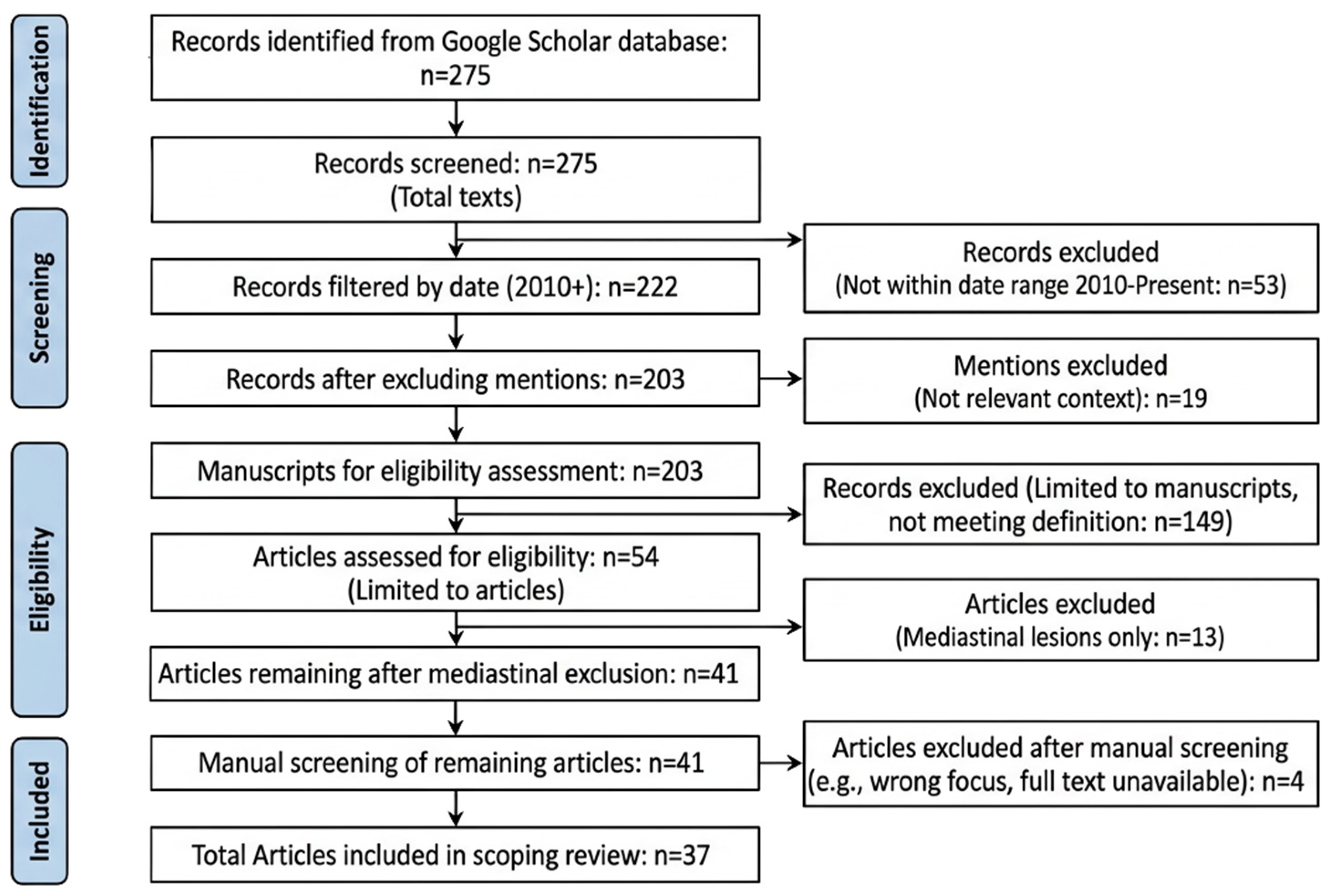
2. Materials and Methods
3. Results
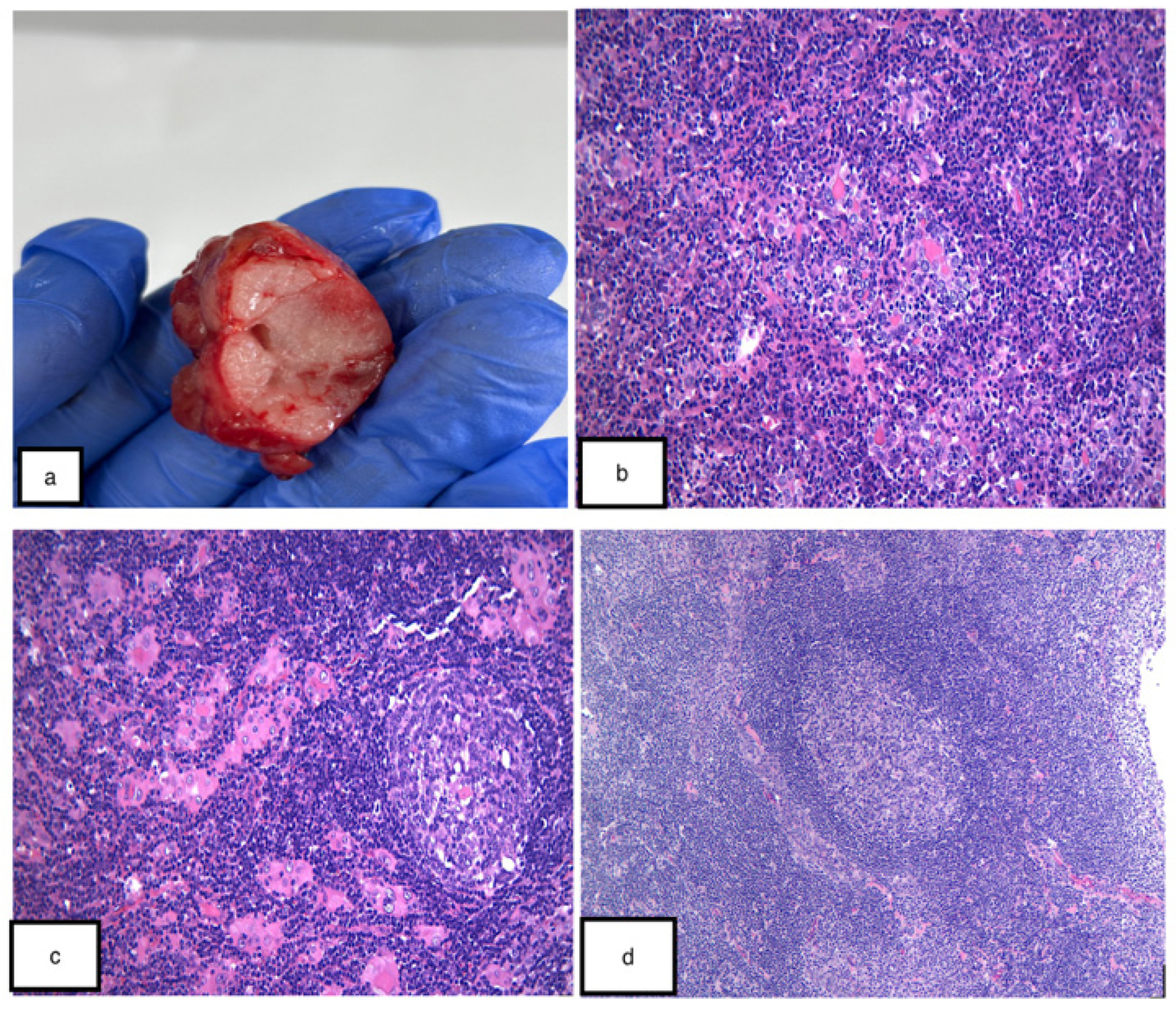
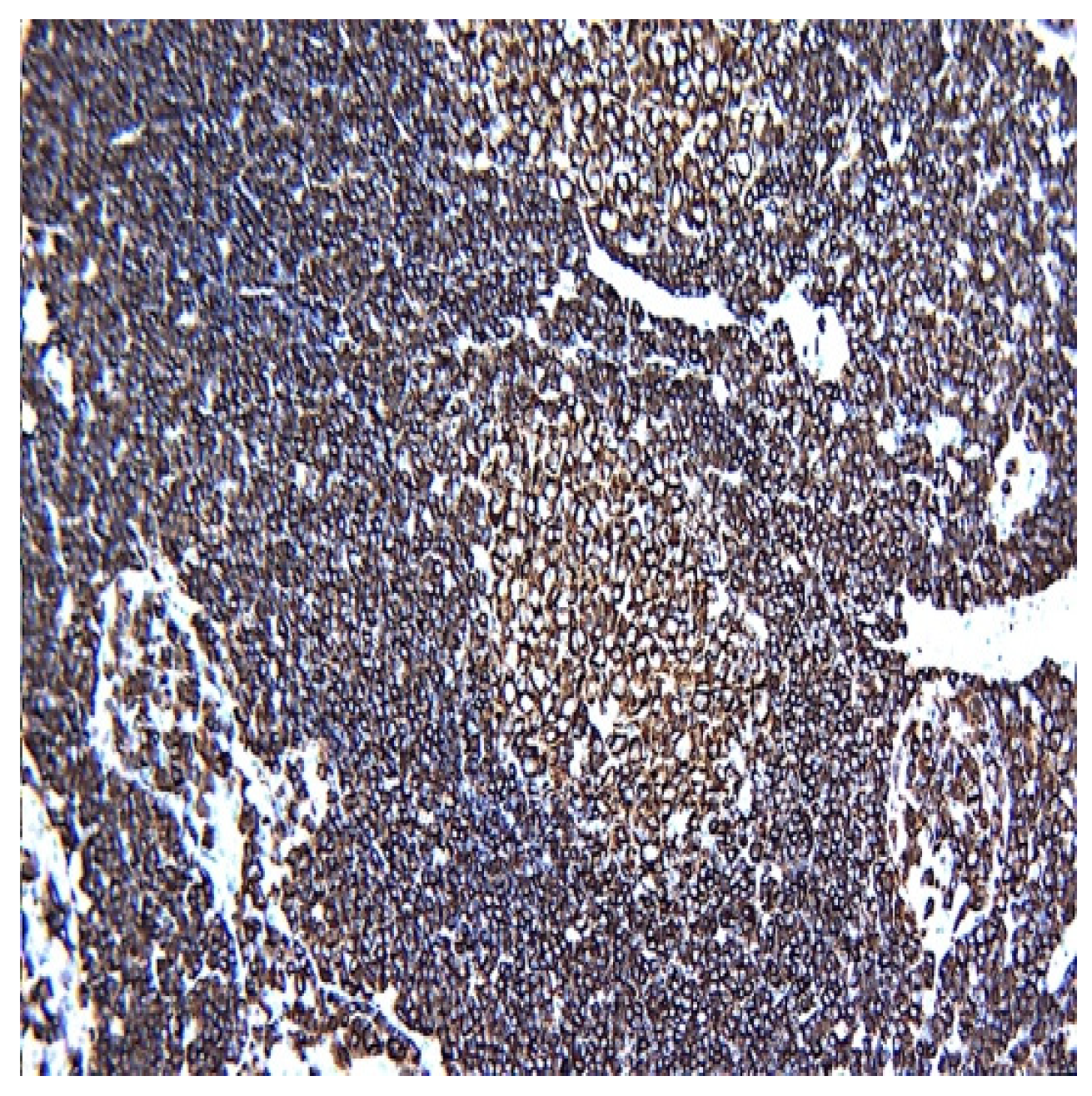
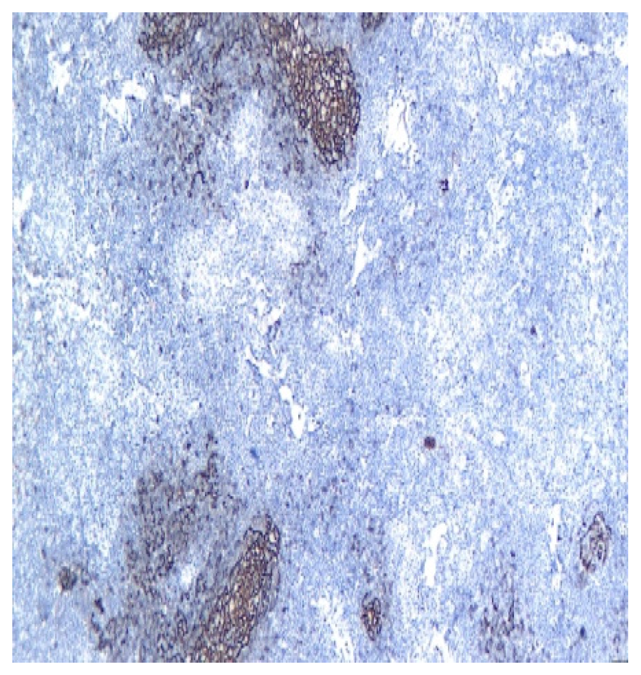
3.1. Immunophenotype and Molecular Features
3.2. Comparison with Minor Salivary Gland Biopsies
3.3. LESA Association and Overlap with Sjögren’s Disease
3.4. Spectrum and Progression to MALT (Extranodal Marginal Zone) Lymphoma
3.5. Differential Diagnoses of Lymphoid-Rich Salivary Gland Lesions
3.6. Characteristics and Critical Appraisal of Included Studies
4. Discussions
4.1. Imaging Modalities
4.2. Type of Biopsy: Diagnostic Challenges and Pitfalls
4.3. Ancillary Molecular and Flow Cytometry Testing for Clonality
4.4. Management Strategy: Surveillance Protocols and Follow-Up
4.5. Existing Guidelines, Consensus Statements, and Identified Gaps
4.6. Special Contexts and Mimics: HIV-Associated Parotid Disease and IgG4-Related Sialadenitis
4.7. LESA Association and Overlap with IgG4-Related Disease
4.8. Multidisciplinary Care Models, Referral Pathways, and Quality Metrics
4.9. Clinical Translation Value
5. Limitations
6. Conclusions
Author Contributions
Funding
Institutional Review Board Statement
Informed Consent Statement
Data Availability Statement
Acknowledgments
Conflicts of Interest
Abbreviations
| ANA | Antinuclear antibody |
| ASCO | American Society of Clinical Oncology |
| BLEC | Benign lymphoepithelial cyst |
| CEUS | Contrast-enhanced ultrasound |
| CNB | Core needle biopsy |
| CT | Computed tomography |
| DLBCL | Diffuse large B-cell lymphoma |
| DWI | Diffusion-weighted imaging |
| EULAR | European Alliance of Associations for Rheumatology |
| FISH | Fluorescence in situ hybridization |
| FNA | Fine-needle aspiration |
| HE | Hematoxylin and eosin |
| HIV | Human immunodeficiency virus |
| IHC | Immunohistochemistry |
| IGH | Immunoglobulin heavy chain |
| IgG4 | Immunoglobulin G4 |
| LEL | Lymphoepithelial lesion |
| LESA | Lymphoepithelial sialadenitis |
| MALT | Mucosa-associated lymphoid tissue |
| MRI | Magnetic resonance imaging |
| MSRSGC | Milan System for Reporting Salivary Gland Cytopathology |
| NGS | Next-generation sequencing |
| PCR | Polymerase chain reaction |
| PET | Positron emission tomography |
| RF | Rheumatoid factor |
| SS | Sjögren’s syndrome |
| TALP | Tumor-associated lymphoid proliferation |
| TCR | T-cell receptor |
| US | Ultrasound |
| EMZBCL | Extranodal marginal zone B-cell lymphoma |
References
- Zabotti, A.; Giovannini, I.; Longhino, S.; Manfrè, V.; Rizzo, M.T.; De Vita, S.; Di Loreto, C.; Quartuccio, L.; Pegolo, E. Nuts and bolts of salivary gland pathology in primary Sjögren’s syndrome. Clin. Exp. Rheumatol. 2023, 41, 2525–2537. [Google Scholar] [PubMed]
- Alunno, A.; Leone, M.C.; Giacomelli, R.; Gerli, R.; Carubbi, F. Lymphoma and Lymphomagenesis in Primary Sjögren’s Syndrome. Front. Med. 2018, 5, 102. [Google Scholar] [CrossRef] [Scilit] [PubMed]
- Valim, V.; Secco, A.; de Oliveira, F.R.; Vázquez, M.; Rosa, B.B.; Macagnani, F.L.; Vargas-Bueno, K.D.; Rojas, E.; Hernández-Delgado, A.; Catalan-Pellet, A.; et al. Parotid gland swelling in primary Sjögren’s syndrome: Activity and other sialadenosis causes. Rheumatology 2022, 61, 2987–2992. [Google Scholar] [CrossRef] [Scilit] [PubMed]
- Kato, H.; Kawaguchi, M.; Ando, T.; Aoki, M.; Kuze, B.; Matsuo, M. CT and MR imaging findings of non-neoplastic cystic lesions of the parotid gland. Jpn. J. Radiol. 2019, 37, 627–635. [Google Scholar] [CrossRef] [Scilit] [PubMed]
- Thompson, L.; Whaley, R. Lymphoepithelial Carcinoma of Salivary Glands. Surg. Pathol. Clin. 2021, 14, 75–96. [Google Scholar] [CrossRef] [Scilit] [PubMed]
- Kalhor, N.; Weissferdt, A.; Moran, C.A. Primary Salivary Gland Type Tumors of the Thymus. Adv. Anat. Pathol. 2017, 24, 15–23. [Google Scholar] [CrossRef] [Scilit] [PubMed]
- Nocturne, G.; Mariette, X. Sjögren Syndrome-associated lymphomas: An update on pathogenesis and management. Br. J. Haematol. 2015, 168, 317–327. [Google Scholar] [CrossRef] [Scilit] [PubMed]
- Wenig, B.M. Lymphoepithelial-like carcinomas of the head and neck. In Seminars in Diagnostic Pathology; WB Saunders: Philadelphia, PA, USA, 2015; Volume 32. [Google Scholar]
- Dong, J.; Zhao, H.; Luo, G.; Yang, X. Research progress on salivary gland mucosa-associated lymphoid tissue lymphoma. J. Prev. Treat. Stomatol. Dis. 2025, 33, 809–818. [Google Scholar] [CrossRef]
- Piyasatukit, N.; Awsakulsutthi, S.; Kintarak, J. Benign lymphoepithelial cyst of parotid glands in HIV-positive patient. J. Med. Assoc. Thai 2015, 98, S141–S145. [Google Scholar] [PubMed]
- Schreuder, M.I.; Brand, M.v.D.; Hebeda, K.M.; Groenen, P.J.T.A.; van Krieken, J.H.; Scheijen, B. Novel developments in the pathogenesis and diagnosis of extranodal marginal zone lymphoma. J. Hematop. 2017, 10, 91–107. [Google Scholar] [CrossRef] [Scilit] [PubMed]
- Papageorgiou, A.; Voulgarelis, M.; Tzioufas, A.G. Clinical picture, outcome and predictive factors of lymphoma in Sjögren syndrome. Autoimmun. Rev. 2015, 14, 641–649. [Google Scholar] [CrossRef] [Scilit] [PubMed]
- Jogai, S. Lymphoreticular pathology of the head and neck region: A review of selected entities. Diagn. Histopathol. 2021, 27, 191–201. [Google Scholar] [CrossRef] [Scilit]
- Barone, F.; Colafrancesco, S.; Campos, J. Histology of Sjögren’s Syndrome. In Sjögren’s Syndrome; Elsevier: Amsterdam, The Netherlands, 2016; pp. 171–187. [Google Scholar]
- Asam, S.; Neag, G.; Berardicurti, O.; Gardner, D.; Barone, F. The role of stroma and epithelial cells in primary Sjögren’s syndrome. Rheumatology 2021, 60, 3503–3512. [Google Scholar] [CrossRef] [Scilit] [PubMed]
- Emfietzoglou, R.; Pettas, E.; Georgaki, M.; Papadopoulou, E.; Theofilou, V.I.; Papadogeorgakis, N.; Piperi, E.; Lopes, M.A.; Nikitakis, N.G. Lymphoepithelial Subtype of Oral Squamous Cell Carcinoma: Report of an EBV-Negative Case and Literature Review. Dent. J. 2022, 10, 165. [Google Scholar] [CrossRef] [Scilit] [PubMed]
- Stergiou, I.E.; Poulaki, A.; Voulgarelis, M. Pathogenetic Mechanisms Implicated in Sjögren’s Syndrome Lymphomagenesis: A Review of the Literature. J. Clin. Med. 2020, 9, 3794. [Google Scholar] [CrossRef] [Scilit] [PubMed]
- Thakral, B.; Zhou, J.; Medeiros, L.J. Extranodal hematopoietic neoplasms and mimics in the head and neck: An update. Hum. Pathol. 2015, 46, 1079–1100. [Google Scholar] [CrossRef] [Scilit] [PubMed]
- El Hussein, S.; Khader, S.N. Cytopathology approach to rare salivary gland lesions with oncocytic features. Diagn. Cytopathol. 2019, 47, 1090–1099. [Google Scholar] [CrossRef] [Scilit] [PubMed]
- Hernandez-Prera, J.C. Update from the 5th Edition of the World Health Organization Classification of Head and Neck Tumors: The Neck and Lymph Nodes, Metastasis, and Melanocytic Tumors. Head. Neck Pathol. 2022, 16, 110–122. [Google Scholar] [CrossRef] [Scilit] [PubMed]
- Trevisani, V.F.M.; Pasoto, S.G.; Fernandes, M.L.M.S.; Lopes, M.L.L.; Fialho, S.C.d.M.S.; Pinheiro, A.C.; dos Santos, L.C.; Appenzeller, S.; Fidelix, T.; Ribeiro, S.L.E.; et al. Recommendations from the Brazilian society of rheumatology for the diagnosis of Sjögren’s syndrome (Part I): Glandular manifestations (systematic review). Adv. Rheumatol. 2019, 59, 58. [Google Scholar] [CrossRef] [Scilit] [PubMed]
- Giannouli, S.; Voulgarelis, M. Predicting progression to lymphoma in Sjögren’s syndrome patients. Expert Rev. Clin. Immunol. 2014, 10, 501–512. [Google Scholar] [CrossRef] [Scilit] [PubMed]
- Parisis, D.; Chivasso, C.; Perret, J.; Soyfoo, M.S.; Delporte, C. Current State of Knowledge on Primary Sjögren’s Syndrome, an Autoimmune Exocrinopathy. J. Clin. Med. 2020, 9, 2299. [Google Scholar] [CrossRef] [Scilit] [PubMed]
- Barone, F.; Campos, J.; Bowman, S.; Fisher, B.A. The value of histopathological examination of salivary gland biopsies in diagnosis, prognosis and treatment of Sjögren’s Syndrome. Swiss Med. Wkly. 2015, 145, w14168. [Google Scholar] [CrossRef] [Scilit] [PubMed]
- Gomez, M.; Yu, W.; Sneige, N. The Milan System for Reporting Salivary Gland Cytopathology: The experience of a tertiary cancer center with emphasis on non-mucinous cysts and improving diagnostic results. J. Am. Soc. Cytopathol. 2024, 13, 59–66. [Google Scholar] [CrossRef] [Scilit] [PubMed]
- Sundling, K.E.; Kurtycz, D.F.I. Standardized terminology systems in cytopathology. Diagn. Cytopathol. 2019, 47, 53–63. [Google Scholar] [CrossRef] [Scilit] [PubMed]
- Miki, Y.; White, H.; Shelton, D.; Poller, D.; Chandra, A. A summary of recent international standardised reporting systems in cytopathology. Diagn. Histopathol. 2021, 27, 431–442. [Google Scholar] [CrossRef] [Scilit]
- Bombardieri, M.; Pitzalis, C. Ectopic lymphoid neogenesis and lymphoid chemokines in Sjogren’s syndrome: At the interplay between chronic inflammation, autoimmunity and lymphomagenesis. Curr. Pharm. Biotechnol. 2012, 13, 1989–1996. [Google Scholar] [CrossRef] [Scilit] [PubMed]
- Stergiou, I.E.; Bakasis, A.D.; Giannouli, S.; Voulgarelis, M. Biomarkers of lymphoma in Sjögren’s syndrome: What’s the latest? Expert Rev. Clin. Immunol. 2022, 18, 1155–1171. [Google Scholar] [CrossRef] [Scilit] [PubMed]
- Rizzo, C.; Grasso, G.; Destro Castaniti, G.M.; Ciccia, F.; Guggino, G. Primary Sjogren Syndrome: Focus on Innate Immune Cells and Inflammation. Vaccines 2020, 8, 272. [Google Scholar] [CrossRef] [Scilit] [PubMed]
- McHugh, J.B. Major and Minor Salivary. In Rosai and Ackerman’s Surgical Pathology E-Book; Elsevier: Amsterdam, The Netherlands, 2017; p. 234. [Google Scholar]
- Goules, A.V.; Tzioufas, A.G. Lymphomagenesis in Sjögren’s syndrome: Predictive biomarkers towards precision medicine. Autoimmun. Rev. 2019, 18, 137–143. [Google Scholar] [CrossRef] [Scilit] [PubMed]
- Ramos-Casals, M.; Brito-Zerón, P.; Bombardieri, S.; Bootsma, H.; De Vita, S.; Dörner, T.; Fisher, B.A.; Gottenberg, J.E.; Hernandez-Molina, G.; Kocher, A.; et al. EULAR recommendations for the management of Sjögren’s syndrome with topical and systemic therapiesA. Nnals Rheum. Dis. 2020, 79, 3–18. [Google Scholar] [CrossRef] [Scilit]
- Geiger, J.L.; Ismaila, N.; Beadle, B.; Caudell, J.J.; Chau, N.; Deschler, D.; Glastonbury, C.; Kaufman, M.; Lamarre, E.; Lau, H.Y.; et al. Management of Salivary Gland Malignancy: ASCO Guideline. J. Clin. Oncol. 2021, 39, 1909–1941. [Google Scholar] [CrossRef] [Scilit] [PubMed]
- Hang, J.F. Milan Reporting System for Salivary Gland Cytopathology. PathologyOutlines.com Website. Available online: https://www.pathologyoutlines.com/topic/salivaryglandsmilandiagnostic.html (accessed on 10 January 2026).
- Triantafyllou, A.; Hunt, J.L.; Devaney, K.O.; Ferlito, A. A perspective of comparative salivary and breast pathology. Part I: Microstructural aspects, adaptations and cellular events. Eur. Arch. Otorhinolaryngol. 2014, 271, 647–663. [Google Scholar] [CrossRef] [Scilit] [PubMed]
- Song, S.; Lao, Z.; Yu, R.; Yu, S.; Li, P.; Yan, Y.; Yang, L.; Liao, G.; Wang, Y.; Zhang, S. Immunotherapy-induced sialadenitis: Sjögren’s syndrome or a new sialadenitis. Front. Immunol. 2026, 17, 1755419. [Google Scholar] [CrossRef] [Scilit] [PubMed]
- Bratiloveanu, M.; Dumitru, M.; Marinescu, A.N.; Serboiu, C.; Patrascu, O.M.; Costache, A.; Vrinceanu, D. Challenges in the Management of Giant Carcinoma Ex-Pleiomorphic Adenoma of the Parotid Gland in a Single Tertiary Center. Medicina 2025, 61, 37. [Google Scholar] [CrossRef] [Scilit] [PubMed]
- Klijanienko, J.; Cozzolino, I. Non Specific Sclerosing Sialadenitis and Sialolithiasis, Cytological Findings. In Encyclopedia of Pathology; van Krieken, J., Ed.; Springer: Cham, Switzerland, 2026. [Google Scholar] [CrossRef] [Scilit]
- Kang, Y.; Lee, H.; Park, Y.J.; Lee, S.T.; Choi, J.R.; Shin, S. Comparative evaluation of two NGS-based assays for somatic hypermutation analysis of IGHV genes in chronic lymphocytic leukemia. Blood Res. 2026, 61, 14. [Google Scholar] [CrossRef] [Scilit] [PubMed]
- Badelita, S.N.; Sandu, H.M.; Coriu, D.; Jercan, A.; Barbu, S.; Zidaru, L.; Himcinschi, M.E.; Dobrea, C.; Popescu, M.; Popa, D.C. Proving Bone Marrow Plasma Cell Clonality by Flow Cytometry: An Important Tool in the Diagnosis of Immunoglobulin Light-Chain Amyloidosis. Biomedicines 2025, 13, 2925. [Google Scholar] [CrossRef] [Scilit] [PubMed]



| Condition | Key Features | Distinguishing Criteria |
|---|---|---|
| LESA (Lymphoepithelial Sialadenitis) [1,11,14,18] | Chronic lymphoplasmacytic infiltrate, acinar atrophy, lymphoepithelial lesions, preserved lobular architecture | Polyclonal infiltrate, absence of overt cytologic atypia |
| MALT Lymphoma [9,11,18] | Atypical lymphoid infiltrate, follicular colonization, lymphoepithelial lesions, light chain restriction | Monoclonality, molecular/cytogenetic aberrations |
| Chronic Sialadenitis [13,18] | Fibrosis, acinar atrophy, ductal dilation, absence of lymphoepithelial lesions | Associated with sialolithiasis, less intense inflammation |
| IgG4-Related Sialadenitis [3] | Dense lymphoplasmacytic infiltrate, storiform fibrosis, obliterative phlebitis, IgG4+ plasma cells | IgG4/IgG ratio > 40%, absence of SS-A/SS-B antibodies |
| Lymphoepithelial Cyst [4,10] | Well-defined cystic lesion, lymphoid stroma, absence of lymphoepithelial lesions | Associated with HIV, cystic architecture |
| Lymphadenoma [19] | Well-circumscribed, uniform epithelial components within lymphoid stroma | Absence of lymphoepithelial lesions |
| Tumor-Associated Lymphoid Proliferation (TALP) [5] | Salivary gland tumor with dense lymphoid infiltrate | Lymphoid infiltrate surrounds, not permeates, tumor |
| Lymphocytic Interstitial Pneumonia [2] | Diffuse interstitial infiltrate, polytypic lymphocytes, plasma cells | Associated with autoimmune disease, pulmonary involvement |
| Sarcoidosis [20] | Non-caseating granulomas, minimal lymphoid infiltrate | Clinical and laboratory evidence of systemic sarcoidosis |
| Author(s) & Year | Study Objective & Design | Methodology | Key Findings | Limitations | Relevance & Critique |
|---|---|---|---|---|---|
| Zabotti et al., 2023 [1] | Review of salivary gland pathology in pSS, including LESA. | Narrative review of histology, biopsy techniques, imaging. | Defines LESA within pSS gland pathology; contrasts labial vs. parotid biopsy. | Review-based; heterogeneous sources. | Strong modern reference for LESA morphology and diagnostic workflow. |
| Alunno et al., 2018 [2] | Review of lymphoma and lymphomagenesis in pSS. | Narrative synthesis of clinical and molecular data. | Links chronic sialadenitis/LESA to MALT lymphoma development. | No unified risk model. | Core reference for LESA as a precursor environment for lymphoma. |
| Valim et al., 2022 [3] | Evaluate parotid swelling in pSS. | Clinical cohort; activity indices, differential diagnoses. | Shows swelling may reflect activity or sialadenosis, not always LESA/lymphoma. | Limited histologic detail. | Important for clinical triage of LESA vs. benign swelling. |
| Kato et al., 2019 [4] | Imaging of non-neoplastic parotid cysts. | Retrospective CT/MRI review. | Defines imaging patterns of benign cysts mimicking LESA. | Not pSS-specific. | Useful for radiologic differential diagnosis in LESA workup. |
| Thompson & Whaley, 2021 [5] | Review of lymphoepithelial carcinoma. | Clinicopathologic review. | Distinguishes carcinoma from LESA/MALT patterns. | Rare tumor; case-based. | Helps avoid misdiagnosing carcinoma as LESA. |
| Kalhor et al., 2017 [6] | Salivary-type tumors of thymus. | Case series + literature review. | Shows lymphoepithelial-like patterns outside salivary glands. | Very rare; peripheral. | Conceptual relevance for lymphoepithelial morphology. |
| Nocturne & Mariette, 2015 [7] | Update on pSS-associated lymphomas. | Narrative review. | Summarizes risk factors (parotid swelling, cryoglobulins, low C4). | Heterogeneous evidence. | Key link between LESA microenvironment and lymphoma. |
| Wenig, 2015 [8] | Review of lymphoepithelial-like carcinomas. | Diagnostic pathology review. | EBV-related and unrelated lymphoepithelial carcinomas described. | Not salivary-specific. | Helps differentiate carcinoma from LESA. |
| Dong et al., 2025 [9] | Review of salivary MALT lymphoma. | Narrative review. | Emphasizes autoimmune sialadenitis/LESA as precursor. | Regional practice differences. | Reinforces LESA–MALT continuum. |
| Piyasatukit et al., 2015 [10] | HIV-associated lymphoepithelial cyst case. | Case report. | Demonstrates LESA-like lesions in HIV. | Single case. | Highlights non-pSS causes of LESA-like pathology. |
| Schreuder et al., 2017 [11] | Review of extranodal MZL pathogenesis. | Molecular + clinical synthesis. | Chronic inflammation drives MZL; salivary glands included. | Broad scope. | Mechanistic basis for LESA → MALT progression. |
| Papageorgiou et al., 2015 [12] | Predictive factors for lymphoma in pSS. | Observational cohort. | Identifies gland enlargement, cryoglobulins, low C4 as predictors. | Referral bias. | Direct clinical relevance for LESA risk stratification. |
| Jogai, 2021 [13] | Review of lymphoreticular pathology in head/neck. | Narrative review. | Summarizes LESA, MALT, lymphomas. | Limited depth. | Good diagnostic overview for LESA. |
| Barone et al., 2016 [14] | Histology of Sjögren’s syndrome. | Book chapter review. | Defines focus score, LESA-like epithelial–lymphoid interactions. | Not primary research. | Foundational histologic reference for LESA. |
| Asam et al., 2021 [15] | Role of stroma/epithelium in pSS. | Translational review. | Shows epithelial/stromal drivers of ectopic lymphoid structures. | Mechanistic focus. | Deepens understanding of LESA microenvironment. |
| Emfietzoglou et al., 2022 [16] | EBV-negative lymphoepithelial SCC case. | Case report + review. | Lymphoepithelial morphology can represent carcinoma. | Single case. | Reinforces need to distinguish carcinoma from LESA. |
| Stergiou et al., 2020 [17] | Review of lymphomagenesis mechanisms in pSS. | Immunologic + genetic synthesis. | Highlights B-cell activation, ectopic GCs, chronic inflammation. | No quantitative risk model. | Mechanistic relevance for LESA → lymphoma. |
| Thakral et al., 2015 [18] | Update on extranodal hematopoietic neoplasms. | Diagnostic review. | Differentiates lymphoma from reactive lymphoid lesions. | Broad scope. | Useful for distinguishing MALT from LESA. |
| El Hussein & Khader, 2019 [19] | Cytology of oncocytic salivary lesions. | Case-based cytology review. | Oncocytic lesions may coexist with lymphoid infiltrates. | Limited LESA focus. | Peripheral but relevant for FNA differential. |
| Hernandez-Prera, 2022 [20] | WHO 5th edition update. | Classification summary. | Updates lymphoma categories and criteria. | Not salivary-specific. | Supports standardized lymphoma classification in LESA contexts. |
| Trevisani et al., 2019 [21] | Diagnostic recommendations for pSS (glandular). | Systematic review + consensus. | Clarifies biopsy indications and interpretation. | Regional nuances. | Important for biopsy use in suspected LESA. |
| Giannouli & Voulgarelis, 2014 [22] | Predicting lymphoma progression in pSS. | Expert review. | Summarizes biomarkers and clinical predictors. | No formal model. | Direct relevance for LESA patient monitoring. |
| Parisis et al., 2020 [23] | Review of pSS as autoimmune exocrinopathy. | Broad narrative review. | Describes glandular inflammation and lymphoid structures. | Broad scope. | Contextualizes LESA within exocrine pathology. |
| Barone et al., 2015 [24] | Value of salivary gland biopsy in pSS. | Narrative review. | Highlights diagnostic/prognostic value of LESA and focus score. | Expert opinion. | Practical guidance for interpreting LESA biopsies. |
| Gomez et al., 2024 [25] | Milan System experience in salivary cytology. | Retrospective cytology study. | Standardized reporting improves diagnostic accuracy. | Cancer-center bias. | Useful for FNA of LESA-like lesions. |
| Sundling & Kurtycz, 2019 [26] | Review of cytopathology terminology systems. | Narrative review. | Benefits of standardized reporting. | Not salivary-specific. | Supports structured reporting of LESA cytology. |
| Miki et al., 2021 [27] | Summary of international cytology reporting systems. | Comparative review. | Defines categories and risk of malignancy. | High-level. | Helps integrate LESA into Milan/Bethesda frameworks. |
| Bombardieri & Pitzalis, 2012 [28] | Review of ectopic lymphoid neogenesis in pSS. | Mechanistic review. | Chemokines drive ectopic GCs → LESA → lymphoma risk. | Pre-omics era. | Mechanistic foundation for LESA biology. |
| Stergiou et al., 2022 [29] | Review of lymphoma biomarkers in pSS. | Expert synthesis. | Identifies candidate biomarkers for early detection. | Limited validation. | Supports precision medicine in LESA patients. |
| Rizzo et al., 2020 [30] | Innate immune cells in pSS. | Translational review. | Innate immunity contributes to chronic gland inflammation. | Limited clinical correlation. | Adds depth to inflammatory drivers of LESA. |
| McHugh, 2017 [31] | Salivary gland pathology overview. | Textbook chapter. | Covers inflammatory and lymphoid lesions including LESA. | Not primary data. | Practical histologic reference. |
| Goules & Tzioufas, 2019 [32] | Predictive biomarkers for lymphoma in pSS. | Narrative review. | Proposes biomarker-based risk stratification. | Evidence evolving. | Relevant for LESA risk prediction. |
| Ramos-Casals et al., 2020 (EULAR) [33] | Management recommendations for pSS. | Systematic review + consensus. | Treatment algorithms for glandular/systemic disease. | Therapy-focused. | Indirect relevance for managing active LESA-like inflammation. |
| Geiger et al., 2021 (ASCO) [34] | Guideline for salivary gland malignancy. | Systematic review + consensus. | Defines management of carcinomas and lymphomas. | Oncology-focused. | Relevant when malignancy arises in LESA background. |
| Hang (PathologyOutlines) [35] | Milan reporting system summary. | Structured online reference. | Clarifies Milan categories and ROM. | Web resource. | Practical for reporting LESA-like FNAs. |
| Triantafyllou et al., 2014 [36] | Comparative salivary vs. breast pathology. | Morphologic review. | Highlights shared microstructural responses to inflammation. | Limited LESA focus. | Conceptual relevance for glandular inflammatory remodeling. |
| Song et al., 2026 [37] | Immunotherapy-induced sialadenitis: Sjögren’s or new entity? | Clinical–pathologic analysis of ICI-treated patients. | Identifies LESA-like sialadenitis triggered by immunotherapy; distinct immunologic signature vs. pSS. | Small cohort; variable regimens. | Crucial for distinguishing true LESA from ICI-induced mimics. |
| Modality | Indication | Notes |
|---|---|---|
| Watchful Waiting [22,30] | Asymptomatic, indolent LESA | Close surveillance for lymphoma transformation |
| Corticosteroids [21,23] | Symptomatic gland swelling, acute exacerbations | Short courses preferred; avoid long-term use |
| Immunosuppressants [24] | Refractory or systemic disease | Cyclophosphamide, azathioprine, methotrexate, mycophenolate |
| Biologics (Rituximab, Belimumab) [29] | Severe, refractory SS or lymphoma | B-cell-targeted therapy; evidence for efficacy in SS |
| Sialendoscopy and Ductal Therapies [31] | Obstructive symptoms, ductal strictures | Minimally invasive, gland-preserving, may include steroid irrigation |
| Surgery (Parotidectomy) [32] | Suspicion or confirmation of malignancy, refractory symptoms | Facial nerve preservation is critical; risk of complications |
| Radiation Therapy [9,11] | Early-stage MALT lymphoma, unresectable disease | Involved field radiation; risk of xerostomia |
| Systemic Chemotherapy [7] | Advanced lymphoma, high-grade transformation | CHOP, rituximab-based regimens |
| Feature | LESA [1,4,11,14] | MALT Lymphoma [37] | IgG4-Related Sialadenitis [3,21,23,31] | HIV-Associated BLEC [4,10,20,31] |
|---|---|---|---|---|
| Clinical Presentation | Recurrent, firm swelling; often bilateral; may have sicca symptoms | Mass lesion, indolent course; may have B symptoms | Painless, bilateral gland enlargement; mild dysfunction | Bilateral parotid swelling, cervical lymphadenopathy |
| Histology | Chronic lymphoplasmacytic infiltrate, acinar atrophy, lymphoepithelial lesions, preserved lobular architecture | Monoclonal B-cell infiltrate, follicular colonization, lymphoepithelial lesions, light chain restriction | Dense lymphoplasmacytic infiltrate, storiform fibrosis, obliterative phlebitis, IgG4+ plasma cells | Cystic spaces lined by stratified epithelium, dense lymphoid tissue |
| Immunophenotype | Polyclonal T and B cells, polytypic plasma cells | CD20+, Bcl-2+, CD10-, CD5-, CD23-, cyclin D1-, SOX11-, light chain restriction | IgG4/IgG ratio > 40%, absence of SS-A/SS-B antibodies | HIV-1 p24 antigen in follicular dendritic cells |
| Molecular | Polyclonal or oligoclonal IGH rearrangement | Monoclonal IGH rearrangement, t(11;18), t(14;18) | Nonspecific | Nonspecific |
| Serology | May have anti-SSA/SSB, RF, ANA | Monoclonal gammopathy, elevated β2 microglobulin | Elevated serum IgG4 | HIV serology positive |
| Imaging | US: hypoechoic, poorly defined; MRI/CT: deep, ill-defined mass | Variable; may mimic benign or inflammatory lesions | US/CEUS: hypoechoic, abundant blood flow | US/CT: multiple cysts |
Disclaimer/Publisher’s Note: The statements, opinions and data contained in all publications are solely those of the individual author(s) and contributor(s) and not of MDPI and/or the editor(s). MDPI and/or the editor(s) disclaim responsibility for any injury to people or property resulting from any ideas, methods, instructions or products referred to in the content. |
© 2026 by the authors. Licensee MDPI, Basel, Switzerland. This article is an open access article distributed under the terms and conditions of the Creative Commons Attribution (CC BY) license.
Share and Cite
Bratiloveanu, M.; Dumitru, M.; Banica, B.; Patrascu, O.M.; Serboiu, C.; Marinescu, A.; Oancea, A.; Vrinceanu, D.; Costache, A. Challenges in Differential Diagnosis and Management of Lymphoepithelial Sialadenitis (LESA): A Scoping Review. Life 2026, 16, 1199. https://doi.org/10.3390/life16071199
Bratiloveanu M, Dumitru M, Banica B, Patrascu OM, Serboiu C, Marinescu A, Oancea A, Vrinceanu D, Costache A. Challenges in Differential Diagnosis and Management of Lymphoepithelial Sialadenitis (LESA): A Scoping Review. Life. 2026; 16(7):1199. https://doi.org/10.3390/life16071199
Chicago/Turabian StyleBratiloveanu, Miruna, Mihai Dumitru, Bogdan Banica, Oana Maria Patrascu, Crenguta Serboiu, Andreea Marinescu, Alina Oancea, Daniela Vrinceanu, and Adrian Costache. 2026. "Challenges in Differential Diagnosis and Management of Lymphoepithelial Sialadenitis (LESA): A Scoping Review" Life 16, no. 7: 1199. https://doi.org/10.3390/life16071199
APA StyleBratiloveanu, M., Dumitru, M., Banica, B., Patrascu, O. M., Serboiu, C., Marinescu, A., Oancea, A., Vrinceanu, D., & Costache, A. (2026). Challenges in Differential Diagnosis and Management of Lymphoepithelial Sialadenitis (LESA): A Scoping Review. Life, 16(7), 1199. https://doi.org/10.3390/life16071199