The Effectiveness of Pleural Cholesterol Levels in Differentiating Transudate from Exudate and Comparison with Light’s Criteria
 , , , , and
Abstract
1. Introduction
2. Materials and Methods
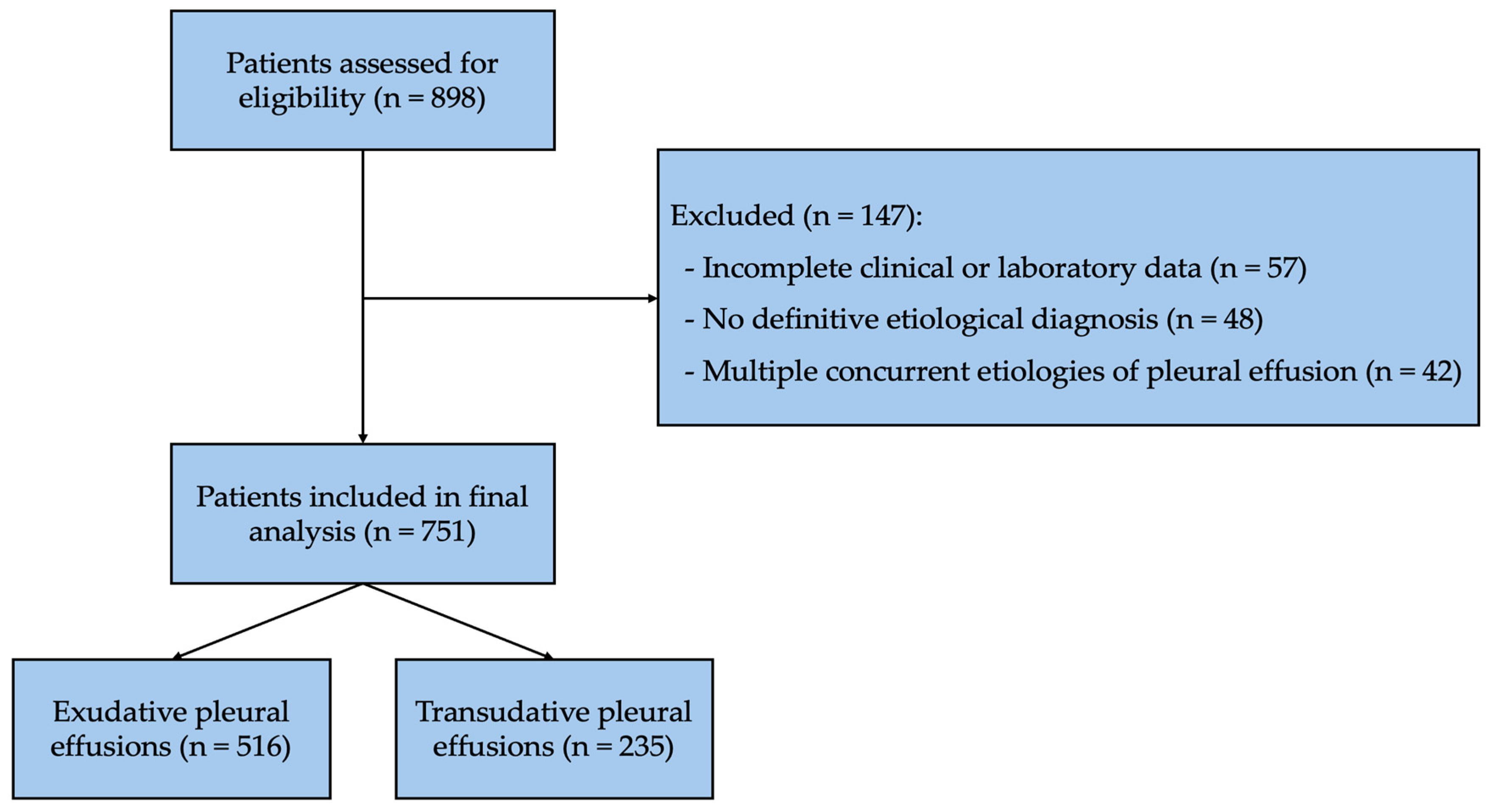
2.1. Study Design and Patient Selection
- Patients older than 18 years
- Patients with a definitively established etiologic diagnosis of pleural effusion
- Patients under 18 years of age
- Patients with missing data
- Patients in whom a definitive clinical and etiologic diagnosis could not be established
- Patients diagnosed with hyperlipidemia and/or using antihyperlipidemic medications
- Patients diagnosed with chylothorax, pseudochylothorax, or hemothorax
- Patients with more than one disease that could lead to the release of both transudative and exudative fluid (such as the coexistence of cancer and atelectasis, cancer and hypoalbuminemia, or heart failure and pneumonia)
- Congestive heart failure: Clinical and/or echocardiographic evidence of cardiac dysfunction, with or without cardiomegaly, consistent with congestive heart failure.
- Malignant pleural effusion: Detection of malignant cells in pleural fluid cytology and/or histopathological confirmation of pleural malignancy.
- Parapneumonic effusion: Clinically and radiologically confirmed pneumonia together with evidence of infection, including positive microbiological culture, positive sputum Gram stain, leukocytosis, or elevated C-reactive protein levels.
- Tuberculous pleural effusion: Demonstration of Mycobacterium tuberculosis by acid-fast bacilli smear or culture from pleural fluid, or identification of necrotizing granulomatous inflammation on pleural biopsy after exclusion of other granulomatous diseases. A favorable clinical response to anti-tuberculosis treatment was also considered supportive evidence.
- Renal failure: Chronic kidney disease with elevated serum urea (>20 mmol/L) or serum creatinine (>167 μmol/L), with or without clinical evidence of volume overload.
- Hypoalbuminemia: Serum albumin level < 20 g/L.
- The ratio of pleural protein to serum protein (protein ratio) ≥ 0.5
- The ratio of pleural LDH to serum LDH (LDH ratio) ≥ 0.6
- Pleural LDH > two-thirds of the upper limit of normal serum LDH
2.2. Statistical Analysis
3. Results
4. Discussion
Study Limitations
5. Conclusions
Author Contributions
Funding
Institutional Review Board Statement
Informed Consent Statement
Data Availability Statement
Conflicts of Interest
References
- Porcel, J.M.; Esquerda, A.; Vives, M.; Bielsa, S. Etiology of pleural effusions: Analysis of more than 3000 consecutive thoracenteses. Arch. Bronconeumol. 2014, 50, 161–165. [Google Scholar] [CrossRef] [Scilit] [PubMed]
- Botana Rial, M.; Perez Pallares, J.; Cases Viedma, E.; Lopez Gonzalez, F.J.; Porcel, J.M.; Rodriguez, M.; Romero Romero, B.; Valdes Cuadrado, L.; Villena Garrido, V.; Cordovilla Perez, R. Diagnosis and Treatment of Pleural Effusion. Recommendations of the Spanish Society of Pulmonology and Thoracic Surgery. Update 2022. Arch. Bronconeumol. 2023, 59, 27–35. [Google Scholar] [CrossRef] [Scilit] [PubMed]
- Lee, J.H.; Choi, C.M.; Park, N.; Park, H.J. Classification of pleural effusions using deep learning visual models: Contrastive-loss. Sci. Rep. 2022, 12, 5532. [Google Scholar] [CrossRef] [Scilit] [PubMed]
- Bintcliffe, O.J.; Hooper, C.E.; Rider, I.J.; Finn, R.S.; Morley, A.J.; Zahan-Evans, N.; Harvey, J.E.; Skyrme-Jones, A.P.; Maskell, N.A. Unilateral Pleural Effusions with More Than One Apparent Etiology. A Prospective Observational Study. Ann. Am. Thorac. Soc. 2016, 13, 1050–1056. [Google Scholar] [CrossRef] [Scilit] [PubMed]
- Feller-Kopman, D.; Light, R. Pleural Disease. N. Engl. J. Med. 2018, 378, 740–751. [Google Scholar] [CrossRef] [Scilit] [PubMed]
- Diacon, A.H.; Brutsche, M.H.; Soler, M. Accuracy of pleural puncture sites: A prospective comparison of clinical examination with ultrasound. Chest 2003, 123, 436–441. [Google Scholar] [CrossRef] [Scilit] [PubMed]
- Light, R.W.; Macgregor, M.I.; Luchsinger, P.C.; Ball, W.C., Jr. Pleural effusions: The diagnostic separation of transudates and exudates. Ann. Intern. Med. 1972, 77, 507–513. [Google Scholar] [CrossRef] [Scilit] [PubMed]
- Heffner, J.E.; Sahn, S.A.; Brown, L.K. Multilevel likelihood ratios for identifying exudative pleural effusions(*). Chest 2002, 121, 1916–1920. [Google Scholar] [CrossRef] [Scilit] [PubMed]
- Valdes, L.; Pose, A.; Suarez, J.; Gonzalez-Juanatey, J.R.; Sarandeses, A.; San Jose, E.; Alvarez Dobana, J.M.; Salgueiro, M.; Rodriguez Suarez, J.R. Cholesterol: A useful parameter for distinguishing between pleural exudates and transudates. Chest 1991, 99, 1097–1102. [Google Scholar] [CrossRef] [Scilit] [PubMed]
- Brown, M.S.; Goldstein, J.L. Receptor-mediated control of cholesterol metabolism. Science 1976, 191, 150–154. [Google Scholar] [CrossRef] [Scilit] [PubMed]
- Costa, M.; Quiroga, T.; Cruz, E. Measurement of pleural fluid cholesterol and lactate dehydrogenase. A simple and accurate set of indicators for separating exudates from transudates. Chest 1995, 108, 1260–1263. [Google Scholar] [CrossRef] [Scilit] [PubMed]
- Gautam, S.; Raj, K.C.S.; Bhattarai, B.; Geetika, K.C.; Adhikari, G.; Gyawali, P.; Rijal, K.; Sijapati, M.J. Diagnostic value of pleural cholesterol in differentiating exudative and transudative pleural effusion. Ann. Med. Surg. 2022, 82, 104479. [Google Scholar] [CrossRef] [Scilit]
- Pg, A.; Ann Abraham, E.; Verma, G.; Prakash, K.; Mounika, P. Pleural Fluid Biomarkers in Focus: The Role of Cholesterol and Lactate Dehydrogenase (LDH) in Differentiating Exudative From Transudative Effusions. Cureus 2025, 17, e87811. [Google Scholar] [CrossRef] [Scilit] [PubMed]
- Hamal, A.B.; Yogi, K.N.; Bam, N.; Das, S.K.; Karn, R. Pleural fluid cholesterol in differentiating exudative and transudative pleural effusion. Pulm. Med. 2013, 2013, 135036. [Google Scholar] [CrossRef] [Scilit] [PubMed]
- Dhandapani, S.; Reddy, S.; Rajagopalan, R. Differentiating pleural effusions: Criteria based on pleural fluid cholesterol. Eurasian J. Pulmonol. 2016, 18, 76–79. [Google Scholar]
- Majmundar, A.; Nouni, H.; Mehta, V.; Lakhani, S.; Lakhani, J.; Darda, S. A comparative study between pleural fluid cholesterol and Light’s criteria in differentiating exudative and transudative pleural effusion. J. Adv. Med. Med. Res. 2023, 38, 28–33. [Google Scholar] [CrossRef] [Scilit]
- Rufino, R.; Marques, B.L.; Azambuja Rde, L.; Mafort, T.; Pugliese, J.G.; da Costa, C.H. Pleural cholesterol to the diagnosis of exudative effusion. Open Respir. Med. J. 2014, 8, 14–17. [Google Scholar] [CrossRef] [Scilit] [PubMed]
- Wilcox, M.E.; Chong, C.A.; Stanbrook, M.B.; Tricco, A.C.; Wong, C.; Straus, S.E. Does this patient have an exudative pleural effusion? The Rational Clinical Examination systematic review. JAMA 2014, 311, 2422–2431. [Google Scholar] [CrossRef] [Scilit] [PubMed]



| Variables | Transudative Pleural Effusion (n: 235) | Exudative Pleural Effusion (n: 516) | p-Value |
|---|---|---|---|
| Age (year) | 76.2 ± 11.6 | 65.2 ± 16.2 | <0.001 |
| Sex F. n (%) | 97 (41.3%) | 194 (37.6%) | 0.337 |
| S. Glucose (mg/dL) | 120 (99–168) | 108 (91–194) | 0.026 |
| S. Albumin (g/dL) | 31.7 ± 6.1 | 34.4 ± 5.8 | <0.001 |
| S. Protein (g/dL) | 64.4 ± 8.1 | 67.6 ± 8.1 | <0.001 |
| S. LDH (IU/L) | 251 (210–297) | 245 (195–320) | 0.413 |
| CRP (mg/L) | 1.9 (0.9–6.1) | 7.8 (2.3–12.0) | <0.001 |
| Sedimentation | 43 ± 29 | 61 ± 31 | 0.005 |
| P. Glucose (mg/dL) | 134 (114–179) | 108 (79–134) | <0.001 |
| P. Albumin (g/dL) | 12.5 ± 4.2 | 25.4 ± 6.0 | <0.001 |
| P. Protein (g/dL) | 22.0 ± 6.9 | 44.6 ± 9.2 | <0.001 |
| P. LDH (IU/L) | 96 (76–120) | 389 (213–750) | <0.001 |
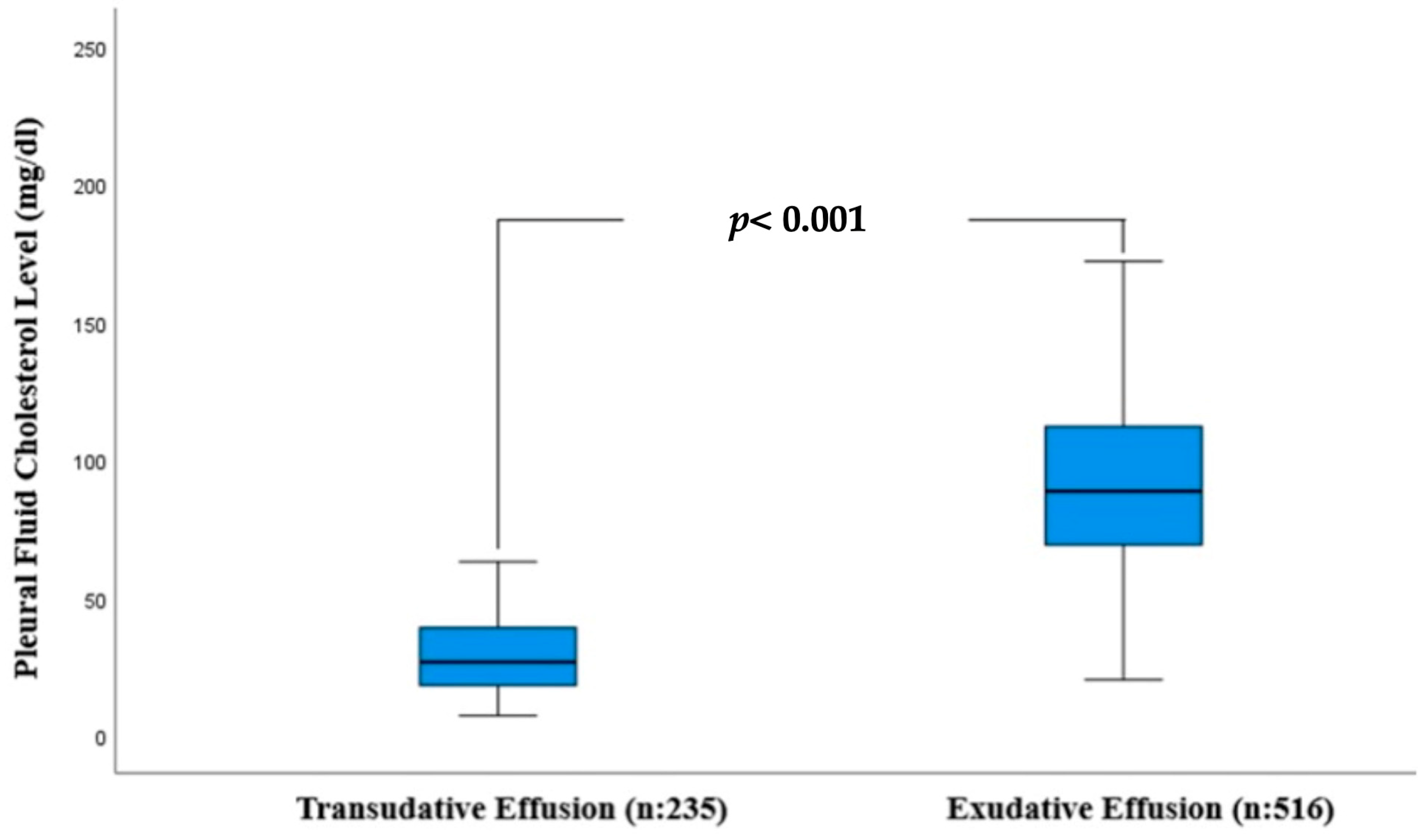
| P. Cholesterol (mg/dL) | 28.9 ± 13.5 | 91.1 ± 30.7 | <0.001 |
| P. Triglycerides (mg/dL) | 16 (12–22) | 33 (24–47) | <0.001 |
| P. Erythrocytes, n | 700 (130–2050) | 3000 (1000–15,200) | <0.001 |
| P. Leukocytes, n | 389 (208–693) | 1633 (769–3541) | <0.001 |
| Area | St. Error | 95% Confidence Interval | p-Value | |
|---|---|---|---|---|
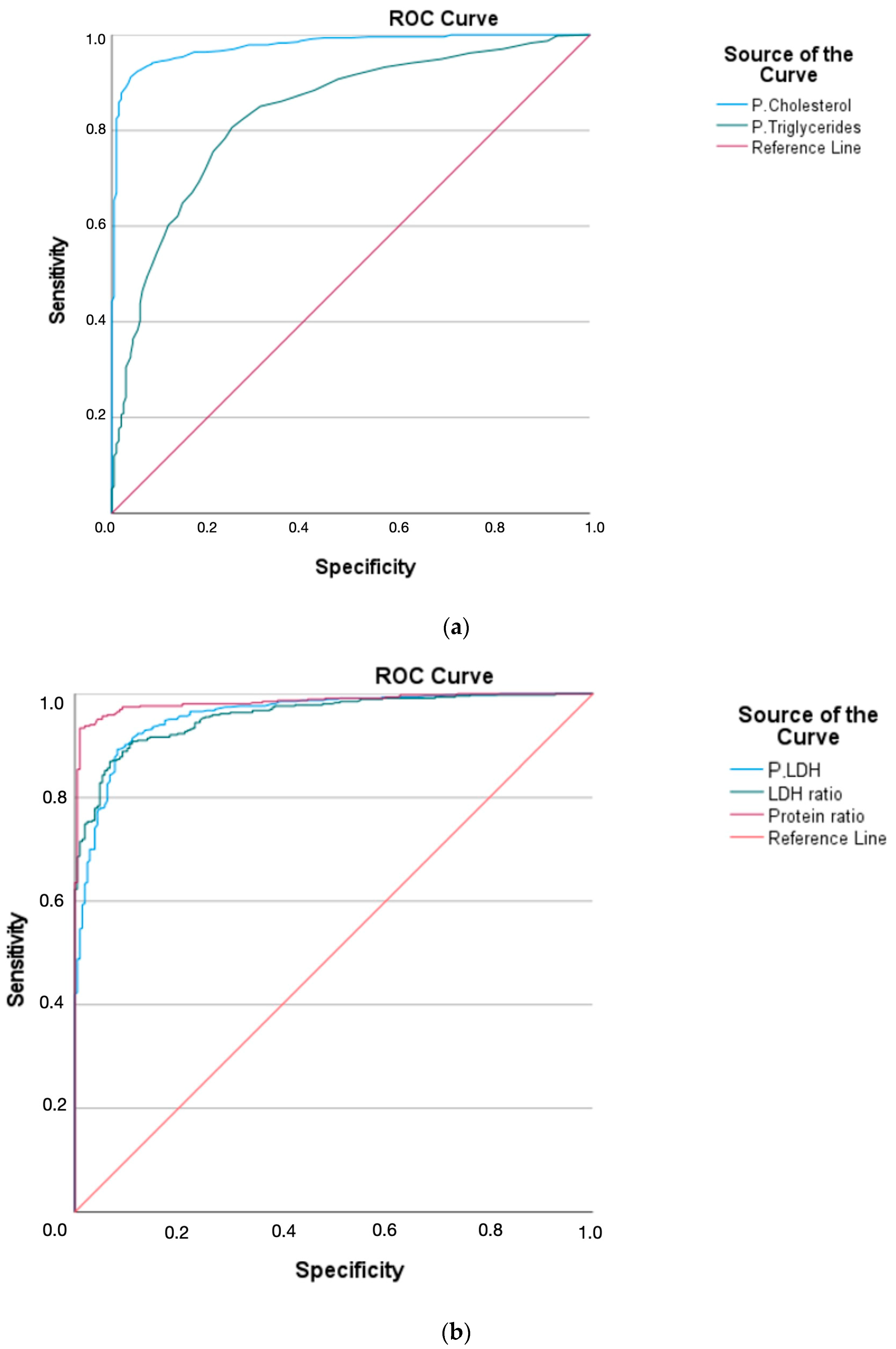
| Protein ratio | 0.985 | 0.004 | 0.977–0.992 | <0.001 |
| LDH ratio | 0.953 | 0.008 | 0.938–0.968 | <0.001 |
| LDH > 2/3 | 0.956 | 0.008 | 0.940–0.972 | <0.001 |
| P. Protein | 0.967 | 0.006 | 0.954–0.979 | <0.001 |
| P. Cholesterol | 0.976 | 0.006 | 0.965–0.987 | <0.001 |
| P. Triglycerides | 0.828 | 0.018 | 0.793–0.863 | <0.001 |
| Cutoff Values | Sensitivity (95% CI) | Specificity (95% CI) | PPV (95% CI) | NPV (95% CI) |
|---|---|---|---|---|
| Protein ratio > 0.5 | 0.944 (0.928–0.961) | 0.949 (0.917–0.974) | 0.976 (0.962–0.989) | 0.884 (0.843–0.924) |
| LDH ratio > 0.6 | 0.893 (0.866–0.919) | 0.884 (0.843–0.924) | 0.946 (0.925–0.966) | 0.786 (0.733–0.838) |
| P. LDH > 2/3 | 0.860 (0.830–0.889) | 0.911 (0.874–0.947) | 0.956 (0.937–0.974) | 0.745 (0.689–0.800) |
| P. Cholesterol ≥ 45 mg/dL | 0.946 (0.926–0.966) | 0.881 (0.839–0.922) | 0.946 (0.925–0.966) | 0.881 (0.839–0.922) |
| P. Cholesterol ≥ 50 mg/dL | 0.922 (0.898–0.945) | 0.949 (0.917–0.974) | 0.975 (0.960–0.989) | 0.848 (0.802–0.893) |
| P. Triglycerides ≥ 30 mg/dL | 0.602 (0.559–0.644) | 0.882 (0.840–0.923) | 0.923 (0.898–0.947) | 0.484 (0.420–0.547) |
| P. Protein ≥ 30 g/L | 0.939 (0.918–0.959) | 0.849 (0.803–0.894) | 0.914 (0.888–0.939) | 0.865 (0.821–0.908) |
| P. Protein ≥ 30 g/L or P. Cholesterol ≥ 50 mg/dL | 0.964 (0.947–0.980) | 0.816 (0.766–0.865) | 0.924 (0.902–0.951) | 0.908 (0.871–0.944) |
| LIGHT | 0.992 (0.984–0.999) | 0.844 (0.797–0.890) | 0.933 (0.910–0.955) | 0.978 (0.959–0.996) |
| Variables | OR | 95% Confidence Interval | p-Value |
|---|---|---|---|
| Age (Years) | 0.961 | 0.922–1.002 | 0.061 |
| Serum protein level (g/L) | 0.982 | 0.919–1.048 | 0.578 |
| P. LDH (IU/L) | 1.019 | 1.012–1.025 | <0.001 |
| P. Cholesterol (≥50 mg/dL) | 126.033 | 34.378–462.056 | <0.001 |
| Presence of malignancy | 246.384 | 24.741–2453.658 | <0.001 |
Disclaimer/Publisher’s Note: The statements, opinions and data contained in all publications are solely those of the individual author(s) and contributor(s) and not of MDPI and/or the editor(s). MDPI and/or the editor(s) disclaim responsibility for any injury to people or property resulting from any ideas, methods, instructions or products referred to in the content. |
© 2026 by the authors. Licensee MDPI, Basel, Switzerland. This article is an open access article distributed under the terms and conditions of the Creative Commons Attribution (CC BY) license.
Share and Cite
Gumus, A.; Senturk Topaloglu, E.; Ozcelik, N.; Kotan, A.; Keskin, H.V.; Topaloglu, O.; Ozyurt, S. The Effectiveness of Pleural Cholesterol Levels in Differentiating Transudate from Exudate and Comparison with Light’s Criteria. Life 2026, 16, 1195. https://doi.org/10.3390/life16071195
Gumus A, Senturk Topaloglu E, Ozcelik N, Kotan A, Keskin HV, Topaloglu O, Ozyurt S. The Effectiveness of Pleural Cholesterol Levels in Differentiating Transudate from Exudate and Comparison with Light’s Criteria. Life. 2026; 16(7):1195. https://doi.org/10.3390/life16071195
Chicago/Turabian StyleGumus, Aziz, Elvan Senturk Topaloglu, Neslihan Ozcelik, Abdurrahman Kotan, Hasan Veysel Keskin, Omer Topaloglu, and Songul Ozyurt. 2026. "The Effectiveness of Pleural Cholesterol Levels in Differentiating Transudate from Exudate and Comparison with Light’s Criteria" Life 16, no. 7: 1195. https://doi.org/10.3390/life16071195
APA StyleGumus, A., Senturk Topaloglu, E., Ozcelik, N., Kotan, A., Keskin, H. V., Topaloglu, O., & Ozyurt, S. (2026). The Effectiveness of Pleural Cholesterol Levels in Differentiating Transudate from Exudate and Comparison with Light’s Criteria. Life, 16(7), 1195. https://doi.org/10.3390/life16071195