


Article

Sex-Specific Association of Toll-like Receptor 8 Polymorphisms with COVID-19 Case Status in a Korean Population

by
Mohammed Zayed
1,2,3,†,
Yong-Chan Kim
4,†,
Chang-Seop Lee
5,6,* and
Byung-Hoon Jeong
1,2,*
1
Korea Zoonosis Research Institute, Jeonbuk National University, Iksan 54531, Republic of Korea
2
Department of Bioactive Material Sciences, Jeonbuk National University, Jeonju 54896, Republic of Korea
3
Department of Surgery, College of Veterinary Medicine, Qena University, Qena 83523, Egypt
4
School of Life Sciences and Biotechnology, Gyeongkuk National University, Andong 36729, Republic of Korea
5
Department of Internal Medicine, Research Institute of Clinical Medicine, Jeonbuk National University, Jeonju 54907, Republic of Korea
6
Biomedical Research Institute, Jeonbuk National University Hospital, Jeonju 54907, Republic of Korea
*
Authors to whom correspondence should be addressed.
These authors contributed equally to this work.
Life 2026, 16(7), 1167; https://doi.org/10.3390/life16071167
Submission received: 19 June 2026 / Revised: 9 July 2026 / Accepted: 13 July 2026 / Published: 14 July 2026
(This article belongs to the Special Issue Genetics and Genomics in Human Health and Disease)

Abstract

Severe acute respiratory syndrome coronavirus 2 (SARS-CoV-2) is the causative pathogen of coronavirus disease 2019 (COVID-19). Toll-like receptor 8 (TLR8), which is located on the X chromosome, plays as a key mediator of the innate immune response. Genetic variation in the form of single-nucleotide polymorphisms (SNPs) within TLR8 has been linked to changes in the transcriptional activity of this gene. Thus, we aimed to identify TLR8 SNPs in the proximal promoter region and investigate whether these SNPs are associated with COVID-19 case status in a Korean population. We performed amplicon sequencing to investigate the genotypes and allele frequencies of regulatory SNPs in COVID-19 patients (n = 191) and the control group (n = 173). Four polymorphic sites, rs5741883, rs186566524, rs3764879, and rs3764880, were identified within the TLR8 proximal promoter. Given the X-linked nature of this locus, allele and genotype frequencies were computed independently by sex. Notably, the minor C allele at rs3764879 occurred at a markedly reduced rate among male patients (10%) relative to male controls (24%), corresponding to an OR of 0.35 (95% CI 0.15–0.8; p = 0.018; q = 0.036). A parallel pattern emerged for rs3764880, where the minor A allele was likewise underrepresented in male patients (9%) versus male controls (24%), yielding an OR of 0.3 (95% CI 0.12–0.7; p = 0.01; q = 0.036). By contrast, neither genotype nor allele distributions differed significantly between female patients and female controls for any of the four variants. These results indicate that the TLR8 polymorphisms rs3764879 and rs3764880 may be associated with COVID-19 case status among Korean males, although further validation in larger, independent cohorts is required.

1. Introduction

Beginning in early 2020, coronavirus disease 2019 (COVID-19), driven by infection with severe acute respiratory syndrome coronavirus 2 (SARS-CoV-2), rapidly became a worldwide pandemic [1,2]. As a result, significant efforts have been initiated to develop diagnostic and therapeutic approaches targeting SARS-CoV-2 [3,4]. The host immune system (especially the innate immune system) serves as the first line of defense against SARS-CoV-2 infection by activating inflammatory pathways upon pathogen detection [5]. Among various pattern recognition receptors, members of the toll-like receptors (TLRs) family play a critical role in the innate immune response and the pathogenesis of COVID-19 [6]. Genetic polymorphisms in genes encoding TLRs have been associated with severe respiratory symptoms in COVID-19 [7]. Notably, TLR8 has been associated with COVID-19 risk and severity [8,9]. However, the association of TLR8 with COVID-19 has not been fully investigated in Korean populations.
TLRs distinguish pathogen-associated and damage-associated molecular patterns developed by the virus and by the host’s injured cells, respectively [10]. Due to TLR signaling, innate immune cells, including neutrophils, monocytes, and natural killer cells, migrate to the infection site to restrain the viral infection [11,12]. The TLR response ultimately stimulates the release of proinflammatory cytokines, such as tumor necrosis factor, interleukin, interferon, and chemokines, by different signaling pathways to restrict infection and stimulate immune responses [13]. In humans, the toll-like receptor family comprises ten identified members (TLR1 through TLR10) [14,15]. Subcellular localization differs across this family: TLR1, 2, 4, 5, 6, and 10 reside on the plasma membrane, whereas TLR3, 7, 8, and 9 are confined to endosomal/endoplasmic reticulum compartments [16]. Recent research has indicated that SARS-CoV-2 stimulates the innate immune system via TLRs and boosts TLR expression, resulting in the elimination of infection [17]. While the TLR response is to promote viral clearance, dysregulated TLR responses may direct continual inflammation and tissue destruction [18]. For instance, overactivation of TLR pathways upregulates a cytokine storm and exacerbates the severity of COVID-19 [6,19].
Among several TLRs, TLR8 recognizes single-stranded RNA and induces inflammatory responses via TLR8-dependent signaling during COVID-19 [20]. COVID-19 showed a different response according to sex; males showed more vulnerability and higher mortality compared to females [21]. Thus, several studies have suggested that immune-related genes located on the X chromosome are important factors in the pathomechanism of COVID-19, and the TLR8 gene is noteworthy because it is located on the X chromosome [21,22,23]. Likewise, TLR8 expression was dramatically increased in patients with severe COVID-19 [24,25]. These results suggest a strong association between the TLR8 gene and the pathomechanism of COVID-19.
Single-nucleotide polymorphisms (SNPs) are variations in DNA sequences that can alter gene transcription and protein function, driving variance across species and human beings [26,27,28]. Since SNP markers may be located across the genome and may be stably passed to progeny, they are employed in genetic and genome-wide association research [29,30]. Numerous SNPs are required to analyze population differences, especially in the case of pandemic diseases such as COVID-19 [31,32,33,34], as they provide information on individuals’ possible responses to certain treatments and susceptibilities to environmental infection [35]. It has been demonstrated that TLR3 rs3775290, TLR4 Asp299Gly and Thr399Ile, TLR7 rs179008, and TLR9 polymorphisms were correlated with the prognosis and susceptibility to SARS-CoV-2 infection [7,36,37]. Alhabibi et al. 2023 showed that TLR2 rs5743708 and TLR9 rs5743836 variants have been significantly associated with the severity of COVID-19 infection [38].
The presence of SNPs in important regulatory regions, such as the promoter region, may explain variances in cytokine production among people [39]. SNPs within TLR genes may reduce individuals’ ability to respond appropriately to TLR ligands, altering susceptibility to infectious diseases. Since SNPs in the TLR8 gene may correlate with TLR8 expression levels, the association between regulatory genetic polymorphisms in TLR8 and COVID-19 susceptibility warrants exploration.
This study set out to examine whether polymorphic variants in the proximal promoter of TLR8 are associated with COVID-19 case status. To this end, we applied amplicon-based sequencing to characterize TLR8 genotypes and allele distributions among Korean COVID-19 patients and controls, analyzing males and females separately.

2. Materials and Methods

2.1. Study Participants

This study enrolled 364 participants overall, divided into a COVID-19 patient group (n = 191) and a control group (n = 173). Patients were identified among individuals admitted to the isolation ward of Jeonbuk National University Hospital (Jeonju-si, Republic of Korea), whereas controls were unrelated, disease-free volunteers obtained through the Korea Biobank Network, with a mean age of 58.7 ± 14.2 years. Eligibility required that participants be male or female, have SARS-CoV-2 infection confirmed via real-time PCR, and self-identify as ethnically Korean; individuals with documented HIV, hepatitis B/C, or chronic pulmonary conditions were excluded from participation. Among the 191 patients (90 male, 101 female; mean age 56.6 ± 15.8 years), 23 had received at least one COVID-19 vaccine dose: 10 with AstraZeneca, 1 with the Janssen vaccine, and 12 with Pfizer-BioNTech (6 single-dose, 6 two-dose recipients). The control cohort included 80 males and 93 females, with the age distribution noted above.

2.2. Ethical Statements

All participants provided written informed consent before enrollment. This study adhered to the ethical principles of the Declaration of Helsinki and received approval from the Institutional Ethics Committee of Jeonbuk National University Hospital (approval code 2020-02-050-067; approval date, 19 October 2023).

2.3. Genomic DNA Extraction

Genomic DNA was extracted from 200 µL whole-blood specimens collected from each participant using a commercial blood DNA isolation kit (Qiagen, Valencia, CA, USA) following the manufacturer’s protocol. DNA yield and purity were quantified by A260/A280 absorbance measurements on a NanoDrop One spectrophotometer (Thermo Fisher Scientific, Waltham, MA, USA). Samples were then stored at −20 °C until further use.

2.4. Polymerase Chain Reaction

A single primer pair spanning the proximal promoter and first exon of TLR8 (Gene ID: 51311) was designed using the Primer3Plus platform (Macrogen, Daejeon, Republic of Korea): forward, 5′-AGCCATTGACTCACTCGTTCA-3′; reverse, 5′-TGGGTCAGAAACCCCATATTC-3′. Each 25-µL PCR reaction contained 1 µL template DNA, 1 µL of each 10 µM primer, 2.5 µL 10× Taq polymerase buffer, 0.5 µL of 0.2 µM dNTPs, 5 µL 5× Band Helper, and 0.25 µL 10× Taq polymerase (BioFACT, Daejeon, Republic of Korea), following the manufacturer’s recommended cycling parameters. Thermocycling (C1000 Touch, Bio-Rad, Hercules, CA, USA) comprised an initial denaturation at 95 °C for 2 min, followed by 35 cycles of 95 °C/20 s, 58 °C/1 min, and 72 °C/1 min, with a final extension at 72 °C for 5 min. Amplicons were resolved on 1% agarose gels, stained with ethidium bromide, and visualized using a Gel Doc XR+ (Bio-Rad). Products were purified with a FavorPrep GEL/PCR kit (Favorgen Biotech, Ping Tung, Taiwan) and bidirectionally sequenced on an ABI 3730 platform (Applied Biosystems, Foster City, CA, USA) using BigDye Terminator v3.1 chemistry (Applied Biosystems) per the manufacturer’s instructions; sequencing was outsourced to Macrogen (Daejeon, Republic of Korea). To confirm genotyping reliability, 10% of samples were randomly re-sequenced independently, yielding 100% concordance with initial calls. The overall call rate reached 100% across all subjects and SNPs.
For genotyping analysis, PCR amplification followed by Sanger DNA sequencing using the chain-termination method (PCR-sequencing) was used to identify SNPs. Finch TV 1.4.0 software (Geospiza Corp., Seattle, WA, USA) was used to analyze the sequencing results [40]. The genotypes and alleles of TLR8 polymorphisms were analyzed in patients and controls to investigate the association between TLR8 polymorphisms and COVID-19 case status.

2.5. Statistical Analysis

Statistical analyses were performed using SPSS version 25.0 (IBM Corp., Armonk, NY, USA). Results were presented as frequencies (percentages) for qualitative variables (genotype, allele, and haplotype distributions) and as mean ± standard deviation (SD) for the quantitative variable age, and compared between groups using Student’s t-test. The chi-square (χ2) test or Fisher’s exact test was used to compare genotype and allele frequencies between the control group and COVID-19 patients. The odds ratio (OR) and 95% confidence interval (CI) for each group were assessed using logistic regression. Age adjustment was not performed because age was statistically balanced between cases and controls overall and within each sex-stratified group. However, residual confounding cannot be completely excluded, as acknowledged in the limitations section. To address multiple comparisons, the Benjamini–Hochberg false discovery rate (FDR) procedure was applied to the primary analysis (allele frequency comparisons of 4 SNPs in male subjects). An FDR threshold of q < 0.05 was used to determine significance. HWE was determined by testing the genotype frequencies at each SNP. Because the TLR8 gene is located on the X chromosome, we tested for HWE only in females. We analyzed LD and haplotypes using Haploview version 4.2 (Broad Institute, Cambridge, MA, USA) based on the observed frequencies of four SNPs. For female subjects, five genetic models were tested for each SNP: the allele model (comparing minor and major allele frequencies), the codominant model (evaluating all three genotypes independently: homozygous major, heterozygous, and homozygous minor), the dominant model (comparing homozygous major versus heterozygous plus homozygous minor), the recessive model (comparing homozygous major and heterozygous group versus homozygous minor), and the overdominant model (comparing homozygous major and homozygous minor versus heterozygous). In contrast, for male subjects, only the allele model was used, as TLR8 is X-linked and hemizygous males carry a single allele, rendering genotype-based models that assume two alleles per individual inapplicable. The sample size was calculated by G*Power (version 3.1.9.2; Germany). The calculation was based on a chi-square test of association in a 2 × 2 contingency table (allele × disease status) under the following parameters: a small-to-medium effect size of w = 0.15 [41], a two-sided significance level of α = 0.05, a statistical power of 80% (1−β = 0.80), one degree of freedom (df = 1), and an approximate allocation ratio of 1:1 (cases to controls). Under these assumptions, the minimum required total sample size was 349 participants. Our enrolled sample of 364 individuals (191 cases, 173 controls) exceeds this threshold. For individual tests, statistical significance was defined as p < 0.05. For the primary male allele comparisons, significance was further evaluated after FDR correction at q < 0.05.

3. Results

3.1. Patients and Samples

Table 1 summarizes specific information regarding the study population. There was no significant difference in age between the COVID-19 patients and the control group (p = 0.194).

3.2. The Location and Genotyping of SNPs

TLR8 genotype and allele distributions were determined via amplicon sequencing across the full cohort (173 controls, 191 patients). Alignment of the resulting sequences confirmed a match to the human TLR8 reference sequence deposited in GenBank under Gene ID 51311. We uncovered a total of four SNPs, including c.-605C>T (rs5741883), c.-418G>A (rs186566524), c.-129G>C (rs3764879), and c.1G>A (rs3764880) (Figure 1A,B). Genotypic distributions of the 4 SNPs were consistent with HWE (p > 0.05). Because the TLR8 gene is located on the X chromosome, only female subjects were included in the HWE test (Table 2). Our study also revealed that minor allele frequency (MAF) was similar to that reported in the public database of the Korean population (Table 2).

3.3. Associations Between COVID-19 Case Status and Genotype, Allele, and Haplotype Frequencies of TLR8 Polymorphisms

To address the study objective, allele and genotype frequencies were compared between female COVID-19 patients and female controls using multiple genetic models (Table 3). The analysis revealed no statistically significant association with COVID-19 case status under any genetic model (p > 0.05).
In males, the C allele of TLR8 rs3764879 was present at frequencies of 24% and 10% in the control and patient groups, respectively. The difference was statistically significant and remained significant after FDR correction (OR 0.35; 95% CI 0.15–0.8; p = 0.018; q = 0.036). For TLR8 rs3764880, the A allele had frequencies of 24% and 9% in the control and patient groups, respectively. The difference was statistically significant and remained significant after FDR correction (OR 0.3; 95% CI 0.12–0.7; p = 0.01; q = 0.036) (Table 4). Table 4 presents the full Benjamini–Hochberg FDR-adjusted q-values for all four SNPs, indicating that only rs3764879 and rs3764880 remained significant after multiple-comparison correction. Because there are only small numbers of male minor-allele carriers, these estimates should be interpreted with caution and confirmed in larger independent cohorts.
In addition, we performed haplotype analysis of four polymorphisms in the TLR8 gene in females using Haploview 4.2. The SNPs in the TLR8 gene revealed three main haplotypes with no significant differences in COVID-19 case status among Korean females (p > 0.05) (Table 5).
Furthermore, LD was analyzed between the identified SNPs of the TLR8 gene with r2 values. In females, the TLR8 rs3764879 showed a strong LD with rs3764880 in both the control and patient groups (r2 = 0.805 and 0.914, Table 6).

4. Discussion

While some patients with COVID-19 demonstrate spontaneous resolution, the factors influencing the features of their immune responses remain mostly unidentified [42]. Host factors, including age, sex, and genetic factors, impact the spontaneous consequence after SARS-CoV-2 infection, the progression to fatality, or a response to treatment [43,44]. The TLR8 gene is critical in the immunological response to SARS-CoV-2. Therefore, TLR7/8 agonists have been suggested as a therapeutic approach for COVID-19 treatment [20,45]. Male sex has been recognized as a potential risk factor for hospitalization and mortality following SARS-CoV-2 infection. On the other hand, females often have a stronger immunological response than males, and X chromosomal genes might explain this tendency [46].
Given the recognized contribution of TLR8 to innate antiviral defense against SARS-CoV-2, we sought to determine whether TLR8 polymorphisms are associated with COVID-19 case status in a Korean cohort. Amplicon sequencing from 173 controls and 191 patients identified four candidate regulatory SNPs in the promoter and first exon of TLR8. Previous studies have suggested that SNPs in TLR8 may exhibit sex-specific effects in genetic association studies [47]. Consistent with these findings, our results revealed a sex-specific association between TLR8 rs3764879 and rs3764880 in males, but not in females. It has been shown that TLR8 rs3764879 and/or rs5741883 are associated with tuberculosis (TB) infection [48], allergic rhinitis [49], Chikungunya virus (CHIKV) infection [50], and systemic lupus erythematosus [51]. Here, we observed that the allele frequencies of TLR8 rs3764879 differed significantly between male COVID-19 patients and the control group, suggesting an association with COVID-19 case status, rather than a confirmed causal role in infection risk. This result is inconsistent with a recent study by Bagci et al. (2023), which reported no association between the TLR8 rs3764879 SNP and COVID-19 [9]. This discrepancy may be attributed to the genetic differences among ethnic groups [52]. Although our control group was not formally matched to cases at the individual level, the two groups had comparable age and sex distributions, and all association analyses were stratified by sex, the primary demographic variable relevant to X-linked gene analysis.
Several studies have investigated the TLR8 rs3764880 polymorphism and its association with susceptibility to TB infection across different ethnicities [53]. For instance, in the Chinese population, the G allele of TLR8 rs3764880 was associated with protection against TB [54]. In the Turkish population, the A allele of the same polymorphism was associated with susceptibility to pulmonary TB [55]. The TLR8 rs3764880 SNP was also found to be associated with protection against the development of acquired immunodeficiency syndrome in both males and females [56]. Our findings indicated that the A allele frequency is significantly higher in the control group than in male COVID-19 patients. Given the strong LD between rs3764879 and rs3764880 (r2 = 0.805–0.914, Table 6), these two SNPs likely tag the same haplotype and should be interpreted as a single genetic signal rather than two independent associations. This result is consistent with findings in Italian, Russian, and Turkish populations [9,57,58].
No prior study has examined a potential link between rs186566524 and COVID-19. Our findings indicate that this variant shows no detectable association with COVID-19 case status in this Korean cohort. It has been indicated that males have higher degrees of mortality and clinical problems from COVID-19 compared to females [59]. Among TLRs, Fallerini and his colleagues showed an association between TLR7 variants (rs189681811, rs147244662, rs149314023, rs200146658, and rs5743781) and COVID-19 in males [60]. Taha et al. identified a link between the TLR4 (Asp299Gly and Thr399Ile) minor alleles 299Gly (G) and 399Ile (T) and COVID-19 severity in a male-dominated sample [36].
We also analyzed the haplotype frequencies of the TLR8 gene in female subjects, comparing the control group and COVID-19 patients, and found no significant differences in haplotype frequencies according to COVID-19 case status (Table 5). In contrast, the individual SNP analysis showed male-specific associations of rs3764879 and rs3764880 with COVID-19 case status (Table 4). A strong genetic linkage was identified between TLR8 rs3764879 and rs3764880 in both the control and patient groups. Notably, other SNPs exhibited weak LD values. TLRs respond to pathogens by stimulating acquired immunity, including the release of proinflammatory cytokines [61]. Among TLRs, interactions between COVID-19 and TLR variants have been explained, mainly for TLR3, TLR4, and TLR7. It has been reported that the TLR2-9 pool has facilitated the clearance of SARS-CoV-2 [62]. TLR3 rs3775290 and TLR7 rs179008 SNPs were indicated to be substantially linked with an extremely high risk of COVID-19 pneumonia, but not with disease outcome [7]. The GG homozygote at the TLR7 rs3853839 SNP was significantly more common among COVID-19 patients (n = 150) than among controls (n = 135). As a result, these SNPs have been associated with SARS-CoV-2 infection, cytokine storms, and higher patient mortality rates [63]. On the other hand, Zayed et al. showed that the TLR7 (rs864058) polymorphism is not associated with SARS-CoV-2 infection in the Korean population [64].
In the post-pandemic genetics era, large-scale GWAS studies have identified strong autosomal susceptibility loci for COVID-19. However, X-linked immune genes such as TLR7 and TLR8 remain underexplored because standard GWAS methods struggle to analyze hemizygous loci. Our research adds to this knowledge by offering population-specific, sex-stratified data from a non-European cohort.
Our study design identified associations between TLR8 genetic variants and confirmed COVID-19 case status (Table 4). Although these results suggested a potential role in the risk of SARS-CoV-2 infection, we cannot rule out that the observed associations reflect differences in disease progression or clinical symptoms rather than direct risk of infection. This study has potential limitations that should be acknowledged. First, the relatively small sample size, limited to Jeonju, South Korea, may introduce bias into participant selection and data analysis. The small number of minor allele carriers among males results in wide confidence intervals, so the reported associations are preliminary and require validation in larger, independent cohorts. Second, age-adjusted logistic regression was not performed. Although age was statistically balanced between groups, the absence of a significant age difference does not completely exclude residual confounding. Residual confounding could influence the observed associations, and future studies with larger sample sizes should address this. Third, the risk of batch effects, arising from using pre-existing biobank samples as controls processed at a different time from the patient samples, cannot be entirely ruled out, despite our efforts to maintain consistent protocols. Fourth, the control participants from the biobank were not verified as SARS-CoV-2-negative, and their infection history, immune status, vaccination status, and COVID-19-related symptoms remain unknown. As a result, some controls could have had previously unrecognized or asymptomatic SARS-CoV-2 infections. Undetected asymptomatic infection among controls could bias the observed odds ratios in either direction, thereby limiting causal interpretation. This limits the interpretation of the observed associations, as they might not reflect true susceptibility but could instead be influenced by differences in infection detection, disease symptoms, or clinical course. Finally, while the genetic homogeneity of our Korean population reduces the risk of population stratification confounding our results, it also limits the applicability of our findings. Functional validation, including quantification of TLR8 expression, eQTL analysis, and cytokine profiling, was beyond the scope of the present association study and is a defined priority for follow-up investigation. Future replication in diverse, multi-ethnic cohorts is necessary to confirm the broader relevance of these TLR8 variants in COVID-19 susceptibility.

5. Conclusions

In conclusion, this study demonstrated a significant risk association between a TLR8 haplotype in the proximal promoter–exon 1 region, tagged by the correlated SNPs rs3764879 and rs3764880 (r2 > 0.8), and COVID-19 case status, with sex-dependent effects in the Korean population. We believe that, in developing future individual risk profiles for SARS-CoV-2 infection, patients’ sex should be taken into account. Further validation in larger independent cohorts is required to confirm these findings.

Author Contributions

Conceptualization, M.Z., Y.-C.K., and B.-H.J.; methodology, M.Z., Y.-C.K., and B.-H.J.; formal analysis, M.Z., Y.-C.K., and B.-H.J.; data curation, M.Z., Y.-C.K., C.-S.L., and B.-H.J.; writing—original draft preparation, M.Z.; writing—review and editing, M.Z., Y.-C.K., C.-S.L., and B.-H.J.; supervision, B.-H.J. All authors have read and agreed to the published version of the manuscript.

Funding

This work was supported by a National Research Foundation of Korea (NRF) grant funded by the Korean government (MSIT) (RS-2025-00517133, RS-2025-23963916, 2022R1C1C2004792). This research was also supported by the Ministry of Education through the Basic Science Research Program (2021R1A6A3A010864) and the Korea Basic Science Institute (National Research Facilities and Equipment Center) grant (RS-2021-NF000550). The experiments were performed in a biosafety cabinet at the BL3 facilities in the Core Facility Center for Zoonosis Research (Core-FCZR). This work was supported by the Regional Innovation System & Education (RISE) program through the Gyeongbuk RISE CENTER, funded by the Ministry of Education (MOE) and the Gyeongsangbuk-do, Republic of Korea (2025-RISE-15-104, and B0080526002355). This work was supported by the Ministry of Education and the NRF of Korea through the Gyeongsang-buk-do RISE (Regional Innovation System & Education) project and the Glocal University Project Group in Gyeongkuk National University-Gyeongbuk Provincial College.

Institutional Review Board Statement

The study was conducted in accordance with the Declaration of Helsinki, and the protocol was approved by the Ethics Committee of Jeonbuk National University Hospital (Project identification code: 2020-02-050-067; approved on 19 October 2023).

Informed Consent Statement

Informed consent was obtained from all subjects involved in the study.

Data Availability Statement

The data presented in this study are available on request from the corresponding authors due to ethical reasons.

Acknowledgments

We thank all the patients who participated in this study.

Conflicts of Interest

The authors declare no conflicts of interest.

References

  1. Hu, B.; Guo, H.; Zhou, P.; Shi, Z.-L. Characteristics of SARS-CoV-2 and COVID-19. Nat. Rev. Microbiol. 2021, 19, 141–154. [Google Scholar] [CrossRef] [Scilit]
  2. Synowiec, A.; Szczepański, A.; Barreto-Duran, E.; Lie, L.K.; Pyrc, K. Severe Acute Respiratory Syndrome Coronavirus 2 (SARS-CoV-2): A Systemic Infection. Clin. Microbiol. Rev. 2021, 34, 10-1128. [Google Scholar] [CrossRef] [Scilit] [PubMed]
  3. Li, G.; Hilgenfeld, R.; Whitley, R.; De Clercq, E. Therapeutic strategies for COVID-19: Progress and lessons learned. Nat. Rev. Drug Discov. 2023, 22, 449–475. [Google Scholar] [CrossRef] [Scilit] [PubMed]
  4. Zayed, M.; Iohara, K. Immunomodulation and Regeneration Properties of Dental Pulp Stem Cells: A Potential Therapy to Treat Coronavirus Disease 2019. Cell Transpl. 2020, 29, 963689720952089. [Google Scholar] [CrossRef] [Scilit] [PubMed]
  5. Diamond, M.S.; Kanneganti, T.-D. Innate immunity: The first line of defense against SARS-CoV-2. Nat. Immunol. 2022, 23, 165–176. [Google Scholar] [CrossRef] [Scilit] [PubMed]
  6. Mabrey, F.L.; Morrell, E.D.; Wurfel, M.M. TLRs in COVID-19: How they drive immunopathology and the rationale for modulation. Innate Immun. 2021, 27, 503–513. [Google Scholar] [CrossRef] [Scilit] [PubMed]
  7. Alseoudy, M.M.; Elgamal, M.; Abdelghany, D.A.; Borg, A.M.; El-Mesery, A.; Elzeiny, D.; Hammad, M.O. Prognostic impact of toll-like receptors gene polymorphism on outcome of COVID-19 pneumonia: A case-control study. Clin. Immunol. 2022, 235, 108929. [Google Scholar] [CrossRef] [Scilit] [PubMed]
  8. Del Carmen Camacho-Rea, M.; Martínez-Gómez, L.E.; Martinez-Armenta, C.; Martínez-Nava, G.A.; Ortega-Peña, S.; Olea-Torres, J.; Herrera-López, B.; Suarez-Ahedo, C.; Vázquez-Cárdenas, P.; Vidal-Vázquez, R.P.; et al. Association of TLR8 Variants in Sex-Based Clinical Differences in Patients with COVID-19. Biochem. Genet. 2024, 63, 2601–2622. [Google Scholar] [CrossRef] [Scilit] [PubMed]
  9. Bagci, G.; Gundogdu, O.; Pektas, A.N.; Bagci, B.; Avci, O.; Gursoy, S.; Kaygusuz, K.; Elaldi, N. The investigation of host genetic variants of toll-like receptor 7 and 8 in COVID-19. Nucleosides Nucleotides Nucleic Acids 2023, 42, 586–602. [Google Scholar] [CrossRef] [Scilit] [PubMed]
  10. Iwasaki, A.; Medzhitov, R. Toll-like receptor control of the adaptive immune responses. Nat. Immunol. 2004, 5, 987–995. [Google Scholar] [CrossRef] [Scilit] [PubMed]
  11. Dai, J.; Wang, Y.; Wang, H.; Gao, Z.; Wang, Y.; Fang, M.; Shi, S.; Zhang, P.; Wang, H.; Su, Y.; et al. Toll-Like Receptor Signaling in Severe Acute Respiratory Syndrome Coronavirus 2-Induced Innate Immune Responses and the Potential Application Value of Toll-like Receptor Immunomodulators in Patients with Coronavirus Disease 2019. Front. Microbiol. 2022, 13, 948770. [Google Scholar] [CrossRef] [Scilit] [PubMed]
  12. Catanzaro, M.; Fagiani, F.; Racchi, M.; Corsini, E.; Govoni, S.; Lanni, C. Immune response in COVID-19: Addressing a pharmacological challenge by targeting pathways triggered by SARS-CoV-2. Signal Transduct. Target. Ther. 2020, 5, 84. [Google Scholar] [CrossRef] [Scilit] [PubMed]
  13. Takeuchi, O.; Akira, S. Pattern recognition receptors and inflammation. Cell 2010, 140, 805–820. [Google Scholar] [CrossRef] [Scilit] [PubMed]
  14. Kawasaki, T.; Kawai, T. Toll-Like Receptor Signaling Pathways. Front. Immunol. 2014, 5, 461. [Google Scholar] [CrossRef] [Scilit] [PubMed]
  15. Kawai, T.; Akira, S. TLR signaling. Cell Death Differ. 2006, 13, 816–825. [Google Scholar] [CrossRef] [Scilit] [PubMed]
  16. Kawai, T.; Akira, S. Toll-like receptors and their crosstalk with other innate receptors in infection and immunity. Immunity 2011, 34, 637–650. [Google Scholar] [CrossRef] [Scilit] [PubMed]
  17. Bortolotti, D.; Gentili, V.; Rizzo, S.; Schiuma, G.; Beltrami, S.; Strazzabosco, G.; Fernandez, M.; Caccuri, F.; Caruso, A.; Rizzo, R. TLR3 and TLR7 RNA Sensor Activation during SARS-CoV-2 Infection. Microorganisms 2021, 9, 1820. [Google Scholar] [CrossRef] [Scilit] [PubMed]
  18. Ebermeyer, T.; Cognasse, F.; Berthelot, P.; Mismetti, P.; Garraud, O.; Hamzeh-Cognasse, H. Platelet Innate Immune Receptors and TLRs: A Double-Edged Sword. Int. J. Mol. Sci. 2021, 22, 7894. [Google Scholar] [CrossRef] [Scilit] [PubMed]
  19. Tang, Y.; Liu, J.; Zhang, D.; Xu, Z.; Ji, J.; Wen, C. Cytokine Storm in COVID-19: The Current Evidence and Treatment Strategies. Front. Immunol. 2020, 11, 1708. [Google Scholar] [CrossRef] [Scilit] [PubMed]
  20. Dyavar, S.R.; Singh, R.; Emani, R.; Pawar, G.P.; Chaudhari, V.D.; Podany, A.T.; Avedissian, S.N.; Fletcher, C.V.; Salunke, D.B. Role of toll-like receptor 7/8 pathways in regulation of interferon response and inflammatory mediators during SARS-CoV2 infection and potential therapeutic options. Biomed. Pharmacother. 2021, 141, 111794. [Google Scholar] [CrossRef] [Scilit] [PubMed]
  21. Pradhan, A.; Olsson, P.E. Sex differences in severity and mortality from COVID-19: Are males more vulnerable? Biol. Sex Differ. 2020, 11, 53. [Google Scholar] [CrossRef] [Scilit] [PubMed]
  22. Schröder, N.W.J.; Schumann, R.R. Single nucleotide polymorphisms of Toll-like receptors and susceptibility to infectious disease. Lancet Infect. Dis. 2005, 5, 156–164. [Google Scholar] [CrossRef] [Scilit] [PubMed]
  23. Skevaki, C.; Pararas, M.; Kostelidou, K.; Tsakris, A.; Routsias, J.G. Single nucleotide polymorphisms of Toll-like receptors and susceptibility to infectious diseases. Clin. Exp. Immunol. 2015, 180, 165–177. [Google Scholar] [CrossRef] [Scilit] [PubMed]
  24. Zheng, M.; Karki, R.; Williams, E.P.; Yang, D.; Fitzpatrick, E.; Vogel, P.; Jonsson, C.B.; Kanneganti, T.-D. TLR2 senses the SARS-CoV-2 envelope protein to produce inflammatory cytokines. Nat. Immunol. 2021, 22, 829–838. [Google Scholar] [CrossRef] [Scilit] [PubMed]
  25. Arefinia, N.; Banafi, P.; Zarezadeh, M.A.; Mousawi, H.S.; Yaghobi, R.; Farokhnia, M.; Sarvari, J. TLR3, TLR7, and TLR8 genes expression datasets in COVID-19 patients: Influences of the disease severity and gender. Data Brief 2024, 54, 110498. [Google Scholar] [CrossRef] [Scilit] [PubMed]
  26. Shastry, B.S. SNPs in disease gene mapping, medicinal drug development and evolution. J. Hum. Genet. 2007, 52, 871–880. [Google Scholar] [CrossRef] [Scilit] [PubMed]
  27. Choi, D.I.; Zayed, M.; Jeong, B.H. Novel Single-Nucleotide Polymorphisms (SNPs) and Genetic Studies of the Shadow of Prion Protein (SPRN) in Quails. Animals 2024, 14, 2481. [Google Scholar] [CrossRef] [Scilit] [PubMed]
  28. Choi, D.-I.; Zayed, M.; Na, E.-J.; Oem, J.-K.; Jeong, B.-H. Novel Insertion/Deletion Polymorphisms and Genetic Studies of the Shadow of Prion Protein (SPRN) in Raccoon Dogs. Animals 2024, 14, 3716. [Google Scholar] [CrossRef] [Scilit] [PubMed]
  29. Robert, F.; Pelletier, J. Exploring the Impact of Single-Nucleotide Polymorphisms on Translation. Front. Genet. 2018, 9, 507. [Google Scholar] [CrossRef] [Scilit] [PubMed]
  30. Choi, D.I.; Zayed, M.; Jeong, C.G.; Oem, J.K.; Jeong, B.H. First Report of IFITM3 Polymorphism Associated with Severe Fever with Thrombocytopenia Syndrome Virus (SFTSV) Infection in the Raccoon Dog. Pak. Vet. J. 2026, 46, 866–875. [Google Scholar] [CrossRef] [Scilit]
  31. Suh, S.; Lee, S.; Gym, H.; Yoon, S.; Park, S.; Cha, J.; Kwon, D.-H.; Yang, Y.; Jee, S.H. A systematic review on papers that study on Single Nucleotide Polymorphism that affects coronavirus 2019 severity. BMC Infect. Dis. 2022, 22, 47. [Google Scholar] [CrossRef] [Scilit] [PubMed]
  32. Li, Y.; Ke, Y.; Xia, X.; Wang, Y.; Cheng, F.; Liu, X.; Jin, X.; Li, B.; Xie, C.; Liu, S.; et al. Genome-wide association study of COVID-19 severity among the Chinese population. Cell Discov. 2021, 7, 76. [Google Scholar] [CrossRef] [Scilit] [PubMed]
  33. Harper, H.; Burridge, A.; Winfield, M.; Finn, A.; Davidson, A.; Matthews, D.; Hutchings, S.; Vipond, B.; Jain, N.; The COVID-19 Genomics UK (COG-UK) Consortium; et al. Detecting SARS-CoV-2 variants with SNP genotyping. PLoS ONE 2021, 16, e0243185. [Google Scholar] [CrossRef] [Scilit] [PubMed]
  34. Nguyen, T.T.D.; Zayed, M.; Choi, D.I.; Lee, C.S.; Jeong, B.H. A Meta-Analysis of the Association between Apolipoprotein E Polymorphisms and SARS-CoV-2 Risk/Severity in the Korean Population. Russ. J. Genet. 2025, 61, 997–1009. [Google Scholar] [CrossRef] [Scilit]
  35. Shastry, B.S. SNPs: Impact on gene function and phenotype. Methods Mol. Biol. 2009, 578, 3–22. [Google Scholar] [CrossRef] [Scilit] [PubMed]
  36. Taha, S.I.; Shata, A.K.; Baioumy, S.A.; Fouad, S.H.; Anis, S.G.; Mossad, I.M.; Moustafa, N.M.; Abdou, D.M.; Youssef, M.K. Toll-Like Receptor 4 Polymorphisms (896A/G and 1196C/T) as an Indicator of COVID-19 Severity in a Convenience Sample of Egyptian Patients. J. Inflamm. Res. 2021, 14, 6293–6303. [Google Scholar] [CrossRef] [Scilit] [PubMed]
  37. Bezemer, G.F.G.; Garssen, J. TLR9 and COVID-19: A Multidisciplinary Theory of a Multifaceted Therapeutic Target. Front. Pharmacol. 2020, 11, 601685. [Google Scholar] [CrossRef] [Scilit] [PubMed]
  38. Alhabibi, A.M.; Hassan, A.S.; Elbaky, N.M.A.; Eid, H.A.; Khalifa, M.; Wahab, M.A.; Althoqapy, A.A.; Abdou, A.E.; Zakaria, D.M.; Nassef, E.M.; et al. Impact of Toll-Like Receptor 2 and 9 Gene Polymorphisms on COVID-19: Susceptibility, Severity, and Thrombosis. J. Inflamm. Res. 2023, 16, 665–675. [Google Scholar] [CrossRef] [Scilit] [PubMed]
  39. Wu, S.; Wang, M.G.; Wang, Y.; He, J.Q. Polymorphisms of cytokine genes and tuberculosis in two independent studies. Sci. Rep. 2019, 9, 2507. [Google Scholar] [CrossRef] [Scilit] [PubMed]
  40. Won, S.Y.; Kim, Y.C.; Jeong, B.H. No Association between Single Nucleotide Polymorphisms (SNPs) of the Interferon-Induced Transmembrane Protein 3 (IFITM3) Gene and the Susceptibility of Alzheimer’s Disease (AD). Medicina 2021, 58, 55. [Google Scholar] [CrossRef] [Scilit] [PubMed]
  41. Jacop, C. Statistical Power Analysis for the Behavioral Sciences, 2nd ed.; Lawrence Erlbaum: Mahwah, NJ, USA, 1988. [Google Scholar]
  42. Brodin, P. Immune determinants of COVID-19 disease presentation and severity. Nat. Med. 2021, 27, 28–33. [Google Scholar] [CrossRef] [Scilit] [PubMed]
  43. Anastassopoulou, C.; Gkizarioti, Z.; Patrinos, G.P.; Tsakris, A. Human genetic factors associated with susceptibility to SARS-CoV-2 infection and COVID-19 disease severity. Hum. Genom. 2020, 14, 40. [Google Scholar] [CrossRef] [Scilit] [PubMed]
  44. Thierry, A.R. Host/genetic factors associated with COVID-19 call for precision medicine. Precis. Clin. Med. 2020, 3, 228–234. [Google Scholar] [CrossRef] [Scilit] [PubMed]
  45. Khalifa, A.E.; Ghoneim, A.I. Potential value of pharmacological agents acting on toll-like receptor (TLR) 7 and/or TLR8 in COVID-19. Curr. Res. Pharmacol. Drug Discov. 2021, 2, 100068. [Google Scholar] [CrossRef] [Scilit] [PubMed]
  46. Spiering, A.E.; de Vries, T.J. Why Females Do Better: The X Chromosomal TLR7 Gene-Dose Effect in COVID-19. Front. Immunol. 2021, 12, 756262. [Google Scholar] [CrossRef] [Scilit] [PubMed]
  47. Salie, M.; Daya, M.; Lucas, L.A.; Warren, R.M.; van der Spuy, G.D.; van Helden, P.D.; Hoal, E.G.; Möller, M. Association of toll-like receptors with susceptibility to tuberculosis suggests sex-specific effects of TLR8 polymorphisms. Infect. Genet. Evol. 2015, 34, 221–229. [Google Scholar] [CrossRef] [Scilit] [PubMed]
  48. Mhmoud, N.A. Association of Toll-like Receptors 1, 2, 4, 6, 8, 9 and 10 Genes Polymorphisms and Susceptibility to Pulmonary Tuberculosis in Sudanese Patients. Immunotargets Ther. 2023, 12, 47–75. [Google Scholar] [CrossRef] [Scilit] [PubMed]
  49. Nilsson, D.; Andiappan, A.K.; Halldén, C.; De Yun, W.; Säll, T.; Tim, C.F.; Cardell, L.O. Toll-like receptor gene polymorphisms are associated with allergic rhinitis: A case control study. BMC Med. Genet. 2012, 13, 66. [Google Scholar] [CrossRef] [Scilit] [PubMed]
  50. Dutta, S.K.; Tripathi, A. Association of toll-like receptor polymorphisms with susceptibility to chikungunya virus infection. Virology 2017, 511, 207–213. [Google Scholar] [CrossRef] [Scilit] [PubMed]
  51. Lee, Y.H.; Choi, S.J.; Ji, J.D.; Song, G.G. Association between toll-like receptor polymorphisms and systemic lupus erythematosus: A meta-analysis update. Lupus 2016, 25, 593–601. [Google Scholar] [CrossRef] [Scilit] [PubMed]
  52. Huang, T.; Shu, Y.; Cai, Y.-D. Genetic differences among ethnic groups. BMC Genom. 2015, 16, 1093. [Google Scholar] [CrossRef] [Scilit] [PubMed]
  53. Varshney, D.; Singh, S.; Sinha, E.; Mohanty, K.K.; Kumar, S.; Barik, S.K.; Patil, S.A.; Katara, P. Systematic review and meta-analysis of human Toll-like receptors genetic polymorphisms for susceptibility to tuberculosis infection. Cytokine 2022, 152, 155791. [Google Scholar] [CrossRef] [Scilit] [PubMed]
  54. Wang, M.G.; Zhang, M.M.; Wang, Y.; Wu, S.Q.; Zhang, M.; He, J.Q. Association of TLR8 and TLR9 polymorphisms with tuberculosis in a Chinese Han population: A case-control study. BMC Infect. Dis. 2018, 18, 561. [Google Scholar] [CrossRef] [Scilit] [PubMed]
  55. Dalgic, N.; Tekin, D.; Kayaalti, Z.; Cakir, E.; Soylemezoglu, T.; Sancar, M. Relationship between toll-like receptor 8 gene polymorphisms and pediatric pulmonary tuberculosis. Dis. Markers 2011, 31, 33–38. [Google Scholar] [CrossRef] [Scilit] [PubMed]
  56. Zaidane, I.; Ouladlahsen, A.; Bensghir, R.; Chihab, H.; Jadid, F.Z.; Fɩhry, R.E.; Baba, H.; Filali, K.M.E.; Oudghiri, M.; Wakrim, L.; et al. An assessment of toll-like receptor 7 and 8 gene polymorphisms with susceptibility to HIV-1 infection, AIDS development and response to antiretroviral therapy. Immunol. Lett. 2020, 227, 88–95. [Google Scholar] [CrossRef] [Scilit] [PubMed]
  57. Azzarà, A.; Cassano, I.; Paccagnella, E.; Tirindelli, M.C.; Nobile, C.; Schittone, V.; Lintas, C.; Sacco, R.; Gurrieri, F. Genetic variants determine intrafamilial variability of SARS-CoV-2 clinical outcomes in 19 Italian families. PLoS ONE 2022, 17, e0275988. [Google Scholar] [CrossRef] [Scilit] [PubMed]
  58. Salamaikina, S.; Karnaushkina, M.; Korchagin, V.; Litvinova, M.; Mironov, K.; Akimkin, V. TLRs Gene Polymorphisms Associated with Pneumonia before and during COVID-19 Pandemic. Diagnostics 2022, 13, 121. [Google Scholar] [CrossRef] [Scilit] [PubMed]
  59. Ancochea, J.; Izquierdo, J.L.; Soriano, J.B. Evidence of Gender Differences in the Diagnosis and Management of Coronavirus Disease 2019 Patients: An Analysis of Electronic Health Records Using Natural Language Processing and Machine Learning. J. Womens Health 2021, 30, 393–404. [Google Scholar] [CrossRef] [Scilit] [PubMed]
  60. Fallerini, C.; Daga, S.; Mantovani, S.; Benetti, E.; Picchiotti, N.; Francisci, D.; Paciosi, F.; Schiaroli, E.; Baldassarri, M.; Fava, F.; et al. Association of Toll-like receptor 7 variants with life-threatening COVID-19 disease in males: Findings from a nested case-control study. eLife 2021, 10, e67569. [Google Scholar] [CrossRef] [Scilit] [PubMed]
  61. Totura, A.L.; Whitmore, A.; Agnihothram, S.; Schäfer, A.; Katze, M.G.; Heise, M.T.; Baric, R.S. Toll-Like Receptor 3 Signaling via TRIF Contributes to a Protective Innate Immune Response to Severe Acute Respiratory Syndrome Coronavirus Infection. mBio 2015, 6, e00638-15. [Google Scholar] [CrossRef] [Scilit] [PubMed]
  62. Manik, M.; Singh, R.K. Role of toll-like receptors in modulation of cytokine storm signaling in SARS-CoV-2-induced COVID-19. J. Med. Virol. 2022, 94, 869–877. [Google Scholar] [CrossRef] [Scilit] [PubMed]
  63. El-Hefnawy, S.M.; Eid, H.A.; Mostafa, R.G.; Soliman, S.S.; Omar, T.A.; Azmy, R.M. COVID-19 susceptibility, severity, clinical outcome and Toll-like receptor (7) mRNA expression driven by TLR7 gene polymorphism (rs3853839) in middle-aged individuals without previous comorbidities. Gene Rep. 2022, 27, 101612. [Google Scholar] [CrossRef] [Scilit] [PubMed]
  64. Zayed, M.; Kim, Y.-C.; Lee, C.-S.; Jeong, B.-H. No Association between SARS-CoV-2 Infection and the Polymorphism of the Toll-like Receptor 7 (TLR7) Gene in Female Population. Diagnostics 2023, 13, 3510. [Google Scholar] [CrossRef] [Scilit] [PubMed]
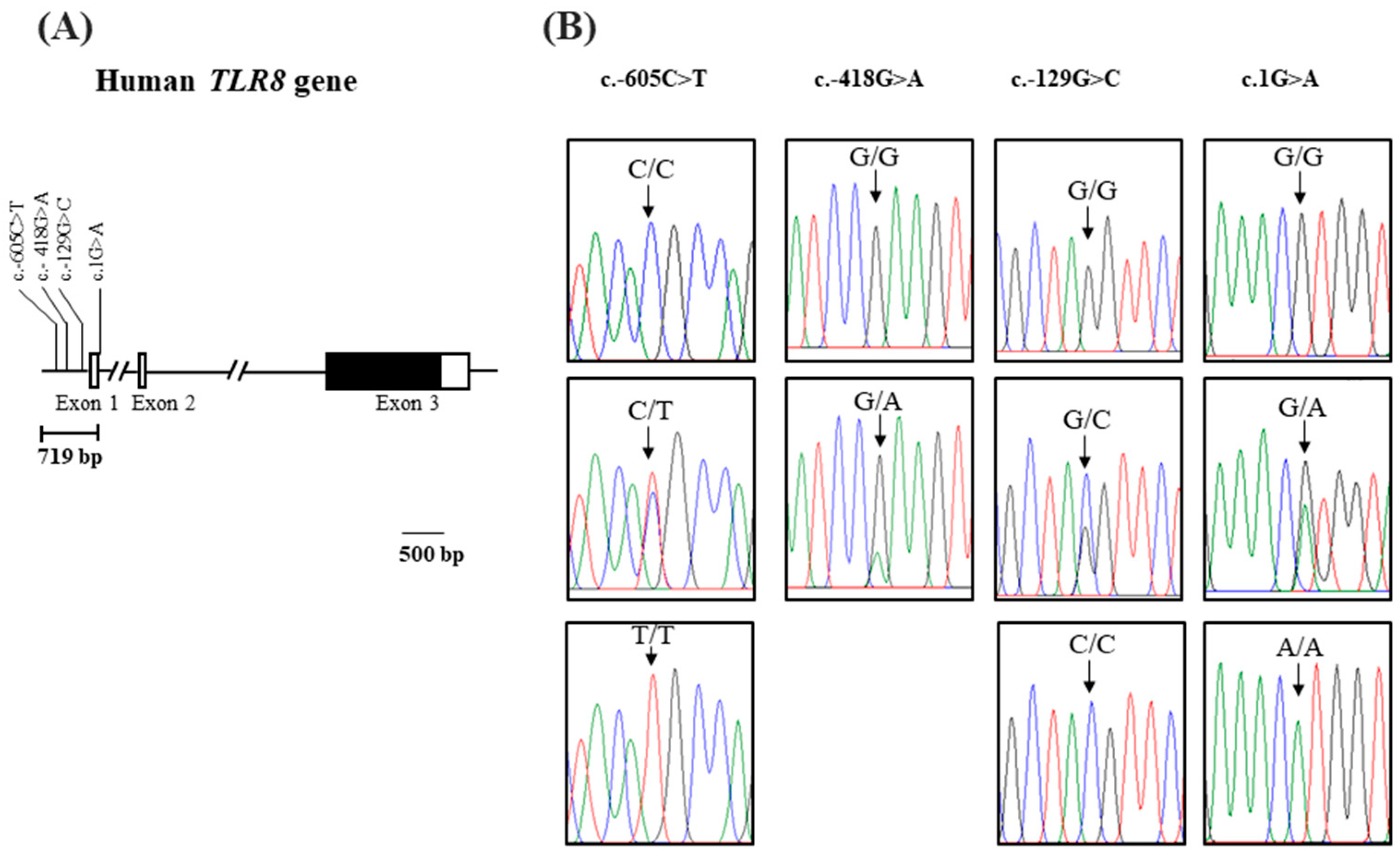
Figure 1. (A) Gene map and polymorphisms identified in the human toll-like receptor 8 gene (TLR8). The sequenced region is shown as horizontal bars (719 bp). The locations of single-nucleotide polymorphisms (SNPs) identified in this study are shown by arrows. A shaded block represents the open reading frame (ORF) in exon 3. (B) Electropherograms of the four SNPsidentified are shown: c.-605C>T, c.-418G>A, c.-129G>C, and c.1G>A. The upper/lower panels show homozygous genotypes; the middle panel shows heterozygous genotypes. Using an ABI 3730 automated sequencer, four colors show individual bases of a DNA sequence (blue: cytosine, red: thymine, black: guanine, and green: adenine).
Life 16 01167 g001
Table 1. Detailed information on the study population.
CharacteristicsControlsCOVID-19 Patientsp-Value
Number, n173191
Sex
Male, n (%)80 (46)90 (47)
Female, n (%)93 (54)101 (53)
Age (year ± SD), overall58.7 ± 14.256.6 ± 15.80.194
Age (year ± SD), male 57.5 ± 12.958.9 ± 14.60.520
Age (year ± SD), female59.7 ± 14.954.8 ± 16.50.086
Table 2. SNPs of TLR8 and HWE in this study.
SNPsLocationNucleotideGroupsHWEMAF
(Korean)
rs5741883 X:12906102 (GRCh38)C>TControl (n = 93)0.6T = 0.02
Patients (n = 101)0.747
rs186566524X:12906289 (GRCh38)G>AControl (n = 93)0.639A = 0.01
Patients (n = 101)0.874
rs3764879X:12906578 (GRCh38)G>CControl (n = 93)0.475C = 0.19
Patients (n = 101)0.274
rs3764880X:12906707 (GRCh38)G>AControl (n = 93)0.295A = 0.19
Patients (n = 101)0.06
Abbreviations: SNPs, single-nucleotide polymorphisms; HWE, Hardy–Weinberg equilibrium; MAF, minor allele frequency.
Table 3. Association analysis between TLR8 polymorphisms and COVID-19 case status in female individuals using different genetic models.
SNPsModelsGenotypeControls, n (%)Patients, n (%)OR (95% CI)p-Value
rs5741883AlleleC
T
180 (97)
6 (3)
192 (95)
10 (5)
Reference
1.5 (0.5–4.3)
0.396
Codominant modelCC
CT
TT
87 (93.5)
6 (6.5)
0 (0.0)
91 (90)
10 (10)
0 (0.0)
Reference
1.6 (0.5–4.5)
 
0.386
 
NA
Dominant modelCC
CT+TT
87 (93.5)
6 (6.5)
91 (90)
10 (10)
Reference
1.6 (0.5–4.5)
0.386
Recessive modelCC+CT
TT
93 (100)
0 (0.0)
101 (100)
0 (0.0)
Reference
 
NA
Overdominant modelCC+TT
CT
87 (93.5)
6 (6.5)
91 (90)
10 (10)
Reference
1.6 (0.5–4.5)
0.386
 
rs186566524AlleleG
A
183 (98)
3 (2)
193 (95.5)
9 (4.5)
Reference
2.8 (0.7–10.6)
0.121
Codominant modelGG
GA
AA
90 (97)
3 (3)
0 (0.0)
92 (91)
9 (9)
0 (0.0)
Reference
2.9 (0.7–11.2)
0.114
 
NA
Dominant modelGG
GA+AA
90 (96.8)
3 (3.2)
92 (91)
9 (9)
Reference
2.9 (0.7–11.2)
0.114
Recessive modelGG+GA
AA
93 (100)
0 (0.0)
101 (100)
0 (0.0)
Reference
 
NA
Overdominant modelGG+AA
GA
90 (97)
3 (3)
92 (91)
9 (9)
Reference
2.9 (0.7–11.2)
0.114
rs3764879Allele
G
C
149 (80)
37 (20)
161 (80)
41 (20)
Reference
1.0 (0.6–1.6)
0.920
Codominant modelGG
GC
CC
58 (62)
33 (36)
2 (2)
63 (62)
35 (35)
3 (3)
Reference
1.0 (0.5–1.7)
1.3 (0.2–8.5)
0.937
 
0.728
Dominant modelGG
GC+CC
58 (62.4)
35 (37.6)
63 (62.4)
38 (37.6)
Reference
1.0 (0.5- 1.7)
0.998
Recessive modelGG+GC
CC
91 (98)
2 (2)
98 (97)
3 (3)
Reference
1.3 (0.2–8.5)
0.720
Overdominant modelGG+CC
GC
60 (64.5)
33 (35.5)
66 (65.3)
35 (34.7)
Reference
0.9 (0.5–1.7)
0.903
rs3764880AlleleG
A
155 (83)
31 (17)
158 (78)
44 (22)
Reference
1.3 (0.8–2.3)
0.203
Codominant modelGG
GA
AA
62 (67)
31 (33)
0 (0.0)
60 (60)
38 (38)
3 (3)
Reference
1.2 (0.7–2.2)
0.434
 
0.193
Dominant modelGG
GA+AA
62 (66.6)
31 (33.4)
60 (59.4)
41 (40.6)
Reference
1.3 (0.7–2.4)
0.296
Recessive modelGG+GA
AA
93 (100)
0 (0.0)
98 (97)
3 (3)
Reference
 
0.212
Overdominant modelGG+AA
GA
62 (66.6)
31 (33.4)
63 (62.4)
38 (37.6)
Reference
1.2 (0.6–2.1)
0.533
Notes: p-value > 0.05, non-significant. Abbreviations: OR, odds ratio; CI, confidence interval. NA, not applicable.
Table 4. Association analysis between TLR8 SNPs and COVID-19 case status in male individuals.
SNPsControls, n (%)Patients, n (%)OR (95% CI)p-Valueq-Value
rs5741883
C
T
79 (99.0)
1 (1.0)
90 (100)
0 (0.0)
Reference
 
0.4540.454
rs186566524
G
A
79 (99.0)
1 (1.0)
87 (96.6)
3 (3.4)
Reference
2.7 (0.27–26.7)
0.3890.454
rs3764879
G
C
61 (76.0)
19 (24.0)
81 (90.0)
9 (10.0)
Reference
0.35 (0.15–0.8)
0.0180.036
rs3764880
G
A
61 (76.0)
19 (24.0)
82 (91.0)
8 (9.0)
Reference
0.3 (0.12–0.7)
0.010.036
Notes: p-value > 0.05 non-significant, p-value < 0.05 significant. Abbreviations: OR, odds ratio; CI, confidence interval. Benjamini–Hochberg FDR correction was applied across all 4 SNP comparisons; rs3764879 and rs3764880 remained significant at q < 0.05, while rs5741883 and rs186566524 are not significant.
Table 5. Comparison of haplotype frequencies of TLR8 polymorphisms associated with COVID-19 case status in Korean females.
HaplotypesFrequency p-Value
Control (n = 186)Patients (n = 202)
CGGG150 (0.806)147 (0.728)0.4679
CGCA23 (0.124)32 (0.158)1.0
TGCA5 (0.027)7 (0.035)0.6780
Other8 (0.043)16 (0.079)-
n = total number of haplotypes. Frequency = proportion of each haplotype among total haplotypes within each group. Other represents pooled rare haplotypes, each present at <5% frequency, combined for statistical comparison due to low individual counts.
Table 6. Linkage disequilibrium (LD) among single-nucleotide polymorphisms (SNPs) of the human toll-like receptor 8 gene (TLR8) in a Korean population.
Femalers5741883rs186566524rs3764879rs3764880
rs5741883-0.0080.1370.132
rs1865665240.042-0.000.00
rs37648790.1340.066-0.914
rs37648800.1670.0820.805-
Bold text indicates strong LD (r2 > 0.3). The above diagonal indicates the LD value in COVID-19 patients. The below diagonal indicates the LD value in controls.
Disclaimer/Publisher’s Note: The statements, opinions and data contained in all publications are solely those of the individual author(s) and contributor(s) and not of MDPI and/or the editor(s). MDPI and/or the editor(s) disclaim responsibility for any injury to people or property resulting from any ideas, methods, instructions or products referred to in the content.


