1. Introduction
Extrahepatic biliary diseases, namely cholecystitis, cholelithiasis, and biliary mucocele, represent important clinical challenges in canine patients and are increasingly recognized in small animal practice [
1,
2,
3,
4]. These pathological conditions primarily involve the liver, gallbladder, and extrahepatic bile ducts, organs with a central role in digestion, bile storage and transport, and metabolic homeostasis [
2,
5].
Clinical presentation is described as variable and nonspecific, particularly in the early stages, as it includes gastrointestinal symptoms such as vomiting, diarrhea, anorexia, and lethargy. A delayed diagnosis can allow disease progression with increased risk of serious complications namely gallbladder rupture, bile peritonitis, and systemic inflammatory responses, all of which are associated with considerable morbidity and, in severe cases, mortality [
6,
7,
8]. Consequently, early recognition of hepatobiliary disease and appropriate diagnostic evaluation are essential to optimize clinical outcomes.
Routine diagnostic investigation typically includes clinical examination and serum biochemical analysis, which can provide valuable information on hepatocellular injury, cholestasis, and systemic involvement [
9]. However, these data alone are often insufficient to characterize structural abnormalities of the biliary system. Over the past decades, abdominal ultrasonography has become a widely used diagnostic tool in the evaluation of hepatobiliary disorders in dogs allowing real-time visualization of the gallbladder, biliary ducts, and surrounding hepatic parenchyma [
5,
9,
10,
11,
12,
13]. This diagnostic tool facilitates the detection of abnormalities such as biliary sludge, gallstones, gallbladder wall thickening or rupture, mucoceles, peritonitis [
6,
7,
10,
11,
12]. Ultrasonography not only assists in identifying hepatobiliary disease, but also provides important information regarding disease severity, extent, and progression, thereby contributing to clinical decision-making and guiding therapeutic interventions [
12,
14,
15]. In addition, ultrasonographic monitoring is frequently used to evaluate disease course and response to treatment in dogs with chronic or progressive biliary conditions [
11,
16,
17].
The bacterial involvement in the pathogenesis of canine extrahepatic biliary disease has been documented in previous studies; however, the reported prevalence of bacterial isolation and the spectrum of microorganisms involved vary considerably [
6,
7,
8,
18,
19]. Some studies report relatively high rates of bacterial culture positivity in bile samples obtained from dogs with hepatobiliary disease, whereas others identify bacteria less frequently, suggesting that infection may represent either a primary pathogenic factor or a secondary complication associated with biliary stasis and structural abnormalities of the gallbladder [
18,
19,
20,
21,
22]. In addition, recent investigations have highlighted the complexity of the gallbladder microbiome, suggesting that microbial communities within the biliary system may play a role in disease development and progression [
23,
24,
25].
Despite this variability, microbiological assessment of bile samples remains clinically relevant, as it allows identification of bacterial organisms and supports targeted antimicrobial therapy.
In vitro antimicrobial susceptibility testing provides information that may assist in antimicrobial selection, which is particularly important in the context of increasing antimicrobial resistance in both veterinary and human medicine [
20,
21,
26,
27]. However, empirical antimicrobial therapy without microbiological confirmation may be associated with suboptimal treatment and the potential selection of resistant organisms.
Ultrasound-guided percutaneous cholecystocentesis represents a minimally invasive and widely accepted technique for obtaining bile samples for microbiological analysis in dogs with suspected hepatobiliary disease [
28,
29,
30]. When performed by experienced clinicians, this procedure is considered safe and can provide suitable samples for culture and
in vitro antimicrobial susceptibility testing of the bacterial isolates. The integration of ultrasonographic findings with microbiological data may therefore contribute to a more comprehensive evaluation of biliary disease, although the relationship between imaging findings and bacterial isolation remains incompletely characterized.
Therefore, the present study aimed to describe the clinical, ultrasonographic, and microbiological findings in dogs with extrahepatic biliary disease and to explore potential associations between ultrasonographic gallbladder patterns and bile culture results. In addition, the in vitro antimicrobial susceptibility profiles of the recovered bacterial isolates were recorded. Given the retrospective design and limited sample size, these analyses were intended to provide descriptive data and generate hypotheses for future investigation.
2. Materials and Methods
2.1. Study Population and Case Selection
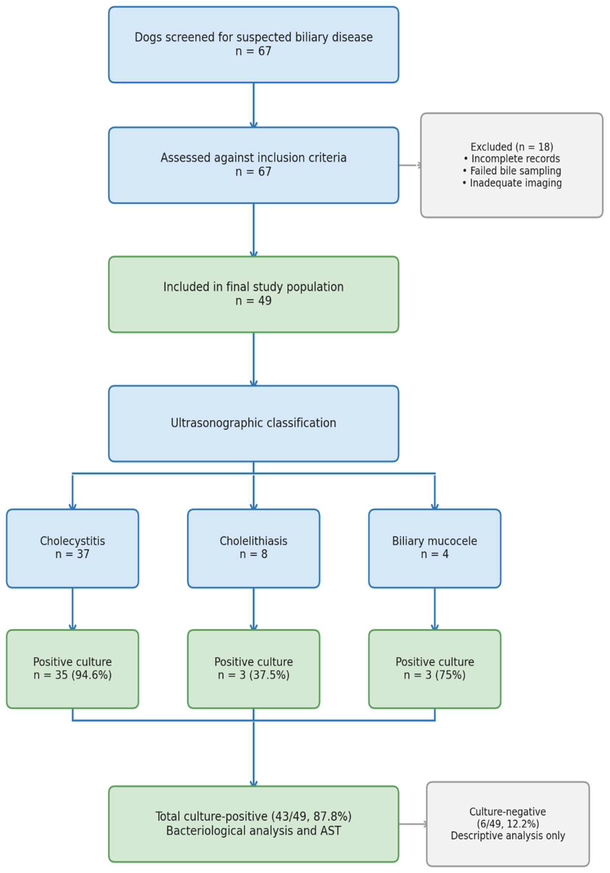
This retrospective case series initially screened 67 client-owned dogs of various breeds and ages that underwent bile sampling for suspected hepatobiliary disease at the Internal Medicine Clinic of the University Veterinary Teaching Hospital, University of Agricultural Sciences and Veterinary Medicine (USAMV) Cluj-Napoca, Romania, between March 2024 and December 2025. All 67 dogs were evaluated based on medical history, clinical examination, complete blood count (CBC), serum biochemistry, and abdominal ultrasonography. Of these, 49 dogs (21 males and 28 females) met all inclusion criteria, had bile cultures performed, and complete datasets and constituted the final study population. The remaining 18 dogs were excluded due to incomplete medical records (17), unsuccessful bile sampling (10), or inadequate imaging for ultrasonographic classification (2); some dogs met more than one exclusion criterion, and therefore counts exceed the total number of excluded cases. These cases were not included in statistical analyses but are reported for transparency of case selection.
A flow diagram illustrating case selection, exclusions, and allocation across ultrasonographic diagnostic groups and culture outcomes is provided in
Figure 1. The flow diagram was created using Microsoft PowerPoint (version 365, Microsoft Corporation, Redmond, WA, USA). Dogs were classified according to the predominant ultrasonographic gallbladder abnormality; however, overlapping imaging findings were occasionally present. In such cases, classification was based on the most clinically relevant or predominant lesion, as determined at the time of evaluation.
Inclusion criteria were as follows: (i) clinical signs compatible with hepatobiliary or gastrointestinal disease (namely vomiting, diarrhea, lethargy, anorexia, jaundice, fever, abdominal pain) or incidental ultrasonographic detection of gallbladder pathology during diagnostic workup for other conditions [
1,
2]; (ii) laboratory results indicating serum biochemical alterations suggestive of hepatobiliary involvement (elevated alanine aminotransferase (ALT), alkaline phosphatase (ALP), gamma-glutamyl transferase (GGT), or total bilirubin) [
5,
9]; (iii) ultrasonographic findings indicative of gallbladder pathology (cholecystitis, cholelithiasis, or biliary mucocele) [
6,
7,
10,
11,
12]; (iv) bile sampling: availability of bile samples obtained via ultrasound-guided percutaneous cholecystocentesis or intraoperative collection for microbiological culture [
28,
29].
Dogs with negative bile cultures were retained in the dataset for descriptive and comparative analyses, whereas only culture-positive samples (43/49 dogs) were included in detailed bacteriological characterization and
in vitro antimicrobial susceptibility testing (
Figure 1). This approach was used to maintain clarity of denominators across different analytical components of the study.
Given the retrospective design and referral hospital setting, the study population represents a clinically selected cohort of dogs with a higher pre-test probability of hepatobiliary disease. This introduces potential referral and selection bias and limits the generalizability of the findings to the broader population of dogs seen in primary care practice.
2.2. Clinical Examination and Laboratory Testing
All dogs underwent complete physical examination. Blood samples were collected from the jugular or cephalic vein for CBC and serum biochemistry analyses that included measurement of alkaline phosphatase (ALP), alanine aminotransferase (ALT), gamma-glutamyl transferase (GGT), and total bilirubin (TB), using a dry chemistry automated analyzer (FUJIFILM DRI-CHEM NX700V
® FUJIFILM Europe GmbH, Düsseldorf, Germany) according to the manufacturer’s instructions. Established canine reference intervals provided by the laboratory were applied [
5,
9,
31].
Biochemical abnormalities were interpreted in the context of the overall clinical and ultrasonographic picture rather than as isolated thresholds. ALP and GGT were considered the primary indicators of cholestatic and biliary disease in dogs, as these enzymes more reliably reflect hepatobiliary dysfunction than ALT alone [
5,
9]. Marked increases in ALT activity, defined as values at least two times greater than the upper limit of the reference interval, were considered supportive but were not required for inclusion when ALP or GGT elevation, hyperbilirubinemia, or compatible clinical and ultrasonographic findings were present [
1,
2,
3,
5,
9]. Dogs exhibiting elevations in one or more of these biochemical markers alongside clinical signs and ultrasonographic evidence of gallbladder disease were considered suitable candidates for further evaluation and bile sampling.
2.3. Abdominal Ultrasonography
Abdominal ultrasonography was performed using a MyLab X8® ultrasound machine (Esaote S.p.A., Genoa, Italy) equipped with a microconvex transducer with a variable frequency range of 3–11 MHz. All ultrasonographic examinations and ultrasound-guided cholecystocentesis procedures were performed by the same experienced operator, which minimized inter-operator variability; however, the retrospective design precluded blinded image review or formal assessment of interobserver agreement.
To ensure optimal restraint and image quality, canine patients were anesthetized, and positioned in right lateral or dorsal recumbency. The ventral abdominal area was clipped to remove hair and cleaned. Ultrasound gel was applied to ensure adequate acoustic coupling. The gallbladder was initially localized by scanning the right cranial abdominal quadrant, and then examined in both longitudinal and transverse planes.
Key ultrasonographic parameters assessed included gallbladder wall thickness and echogenicity, luminal content and echogenicity, the presence of sediment or sludge, gallstones, pericholecystic fluid, and gallbladder size and contour. Color Doppler imaging was used to evaluate vascularization around the gallbladder and to identify evidence of inflammation or vascular compromise. Representative images were stored for review and documentation.
Gallbladder lesions were categorized based on ultrasonographic criteria previously described [
6,
7,
10,
12,
14,
32,
33,
34].
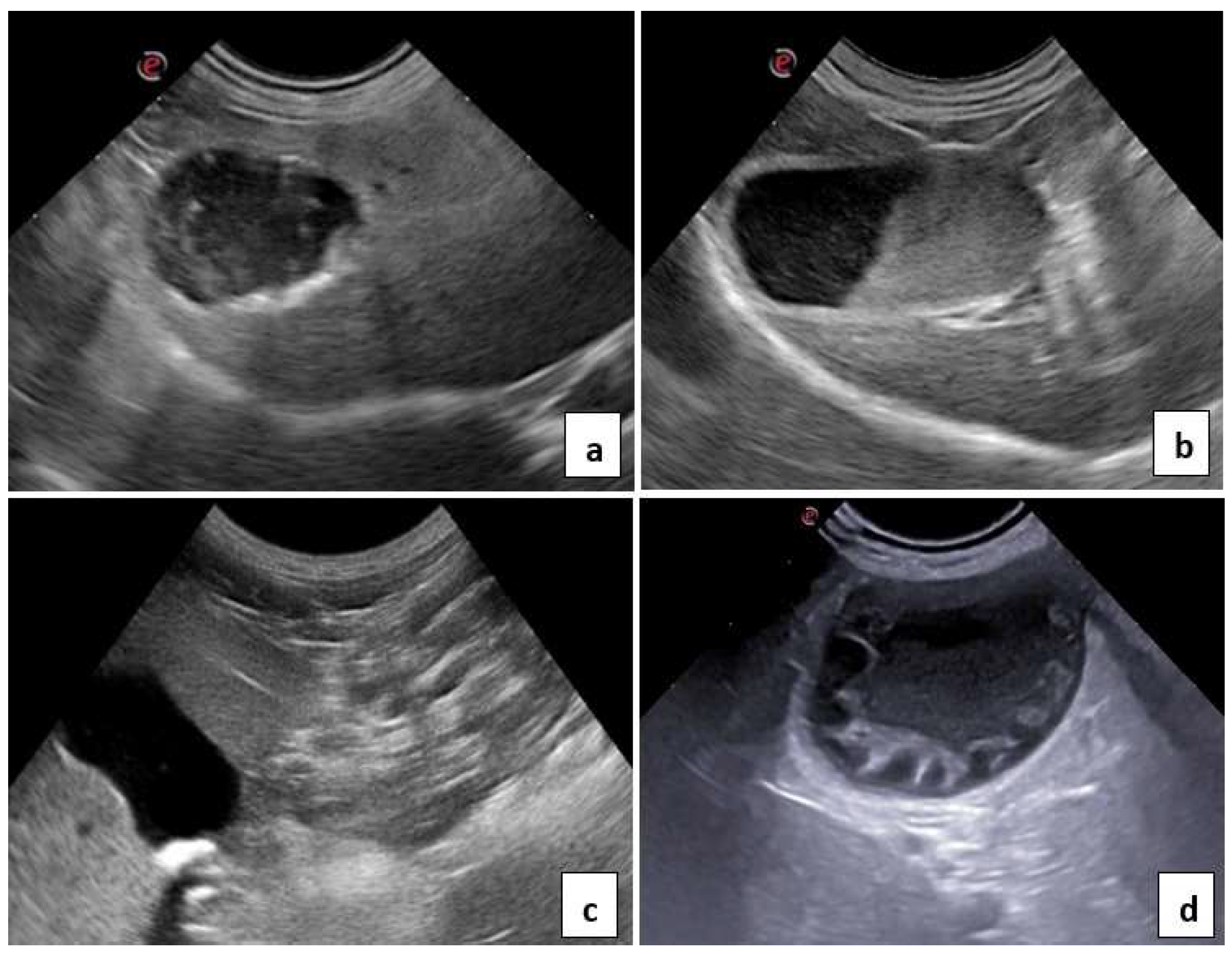
Cholecystitis: Gallbladder wall thickening (≥3 mm or subjectively increased relative to breed- and size-adjusted expected values), often irregular, and occasionally accompanied by a hypoechoic subserosal halo consistent with edema and inflammation, was considered supportive of cholecystitis when interpreted together with luminal abnormalities and pericholecystic inflammatory changes. The gallbladder lumen frequently contained echogenic material compatible with biliary sludge and/or purulent content, resulting in a “dirty” appearance. Inflammation was also indicated by variable amounts of pericholecystic fluid.
Cholelithiasis: Presence of one or more strongly echogenic intraluminal foci casting clean distal acoustic shadows, consistent with gallstones. Gallbladder wall thickness and lumen distension were recorded but could be normal or increased. Evidence of cystic duct obstruction or additional inflammatory changes was noted when present.
Biliary mucocele: Marked gallbladder enlargement with echogenic, immobile, and often stratified bile producing a characteristic stellate or “kiwi fruit” pattern in cross-section. In most cases, the gallbladder wall was intact, but wall thinning or irregularity could be present. Due to the risk of rupture and bile peritonitis, these cases were considered contraindicated for percutaneous cholecystocentesis and were managed surgically.
Dogs that met ultrasonographic criteria for cholecystitis, cholelithiasis, or biliary mucocele were included in the study for further analysis.
2.4. Ultrasound-Guided Cholecystocentesis
Dogs diagnosed ultrasonographically with cholecystitis or cholelithiasis were scheduled for ultrasound-guided percutaneous cholecystocentesis for diagnostic bile sampling. Dogs with biliary mucocele underwent surgical cholecystectomy, and bile samples were collected directly from the gallbladder intraoperatively.
In 45 dogs, ultrasound-guided cholecystocentesis was performed under general anesthesia. Four dogs with biliary mucocele were not subjected to cholecystocentesis due to the perceived high risk of gallbladder rupture; in these cases, bile was aspirated directly from the intact gallbladder using a sterile needle and syringe immediately after ligation of the cystic duct and before excision of the gallbladder.
All 45 dogs scheduled for percutaneous cholecystocentesis underwent the procedure under general anesthesia following a 10–12 h fasting period. Intravenous isotonic crystalloids were administered to maintain adequate hydration and support normal systolic blood pressure. Sedation and anxiolysis were achieved with midazolam (Midazolam Panpharma® 5 mg/mL, Chiesi Pharmaceuticals GmbH, Viena, Austria) 0.2–0.5 mg/kg IV, followed by propofol (Propofol-Lipuro® 1%, B. Braun, Melsungen, Germany) 10 mg/mL 2–6 mg/kg IV for anesthesia induction and intubation. Isoflurane (Isoflutek® 1000 mg/g, Laboratorios Karizoo S.A., Barcelona, Spain) in 100% oxygen provided inhalational anesthesia, titrated to maintain appropriate depth. Intraoperative analgesia was achieved through intravenous administration of fentanyl (Fentanyl Torrex® 0.05 mg/1 mL, Chiesi Pharmaceuticals GmbH, Viena, Austria) 2–5 μg/kg initial dose. The fentanyl dose was titrated throughout the procedure to provide appropriate analgesia for each patient. Heart rate, respiratory rate, temperature, capnography, pulse oximetry, and blood pressure were continuously monitored.
After induction of general anesthesia, each dog was positioned in dorsal recumbency. The upper right abdominal quadrant was shaved and aseptically prepared with isopropyl alcohol and chlorhexidine. The microconvex transducer was placed in a sterile sheath and sterile ultrasound gel was applied. The gallbladder was located ultrasonographically, and a safe percutaneous window traversing hepatic parenchyma was identified.
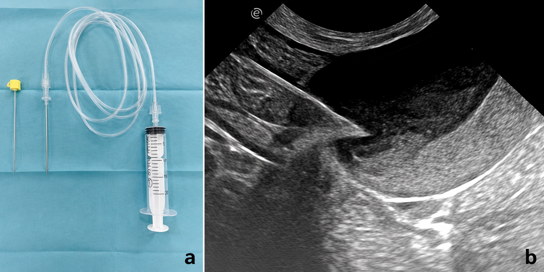
A 22 G (0.90 mm) Quincke spinal needle 75 mm in length (Spinocan® B. Braun, Melsungen, Germany), was advanced under ultrasound guidance through the liver parenchyma into the gallbladder lumen. The Quincke needle was chosen for its favorable balance between diameter and length, which facilitated precise guidance. Before needle insertion, the stylet was withdrawn and a sterile extension tube (100 cm) was attached to the needle hub. A sterile 10 mL syringe was connected to the distal end of the extension tube for bile aspiration. This arrangement allowed the same operator to manage both the transducer and the puncture needle while an assistant aspirated bile as soon as the needle tip was confirmed within the gallbladder lumen.
Between 2 and 4 mL of bile were aspirated from each dog, depending on body size and degree of gallbladder distension (
Figure 2a,b). Traversing the hepatic parenchyma was intentionally chosen to allow the needle track to be partially “wiped” on withdrawal, theoretically reducing the risk of peritoneal contamination from a potentially infected gallbladder.
After cholecystocentesis, dogs were monitored clinically and ultrasonographically at 2, 4, 8, 12, 24, and 48 h post-procedure to detect possible complications such as gallbladder laceration, hemoperitoneum, or free fluid. Post-procedural analgesia was provided using buprenorphine, 20 µg/kg IV (Buprecare Multidose 0.3 mg/mL, Animacare, York, UK). Prophylactic antibiotics were not administered solely due to the procedure [
28,
29].
All abdominal ultrasonographic examinations and ultrasound-guided cholecystocentesis procedures were performed by the same operator, a veterinarian with 13 years of experience in small animal ultrasonography, which helped to minimize inter-operator variability in image acquisition and interpretation.
2.5. Microbiological Culture and Identification
Bile samples were processed within minutes of collection under aseptic conditions with the aim of recovering both aerobic and anaerobic bacteria. Thus, each sample was inoculated onto 5% Columbia blood agar and MacConkey agar plates (Bio-Rad, Marnes-la-Coquette, France) for aerobic culture and incubated at 37 °C for 24–48 h. MacConkey agar was included to facilitate the recovery and differentiation of Gram-negative enteric bacteria based on lactose-fermenting ability. For anaerobic culture, samples were inoculated onto 5% Columbia blood agar plates and incubated at 37 °C in an anaerobic jar system with a commercial gas-generating kit to achieve an oxygen-free atmosphere. Anaerobic conditions were verified using an anaerobic indicator strip and the plates were monitored for up to 72 h. No obligate anaerobic reference strain was used for validation. Although this anaerobic culture protocol is routinely used in veterinary diagnostic laboratories, the absence of specialized pre-reduced anaerobic media, prolonged incubation and formal validation using a dedicated obligate anaerobic reference strain may have limited the recovery of fastidious obligate anaerobes.
Initial identification of the bacterial isolates was based on conventional veterinary microbiology criteria, including Gram staining, colony morphology, hemolysis patterns, and standard biochemical characteristics [
16]. Identification at the genus and species levels was subsequently performed using matrix-assisted laser desorption/ionization time-of-flight mass spectrometry (MALDI-TOF MS) [
35,
36,
37]. For MALDI-TOF MS analysis, a small portion of a single, well-isolated fresh colony obtained from pure 18–24 h culture was transferred with a sterile microbiological loop onto an individual spot of a polished steel target plate to form a thin smear film. After air drying, each spot was overlaid with 1 µL of α-cyano-4-hydroxycinnamic acid (HCCA) matrix solution and allowed to air-dry completely prior to analysis. Protein extraction using an ethanol–formic acid protocol was performed according to the manufacturer’s recommendations to improve spectral quality when direct smear spectra were insufficient for reliable identification. Mass spectra were acquired using a MALDI-TOF Biotyper Sirius IVD
® system (Bruker Daltonics, Bremen, Germany), and spectral interpretation was performed using the MALDI Biotyper™ software (version 3.1) platform with automated comparison against the Bruker reference database. The identification scores were interpreted according to the manufacturer’s recommended criteria, with log(score) values ≥2.0 considered reliable for species-level identification and scores between 1.7 and 1.99 considered reliable for genus-level identification only [
35,
36,
37]. All isolates included in the study achieved MALDI-TOF identification scores considered acceptable according to the manufacturer-recommended interpretive criteria. Isolates yielding genus-level scores only were reported conservatively according to the highest-confidence identification obtained. Prior to each analytical session, the instrument was calibrated using the Bruker Bacterial Test Standard (BTS) according to the manufacturer’s instructions as part of routine laboratory quality-control procedures. Internal quality-control procedures were performed in accordance with laboratory standard operating protocols using manufacturer-recommended calibration standards and reference control organisms, including
Escherichia coli ATCC 8739 for calibration and identification verification. All bacterial isolates recovered from bile samples were identified using the same MALDI-TOF MS workflow described above.
At the time of bile sampling, none of the dogs had received systemic antimicrobial therapy. Antimicrobials were deliberately withheld until after bile collection in order to avoid influencing culture results and in vitro susceptibility testing.
Of the 49 dogs retained in the study, 43 had positive bile cultures and formed the basis for the detailed microbiological analyses.
2.6. In Vitro Antimicrobial Susceptibility Testing
The
in vitro antimicrobial susceptibility testing was performed using the Kirby–Bauer disk diffusion method on Mueller–Hinton agar (Bio-Rad, Marnes-la-Coquette, France) according to EUCAST and CLSI recommendations. For fastidious isolates requiring enriched media, Mueller–Hinton agar supplemented with 5% sheep blood was used when appropriate. Each bacterial strain was prepared as an inoculum by suspending 24 h colonies in sterile saline and adjusting the turbidity to 0.5 McFarland standard. Then, the standardized inoculum was evenly distributed across the agar surface using a sterile swab to obtain a confluent bacterial lawn. Commercial antimicrobial disks (Bio-Rad, Marnes-la-Coquette, France; 6.5 mm diameter) impregnated with predefined antibiotic concentrations were applied onto inoculated plates according to the manufacturer’s instructions. The following antimicrobial agents and disk concentrations were tested: amoxicillin–clavulanic acid (AMC, 20/10 µg), ampicillin (AMP, 10 µg), marbofloxacin (MAR, 5 µg), gentamicin (GEN, 10 µg), cefalexin (CLE, 30 µg), doxycycline (DOX, 30 µg), trimethoprim–sulfamethoxazole (TRS, 1.25/23.75 µg), and clindamycin (CLI, 2 µg) [
26,
27]. This panel was selected to include representatives of the major antimicrobial classes commonly used in small animal practice for hepatobiliary infections, including β-lactams (AMC, AMP, CLE), a fluoroquinolone (MAR), an aminoglycoside (GEN), a tetracycline (DOX), a potentiated sulfonamide (TRS), and a lincosamide (CLI).
After incubation at 37 °C for 18–24 h under aerobic conditions, inhibition zone diameters were measured in millimeters and interpreted primarily according to EUCAST veterinary clinical breakpoints [
38]; when species-specific EUCAST breakpoints were unavailable, CLSI veterinary standards (VET01) were applied [
39]. Quality-control procedures were performed periodically using reference strains recommended by CLSI and EUCAST, including
Escherichia coli ATCC 25922 and
Staphylococcus aureus ATCC 25923.
Detailed bacteriological and antimicrobial susceptibility analyses were performed exclusively on the 43 culture-positive bile samples obtained from the 49 included dogs.
2.7. Statistical Analysis
Data were analyzed using Epi Info™ 7 (Centers for Disease Control and Prevention, Atlanta, GA, USA). Descriptive statistics included means and ranges for continuous variables and frequencies and percentages for categorical variables. The chi-square test or Fisher’s exact test (when expected counts were <5) was used to evaluate associations between ultrasonographic diagnosis (cholecystitis, cholelithiasis, biliary mucocele) and categorical outcomes, including culture positivity and specific bacterial isolates. A p-value of ≤0.05 was considered statistically significant; however, given the small sample sizes, particularly for the cholelithiasis (n = 8) and biliary mucocele (n = 4) groups, all statistical analyses were considered exploratory and hypothesis-generating rather than confirmatory, therefore, these results should be interpreted with caution. No correction for multiple comparisons was applied as the analyses were exploratory in nature and not intended for formal hypothesis testing.
3. Results
A total of 67 dogs were initially screened based on clinical and ultrasonographic criteria. Of these, 49 dogs, for which bile culture was performed and complete data were available, were included in the final analysis. The study population comprised 49 client-owned adult dogs (21 males and 28 females) of various breeds, with a mean age of 10.8 years. The breed distribution included both purebred and mixed-breed dogs; however, detailed breed-specific analysis was not performed due to the small sample size and heterogeneous breed representation.
3.1. Clinical and Laboratory Findings
Regarding the clinical examination, gastrointestinal signs were the most frequently reported clinical manifestations. Diarrhea was identified in 10 dogs (20.4%), followed by vomiting in 9 dogs (18.4%). Reduced appetite was observed in 7 cases (14.3%).
General systemic signs were also commonly recorded, with weakness and lethargy each present in 8 dogs (16.3%). Abdominal pain was documented in 5 cases (10.2%). Less frequently, jaundice was noted in 3 dogs (6.1%), while fever was identified in only 2 cases (4.1%).
Notably, 4 dogs (8.2%) were asymptomatic at the time of diagnosis, with biliary disease detected incidentally during diagnostic investigations performed for other reasons.
Overall, the clinical presentation of biliary disorders in dogs was variable and often nonspecific, with gastrointestinal and general systemic signs predominating.
Serum biochemistry results are summarized in
Table 1. Elevated ALP activity was observed in 46/49 dogs (93.9%), with an average value of 493 U/L (range 93–1200 U/L; reference interval 10–84 U/L). Increased ALT activity was detected in 22/49 dogs (44.9%), with an average value of 171.68 U/L (range 22–960 U/L; reference interval 5–65 U/L). GGT activity was increased in 40/49 dogs (81.6%), with an average value of 19.59 U/L (range 2.80–28 U/L; reference interval 2–10 U/L). Hyperbilirubinemia was present in 7/49 dogs (14.3%), with an average total bilirubin concentration of 2.5 mg/dL (range 0.5–8.3 mg/dL; reference interval 0.1–0.4 mg/dL).
Hematological abnormalities were observed in dogs with suspected hepatobiliary disease undergoing biliary culture testing. Leukocytosis was identified in 18.4% of cases (9/49 dogs), while leukopenia was present in 4.1% (2/49 dogs). A left shift was detected in 12.2% of the dogs (6/49), suggesting an inflammatory or infectious process in a subset of patients.
3.2. Ultrasonographic Findings
Abdominal ultrasonography revealed gallbladder abnormalities consistent with cholecystitis in 37/49 dogs (75.5%), cholelithiasis in 8/49 (16.3%), and biliary mucocele in 4/49 (8.2%) (
Table 2,
Figure 3). In two dogs, overlapping ultrasonographic features compatible with more than one gallbladder abnormality were observed; however, each dog was assigned to the predominant ultrasonographic diagnosis determined by overall imaging appearance to avoid duplicate case allocation across diagnostic categories.
3.3. Bile Culture Results
In total, bile samples from 67 dogs that fulfilled the screening criteria were submitted for microbiological culture. Among the 49 dogs that met all inclusion criteria and were retained in the study, 43/49 bile samples (87.8%) were microbiologically positive, whereas 6/49 (12.2%) yielded no growth. Considering all screened dogs, 43/67 (64.2%) had positive cultures and 24/67 (35.8%) had negative culture results. Detailed bacteriological and antimicrobial susceptibility analyses were performed on the 43 culture-positive samples from the 49 included dogs. Of the 43 culture-positive samples, 39 (90.7%) yielded a single bacterial species, while 4 (9.3%) showed polymicrobial growth (two species per sample). Only aerobic bacteria were isolated; all anaerobic cultures were negative.
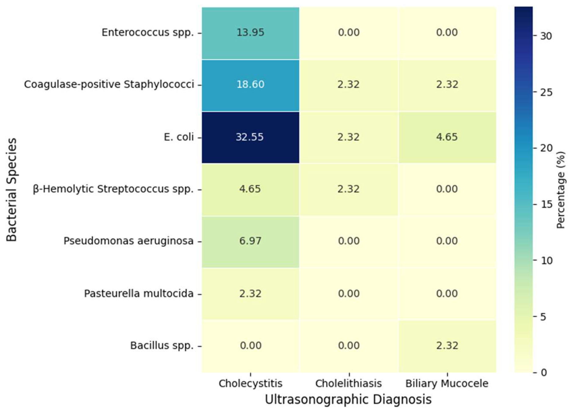
Escherichia coli was the most frequently isolated organism among the 43 culture-positive samples, followed by coagulase-positive
Staphylococci,
Enterococcus spp., β-hemolytic
Streptococcus spp.,
Pseudomonas aeruginosa,
Pasteurella multocida, and
Bacillus spp. The frequencies and distributions of bacterial isolates according to ultrasonographic diagnosis are summarized in
Figure 4 and
Table 3. Each dog was assigned to a single ultrasonographic diagnostic category based on the predominant gallbladder finding. Odds ratios (OR) and Fisher’s exact test
p-values were calculated as exploratory analyses for the two most frequently isolated organisms (
E. coli and Coagulase-positive
Staphylococcus spp., with OR >1 indicative for increased likelihood of bacteria isolation. An association between
E. coli and cholecystitis was observed (Fisher’s exact test,
p = 0.02) (
Table 3); however, this finding should be interpreted cautiously given the exploratory nature of the analysis and small subgroup sizes.
3.4. In Vitro Antimicrobial Susceptibility Testing
The
in vitro antimicrobial susceptibility patterns of the isolated organisms are summarized in
Table 4. For
Enterococcus spp., higher susceptibility rates were recorded for amoxicillin–clavulanic acid, ampicillin, and gentamicin, whereas to doxycycline and clindamycin was variable. Mixed cultures of coagulase-positive
Staphylococci and
Bacillus spp. exhibited susceptibility to amoxicillin–clavulanic acid, marbofloxacin, gentamicin, doxycycline, and clindamycin, while being resistant to ampicillin and trimethoprim–sulfamethoxazole.
Escherichia coli isolates displayed higher susceptibility rates to marbofloxacin and gentamicin and moderate susceptibility to amoxicillin–clavulanic acid and ampicillin, whereas lower susceptibility was observed for trimethoprim–sulfamethoxazole and clindamycin. Pseudomonas aeruginosa isolates were resistant to most tested antimicrobials, with only partial susceptibility to marbofloxacin and gentamicin. β-Hemolytic Streptococcus spp. showed excellent susceptibility to amoxicillin and ampicillin and variable responses to other agents. Pasteurella multocida was susceptible to most tested antimicrobials, except gentamicin and trimethoprim–sulfamethoxazole.
Overall, the antibiotics with the highest inhibitory activity across the tested bacterial species were amoxicillin–clavulanic acid, marbofloxacin, and gentamicin. In contrast, trimethoprim–sulfamethoxazole and clindamycin showed the lowest overall effectiveness, particularly in infections involving
Pseudomonas aeruginosa (
Table 4).
All dogs received individualized medical and/or surgical management according to the underlying diagnosis. All 49 dogs survived to hospital discharge. No standardized follow-up after discharge was performed, and long-term outcomes could not be systematically assessed.
4. Discussion
This study described the clinical, blood biochemistry, ultrasonographic and microbiological findings in 49 dogs with extrahepatic biliary disease and explored potential associations between ultrasonographic diagnoses and bile culture results in a clinically selected referral population. Because the study was retrospective and performed at a tertiary referral hospital, the findings should be interpreted within the context of a selected cohort of dogs with concurrent clinical, biochemical, and ultrasonographic evidence of gallbladder disease. Of the 67 initially screened dogs evaluated for suspected hepatobiliary disease, 49 fulfilled all inclusion criteria and underwent bile culture, whereas only the 43 culture-positive cases were included in the detailed microbiological and in vitro antimicrobial susceptibility analyses. Consequently, the observed culture-positive rate reflects this selected study population and should not be generalized to all dogs with suspected hepatobiliary disease.
Within this selected cohort, the clinical presentation was diverse and often non-specific—characterized mainly by bilious vomiting, diarrhea, lethargy, and reduced appetite—highlights the difficulty of diagnosing hepatobiliary disease based solely on clinical signs. These manifestations overlap considerably with those reported in gastrointestinal, pancreatic, and systemic inflammatory disorders, further emphasizing the limited specificity of clinical signs for hepatobiliary disease.
Marked elevations in ALP and GGT, and to a lesser extent ALT, were common among the included dogs and supported the presence of hepatobiliary dysfunction. Biochemical abnormalities were heterogeneous across cases, reflecting the exploratory clinical nature of the study population and the fact that inclusion was based on a composite profile rather than any single enzyme threshold. These heterogeneous changes reflect the variable clinical expression of hepatobiliary disease in this population. In contrast, hyperbilirubinemia was less consistently identified, suggesting that bilirubin may be less sensitive for early or less severe disease compared with cholestatic enzymes. Hematologic changes, such as leukocytosis, leukopenia, and left shift, were variably present and likely reflect systemic inflammatory responses rather than being specific for a particular biliary pathology.
Ultrasonography proved essential in defining the type of gallbladder lesion, allowing differentiation among cholecystitis, cholelithiasis, and biliary mucocele based on well-established imaging criteria. The predominance of cholecystitis in this series is consistent with previous reports in dogs with extrahepatic biliary disease and emphasizes the importance of careful evaluation of gallbladder wall thickness, echotexture, pericholecystic fluid, and luminal content.
The substantial proportion of positive aerobic bile cultures (87.8%), particularly in dogs with ultrasonographic evidence of cholecystitis and biliary mucocele, supports a potential association between bacterial isolation and canine gallbladder disease. Our finding that
E. coli and coagulase-positive
Staphylococci were the most frequently isolated organisms is broadly consistent with previous studies reporting intestinal and skin/oral commensals as major contributors to biliary infection in dogs [
6,
7,
8,
18,
19,
20,
40].
It should be emphasized that this proportion reflects a highly selected referral population and a subset of dogs (43/49) with both ultrasonographic evidence of gallbladder disease and positive bile cultures. As such, the observed culture-positive rate should not be generalized to all dogs with suspected hepatobiliary disease in primary care settings. Direct comparison of culture-positive rates with prior studies is further limited because our inclusion criteria required concurrent clinical signs, biochemical abnormalities, and ultrasonographic evidence of gallbladder disease, which selects for dogs with clinically relevant biliary pathology.
The variation in bacterial prevalence across different ultrasonographic diagnoses suggests that distinct pathophysiological mechanisms may underlie these conditions. Cholecystitis appears to represent a primarily infectious process, whereas in cholelithiasis and biliary mucocele bacterial colonization may be secondary to biliary stasis and altered gallbladder motility. The relatively high proportion of positive cultures in biliary mucocele cases supports previous observations that bacterial infection can be associated with mucoceles, although this association is inconsistent among reports and may depend on methodology (e.g., culture vs. FISH vs. 16S rRNA sequencing) [
7,
19,
20,
21].
Notably, all anaerobic cultures in this study were negative, whereas other authors have reported occasional anaerobic isolates (e.g.,
Bacteroides spp.,
Clostridium spp.) in similar clinical contexts [
18]. Differences in culture methods, incubation duration, population characteristics, and prior antimicrobial exposure might explain these discrepancies. In the present study, although anaerobic incubation for up to 72 h is generally considered sufficient for the recovery of most clinically relevant obligate anaerobes, the absence of specialized pre-reduced anaerobic media and prolonged incubation protocols may have limited the detection of fastidious or slow-growing anaerobic organisms. Therefore, negative anaerobic culture results should be interpreted cautiously. Furthermore, advanced molecular methods, such as fluorescence in situ hybridization and 16S rRNA gene sequencing, have demonstrated the presence of bacterial DNA in culture-negative biliary samples, suggesting that conventional culture may underestimate the true prevalence and diversity of biliary microbiota [
23,
24,
25].
The use of MALDI-TOF MS facilitated rapid bacterial identification with high confidence for most isolates [
35,
36,
37].
The
in vitro antimicrobial susceptibility results provide descriptive information that may be valuable in guiding empirical therapy in similar clinical contexts. Higher susceptibility rates were observed for three broad spectrum antibiotics, namely amoxicillin–clavulanic acid, marbofloxacin, and gentamicin, across the isolates evaluated in this study. In contrast, lower susceptibility rates were recorded towards trimethoprim–sulfamethoxazole and clindamycin, particularly among Gram-negative isolates. Notably,
Pseudomonas aeruginosa strains displayed reduced susceptibility to most tested antimicrobials, a finding consistent with its well-documented intrinsic and acquired multidrug resistance mechanisms. This organism is a recognized opportunistic pathogen frequently associated with infections that are challenging to manage therapeutically due to limited antimicrobial options and the ability to rapidly develop resistance during therapy, which may contribute to poorer clinical outcomes [
41].
However, these findings should be interpreted cautiously due to the limited number of isolates for several species and their heterogeneous distribution. In particular, results for Pseudomonas aeruginosa and Pasteurella multocida are based on very small sample sizes and should be considered descriptive only.
Therefore, while these data may support antimicrobial selection in similar cases, they do not replace culture-guided therapy, which remains essential for optimal antimicrobial stewardship in canine hepatobiliary infections. These findings further reinforce the importance of performing bile culture and susceptibility testing whenever feasible, rather than relying solely on empirical broad-spectrum therapy. Rational antimicrobial selection based on culture and susceptibility results is essential to maximize therapeutic success and minimize the emergence of antimicrobial resistance [
26,
27].
Overall, these findings further emphasize the importance of a multimodal diagnostic approach integrating clinical assessment, laboratory testing, abdominal ultrasonography, and microbiological evaluation.
Nevertheless, this study has several limitations. Its retrospective nature and reliance on clinical case selection introduce potential selection bias. The biochemical inclusion criterion as originally stated implied a strict ALT ≥2× ULN threshold; however, inclusion was applied on the basis of a composite clinical, biochemical, and ultrasonographic profile, with ALP and GGT serving as primary indicators of biliary disease. This discrepancy between the stated criterion and the criterion as applied represents a limitation of the study design and is acknowledged accordingly. The absence of a healthy control group limits the ability to distinguish between true infection and possible colonization or contamination. Skin or needle-track cultures were not performed; therefore, contamination during percutaneous sampling cannot be completely excluded, particularly for organisms commonly associated with skin flora such as coagulase-positive Staphylococci or Bacillus spp. Still, the use of aseptic sampling procedures and the consistent recovery of bacteria from dogs with concurrent ultrasonographic gallbladder abnormalities support the potential clinical relevance of the bacterial isolates. The sample size, while reasonable for a veterinary study, may limit the statistical power to detect associations for less common bacterial species. The small number of dogs in the cholelithiasis (n = 8) and biliary mucocele (n = 4) subgroups limited the statistical power of between-group comparisons, and the significant association identified between E. coli and cholecystitis should be considered exploratory until confirmed in larger cohorts. No correction for multiple comparisons was applied given the exploratory nature of these analyses. Given the number of comparisons performed, the risk of type I error cannot be excluded. Information on comorbidities such as endocrinopathies, pancreatitis, or prior corticosteroid administration—conditions recognized as risk factors for canine biliary disease—was not systematically recorded and therefore could not be analyzed as potential confounding factors. The study was conducted at a single university referral center, which may introduce referral bias and limit generalizability to primary-care settings. All ultrasonographic examinations and cholecystocentesis procedures were performed by a single experienced operator who was not blinded to clinical or biochemical findings; this ensures procedural consistency but precludes assessment of inter-observer agreement and may introduce operator bias. Histopathological confirmation of ultrasonographic diagnoses was available only for dogs that underwent surgery for biliary mucocele; the positive predictive value of the ultrasonographic criteria for cholecystitis and cholelithiasis in non-surgical cases therefore remains unknown. Histopathological confirmation of ultrasonographic diagnoses was available only for dogs that underwent surgery for biliary mucocele; the positive predictive value of the ultrasonographic criteria for cholecystitis and cholelithiasis in non-surgical cases therefore remains unknown. The gallbladder wall thickness threshold of ≥3 mm was applied uniformly across all breeds and body sizes; breed- and size-specific ultrasonographic reference values were not available for this cohort, which may have influenced the diagnostic classification of cholecystitis in some cases. In addition, advanced molecular techniques were not employed; thus, some fastidious or non-culturable organisms may have been missed.
Future research should focus on prospective, multicenter studies including larger populations and standardized diagnostic and sampling protocols [
38,
39,
42]. Integration of culture-based methods with molecular diagnostics could provide a more complete picture of the biliary microbiome in health and disease. Detailed investigation of bacterial virulence factors and resistance mechanisms, as well as pharmacokinetic–pharmacodynamic studies of antimicrobials in bile, would further refine treatment recommendations.
5. Conclusions
In this selected referral population of dogs with clinical, biochemical and ultrasonographic evidence of extrahepatic biliary disease, bacteria were recovered from bile samples particularly for cholecystitis. The most frequently isolated organisms were represented by Escherichia coli (46.5%), coagulase-positive Staphylococcus spp. (30.2%), Enterococcus spp., β-hemolytic Streptococcus spp., Pseudomonas aeruginosa, Bacillus spp., and Pasteurella multocida. However, these findings should be interpreted cautiously as it reflects only the subset of dogs that fulfilled all inclusion criteria and underwent bile culture, and therefore is not representative of all dogs with suspected hepatobiliary disease. Moreover, bacterial presence does not necessarily indicate clinically significant infection.
Amoxicillin–clavulanic acid, marbofloxacin, and gentamicin demonstrated higher in vitro susceptibility rates, whereas trimethoprim–sulfamethoxazole and clindamycin exhibited lower in vitro antibacterial activity, particularly against Pseudomonas aeruginosa and certain Gram-negative species. These results may guide empirical antimicrobial selection in similar referral canine cases, but should be interpreted alongside local resistance patterns and individual culture results to optimize therapy and reduce the risk of antimicrobial resistance.
The combination of detailed ultrasonographic assessment and microbiological evaluation of bile may provide a more comprehensive, practical, and clinically informative approach for the diagnosis and management of canine hepatobiliary disorders. Ultrasound-guided percutaneous cholecystocentesis proved feasible, enabling targeted sampling with minimal apparent morbidity. Integrating imaging and culture data may support more informed clinical decisions and prudent antimicrobial usage.
Despite these encouraging results, prospective studies involving larger populations are warranted to confirm these findings, define the bacterial prevalence and clinical significance of bacterial colonization or infection, and further refine evidence-based diagnostic treatment strategies for dogs with extrahepatic biliary disease.





 ,
,