


Abstract
Callistosporiaceae is a recently established family within the suborder Tricholomatineae, encompassing tricholomatoid, collybioid or pleurotoid morphological forms. While most species of this family exhibit a saprotrophic lifestyle and have been predominantly documented in Europe and Americas, records from Asia remain comparatively sparse. In this study, Pulchragaricus rhodophyllus gen. et sp. nov., discovered in a Pinaceae and Fagaceae mixed forest in southwestern China, is described based on a comprehensive approach integrating both multigene phylogenetic analyses and morphological methods. A concatenated dataset comprising nuclear ribosomal DNA (ITS, LSU) and protein-coding genes (rpb2, tef1-α) provides robust statistical support for the placement of Pulchragaricus within Callistosporiaceae. Morphologically, this new taxon is characterized by a tomentose and yellowish-brown pileus, pink to pinkish lamellae, a solid and basally tapering stipe, broadly ellipsoid to ellipsoid basidiospores, sparse and subclavate cheilocystidia, and plentiful clamp connections. This discovery not only broadens the known diversity and distribution of the poorly documented Chinese Callistosporiaceae, but also offers a potential clue for understanding the evolutionary origins of the ectomycorrhizal symbiosis within the family.
1. Introduction
The family Callistosporiaceae Vizzini et al. was recently segregated from the Biannulariaceae Jülich (≡Catathelasmataceae Wasser). It currently comprises six genera: Anupama K.N.A. Raj et al., Callistosporium Singer, Guyanagarika Sánchez-García et al., Macrocybe Pegler & Lodge, Pseudolaccaria Vizzini et al., and Xerophorus (Bon) Vizzini et al. [1]. Members of this family are typically characterized by tricholomatoid, collybioid, or pleurotoid basidiomata, adnate to sinuate lamellae, and ellipsoid basidiospores. Ecologically, they exhibit predominantly saprotrophic lifestyles, with the notable exception of Guyanagarika. Phylogenetic analyses place Callistosporiaceae close to Tricholomataceae s. str. within the suborder Tricholomatineae Aime et al. [1,2]. To date, approximately 40 species have been described in this family [1,3,4].
Among them, Callistosporium is both the type genus and the most species-rich, containing around 25 collybioid to pleurotoid species that are primarily lignicolous and distributed across Europe, the Americas, and North Africa [1,5,6]. In stark contrast to the relatively small basidiomata of Callistosporium, the genus Macrocybe produces some of the largest basidiomata within the Basidiomycota. Pantropically distributed and comprising eight species [7], Macrocybe includes species such as M. sardoa Vizzini et al., a specimen of which was reported from India with a pileus exceeding 50 cm in diameter [8]. The remaining genera are mostly species-poor or monotypic, displaying terricolous or lignicolous habits. Notably, Guyanagarika represents the only verified ectomycorrhizal lineage within the Callistosporiaceae, forming symbiotic associations with Pakaraimaea Maguire & P.S. Ashton and Dicymbe Spruce ex Benth. & Hook.f. [3] (summarized in Table 1).
Table 1.
Diversity, ecology, and distribution of currently recognized genera within the family Callistosporiaceae.
Records of Callistosporiaceae in China remain sparse. Although Macrocybe gigantea (Massee) Pegler & Lodge is cultivated on a large scale in China [11], the taxonomic literature concerning this species remains scarce. Recently, Xu et al. [5] described a new species, Callistosporium subpetaloideum J. Z. Xu & Yu Li, from Guizhou Province, thereby expanding the family’s known distribution. In a broader systematic context, Mou and Bau [12] erected the family Asproinocybaceae T. Bau & G.F. Mou to accommodate the genera Asproinocybe R. Heim and Tricholosporum Guzmán, which form the sister clade to Callistosporiaceae. In this study, we collected three noteworthy specimens from the mixed forest with Fagaceae Dumort. and Pinaceae Spreng. ex F. Rudolphi (Pinus kesiya var. langbianensis (A. Chev.) Gaussen ex Bui) in Pu’er City, southwestern Yunnan, China. Morphologically, these specimens exhibit tricholomatoid basidiomata with pinkish lamellae and robust stipes, superficially resembling Agaricus L. or Leucopaxillus Boursier. However, combined morphological and multigene phylogenetic analyses revealed them to represent a novel genus and species sister to Guyanagarika. The discovery of this putative ectomycorrhizal taxon provides a potential phylogenetic link, expanding our understanding of the family’s diversity in China and offering a tentative clue for the evolutionary origins of symbioses within the Callistosporiaceae.
2. Materials and Methods
2.1. Sampling and Morphological Studies
Details of the voucher specimens and sequences used in the phylogenetic analyses are listed in Table S1. Three specimens were collected from Pu’er City, Yunnan Province, China, during separate collecting trips in July and September (for the location map, please see Figure S2). Macromorphological descriptions were based on field notes and digital images. An Olympus TG-5 camera (Olympus Corporation, Tokyo, Japan) was used to record the colors of basidiomata in the field under natural light conditions. Hex triplet color codes were then extracted from the photographs with reference to ColorHexa (https://www.colorhexa.com, accessed on 24 February 2026) and are used here as approximate, visually matched descriptors, not as standardized colorimetric data. Traditional color standards, such as the Methuen Handbook of Colour, have known limitations, including edition-related discrepancies in hue [13]. Digital hex triplet color codes could provide a quantifiable and reproducible alternative that can facilitate cross-study comparisons. The size of basidiomata was defined with the standard proposed by Bas [14]. All voucher specimens have been deposited in the Cryptogamic Herbarium of the Kunming Institute of Botany, Chinese Academy of Sciences (KUN-HKAS).
For micromorphological observations, dried specimens were rehydrated and mounted in 5% KOH, deionized water, 1% aqueous Congo red, or Melzer’s reagent, and subsequently examined using a ZEISS Axio Scope A1 microscope (Zeiss Technology (Suzhou) Co., Ltd., Suzhou, China) under oil immersion at ×1000 magnification. Basidiospore measurements were obtained from images captured by a ZEN AxioCam ERc5s camera with the ZEN 2 imaging software (Carl Zeiss Microscopy GmbH, Jena, Germany). Note that basidiospore dimensions exclude the hilar appendix. It is worth noting that the specimen KUN-HKAS 154740 consists of a single immature basidioma, which did not provide sufficient mature basidiospores for reliable measurements. Therefore, the statistical data for basidiospores were derived entirely from specimens: KUN-HKAS 154741, which contained one mature basidioma, and KUN-HKAS 154742, which contained two mature basidiomata.
The notation [n/m/p] indicates that n basidiospores were measured from m basidiomata across p specimens. Measurements are presented as (a) b–c (d), where the range b–c encompasses at least 90% of the measured values, and the extremes are given in parentheses (a and d). Lm and Wm denote the mean length and width of the basidiospores (±standard deviation), respectively. The length/width ratio of an individual basidiospore is represented by Q, and the mean Q of all basidiospores (±standard deviation) is denoted by Qm.
2.2. DNA Extraction, Amplification, and Sequencing
Total genomic DNA was extracted from dried basidiomata using the cetyltrimethylammonium bromide (CTAB) method as described by Doyle and Doyle [15]. For phylogenetic analyses, four nuclear loci—including two ribosomal RNA regions and two protein-coding genes—were amplified: the internal transcribed spacer region (ITS), the large subunit of nuclear ribosomal RNA (LSU), the second largest subunit of RNA polymerase II (rpb2), and the translation elongation factor 1-alpha (tef1-α). ITS and LSU were selected for their broad utility in fungal phylogenetics and the availability of universal primers [16], whereas rpb2 and tef1-α were included to provide a higher proportion of informative sites and robust nucleotide variation for resolving closely related lineages [1,12,17].
PCR amplifications were performed using the primer pairs ITS5/ITS4 for ITS, LR0R/LR5 for LSU, bRPB2-6F/bRPB2-7.1R for rpb2, and EF1-983F/EF1-1953R for tef1-α. Detailed amplification protocols followed previously established methods [18,19,20]. Specifically, PCR amplification was carried out in a 25 μL reaction mixture containing 2.5 μL of amplification buffer (with MgCl2), 2.5 μL of dNTP mixture (200 μM), 0.5 μL of Taq DNA polymerase (2.5 U/μL), 1 μL each of forward and reverse primer (10 μM), 0.5–2 μL of DNA template (depending on concentration), and ddH2O to adjust the final volume to 25 μL. The PCR protocol was executed as follows: initial denaturation at 95 °C for 5 min, 40 cycles of 95 °C for 30 s, annealing at an optimized temperature (Ta) for 2 min, and 72 °C for 1 min, followed by a final extension at 72 °C for 10 min. The optimal Ta was 52 °C for ITS, LSU, and rpb2, and 56 °C for tef1-α. The resulting PCR products were purified using a Gel Extraction and PCR Purification Combo Kit (spin-column, BioTeke Corporation, Beijing, China) and subsequently sequenced on an ABI-3730-XL DNA analyzer (Applied Biosystems, Foster City, CA, USA) utilizing the aforementioned amplification primers.
All newly generated sequences were deposited in GenBank (NCBI); corresponding accession numbers and detailed specimen information are summarized in Table S1. For phylogenetic analyses, available sequences of Callistosporiaceae and related families (Asproinocybaceae) were retrieved from GenBank. Besides outgroups, all downloaded sequences were screened to exclude those of insufficient length (e.g., ITS < 400 bp, LSU < 500 bp), with a high proportion of ambiguous bases, or from specimens with questionable identification (e.g., sequences labeled as “uncultured fungus”, lacking voucher information, or showing conflicting taxonomic assignments in preliminary BLAST searches). Outgroups were chosen following recent phylogenies of the Agaricales, with priority given to taxa with complete multi-locus data. After this quality filtering, taxa were selected based on (i) representation of major lineages within Callistosporiaceae and Asproinocybaceae, and (ii) preference for taxa with more available loci (ITS, LSU, rpb2, tef1-α) to minimize missing data. Due to extensive missing data in rpb2 and tef1-α for many retained taxa, an additional phylogenetic tree based solely on ITS and LSU was constructed to confirm the placement of the new samples with a broader taxon sampling.
2.3. Phylogenetic Analyses
Raw trace files from bi-directional sequencing were assembled and manually edited using Sequencher v4.1.4 (Gene Codes Corp., Ann Arbor, MI, USA) to correct ambiguous bases. All subsequent dataset handling and phylogenetic analyses were integrated within the PhyloSuite v2 platform [21]. Newly generated sequences were combined with corresponding reference sequences downloaded from GenBank. Sequences for each locus were aligned using the “--auto” strategy in MAFFT v7.505 [22] and subsequently inspected and trimmed manually in BioEdit v7.7.1 [23].
To assess topological congruence, Maximum Likelihood (ML) trees were preliminarily inferred for each single-gene alignment. If no conflicting nodes were recovered with bootstrap support ≥75% in any of the single-gene trees (i.e., no mutually exclusive clades received ≥75% BS across different single-gene phylogenies), the four individual alignments were concatenated into a single combined dataset (ITS-LSU-rpb2-tef1-α), with missing sequences coded as “?”. Based on the established genetic relationships within the Tricholomatineae, Lepista nebularis (Batsch) Harmaja, L. nuda (Bull.) Cooke, and Clitocybe dealbata (Sowerby) P. Kumm. were selected as outgroups, all of which were represented by complete sequences for the four targeted loci. All phylogenetic analysis documents (including codes and settings) are provided as Supplementary File S1.
Best-fit models for each data partition were selected using the ModelFinder functionality implemented within IQ-TREE v3.0.1 [24,25] according to the Bayesian Information Criterion (BIC). The selected models were TVM + F + I + G4 for ITS, TN + F + I + R2 for LSU, TIM3 + I + G4 for rpb2, and TIM2e + I + G4 for tef1-α. The ML phylogeny of the concatenated dataset was inferred using IQ-TREE under an edge-linked proportional partition model with 1000 standard bootstrap replicates. Bayesian Inference (BI) was conducted in MrBayes v3.2.7a [26]. Since the best-fit models selected for IQ-TREE are not directly implementable in MrBayes, the general time-reversible model with invariant sites and gamma-distributed rate heterogeneity (GTR + I + G) was applied to all partitions with unlinked substitution parameters, following common practice in phylogenetic studies. Two parallel runs with four Markov chains were executed for 2,000,000 generations, sampling every 1000th generation. Convergence was assessed by ensuring the average standard deviation of split frequencies < 0.01 and effective sample sizes (ESS) > 200 for all parameters after discarding the first 25% as burn-in. Lastly, pairwise intergeneric genetic distances were calculated using the uncorrected p-distance method in MEGA11 [27], with pairwise deletion of gaps and missing data. All other parameters were kept at their default settings.
3. Results
3.1. Phylogenetic Results
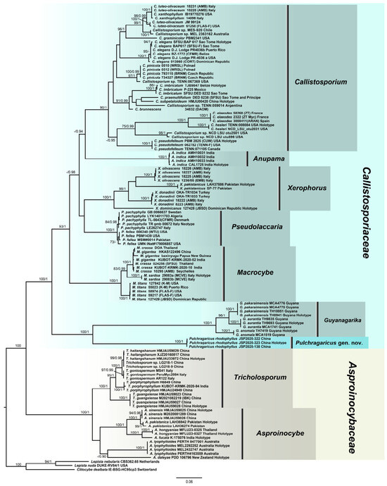
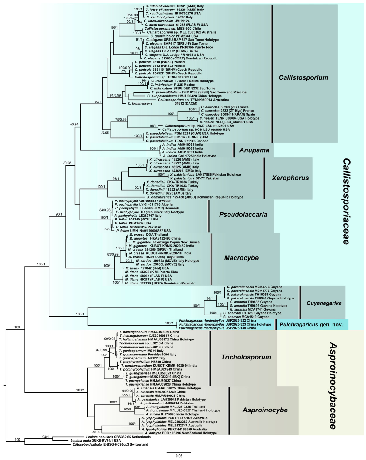
No topological inconsistencies were detected either among the single-gene trees (ITS, LSU, rpb2, and tef1-α) and the concatenated multigene datasets (ITS-LSU and ITS-LSU-rpb2-tef1-α), or between the ML and BI analyses for concatenated multigene datasets (see detailed in Figure S1A–D). Consequently, only the phylogenetic tree inferred from the ML analysis is presented, with statistical support values from both ML (bootstrap support, BS) and BI (posterior probabilities, PP) displayed on the respective nodes (Figure 1). Given the limited availability of rpb2 and tef1-α sequences for certain reference taxa, a combined tree based solely on the ITS and LSU dataset is also provided in Figure S1E.
Figure 1.
Phylogenetic relationships among representative genera in Callistosporiaceae and Asproinocybaceae inferred from a multigene dataset (ITS-LSU-rpb2-tef1-α) using both ML and BI. Only the ML tree is shown. Support values are indicated on branches as ML bootstrap (BS)/Bayesian posterior probability (PP); BS > 70% and PP > 0.90 are indicated on branches. Sequences derived from type specimens (holotype or neotype) are labeled accordingly. The phylogenetic tree was rooted with Lepista nebularis, L. nuda, and Clitocybe dealbata. A new genus and species are highlighted in cyan. The proportion of missing data for each gene partition is summarized in Table S1.
For the four-gene concatenated matrix, a total of 118 terminal taxa were assembled, comprising 106 sequences for ITS, 92 for LSU, 37 for rpb2, and 25 for tef1-α. The final aligned dataset comprised 4238 sites (including gaps), distributed as follows: 1018 bp for ITS, 1484 bp for LSU, 1056 bp for rpb2, and 680 bp for tef1-α.
Phylogenetic analyses (Figure 1) placed the newly discovered monophyletic lineage within the Callistosporiaceae with maximum statistical support (BS = 100%, PP = 1.00), positioning it as sister to the genus Guyanagarika. These results robustly confirm that our samples represent a novel genus and species within the family in the phylogenetic tree.
For the four-gene dataset, intergeneric distances were calculated using the uncorrected p-distance (Table 2). The divergence between Pulchragaricus and its phylogenetically closest relative, Guyanagarika, is 0.186 (18.6% nucleotide difference). This value falls within the range of intergeneric distances among well-established genera in Callistosporiaceae (min: Anupama vs. Callistosporium = 0.111; max: Guyanagarika vs. Macrocybe = 0.206). Furthermore, even the smallest distance between Pulchragaricus and any other genus in the dataset (Pulchragaricus vs. Anupama = 0.156) exceeds several accepted intergeneric distances, such as those between Callistosporium and Macrocybe (0.145) or Callistosporium and Pseudolaccaria (0.122). This indicates that the molecular divergence of Pulchragaricus is consistent with, or in some comparisons greater than, typical generic separation within these families. It should be noted that these p-distances were calculated from a concatenated alignment with a substantial proportion of missing data (e.g., rpb2 and tef1-α), and are therefore considered as additional evidence. Although Pulchragaricus is currently monotypic and intrageneric distances could not be calculated, the substantial genetic gap separating it from its sister lineage, together with its phylogenetic distinctness and the morphological characteristics discussed below, supports its recognition at the generic rank.
Table 2.
Intergeneric uncorrected p-distances among genera of Callistosporiaceae based on the four-gene dataset.
3.2. Taxonomy
Pulchragaricus S.P. Jian gen. nov.
MycoBank MB863543
Type species: Pulchragaricus rhodophyllus S.P. Jian sp. nov.
Etymology: Pulchragaricus is derived from the Latin adjective stem pulchr- (from pulcher, beautiful) and the generic name Agaricus, referring to the beautiful basidiomata that resemble species of Agaricus.
Diagnosis: Pulchragaricus is distinguished from the phylogenetically closest genera Guyanagarika and Macrocybe by the following combination of characteristics: tricholomatoid and medium-sized basidiomata, a tomentose and yellowish brown pileus with an involute margin, crowded, pink to pinkish lamellae, subclavate cheilocystidia, and broadly ellipsoid to ellipsoid basidiospores.
Description: Basidiomata tricholomatoid. Pileus—hemispherical to convex; surface—tomentose, yellowish brown; margin—involute; context—white. Lamellae—crowded, adnate, pink to pinkish. Stipe—central and solid; basal mycelium—white. Basidiospores—broadly ellipsoid to ellipsoid, smooth, inamyloid. Cheilocystidia—rare and subclavate. Pleurocystidia—absent. Clamp connections present in all parts.
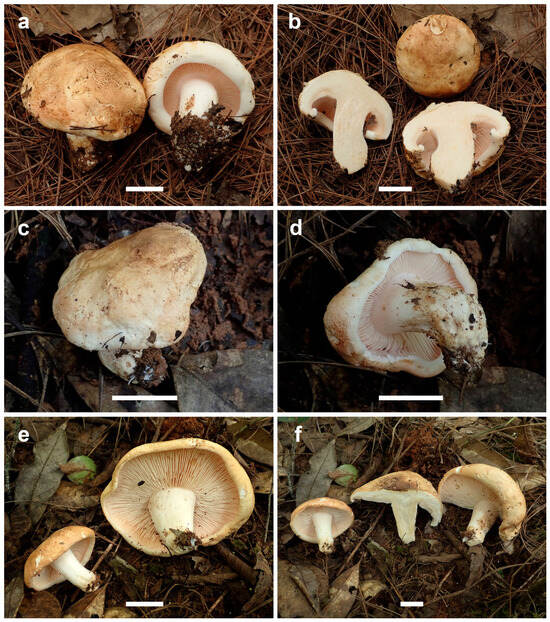
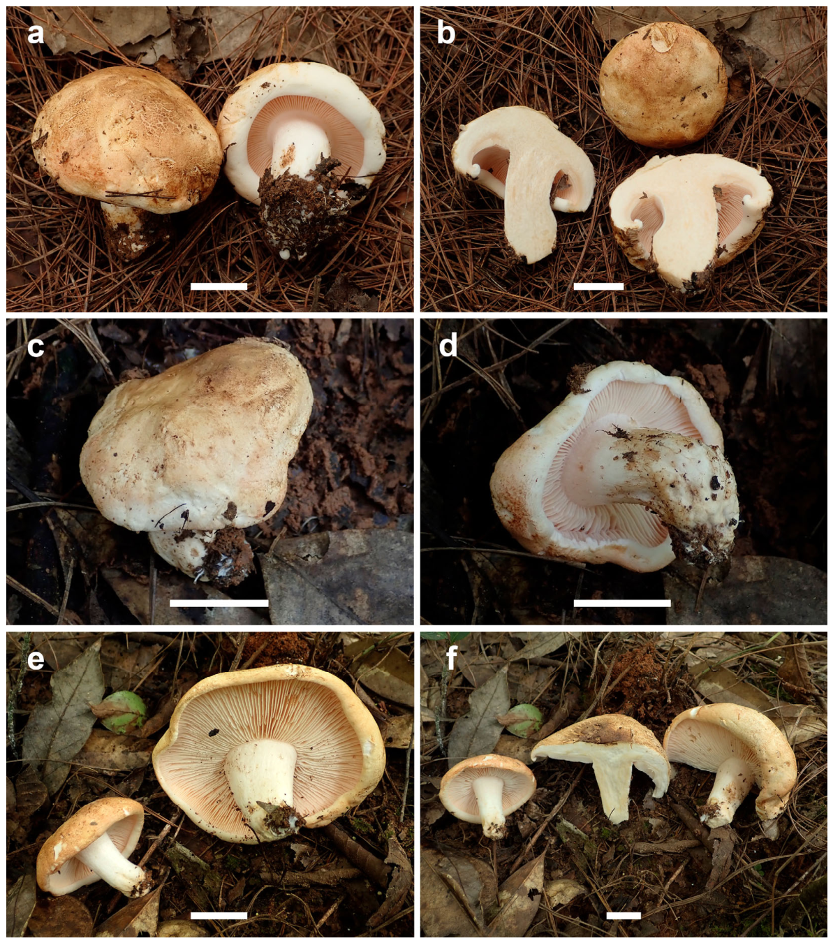
Figure 2.
Fresh basidiomata of Pulchragaricus rhodophyllus. Colors of basidiomata are as recorded in the field (Pu’er City, China) under natural light conditions. KUN-HKAS 154740: (a) pileus, lamellae, and stipe; (b) context. KUN-HKAS 154741: (c) pileus; (d) lamellae and stipe. KUN-HKAS 154742 (holotype): (e) pileus, lamellae, and stipe. (f) Context. Scale bars = 2 cm.
Figure 3.
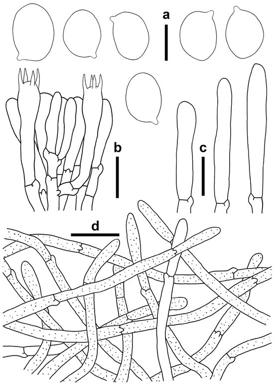
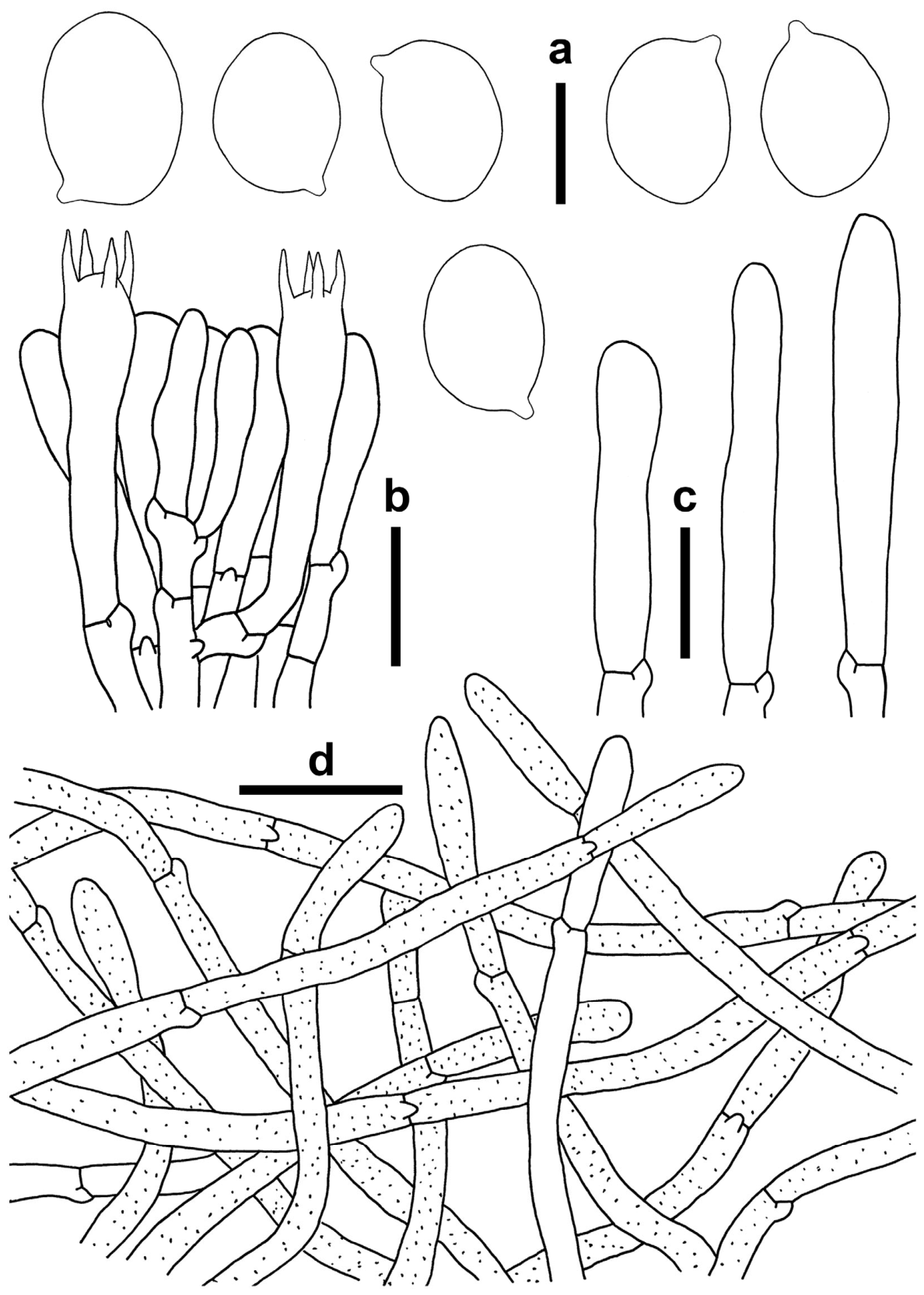
Microscopic structures of Pulchragaricus rhodophyllus (KUN-HKAS 154742, holotype). (a) Basidiospores. (b) Hymenium and subhymenium. (c) Cheilocystidia. (d) Pileipellis (suprapellis). All sections mounted in 5% KOH. Scale bars: 5 μm (a); 10 μm (b,c); 20 μm (d).
MycoBank MB863544
Holotype: China, Yunnan Province, Pu’er City, Simao District, 17 km along Laosilan Road away from the city, 100°49′12.85″ E, 22°45′27.63″ N (datum WGS84, coordinate uncertainty: ca. 50 m), alt. 1254.3 m, scattered on soil, in the subtropical mixed forest (Pinus kesiya var. langbianensis and Fagaceae), 16 September 2025, collected by S.P. Jian & X.J. Xu, JSP2025-323 (KUN-HKAS 154742). GenBank: ITS = PZ267112; LSU = PZ229067; rpb2 = PZ233666; tef1-α = PZ233669.
Etymology: “rhodo-” (rose) and “-phyllus” (leaf, here lamellae), referring to the pink-colored lamellae when young.
Diagnosis: Pulchragaricus rhodophyllus is distinguished from morphologically similar species of Callistosporiaceae by the following combination of characteristics: tricholomatoid basidiomata, yellowish brown pileus with involute margin, pink to pinkish lamellae, broadly ellipsoid basidiospores, sparse and subclavate cheilocystidia, and plentiful clamp connections.
Description: Basidiomata—medium-sized, tricholomatoid. Pileus—5.3–8.2 cm in diam., hemispherical to subhemispherical when young, then convex; surface—soft, tomentose, sometimes with finely net-like cracks at the center, brownish (#b7814b) to brown (#754e34) near the center, yellowish brown (#ecd19c) to faint yellow (#e5c7a1) towards the margin, sometimes uniformly pale brown (#ceb997); margin—persistently involute; context—1.5–2.1 cm thick at the center, white with yellowish (#e0d1b2) to dirty white (#dfdec1), unchanging in color when injured. Lamellae—crowded, emarginate to adnate, pink (#c78b65) when young, then pinkish (#c4af8b) to pale pink (#d5ae90), darkening after drying, edge entire and concolorous or somewhat paler, lamellulae numerous, unchanging in color when injured. Stipe—3.7–5.0 × 1.2–1.9 cm, central, tapering downwards, and solid; surface—glabrous, white (#f5f8ed) to whitish (#eff0df); context—concolorous with pileus context; basal mycelium—white (#adb6b2). Its odor is fragrant, and its taste is mild.
Basidiospores: [65/3/2] 6.5–8 (8.5) × 4.5–6 (6.5) μm, Lm × Wm = 7.03 (±0.42) × 5.16 (±0.34) μm, Q = 1.20–1.60, Qm = 1.37 ± 0.09, hyaline in KOH, broadly ellipsoid to ellipsoid in side and face view, smooth, thin-walled, inamyloid. Basidia: 35–41 × 7–10 μm, clavate, four-spored; sterigmata 4–8 μm long. Cheilocystidia: sparse, 45–60 × 7–9 μm, cylindrical to subclavate, thin-walled, and fragile. Pleurocystidia: absent. Hymenophoral trama: more or less bilateral (divergent), made of cylindrical, parallel, smooth hyphae 4–8 μm wide, with yellow to yellowish-brown pigments; thromboplera (oleiferous hyphae sensu Clémençon [28]): abundant, luminous yellow. Pileipellis: usually two-layered. Suprapellis: about 90–160 μm thick, consisting of loose, erect, smooth, thin-walled, interwoven or irregularly arranged hyphae with yellowish-brown pigments, 4–9 μm wide. Subpellis—made up of subregular to regular, compactly arranged, thin-walled, hyaline, smooth, and cylindrical hyphae 5–12 μm wide, with abundant luminous yellow thromboplera; pileal trama—interwoven or irregular, composed of hyaline, thin-walled, cylindrical hyphae with a diameter of 5–10 (17) μm, occasionally distributed with white to yellowish, reflective thromboplera. Stipitipellis is a cutis composed of regular, compactly arranged, thin-walled, and hyaline hyphae with a diameter of (3.5) 4–13 (15) μm; stipe trama composed of regular, compactly arranged, thin-walled, and yellowish-brown hyphae 5–16 μm, with luminous yellow thromboplera. Caulocystidia—absent. Clamp connections present in all parts.
Ecology and distribution: Solitary or scattered on soil under the subtropical mixed forests (Pinaceae & Fagaceae). Known from Yunnan Province, China, fruiting from July to September.
Additional specimens examined: China, Yunnan Province, Pu’er City, Simao District, 17 km along Laosilan Road away from the city, 100°49′15.18″ E, 22°45′27.85″ N (datum WGS84, coordinate uncertainty: ca. 50 m), alt. 1303.5 m, scattered on soil, in the subtropical pine forest (dominated by Pinus kesiya var. langbianensis), 15 July 2025, collected by S.P. Jian & X.J. Xu, JSP2025-138 (KUN-HKAS 154740); ibid., 100°49′13.22″ E, 22°45′28.00″ N (datum WGS84, coordinate uncertainty: ca. 50 m), alt. 1254.6 m, scattered on soil, in the subtropical mixed forest (Pinus kesiya var. langbianensis and Fagaceae), 16 September 2025, collected by S.P. Jian & X.J. Xu, JSP2025-322 (KUN-HKAS 154741).
Notes: Pulchragaricus rhodophyllus phylogenetically belongs to Callistosporiaceae, but it could be easily distinguished from species of other genera in this family (see Discussion for details). In general, P. rhodophyllus is morphologically more similar to Agaricus fissuratus F.H. Møller, Guyanagarika pakaraimensis Sánchez-García et al., Lepista panaeolus (Fr.) P. Karst., Leucopaxillus alboalutaceus (F.H. Møller & Jul. Schäff.) F.H. Møller, and Pseudoclitopilus rhodoleucus (Sacc.) Vizzini & Contu (synonym: Leucopaxillus rhodoleucus (Sacc.) Kühner). Agaricus fissuratus could be confused with Pulchragaricus rhodophyllus because both have a yellow pileus and pink lamellae. However, A. fissuratus differs from P. rhodophyllus by its radially cracked pileus, free lamellae, membranous ring, larger basidiospores, and spherical to broadly clavate cheilocystidia (7–45 × 5–27 μm) [29,30]. Guyanagarika pakaraimensis is characterized by tricholomatoid basidiomata and a solid, downwards-tapering stipe, which could be confused with P. rhodophyllus. Nevertheless, the orange pileus, pale yellow lamellae, longer stipe (5.5–10 cm long), and absence of cheilocystidia distinguish G. pakaraimensis from P. rhodophyllus [3].
Lepista panaeolus is another species similar to P. rhodophyllus in basidiomata size, with crowded pinkish-gray lamellae, and a robust stipe. However, the plano-convex to applanate pileus with brown marginal droplets, brownish lamellae, and warty basidiospores distinguishes L. panaeolus from P. rhodophyllus [31,32]. Leucopaxillus alboalutaceus is notable for its bitter taste, almost white pileus, and amyloid basidiospores with warty ornamentation, thus differing from P. rhodophyllus [33,34]. Lastly, Pseudoclitopilus rhodoleucus is a rare species distributed across Eurasia. It could be confused with P. rhodophyllus due to its pink to pinkish lamellae, robust stipe, and clamp connections. However, Pseudoclitopilus rhodoleucus has a white pileus, amyloid warty basidiospores, and lacks cheilocystidia [34,35,36].
4. Discussion
In the present four-locus dataset, only 37 of the 118 included taxa had sequences for rpb2, and 25 had sequences for tef1-α, resulting in a substantial proportion of missing data. It has been suggested that extensive missing data can sometimes produce inflated support values or misleading relationships [37]. However, several lines of evidence indicate that the missing sequences do not compromise the support for the new genus. Firstly, the clade corresponding to the new genus received maximum support (BS = 100%, PP = 1.0) in the four-locus dataset (Figure 1) and was consistent (BS = 100%) across four single-gene analyses (Figure S1A–D). Secondly, the new genus remained monophyletic with equally strong support (BS = 100%, PP = 1.0) when performing phylogenetic analysis using only a two-locus dataset (ITS-LSU), excluding the two most incomplete genes (rpb2 and tef1-α) (Figure S1E). Thirdly, the missing sequences are not concentrated in any particular genus or lineage, but are instead scattered across the phylogeny (Table S1). Such scattered distribution of missing data has a limited impact on phylogenetic accuracy when the overall number of characters is large [38]. Taken together, the molecular delineation of the new genus is robust and is further corroborated by the morphological analysis, indicating that the phylogenetic result for this group is not an artifact of sparse gene sampling.
Species of Callistosporiaceae exhibit diverse colors, ranging from white, yellow, and brown to purple [1]. The trophic habits of species in this family are predominantly saprotrophic (on soil or rotten wood), with ectomycorrhizal symbiosis confirmed only in Guyanagarika. Numerous species of this family have been reported from Europe and the Americas, whereas species from China are rarely documented. In this study, we describe a new genus and species, P. rhodophyllus, which is a putatively ectomycorrhizal fungus based only on its occurrence in forests dominated by Pinaceae and Fagaceae. Direct anatomical evidence, such as Hartig net and mantle structures on host root tips, has yet to be obtained. Future studies employing root-tip sectioning or stable isotope tracing (e.g., 13C or 15N labeling) would help confirm the ectomycorrhizal status of this species and provide insights into the evolution of symbioses within Callistosporiaceae.
In the phylogenetic tree, Pulchragaricus is closely related to Asproinocybe, Callistosporium, Guyanagarika, and Macrocybe. However, it can be distinguished from all of them by a unique combination of characteristics: tricholomatoid, medium-sized basidiomata with a soft, yellowish-brown pileus and pinkish lamellae, subclavate cheilocystidia, and ellipsoid basidiospores. No single allied genus shares this entire suite of traits, which represents a clear morphological discontinuity supporting its recognition at the generic rank. Firstly, Asproinocybe was characterized by its pink to violet lamellae that turn reddish when injured, and tuberculate to stellate basidiospores, thus differing from Pulchragaricus [12,39,40]. Callistosporium has collybioid to pleurotoid habits and a smaller pileus (usually < 5 cm in diam.) that is yellow, brown, or brick red. It is lignicolous on rotting wood or wood debris, which significantly differs from Pulchragaricus [1,6,41]. Previously, Guyanagarika has been reported only from Guyana and is characterized by its tricholomatoid basidiomata, orange to dark orange pileus, and solid stipe. It differs from Pulchragaricus in having a pileus with an incurved to crenulate margin, pale yellow to orange lamellae, and lacking cheilocystidia [3]. Macrocybe is usually pantropically distributed and has a tricholomatoid habit. However, its larger basidiomata (>10 cm when mature), white, cream, or grayish pileus, and pale lamellae differ from those of Pulchragaricus [1,7].
Morphologically, Pulchragaricus could be confused with Clitocybe (Fr.) Staude, Leucopaxillus, Pseudoclitopilus Vizzini & Contu, and Tricholoma (Fr.) Staude, but is readily distinguished by the combination of a yellowish brown pileus, pink to pinkish lamellae that are emarginate to adnate, and inamyloid, smooth basidiospores. Specifically, Clitocybe differs in its white to yellowish pileus, decurrent, white to yellowish lamellae [42]. Leucopaxillus has a white pileus, white, decurrent lamellae, and amyloid, warty basidiospores [34,43], while Pseudoclitopilus, though also possessing pinkish lamellae, shares with Leucopaxillus the decurrent lamellae and amyloid, warty basidiospores, thus clearly distinct from Pulchragaricus [35,36,44]. The last genus, Tricholoma, is distinguished by its colorful pileus (e.g., white, grayish, brown, or yellow), adnexed to emarginate lamellae that are white, yellow, or gray [43,45,46], differing from Pulchragaricus. A full comparison of these genera with their diagnostic characteristics is provided in Table 3.
Table 3.
Comparison of morphological characteristics among Pulchragaricus and morphologically similar or phylogenetically related genera.
One limitation of the present study should be noted: specimen KUN-HKAS 154740 was immature and was therefore excluded from basidiospore size statistics and other microscopic character measurements; consequently, the description of mature basidiospores is based on the remaining collections. Additional fully mature collections would help confirm the range of intraspecific variability (e.g., the coloration of the basidiomata and variability of cheilocystidia) reported here.
5. Conclusions
In summary, Pulchragaricus can be distinguished from closely related genera by both molecular phylogenetic evidence and a combination of morphological traits. The key characteristics of this new genus are as follows: tricholomatoid basidiomata, a tomentose and yellowish-brown pileus with an involute margin, crowded and pinkish lamellae, subclavate cheilocystidia, broadly ellipsoid to ellipsoid, inamyloid basidiospores, and clamp connections present throughout. It should be noted, however, that the current dataset is built upon a limited number of collections, that several included taxa are represented by incomplete loci, and that the ecology of Pulchragaricus remains unconfirmed. Although only a single species is described to date, continued fieldwork and more comprehensive integrative analyses are likely to reveal additional species diversity. An expanded taxon and characteristic sampling will also be essential to more rigorously assess the phylogenetic significance of Pulchragaricus within Callistosporiaceae.
Supplementary Materials
The following supporting information can be downloaded at https://www.mdpi.com/article/10.3390/life16060899/s1: Figure S1: Phylogenetic relationships among representative genera in Callistosporiaceae and Asproinocybaceae inferred from each individual gene (ITS, LSU, rpb2, and tef1-α, Figure S1A–D) using ML, and a multigene dataset (ITS-LSU, Figure S1E) using both ML and BI. Sequences derived from type specimens (holotype or neotype) are labeled accordingly; Figure S2: Sample location map (Yunnan, China). The sampling locations of basidiomata are indicated by the blue triangle; Table S1: Collection information of voucher specimens and GenBank accession numbers for sequences used in phylogenetic analyses. N or H in parentheses means the neotype or holotype specimen. Sequences newly generated in this study are shown in bold. Supplementary File S1: The original data of phylogenetic analysis in this research.
Author Contributions
Conceptualization, C.Z. and S.J.; methodology, S.J.; software, S.J. and T.Y.; validation, S.J.; formal analysis, Y.F.; investigation, J.L. and X.X.; resources, F.G., T.Y. and X.X.; data curation, S.J.; writing—original draft preparation, S.J.; writing—review and editing, C.Z.; visualization, W.A.; supervision, C.Z.; project administration, C.Z.; funding acquisition, S.J., X.X. and C.Z. All authors have read and agreed to the published version of the manuscript.
Funding
This research was funded by the Doctoral Launch Program in 2025, Yunnan Institute of Tropical Crops (No. ZY11), the Funds of Sci-Tech Innovation System Construction for Tropical Crops of Yunnan Province (No. 727-18), Xishuangbanna Prefecture Sci-Tech Innovation Program (No. 2025kjcx004), and Yunnan Fundamental Research Projects (No. 202401BD070001-125 and No. 202501BD070001-110).
Institutional Review Board Statement
Not applicable.
Informed Consent Statement
Not applicable.
Data Availability Statement
The sequence data generated in this study can be obtained from NCBI GenBank (http://www.ncbi.nlm.nih.gov/, accessed on 24 February 2026). The data included in this study can be made available by contacting the authors.
Acknowledgments
The authors are very grateful to Latin experts Shaun Pennycook (Manaaki Whenua-Landcare Research), Konstanze Bensch (Westerdijk Fungal Biodiversity Institute), Yanchun Li, and Xianghua Wang (Kunming Institute of Botany, Chinese Academy of Sciences) for their consultation regarding the code of nomenclature for fungi. The authors are also grateful to Xuan Chen (Kunming Institute of Botany, Chinese Academy of Sciences), Geng-Shen Wang (Southwest Forestry University), and Ya-Jun Hou for providing some constructive information and suggestions. The authors thank the herbaria KUN-HKAS for providing storage of materials.
Conflicts of Interest
The authors declare no conflicts of interest.
References
- Vizzini, A.; Consiglio, G.; Marchetti, M.; Alvarado, P. Insights into the Tricholomatineae (Agaricales, Agaricomycetes): A new arrangement of Biannulariaceae and Callistosporium, Callistosporiaceae fam. nov., Xerophorus stat. nov., and Pleurocollybia incorporated into Callistosporium. Fungal Divers. 2020, 101, 211–259. [Google Scholar] [CrossRef] [Scilit]
- Wang, G.-S.; Cai, Q.; Hao, Y.-J.; Bau, T.; Chen, Z.-H.; Li, M.-X.; David, N.; Kraisitudomsook, N.; Yang, Z.-L. Phylogenetic and taxonomic updates of Agaricales, with an emphasis on Tricholomopsis. Mycology 2023, 15, 180–209. [Google Scholar] [CrossRef] [Scilit]
- Sánchez-García, M.; Henkel, T.W.; Aime, M.C.; Smith, M.E.; Matheny, P.B. Guyanagarika, a new ectomycorrhizal genus of Agaricales from the Neotropics. Fungal Biol. 2016, 120, 1540–1553. [Google Scholar] [CrossRef] [Scilit]
- Raj, K.N.A.; Latha, K.P.D.; Leelavathy, K.M.; Manimohan, P. Anupama: A new genus of Biannulariaceae (Agaricales) from tropical India. Mycol. Prog. 2019, 18, 659–669. [Google Scholar] [CrossRef] [Scilit]
- Xu, J.; Yu, X.; Idrees, M.; Zhang, C.; Li, Y. Morphology and phylogeny reveal Callistosporium subpetaloideum sp. nov. (Callistosporiaceae) from China. Curr. Microbiol. 2021, 78, 2122–2129. [Google Scholar] [CrossRef] [Scilit]
- Singer, R. New genera of fungi. Mycologia 1944, 36, 358–368. [Google Scholar] [CrossRef] [Scilit]
- Pegler, D.N.; Lodge, D.J.; Nakasone, K.K. The pantropical genus Macrocybe gen. nov. Mycologia 1998, 90, 494–504. [Google Scholar] [CrossRef] [Scilit]
- Kantharaja, R.; Krishnappa, M. Macrocybe sardoa (Callistosporiaceae, Agaricales) a new record from India. Mycopathol. Res. 2021, 59, 279–283. [Google Scholar]
- Lavorato, C.; Vizzini, A.; Ge, Z.-W.; Contu, M. Redescription of Clitocybe umbrinopurpurascens (Basidiomycota, Agaricales) and revision of Neohygrophorus and Pseudoomphalina. Phytotaxa 2015, 219, 43–57. [Google Scholar] [CrossRef] [Scilit]
- Noordeloos, M.E. Entolomataceae. In Flora Agaricina Neerlandica; Bas, C., Kuyper, T.W., Noordeloos, M.E., Vellinga, E.C., Eds.; A.A. Balkema: Rotterdam, The Netherlands, 1988; Volume 1, p. 182. [Google Scholar]
- Yang, S.-L.; Nie, J.; Ye, Y.-S.; Mo, M.-H.; Jiang, X.-Y.; Huang, Y.-H.; Zheng, J.-R.; Liu, Y.; Yuan, J.-Y. Research status and prospect forecast of Tricholoma giganteum. J. Anhui Agric. Sci. 2016, 44, 9–12. [Google Scholar] [CrossRef]
- Mou, G.F.; Bau, T. Asproinocybaceae fam. nov. (Agaricales, Agaricomycetes) for accommodating the genera Asproinocybe and Tricholosporum, and description of Asproinocybe sinensis and Tricholosporum guangxiense sp. nov. J. Fungi 2021, 7, 1086. [Google Scholar] [CrossRef] [Scilit]
- Christensen, M.; Miller, S.L.; Tuthill, D. Colour standards: A review and evaluation in relation to Penicillium taxonomy. Mycol. Res. 1994, 98, 635–644. [Google Scholar] [CrossRef] [Scilit]
- Bas, C. Morphology and subdivision of Amanita and a monograph of its section Lepidella. Persoonia 1969, 5, 285–579. [Google Scholar]
- Doyle, J.J.; Doyle, J.L. A rapid DNA isolation procedure for small quantities of fresh leaf tissue. Phytochem. Bull. 1987, 19, 11–15. [Google Scholar]
- White, T.J.; Bruns, T.; Lee, S.; Taylor, J. Amplification and Direct Sequencing of Fungal Ribosomal RNA Genes for Phylogenetics. In PCR Protocols: A Guide to Methods and Applications; Innis, M.A., Gelfand, D.H., Sninsky, J.J., White, T.J., Eds.; Academic Press: San Diego, CA, USA, 1990; pp. 315–322. [Google Scholar]
- Matheny, P.B.; Curtis, J.M.; Hofstetter, V.; Aime, M.C.; Moncalvo, J.M.; Ge, Z.W.; Yang, Z.L.; Slot, J.C.; Ammirati, J.F.; Baroni, T.J. Major clades of Agaricales: A multilocus phylogenetic overview. Mycologia 2006, 98, 982–995. [Google Scholar] [CrossRef]
- Jian, S.; Chen, X.; Yang, T.; Xu, X.; Gao, F.; Fang, Y.; Liu, J.; Zhang, C. Biotrophic and saprophytic fungi from the Rhodocybe-Clitopilus clade (Entolomataceae): Two new species and one new record in subtropical China. MycoKeys 2025, 116, 227–254. [Google Scholar] [CrossRef] [Scilit]
- Matheny, P.B. Improving phylogenetic inference of mushrooms with RPB1 and RPB2 nucleotide sequences (Inocybe; Agaricales). Mol. Phylogen. Evol. 2005, 35, 1–20. [Google Scholar] [CrossRef] [Scilit]
- Matheny, P.B.; Wang, Z.; Binder, M.; Curtis, J.M.; Lim, Y.W.; Nilsson, R.H.; Hughes, K.W.; Hofstetter, V.; Ammirati, J.F.; Schoch, C.L.; et al. Contributions of RPB2 and TEF1 to the phylogeny of mushrooms and allies (Basidiomycota, Fungi). Mol. Phylogen. Evol. 2007, 43, 430–451. [Google Scholar] [CrossRef] [Scilit] [PubMed]
- Zhao, D.; Ye, T.; Gao, F.; Jakovlić, I.; La, Q.; Tong, Y.; Liu, X.; Song, R.; Liu, F.; Lian, Z.M.; et al. PhyloSuite v2: The development of an all-in-one, efficient and visualization-oriented suite for molecular dating analysis and other advanced features. iMeta 2025, 4, e70095. [Google Scholar] [CrossRef] [Scilit]
- Katoh, K.; Standley, D.M. MAFFT multiple sequence alignment software version 7: Improvements in performance and usability. Mol. Biol. Evol. 2013, 30, 772–780. [Google Scholar] [CrossRef] [Scilit] [PubMed]
- Hall, T.A. BioEdit: A user-friendly biological sequence alignment editor and analysis program for Windows 95/98/NT. Nucleic Acids Symp. Ser. 1999, 41, 95–98. [Google Scholar]
- Kalyaanamoorthy, S.; Minh, B.Q.; Wong, T.K.F.; von Haeseler, A.; Jermiin, L.S. ModelFinder: Fast model selection for accurate phylogenetic estimates. Nat. Methods 2017, 14, 587–589. [Google Scholar] [CrossRef] [Scilit]
- Nguyen, L.-T.; Schmidt, H.A.; von Haeseler, A.; Minh, B.Q. IQ-TREE: A fast and effective stochastic algorithm for estimating maximum-likelihood phylogenies. Mol. Biol. Evol. 2015, 32, 268–274. [Google Scholar] [CrossRef] [Scilit]
- Ronquist, F.; Teslenko, M.; van der Mark, P.; Ayres, D.L.; Darling, A.; Höhna, S.; Larget, B.; Liu, L.; Suchard, M.A.; Huelsenbeck, J.P. MrBayes 3.2: Efficient bayesian phylogenetic inference and model choice across a large model space. Syst. Biol. 2012, 61, 539–542. [Google Scholar] [CrossRef] [Scilit] [PubMed]
- Tamura, K.; Stecher, G.; Kumar, S. MEGA11: Molecular evolutionary genetics analysis version 11. Mol. Biol. Evol. 2021, 38, 3022–3027. [Google Scholar] [CrossRef] [Scilit]
- Clémençon, H. Cytology and Plectology of the Hymenomycetes; Gebr. Borntraeger Verlagsbuchhanddlung: Stuttgart, Germany, 2012; p. 520. [Google Scholar]
- Kerrigan, R.W. Agaricus of North America. Mem. N. Y. Bot. Gard. 2016, 114, 1–574. [Google Scholar]
- Parra, L.A. Agaricus L. Allopsalliota Nauta & Bas, Part 2; Fungi Europaei; Candusso Edizioni s.a.s.: Alassio, Italy, 2013; p. 1168. [Google Scholar]
- Xu, Y.-D.; Jiang, L.; Yang, Z.L.; Liu, J.-W.; Cai, Q.; Xiao, Y.-Q.; Chen, Z.-H.; He, Z.-M. Lepista cremeoinvoluta and L. multiformis (Clitocybaceae, Agaricales), a new species and a newly recorded species from Southwestern China. Mycol. Prog. 2024, 23, 47. [Google Scholar] [CrossRef] [Scilit]
- Noordeloos, M.E.; Kuyper, T.W. Lepista. In Flora Agaricina Neerlandica; Bas, C., Kuyper, T.W., Noordeloos, M.E., Vellinga, E.C., Eds.; A.A. Balkema: Rotterdam, The Netherlands, 1995; Volume 3, p. 183. [Google Scholar]
- Ercole, E.; Vizzini, A. Leucopaxillus agrippinae, una nuova specie raccolta in Sicilia. Riv. Di Micol. 2012, 2, 99–114. [Google Scholar]
- Læssøe, T.; Petersen, J.H. Fungi of Temperate Europe; Princeton University Press: Princeton, NJ, USA, 2019; p. 813. [Google Scholar]
- Singer, R.; Smith, A.H. Additional notes on the genus Leucopaxillus. Mycologia 1947, 39, 725–736. [Google Scholar] [CrossRef] [Scilit]
- Vizzini, A.; Ercole, E.; Contu, M. A contribution to the ITS-LSU phylogeny of the genus Leucopaxillus (/tricholomatoid clade, Agaricales), with three new genera and notes on Porpoloma. Mycosphere 2012, 3, 79–90. [Google Scholar] [CrossRef] [Scilit]
- Lemmon, A.R.; Brown, J.M.; Stanger-Hall, K.; Lemmon, E.M. The effect of ambiguous data on phylogenetic estimates obtained by maximum likelihood and bayesian inference. Syst. Biol. 2009, 58, 130–145. [Google Scholar] [CrossRef] [Scilit] [PubMed]
- Wiens, J.J.; Morrill, M.C. Missing data in phylogenetic analysis: Reconciling results from simulations and empirical data. Syst. Biol. 2011, 60, 719–731. [Google Scholar] [CrossRef] [Scilit] [PubMed]
- Lv, T.; Wang, S.; Wu, X.; Chen, D.; Ao, C.; Luo, H.; Li, E.; Tang, S.; Li, S. Asproinocybe hongyaniae sp. nov. (Agaricales, Basidiomycota) in Thailand. Phytotaxa 2024, 640, 16–26. [Google Scholar] [CrossRef] [Scilit]
- Ali, M.; Niazi, A.R.; Ashraf, U.; Anam, A.; Afshan, N.u.S.; Aime, M.C.; Khalid, A.N. Asproinocybe pakistanica sp. nov. and A. daleyae: First representatives of the genus Asproinocybe from Pakistan. Nord. J. Bot. 2026, 2026, e04698. [Google Scholar] [CrossRef] [Scilit]
- Baroni, T.J.; Bocsusis, N.; Lodge, D.J.; Lindner, D. A new species of Pleurocollybia (Tricholomataceae; Agaricales; Basidiomycetes) from Belize. Mycotaxon 2008, 103, 353–363. [Google Scholar] [CrossRef] [Scilit]
- He, Z.-M.; Chen, Z.-H.; Bau, T.; Wang, G.-S.; Yang, Z.L. Systematic arrangement within the family Clitocybaceae (Tricholomatineae, Agaricales): Phylogenetic and phylogenomic evidence, morphological data and muscarine-producing innovation. Fungal Divers. 2023, 123, 1–47. [Google Scholar] [CrossRef] [Scilit]
- Singer, R. The Agaricales in Modern Taxonomy, 4th ed.; Koeltz Scientific Books: Koenigstein, Germany, 1986; p. 981. [Google Scholar]
- Pegler, D.N.; Young, T.W.K. Basidiospore form in the British Leucopaxilleae. Kew Bull. 1973, 28, 365–379. [Google Scholar] [CrossRef] [Scilit]
- Noordeloos, M.E. Tricholoma. In Flora Agaricina Neerlandica; Bas, C., Kuyper, T.W., Noordeloos, M.E., Vellinga, E.C., Eds.; A.A. Balkema: Rotterdam, The Netherlands, 1999; Volume 4, p. 189. [Google Scholar]
- Ding, X.X.; Xu, X.; Cui, Y.Y.; Kost, G.; Wang, P.M.; Yang, Z.L. A fifty-locus phylogenetic analysis provides deep insights into the phylogeny of Tricholoma (Tricholomataceae, Agaricales). Persoonia 2023, 50, 1–26. [Google Scholar] [CrossRef] [Scilit]

Disclaimer/Publisher’s Note: The statements, opinions and data contained in all publications are solely those of the individual author(s) and contributor(s) and not of MDPI and/or the editor(s). MDPI and/or the editor(s) disclaim responsibility for any injury to people or property resulting from any ideas, methods, instructions or products referred to in the content. |
© 2026 by the authors. Licensee MDPI, Basel, Switzerland. This article is an open access article distributed under the terms and conditions of the Creative Commons Attribution (CC BY) license.