Update on Repetitive Transcranial Magnetic Stimulation in Post-Stroke Cognitive Rehabilitation: A Systematic Review of Randomized Clinical Trials
 , , ,
, , ,  ,
,  , , , , ,
  and
and 
Abstract
1. Introduction
2. Materials and Methods
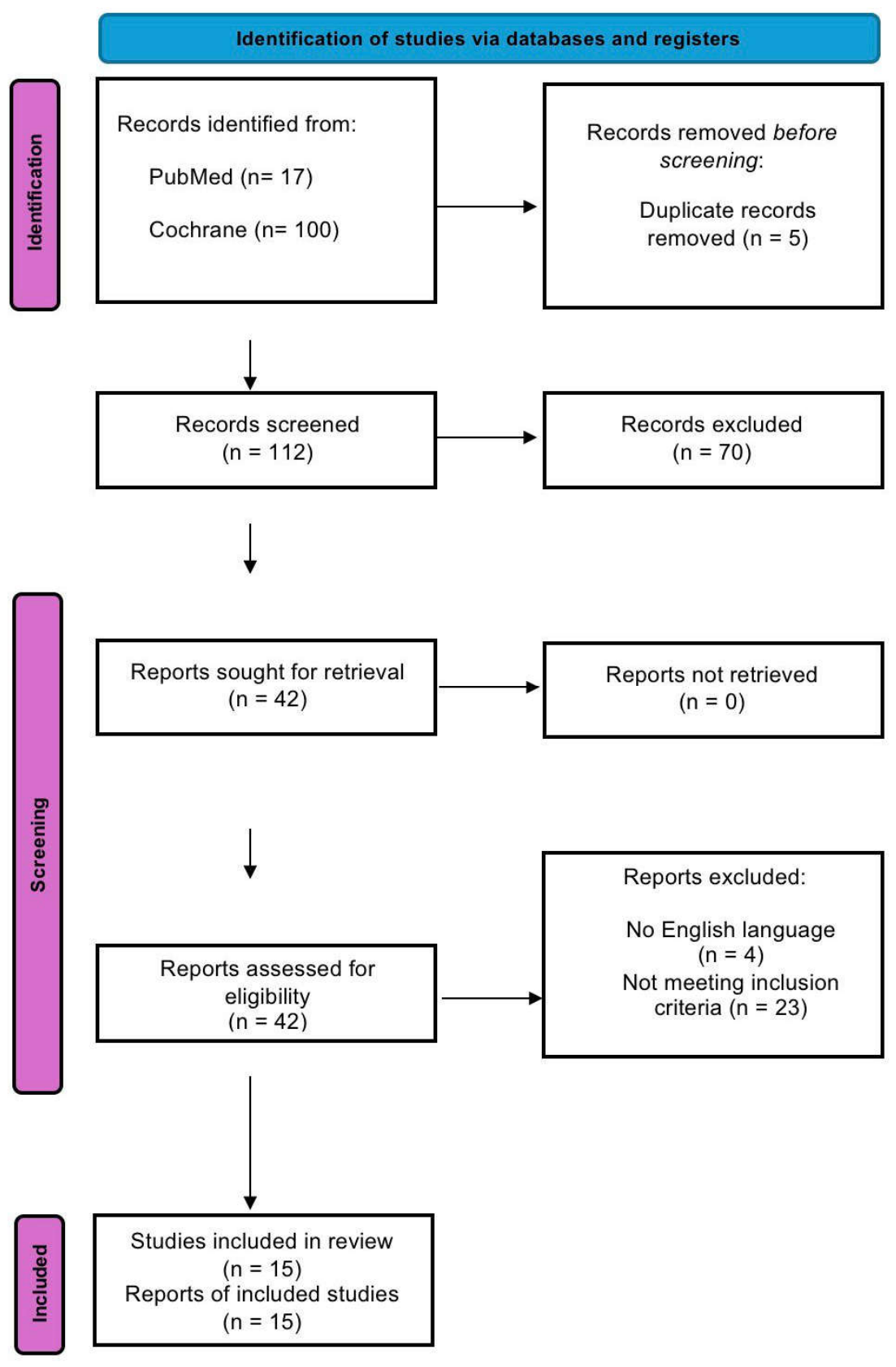
2.1. Inclusion/Exclusion Criteria and Selection Strategy
- -
- Population: patients with stroke (ischaemic or haemorrhagic), regardless of phase (acute, subacute, or chronic).
- -
- Intervention: rTMS, including iTBS protocols, administered using any protocol (frequency, duration, brain target, etc.).
- -
- Comparison: no treatment, placebo (sham TMS) or other forms of standard cognitive rehabilitation.
- -
- Outcome: improvement in cognitive functions, assessed using standardised neuropsychological tests.
2.2. Search Strategy
2.3. Risk of Bias Assessment
3. Results
3.1. Cognitive Domains Involved
3.2. Stimulation Protocols and Comparisons
3.3. Integration with Cognitive Training and Durability of Effects
3.4. Safety and Tolerability
3.5. Risk of Bias
3.6. Heterogeneity of Stimulation Protocols and Outcome Patterns
4. Discussion
4.1. Main Findings
4.2. Summary, Limitations, and Perspectives
5. Conclusions
Supplementary Materials
Author Contributions
Funding
Institutional Review Board Statement
Informed Consent Statement
Data Availability Statement
Conflicts of Interest
References
- Feigin, V.L.; Norrving, B.; Mensah, G.A. Global burden of stroke. Circ. Res. 2017, 120, 439–448. [Google Scholar] [CrossRef] [Scilit] [PubMed]
- Huang, Y.-Y.; Chen, S.-D.; Leng, X.-Y.; Kuo, K.; Wang, Z.-T.; Cui, M.; Tan, L.; Wang, K.; Dong, Q.; Yu, J.-T. Post-stroke cognitive impairment: Epidemiology, risk factors, and management. J. Alzheimers Dis. 2022, 86, 983–999. [Google Scholar] [CrossRef] [Scilit] [PubMed]
- Pendlebury, S.T.; Rothwell, P.M. Incidence and prevalence of dementia associated with transient ischemic attack and stroke: Analysis of the population-based Oxford Vascular Study. Lancet Neurol. 2019, 18, 248–258.3. [Google Scholar] [CrossRef] [Scilit]
- Levine, D.A.; Galecki, A.T.; Langa, K.M.; Unverzagt, F.W.; Kabeto, M.U.; Giordani, B.; Wadley, V.G. Trajectory of cognitive decline after incident stroke. JAMA 2015, 314, 41–51. [Google Scholar] [CrossRef] [Scilit]
- Murray, A.D.; Staff, R.T.; McNeil, C.J.; Salarirad, S.; Ahearn, T.S.; Mustafa, N.; Whalley, L.J. The balance between cognitive reserve and brain imaging biomarkers of cerebrovascular and Alzheimer’s diseases. Brain 2011, 134, 3687–3696. [Google Scholar] [CrossRef] [Scilit]
- Ihle-Hansen, H.; Hagberg, G.; Fure, B.; Thommessen, B.; Fagerland, M.W.; Øksengård, A.R.; Engedal, K.; Selnes, P. Association between total-tau and brain atrophy one year after first-ever stroke. BMC Neurol. 2017, 17, 107. [Google Scholar] [CrossRef] [Scilit]
- Revet, M.; Immerzeel, J.; Voogt, L.; Paulis, W. Patients with neuropsychological disorders short after stroke have worse functional outcome: A systematic review and meta-analysis. Disabil. Rehabil. 2021, 43, 2233–2252. [Google Scholar] [CrossRef] [Scilit]
- Nys, G.M.; van Zandvoort, M.J.; de Kort, P.L.; Jansen, B.P.; de Haan, E.H.; Kappelle, L.J. Cognitive disorders in acute stroke: Prevalence and clinical determinants. Cerebrovasc. Dis. 2007, 23, 408–416. [Google Scholar] [CrossRef] [Scilit]
- Figorilli, M.; Lanza, G.; Congiu, P.; Lecca, R.; Casaglia, E.; Mogavero, M.P.; Puligheddu, M.; Ferri, R. Neurophysiological Aspects of REM Sleep Behavior Disorder (RBD): A Narrative Review. Brain Sci. 2021, 11, 1588. [Google Scholar] [CrossRef] [Scilit] [PubMed]
- Jannati, A.; Oberman, L.M.; Rotenberg, A.; Pascual-Leone, A. Assessing the mechanisms of brain plasticity by transcranial magnetic stimulation. Neuropsychopharmacology 2023, 48, 191–208. [Google Scholar] [CrossRef] [Scilit]
- Lefaucheur, J.P.; Aleman, A.; Baeken, C.; Benninger, D.H.; Brunelin, J.; Di Lazzaro, V.; Filipović, S.R.; Grefkes, C.; Hasan, A.; Hummel, F.C.; et al. Evidence-based guidelines on the therapeutic use of repetitive transcranial magnetic stimulation (rTMS): An update (2014–2018). Clin. Neurophysiol. 2020, 131, 474–528. [Google Scholar] [CrossRef] [Scilit]
- Chou, Y.H.; That, V.T.; Sundman, M. A systematic review and meta-analysis of rTMS effects on cognitive enhancement in mild cognitive impairment and Alzheimer’s disease. Neurobiol. Aging 2020, 86, 1–10. [Google Scholar] [CrossRef] [Scilit]
- Bella, R.; Ferri, R.; Cantone, M.; Pennisi, M.; Lanza, G.; Malaguarnera, G.; Spampinato, C.; Giordano, D.; Raggi, A.; Pennisi, G. Motor cortex excitability in vascular depression. Int. J. Psychophysiol. 2011, 82, 248–253. [Google Scholar] [CrossRef] [Scilit]
- Singh, N.; Saini, M.; Kumar, N.; Srivastava, M.V.P.; Mehndiratta, A. Evidence of neuroplasticity with robotic hand exoskeleton for post-stroke rehabilitation: A randomized controlled trial. J. Neuroeng. Rehabil. 2021, 18, 76. [Google Scholar] [CrossRef] [Scilit] [PubMed]
- Koch, G.; Bonnì, S.; Pellicciari, M.C.; Casula, E.P.; Mancini, M.; Esposito, R.; Ponzo, V.; Picazio, S.; Di Lorenzo, F.; Serra, L.; et al. Transcranial magnetic stimulation of the precuneus enhances memory and neural activity in prodromal Alzheimer’s disease. Neuroimage 2018, 169, 302–311. [Google Scholar] [CrossRef] [Scilit] [PubMed]
- Bella, R.; Ferri, R.; Pennisi, M.; Cantone, M.; Lanza, G.; Malaguarnera, G.; Spampinato, C.; Giordano, D.; Alagona, G.; Pennisi, G. Enhanced motor cortex facilitation in patients with vascular cognitive impairment-no dementia. Neurosci. Lett. 2011, 503, 171–175. [Google Scholar] [CrossRef] [Scilit]
- Jin, X.; Xu, C.Y.; Fei, J.F.; Fang, Y.; Sun, C.H. Alzheimer’s disease with depressive symptoms: Clinical effect of intermittent theta burst stimulation repetitive transcranial magnetic stimulation. World J. Psychiatry 2024, 14, 1216–1223. [Google Scholar] [CrossRef] [Scilit]
- Nicoletti, V.G.; Fisicaro, F.; Aguglia, E.; Bella, R.; Calcagno, D.; Cantone, M.; Concerto, C.; Ferri, R.; Mineo, L.; Pennisi, G.; et al. Challenging the pleiotropic effects of repetitive transcranial magnetic stimulation in geriatric depression: A multimodal case series study. Biomedicines 2023, 11, 958. [Google Scholar] [CrossRef] [Scilit]
- Zhang, L.; Gao, S.; Wang, C.; Li, Y.; Yuan, H.; Cao, L.; Gao, C. Efficacy of repetitive transcranial magnetic stimulation in post stroke cognitive impairment: An overview of systematic reviews. Front. Neurol. 2024, 15, 1378731. [Google Scholar] [CrossRef] [Scilit]
- Chervyakov, A.V.; Chernyavsky, A.Y.; Sinitsyn, D.O.; Piradov, M.A. Possible mechanisms underlying the therapeutic effects of transcranial magnetic stimulation. Front. Hum. Neurosci. 2015, 9, 303. [Google Scholar] [CrossRef] [Scilit]
- Xu, B.; Lin, C.; Wang, Y.; Wang, H.; Liu, Y.; Wang, X. Using dual target rTMS, single target rTMS, or sham rTMS on post stroke cognitive impairment. J. Integr. Neurosci. 2024, 23, 161. [Google Scholar] [CrossRef] [Scilit]
- Liu, Y.; Yin, M.; Luo, J.; Huang, L.; Zhang, S.; Pan, C.; Hu, X. Effects of transcranial magnetic stimulation on the performance of the activities of daily living and attention function after stroke: A randomized controlled trial. Clin. Rehabil. 2020, 34, 1465–1473. [Google Scholar] [CrossRef] [Scilit] [PubMed]
- Yin, M.; Liu, Y.; Zhang, L.; Zheng, H.; Peng, L.; Ai, Y.; Luo, J.; Hu, X. Effects of rTMS treatment on cognitive impairment and resting state brain activity in stroke patients: A randomized clinical trial. Front. Neural Circuits 2020, 14, 563777. [Google Scholar] [CrossRef] [Scilit] [PubMed]
- Liu, Y.; Ai, Y.; Cao, J.; Cheng, Q.; Hu, H.; Luo, J.; Zeng, L.; Zhang, S.; Fang, J.; Huang, L.; et al. High frequency rTMS broadly ameliorates working memory and cognitive symptoms in stroke patients: A randomized controlled trial. Neurorehabil. Neural Repair 2024, 38, 729–741. [Google Scholar] [CrossRef] [Scilit] [PubMed]
- Li, W.; Wen, Q.; Xie, Y.H.; Hu, A.L.; Wu, Q.; Wang, Y.X. Improvement of poststroke cognitive impairment by intermittent theta bursts: A double blind randomized controlled trial. Brain Behav. 2022, 12, e2569. [Google Scholar] [CrossRef] [Scilit]
- Chu, M.; Zhang, Y.; Chen, J.; Chen, W.; Hong, Z.; Zhang, Y.; Yu, H.; Zhang, F.; Ye, X.; Li, J.; et al. Efficacy of intermittent theta burst stimulation and transcranial direct current stimulation in treatment of post stroke cognitive impairment. J. Integr. Neurosci. 2022, 21, 130. [Google Scholar] [CrossRef] [Scilit]
- Hu, A.M.; Huang, C.Y.; He, J.G.; Wu, L. Effect of repetitive transcranial magnetic stimulation combined with transcranial direct current stimulation on post stroke dysmnesia: A preliminary study. Clin. Neurol. Neurosurg. 2023, 231, 107797. [Google Scholar] [CrossRef] [Scilit]
- Li, H.; Ma, J.; Zhang, J.; Shi, W.Y.; Mei, H.N.; Xing, Y. Repetitive transcranial magnetic stimulation (rTMS) modulates thyroid hormones level and cognition in the recovery stage of stroke patients with cognitive dysfunction. Med. Sci. Monit. 2021, 27, e931914-1–e931914-10. [Google Scholar] [CrossRef] [Scilit]
- Song, X.; Fu, J.; Yao, Y.; Shu, Y.; Wang, Z.; Chen, X.; Ma, L.; Shen, F.; Sun, X.; Ma, X.; et al. The impact of high frequency rTMS treatment on brain activity in PSCI patients: A TMS EEG study. Front. Neurol. 2025, 16, 1582437. [Google Scholar] [CrossRef] [Scilit]
- Hu, G.; Zhang, L.; Sun, X.; Wang, L.; Xu, Q.; Li, Q.; Huang, W.; Xiao, Y. Effect of high frequency (5 Hz) rTMS stimulating left DLPFC combined with galantamine on cognitive impairment after ischemic stroke and serum homocysteine and neuron specific enolase. Front. Neurol. 2024, 15, 1345832. [Google Scholar] [CrossRef] [Scilit]
- Tsai, P.Y.; Lin, W.S.; Tsai, K.T.; Kuo, C.Y.; Lin, P.H. High frequency versus theta burst transcranial magnetic stimulation for the treatment of poststroke cognitive impairment in humans. J. Psychiatry Neurosci. 2020, 45, 262–270. [Google Scholar] [CrossRef] [Scilit]
- Li, Y.; Luo, H.; Yu, Q.; Yin, L.; Li, K.; Li, Y.; Fu, J. Cerebral functional manipulation of repetitive transcranial magnetic stimulation in cognitive impairment patients after stroke: An fMRI study. Front. Neurol. 2020, 11, 977. [Google Scholar] [CrossRef] [Scilit]
- Cha, B.; Kim, J.; Kim, J.M.; Choi, J.W.; Choi, J.; Kim, K.; Cha, J.; Kim, M. Therapeutic effect of repetitive transcranial magnetic stimulation for post stroke vascular cognitive impairment: A prospective pilot study. Front. Neurol. 2022, 13, 813597. [Google Scholar] [CrossRef] [Scilit]
- Yingli, B.; Zunke, G.; Wei, C.; Shiyan, W. Cerebral activity manipulation of low frequency repetitive transcranial magnetic stimulation in post stroke patients with cognitive impairment. Front. Neurol. 2022, 13, 951209. [Google Scholar] [CrossRef] [Scilit]
- Li, K.; Mo, D.; Yu, Q.; Feng, R.; Li, Y. Effect of repetitive transcranial magnetic stimulation on post stroke comorbid cognitive impairment and depression: A randomized controlled trial. J. Alzheimers Dis. 2024, 101, 337–352. [Google Scholar] [CrossRef] [Scilit] [PubMed]
- Li, X.-Y.; Hu, R.; Lou, T.-X.; Liu, Y.; Ding, L. Global research trends in transcranial magnetic stimulation for stroke (1994–2023): Promising, yet requiring further practice. Front Neurol. 2024, 15, 1424545. [Google Scholar] [CrossRef] [Scilit]
- Ahn, H.C.; Kim, K.T. Case report: Improved behavioral and psychiatric symptoms with repetitive transcranial magnetic stimulation at the bilateral DLPFC combined with cognitive and behavioral therapy in a patient with unilateral thalamic hemorrhage. Front. Neurol. 2022, 13, 880161. [Google Scholar] [CrossRef] [Scilit] [PubMed]
- De Witte, L.; Brouns, R.; Kavadias, D.; Engelborghs, S.; De Deyn, P.P.; Mariën, P. Cognitive, affective and behavioural disturbances following vascular thalamic lesions: A review. Cortex 2011, 47, 273–319. [Google Scholar] [CrossRef] [Scilit] [PubMed]
- Lanza, G.; Fisicaro, F.; Dubbioso, R.; Ranieri, F.; Chistyakov, A.V.; Cantone, M.; Pennisi, M.; Grasso, A.A.; Bella, R.; Di Lazzaro, V. A comprehensive review of transcranial magnetic stimulation in secondary dementia. Front. Aging Neurosci. 2022, 14, 995000. [Google Scholar] [CrossRef] [Scilit]
- Fisicaro, F.; Lanza, G.; Pennisi, M.; Vagli, C.; Cantone, M.; Pennisi, G.; Ferri, R.; Bella, R. Moderate mocha coffee consumption is associated with higher cognitive and mood status in a non demented elderly population with subcortical ischemic vascular disease. Nutrients 2021, 13, 536. [Google Scholar] [CrossRef] [Scilit] [PubMed]
- Li, K.P.; Wu, J.J.; Zhou, Z.L.; Xu, D.S.; Zheng, M.X.; Hua, X.Y.; Xu, J.G. Noninvasive Brain Stimulation for Neurorehabilitation in Post-Stroke Patients. Brain Sci. 2023, 13, 451. [Google Scholar] [CrossRef] [Scilit] [PubMed]
- Page, M.J.; McKenzie, J.E.; Bossuyt, P.M.; Boutron, I.; Hoffmann, T.C.; Mulrow, C.D.; Shamseer, L.; Tetzlaff, J.M.; Akl, E.A.; Brennan, S.E.; et al. The PRISMA 2020 statement: An updated guideline for reporting systematic reviews. BMJ 2021, 372, n71. [Google Scholar] [CrossRef] [Scilit] [PubMed]



| N. | Authors (Year) | Disease Model (Stroke Phase) | Type of Study | Sample Size | Intervention | Control | Outcome | Follow-Up | Main Findings |
|---|---|---|---|---|---|---|---|---|---|
| 1 | Xu et al. (2024) [21] | PSCI (subacute/chronic; timing unspecified) | 3-arm RCT, single-blind | 48 patients (n = 15 dual-target, n = 15 single-target, n = 18 sham) | rTMS 10 Hz, 80% RMT for 4 weeks (5 sessions/week) on L-DLPFC + M1; 2000 pulses on L-DLPFC and 1200 on M1 in the double target group; real L-DLPFC + sham M1 in the single target group | Sham rTMS on L-DLPFC + M1 with coil tilted at 90° but same parameters, plus conventional rehabilitation in all groups | MoCA-BJ (primary outcome); MBI, TMT-A/B, DST (sequence and reverse sequence); serum levels of brain-derived neurotrophic factor and vascular endothelial growth factor | 4 weeks (pre-treatment assessment and after 20 sessions) | Double-target rTMS significantly increased MoCA scores compared to sham and single target, reduced TMT-A times, and improved reverse DST in the active groups; brain-derived neurotrophic factor increased in both real groups, while vascular endothelial growth factor increased only in the double-target group and was higher than in the sham group. |
| 2 | Liu et al. (2020) [22] | Stroke outcomes with attention dysfunction (subacute) | Randomized, prospective, parallel, double-blind trial | 58 patients (n = 29 active TMS, n = 29 sham), mean age ≈58 years | High-frequency TMS at 10 Hz on the left DLPFC (point F3), 90% RMT, 700 pulses per session, 5 days/week for 4 weeks, combined with comprehensive cognitive training on a touchscreen | Sham TMS with 90° coil at the same site and same parameters, plus the same cognitive training programme | FIM (motor, cognitive, total) for ADL; MMSE; TMT-A (time and errors); DST; Digit Span (forward and backward) | 4 weeks (baseline and end-of-cycle assessment) | The TMS group showed significantly greater post-treatment gains in motor, cognitive and total FIM, MMSE, TMT-A performance (shorter time and fewer errors), DST, and Digit Span compared to sham, with no serious adverse events, indicating that 10 Hz TMS + cognitive training improves activities of daily living and attention after stroke. |
| 3 | Yin et al. (2020) [23] | PSCI (subacute) | Randomized clinical trial with rTMS group vs. control group without stimulation | 34 PSCI patients: n = 16 rTMS, n = 18 control; fMRI subsample n = 14 (7 + 7) | 10 Hz rTMS on L-DLPFC, 80% RMT, 2000 pulses per session (5 s × 40 trains, 25 s interval), once daily, 5 days per week for 4 weeks, followed by 30 min of computerized cognitive rehabilitation; standard drug therapy for all | Coil positioned perpendicular to the scalp (no-stim) at the same site, same session times, same cognitive rehabilitation, and drug therapy | MoCA (primary outcome); Stroop Test, times, and errors); RBMT; MBI; in fMRI subgroup: amplitude of low-frequency fluctuations and functional connectivity at rest | Assessments at baseline, after 2 weeks and after 4 weeks (end of 20 sessions) | rTMS resulted in significantly greater increases in MoCA, RBMT and MBI scores compared to the control group, with improvements also in Victoria Stroop Test times and errors, especially in the most complex conditions. These changes were associated with increased amplitude of low-frequency fluctuations in the left medial prefrontal cortex and greater functional connectivity between the left medial prefrontal cortex, right medial prefrontal cortex, and right ventral anterior cingulate cortex, correlating with cognitive and functional improvements. |
| 4 | Liu et al. (2024) [24] | Post-stroke working memory Deficits (subacute) | Randomised, parallel, double-blind, sham-controlled trial | 100 patients with stroke and working memory deficits randomized (50 rTMS, 50 sham); 82 completed the trial | 10 Hz rTMS on the left DLPFC localized with MRI neuronavigation; 90% RMT, 1280 pulses per session, 18 min, 1 session/day for 14 consecutive days (2 weeks) | Sham rTMS with placebo coil identical in appearance, sound, and vibration but without effective magnetic field, same parameters and duration | Primary: accuracy in the N back visual test Secondary: MMSE, MoCA, ADL, IADL, DST, Wisconsin Card Sorting Test, Symbol Digit Modalities Test, fNIRS (oxygenated hemoglobin and FC in DLPFC, PMC, superior parietal lobule), adverse events. | Assessments at baseline (T0), at the end of the intervention (week 2) and 4 weeks after completion (week 6) | The rTMS group showed significantly greater improvements in all levels of the N back test at 2 and 6 weeks compared to sham, with partial maintenance of the effect at 6 weeks; MMSE, MoCA, IADL, attention and executive function (DST, Wisconsin Card Sorting Test, Symbol Digit Modalities Test) also improved, along with an increase in oxygenated hemoglobin and connectivity between the left DLPFC, right PMC and right superior parietal lobule; side effects (mainly mild headache) were frequent but well tolerated and similar between groups. |
| 5 | Li et al. (2022) [25] | PSCI (subacute) | Prospective, single-centre, randomised, double-blind, pseudo-controlled RCT | 60 PSCI patients enrolled; n = 30 assigned to i or iTBS, n = 30 to sham; 58 completed the study (28 iTBS, 30 sham) | iTBS on the left DLPFC (point F3, 10–20 system); intensity 100% RMT; triplets at 50 Hz repeated at 5 Hz, pattern 2 s on/8 s off, total 600 pulses in 192 s; 1 session/day, 5 days/week for 2 weeks, combined with donepezil 10 mg/day and standard cognitive training | Sham iTBS with coil rotated 90° on the same area (minimum stimulation), same parameters and same pharmacological l and cognitive rehabilitation | MMSE (global cognition); Oxford Cognitive Screen (modules: picture naming, semantics, orientation, visual field, sentence reading, number/calculation, imitation, memory, executive task); ERP P300 (latency and amplitude) | Baseline assessments (T0) and immediately after 10 sessions (T1, end of 2 weeks); no long-term follow-up | Both groups showed improvements, but iTBS produced significantly greater increases in MMSE, more marked improvement in semantics and executive tasks on the Oxford Cognitive Screen, and a greater reduction in P300 latency with increased amplitude, indicating a favorable effect on global cognition, executive functions, and processing speed; no serious adverse events, only a transient episode of sneezing |
| 6 | Chu et al. (2022) [26] | PSCI (subacute/chronic; unspecified) | Prospective, randomised, single-blind, 3-arm controlled study (iTBS + training, tDCS + training, cognitive training alone) | 60 PSCI patients: n = 21 iTBS, n = 19 tDCS, n = 20 control; 30 sessions over 6 weeks for all | iTBS on left DLPFC (F3, 10–20 system): 70% RMT, triplets at 50 Hz repeated at 5 Hz, 2 s on/8 s off, 600 pulses in 3 min 20 s, 1 session/day for 30 weekdays; after each session, computer-assisted cognitive rehabilitation (attention, executive function, memory, calculation, reasoning) 30 min | tDCS: anode on left DLPFC (F3), cathode on contralateral shoulder, 2 mA for 20 min, 30 sessions + same cognitive training; control group: cognitive training only (30 min, 5 ×/week for 6 weeks) | Primary: LOTCA (total and subscales: orientation, visuospatial perception, visuomotor organization, operational thinking, attention); Secondary: MBI for ADL; subgroup (n = 7 iTBS, n = 7 tDCS) undergoing fNIRS during verbal fluency test for Oxygenated hemoglobin and prefrontal activation patterns | Assessments at baseline and at the end of the 6-week treatment period; no long-term follow-up | All groups improved their LOTCA scores, but the increase was significantly greater with iTBS + training and tDCS + training than with training alone, particularly for visuomotor organization and operational thinking, and for attention only in the iTBS group. MBI increased significantly in the iTBS and tDCS groups but not in the control group, and iTBS/tDCS showed higher final MBI scores; at fNIRS, iTBS activated the left DLPFC, frontopolar cortex and Broca’s area, while tDCS mainly activated the frontopolar cortex, suggesting partially different mechanisms of cognitive improvement. |
| 7 | Hu et al. (2023) [27] | Post-stroke memory Deficit (chronic) | Preliminary randomized 3-arm study (sham, rTMS, rTMS + tDCS) with blinded assessor | 34 patients with postictal memory deficit: n = 12 sham, n = 12 rTMS, n = 10 rTMS + tDCS | rTMS: 5 Hz on left DLPFC (F3), 80% RMT, 1200 pulses/day (5 s trains with 25 s pauses), 20 min, 5 days/week for 4 weeks; in the combined | Sham group: same cognitive rehabilitation but without rTMS or tDCS; all groups received computerized cognitive training focused on memory, motor rehabilitation, and ADL. rTMS + tDCS group, rTMS as above + simultaneous tDCS with anode on the affected temporal lobe (T5/T6) and cathode on the contralateral posterior parietal cortex (P3/P4), 1.2 mA for 20 min | Total MoCA and sub-items (in particular, delayed recall); total RBMT 3 and individual daily memory tests; mismatch negativity ERP (latency) and P300 ERP (latency and amplitude) recorded at Cz. | Assessments on the day before the start (PRE) and the day after the end of the 4-week treatment (POST); no long-term follow-up | All groups improved their MoCA scores, but the rTMS and, above all, rTMS + tDCS groups showed greater increases and a more marked improvement in delayed recall; in the RBMT, several items (belongings, orientation, recall of stories and routes, in particular delayed route recall) improved only in the active groups, with a significant advantage for the combination compared to rTMS alone; mismatch negativity and P300 latencies shortened and P300 amplitude increased in the active groups, with more pronounced changes in the rTMS + tDCS group, and these changes were correlated with RBMT scores, indicating that bimodal stimulation is more effective than cognitive rehabilitation alone or rTMS alone for post-stroke iTBS amnesia. |
| 8 | Li et al. (2021) [28] | Cognitive impairment in the post-stroke recovery phase (subacute) | Single-centre, parallel-group RCT, rTMS vs. sham, with conventional rehabilitation in both groups | 70 randomised patients (35 rTMS, 35 sham); final analysis on 65 patients: n = 33 rTMS, n = 32 sham | Low frequency (1 Hz) rTMS on the contralateral DLPFC (F3/F4, 10–20 system), intensity 90% MT, 1000 pulses in 20 min (10 s trains with 3 s pauses), 1 session/day, 5 days/week for 4 weeks (20 sessions) + standard medical therapy, motor rehabilitation, and structured cognitive training 30 min/day | Sham rTMS on the same area with coil rotated 90° (ineffective field), same time parameters and same pharmacological, motor, and cognitive rehabilitation | MMSE, MoCA total and subdomains (visuospatial, memory, language, attention), MBI for ADL; thyroid hormones and TSH; correlation and regression analysis between hormones and MoCA | Assessments one day before the start and one day after the end of the 4-week treatment; no long-term follow-up | At baseline, T3, FT3, and TSH were positively correlated with MoCA scores, while thyroid hormones were not; after 4 weeks, both groups improved MoCA, MBI, and cognitive domains, but the increase in MoCA, MBI, and the visuospatial, memory, and attention subscales was significantly greater in the rTMS group; rTMS resulted in a more marked increase in thyroid hormones and TSH compared to sham, and in rTMS patients, increases in thyroid hormones were associated with gains in MoCA and specific domains (visuospatial, memory, attention), suggesting a possible role mediated by the hypothalamic-pituitary-thyroid axis in post-stroke cognitive modulation. |
| 9 | Song et al. (2025) [29] | Cognitive impairment in the subacute post-stroke phase (subacute) | Randomised study, real vs. sham rTMS, with extensive neuropsychological, neurophysiological, and functional connectivity assessment | 28 patients with first stroke and cognitive deficits, assigned to real or sham rTMS; 10 sessions over 2 weeks | High-frequency 10 Hz neuronavigated rTMS on the left DLPFC (80% rMT, 5 s trains with 25 s pauses, 1500 pulses per session), for 10 sessions over 2 weeks, in addition to standard rehabilitation | Sham rTMS with the same procedure but without effective magnetic field output, plus the same standard rehabilitation | Clinical scales (Korean version): MMSE, MoCA, ADL, EQ-5D (QoL), GDS (depression), Continuous Performance Test; and Vascular Cognitive Impairment Harmonization Standards (executive functions/memory); neurophysiology: motor evoked potential and ERP (P300); neuroimaging: rs fMRI and diffusion tensor imaging (cingulate). | Assessments at baseline, 1 month and 3 months after stroke; neurophysiology and neuroimaging at baseline and 3 months | The rTMS group showed significantly greater improvements in MMSE, MoCA, MBI, and GDS compared to sham, with the benefit on MoCA maintained at 3 months; vascular cognitive impairment Harmonization Standards Z scores for executive functions and memory increased more in the rTMS group; neurophysiologically, an increase in intracortical inhibition and fronto-central P300 amplitude was observed, while rs fMRI showed increased connectivity in the cingulate, supramarginal gyrus, cerebellar crus II, precentral and temporal areas; changes in MoCA positively correlated with the anisotropy fraction of the cingulate, suggesting that cognitive improvement is mediated by modulation of the fronto-cingulate networks. |
| 10 | Hu et al. (2024) [30] | Cognitive impairment/v vascular dementia after first ischemic stroke (within 6 months) (subacute) | Three-arm RCT: rTMS + cognitive rehabilitation vs. galantamine + cognitive rehabilitation vs. rTMS + galantamine + cognitive rehabilitation; no sham/placebo group for ethical reasons | 90 patients (30 per group), aged 50–80 years, first ischaemic stroke; all diagnosed with vascular dementia according to 2016 Chinese guidelines | high-frequency (5 Hz) rTMS on the left DLPFC (80% MT, 3000 pulses/day), 1 session/day, 5×/week for 4 weeks, plus structured cognitive rehabilitation, standard medical therapy, and motor rehabilitation | Oral galantamine (second generation, acetylcholinesterase inhibitor) + same cognitive and motor rehabilitation; combined group: rTMS 5 Hz + galantamine + cognitive and motor rehabilitation according to the same protocols | MMSE, MoCA (8 cognitive domains), Fugl Meyer motor, modified Barthel index for ADL; serum markers: homocysteine and neuron-specific enolase as indices of neuronal damage | Assessments before initiation and after 4 weeks of treatment; no long-term follow-up reported | All three groups showed significant improvements in MMSE, MoCA, Fugl Meyer, and Barthel scores and a reduction in neuron-specific enolase and homocysteine compared to baseline, but the combined rTMS + galantamine + cognitive rehabilitation group achieved the highest cognitive and motor scores and the greatest reduction in homocysteine and neuron-specific enolase, with significant differences compared to the rTMS-only and galantamine-only groups. The authors hypothesized a synergistic effect through more extensive activation of the cholinergic system and modulation of synaptic plasticity and neurovascular inflammation. |
| 11 | Tsai et al. (2020) [31] | PSCI after left hemispheric stroke (>3 months) (subacute/chronic)) | Randomised, controlled, double-blind, 3-arm RCT (5 Hz rTMS vs. iTBS vs. sham) | 44 enrolled, assigned to 5 Hz rTMS (n = 14), iTBS (n = 15), sham (n = 15); final analysis: 5 Hz n = 11 (3 dropouts), iTBS n = 15, sham n = 15 | 44 enrolled, assigned to 5 Hz rTMS (n = 14), iTBS (n = 15), sham (n = 15); final analysis: 5 Hz n = 11 (3 dropouts), iTBS n = 15, sham n = 15 | iTBS: burst of 3 pulses at 50 Hz repeated at 5 Hz, 2 s on/8 s off, 600 total pulses in 190 s, intensity 80% RMT, same number of sessions; sham: same procedure and positioning with placebo coil (<5% output) | RBANS battery (total and indices: immediate memory, visuospatial/constructive, language, attention, delayed memory) and Beck Depression Inventory, assessed at baseline and 1 day after the 10th session | No long-term follow-up; assessment only before and immediately after treatment | Both active groups (5 Hz and iTBS) showed significant increases in total RBANS scores compared to baseline, while sham remained unchanged; compared to sham, both 5 Hz and iTBS resulted in greater improvement in total RBANS; 5 Hz rTMS markedly improved attention and delayed memory and was superior to iTBS in the attention domain, while iTBS mainly improved immediate memory, language and delayed memory; depression did not change significantly; Patients without hypertension appeared to respond better to treatment, suggesting that both 5 Hz rTMS and iTBS on the left DLPFC are effective for PSCI, with a possible advantage of 5 Hz on attention. |
| 12 | Li et al. (2020) [32] | Cognitive impairment after first haemorrhagic stroke (basal ganglia/corona radiata) (subacute) | Prospective, single-centre, randomised, double-blind, sham-controlled study with resting fMRI | 30 patients with haemorrhagic stroke SCI: n = 15 rTMS, n = 15 control; all within 3 months of the event, age 50–75 years | High-frequency 5 Hz rTMS on the left DLPFC (F3, 10–20), 100% MT, 50 trains of 40 pulses (2000 pulses/session) with 25 s interval, 20 min/day, 5×/week for 3 weeks (15 sessions) + structured multimodal cognitive training 30 min/day | Sham rTMS with the same procedure but with the coil perpendicular to the scalp (ineffective field), same cognitive training for 3 weeks | MMSE and MoCA (Chinese version) for global cognitive function and domains; rs fMRI before and after 3 weeks for fractional amplitude of low-frequency fluctuations (local spontaneous activity) and seed-based functional connectivity from the left DLPFC | Clinical assessments and fMRI at baseline and after 3 weeks of treatment; no follow-up beyond the intervention period | Both groups improved on the MMSE and MoCA, but the rTMS group had a significantly greater cognitive increase; at rs-fMRI, rTMS increased fractional amplitude of low-frequency fluctuations in the superior temporal gyrus, inferior frontal gyrus, and para-hippocampal gyrus and reduced it in the middle temporal gyrus, middle frontal gyrus and fusiform gyrus; DLPFC-precuneus, DLPFC-middle/inferior frontal, DLPFC-inferior temporal gyrus, and DLPFC-marginal gyrus connectivity increased, while DLPFC-middle temporal gyrus and DLPFC-thalamus connectivity decreased. The increase in FC between DLPFC and precuneus/frontal/marginal correlated with MoCA improvement, indicating that cognitive benefit was mediated by a reorganization of fronto-temporal and default mode networks. |
| 13 | Cha et al. (2022) [33] | PSCI in the chronic phase, with post-stroke depression (chronic) | Prospective pilot study, single arm with historical control group; high frequency rTMS on ipsilesional DLPFC, 10 sessions | 10 PSCI patients (6 ischemic strokes, 4 hemorrhagic strokes), PSCI duration ≥6 months (mean ~30 months), all with depression (GeDS ≥10) | High-frequency (20 Hz) rTMS on ipsilesional DLPFC: 100% RMT, 5-s trains with 55-s intervals for 20 min (2000 pulses/session), 5×/week for 2 weeks (10 sessions total); no change in usual rehabilitation, instructions for homebased cognitive training | No parallel sham group; comparison with 11 chronic PSCI patients without rTMS, with repeated MMSE after ~14 months | Cognitive tests: MMSE, MoCA, Wechsler Adult Intelligence Scale-IV, auditory verbal learning test, Complex Figure Test, Memory Quotient, global GDS, CDR-SB, motor scales (Berg Balance Scale, Manual Function Test; Fugl Meyer), ADL (MBI, IADL), QoL (Stroke Specific QoL); biomarkers: mRNA IL-6, IL-1beta, tumor necrosis factor-alpha, Transforming Growth Factor beta and C-reactive protein; cognitive fMRI on 2 patients | Assessments at baseline, 2 weeks (end of rTMS) and 14 weeks; historical control with MMSE at baseline and ~14 months | After 10 sessions, IQ, Memory Quotient, auditory verbal learning test, CFT, QoL and Manual Function Test improved significantly; at 14 weeks, maintenance/further improvement in auditory verbal learning test, CFT and Memory Quotient was observed, along with an increase in MMSE and MoCA and a reduction in CDR-SB, with late motor improvements (Berg Balance Scale, Trunk Impairment Scale); the historical group showed no significant changes in MMSE, while rTMS patients did; pro-inflammatory cytokines (IL-6, IL-1beta, tumor necrosis factor-alpha, transforming growth factor beta were reduced immediately after rTMS, with Interleukin-1 beta still low at 3 months, and the reduction in IL-6 strongly correlated with gains in auditory verbal learning test and CFT; fMRI in 2 patients showed greater post-rTMS activation in areas related to language, memory and executive control (angular gyrus, medial frontal cortex, hippocampus), suggesting that high-frequency rTMS on the ipsilesional DLPFC may induce lasting cognitive improvement mediated by anti-inflammatory responses and network reorganization. |
| 14 | Yingli et al. (2020) [34] | PSCI within 6 months of stroke (ischaemic or hemorrhagic) (subacute)) | Prospective, single-centre, double-arm (1 Hz rTMS vs. sham) RCT on a background of conventional cognitive training | 36 patients with PSCI, aged 38–75 years: n = 18 rTMS, n = 18 control; groups balanced for age, sex, stroke type, and side of lesion (predominantly left hemisphere) | Low- frequency 1 Hz rTMS on the DLPFC of the unaffected hemisphere (F3 or F4, 10–20 system), 80% MT, 30 sequences of 20 pulses (600 pulses/session ), 1 session/day, 5×/week for 8 weeks, combined with structured cognitive rehabilitation 30 min/day | Sham rTMS with same parameters and positioning but Coil perpendicular to the skull (ineffective field), same cognitive rehabilitation, and basic medical therapy | LOTCA (global cognitive functions and executive, visuospatial, attention subdomains, etc.); ERP P300 (latency and amplitude) as a neurophysiological index of cognitive processing; assessed pre- and post-8 weeks | Measurements at baseline and after 8 weeks of treatment; no long-term follow-up is planned. | After 8 weeks, both groups showed an increase in LOTCA scores and a shortening of P300 latency with increased amplitude, but the improvements were significantly greater in the rTMS group than in the sham group. The authors conclude that 1 Hz rTMS on the contralateral DLPFC, added to cognitive training, can enhance cognitive recovery in PSCI, probably by modulating cortical excitability and LTP-like plasticity of cognitive circuits. |
| 15 | Li et al. (2024) [35] | PSCCID: cognitive impairment + depression after first stroke (within 12 weeks) (subacute) | Prospective, single-centre, randomised, double-blind, controlled RCT (rTMS vs. sham) with rs-fMRI and P300 | 30 PSCCID patients, aged 45–75, righthanded: n = 15 rTMS, n = 15 sham, groups balanced for age, stroke type and side, duration, education | High-frequency (10 Hz) rTMS on the left DLPFC (8 coil): 100% RMT, 39 trains of 30 pulses (1170 pulses/session) with 28 s intervals, 20 min/session, 5×/week for 4 weeks (20 sessions) + standard drug therapy (including sertraline 50 mg/day) and conventional motor, cognitive, psychological rehabilitation | Sham rTMS with the same procedure but coil perpendicular to the skull (ineffective field) + same medical therapy (including sertraline) and standard rehabilitation | Cognition: MMSE; depression: HDRS-17; neurophysiology: P300 (amplitude and latency at Pz); rs fMRI (functional connectivity analysis within the default mode network) pre- and post-cycle | Clinical assessments, P300 and rs fMRI at baseline and after 4 weeks of treatment; no follow-up beyond immediate post-intervention | Both groups showed improvements in MMSE, HDRS-17 and P300 parameters, but the rTMS group had significantly greater increases in both cognition and depression compared to sham; in the default mode network, rTMS increased connectivity between the left temporal pole/left parahippocampus and right lateral temporal cortex/right retrosplenial cortex, with these FC indices positively correlated with MMSE scores and some P300 characteristics; The authors conclude that 10 Hz stimulation of the left DLPFC is effective in simultaneously improving cognitive deficits and depressive symptoms in PSCCID, probably through compensatory remodeling of connections within the default mode network. |
| N. | Authors (Year) | Randomisation | Intervention Deviations | Missing Data | Outcome Measurement | Outcome Selection | Overall Risk |
|---|---|---|---|---|---|---|---|
| 1 | Xu et al. (2024) [21] | Moderate | Low | Moderate | Low | Low | Moderate |
| 2 | Liu et al. (2020) [22] | Low | Low | Low | Low | Low | Low |
| 3 | Yin et al. (2020) [23] | Low | Low | Low | Low | Low | Low |
| 4 | Liu et al. (2024) [24] | Low | Low | Low | Low | Low | Low |
| 5 | Li et al. (2022) [25] | Low | Low | Low | Low | Low | Low |
| 6 | Chu et al. (2022) [26] | Low | Low | Low | Moderate | Low | Moderate |
| 7 | Hu et al. (2023) [27] | Moderate | Moderate | Low | Moderate | Low | Moderate |
| 8 | Li et al. (2021) [28] | Moderate | Low | Low | Low | Low | Low |
| 9 | Song et al. (2025) [29] | Moderate | Moderate | Low | Moderate | Moderate | Moderate |
| 10 | Hu et al. (2024) [30] | Moderate | Low | Low | Moderate | Low | Moderate |
| 11 | Tsai et al. (2020) [31] | Low | Low | Low | Low | Low | Low |
| 12 | Li et al. (2020) [32] | Low | Low | Low | Low | Low | Low |
| 13 | Cha et al. (2022) [33] | High | High | Low | Moderate | Low | High |
| 14 | Yingli et al. (2022) [34] | Moderate | Low | Low | Moderate | Low | Moderate |
| 15 | Li et al. (2024) [35] | Low | Low | Low | Low | Low | Low |
Disclaimer/Publisher’s Note: The statements, opinions and data contained in all publications are solely those of the individual author(s) and contributor(s) and not of MDPI and/or the editor(s). MDPI and/or the editor(s) disclaim responsibility for any injury to people or property resulting from any ideas, methods, instructions or products referred to in the content. |
© 2026 by the authors. Licensee MDPI, Basel, Switzerland. This article is an open access article distributed under the terms and conditions of the Creative Commons Attribution (CC BY) license.
Share and Cite
Tringali, D.N.; Ferlito, R.; Bella, R.; Cantone, M.; Chiaramonte, R.; Ferri, R.; Fisicaro, F.; Iacona, M.; Mogavero, M.P.; Pennisi, M.; et al. Update on Repetitive Transcranial Magnetic Stimulation in Post-Stroke Cognitive Rehabilitation: A Systematic Review of Randomized Clinical Trials. Life 2026, 16, 700. https://doi.org/10.3390/life16040700
Tringali DN, Ferlito R, Bella R, Cantone M, Chiaramonte R, Ferri R, Fisicaro F, Iacona M, Mogavero MP, Pennisi M, et al. Update on Repetitive Transcranial Magnetic Stimulation in Post-Stroke Cognitive Rehabilitation: A Systematic Review of Randomized Clinical Trials. Life. 2026; 16(4):700. https://doi.org/10.3390/life16040700
Chicago/Turabian StyleTringali, Davide N., Rosario Ferlito, Rita Bella, Mariagiovanna Cantone, Rita Chiaramonte, Raffaele Ferri, Francesco Fisicaro, Michele Iacona, Maria P. Mogavero, Manuela Pennisi, and et al. 2026. "Update on Repetitive Transcranial Magnetic Stimulation in Post-Stroke Cognitive Rehabilitation: A Systematic Review of Randomized Clinical Trials" Life 16, no. 4: 700. https://doi.org/10.3390/life16040700
APA StyleTringali, D. N., Ferlito, R., Bella, R., Cantone, M., Chiaramonte, R., Ferri, R., Fisicaro, F., Iacona, M., Mogavero, M. P., Pennisi, M., Vecchio, M., & Lanza, G. (2026). Update on Repetitive Transcranial Magnetic Stimulation in Post-Stroke Cognitive Rehabilitation: A Systematic Review of Randomized Clinical Trials. Life, 16(4), 700. https://doi.org/10.3390/life16040700