Pontine Microtubular Signal Intensity in Hemifacial Spasm: Association with Outcome After Microvascular Decompression Surgery
 , ,
, ,  ,
,
Abstract
1. Introduction
2. Materials and Methods
2.1. Clinical and Surgical Data
2.2. MR Imaging: High-Resolution Proton Density-Weighted Images
2.3. Image Analysis
2.4. Statistical Analysis
3. Results
3.1. Clinical and Surgical Findings
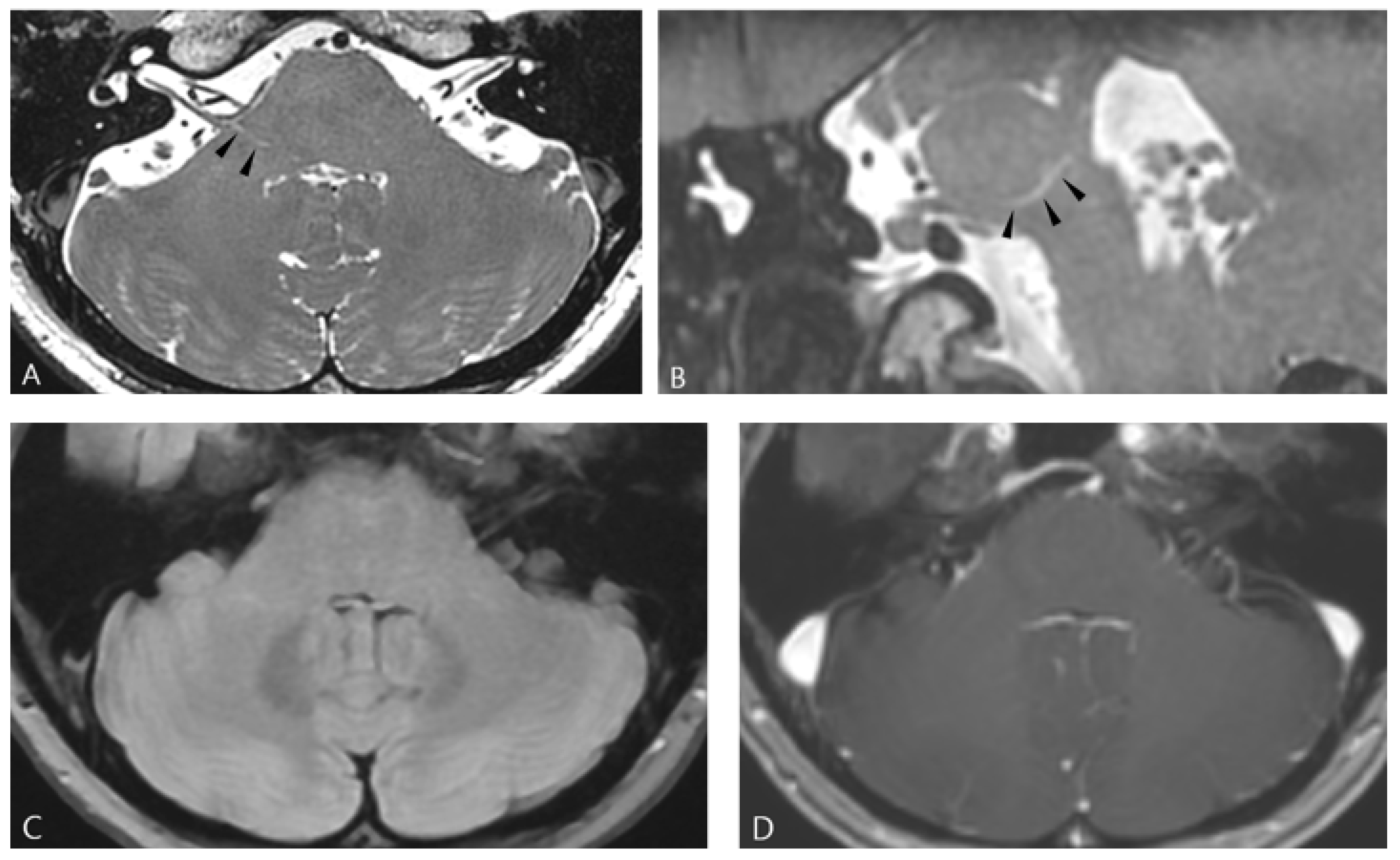
3.2. Presence of Pontine Hyperintense Lesion on High-Resolution Proton-Density Imaging
3.3. Group Comparison Between Patients with and Without Ipsilateral Pontine MSI
3.4. Relationship Between the Pontine Hyperintense Lesion and Clinical Outcome
4. Discussion
5. Conclusions
Author Contributions
Funding
Institutional Review Board Statement
Informed Consent Statement
Data Availability Statement
Acknowledgments
Conflicts of Interest
References
- Park, J.S.; Park, K. Operative Findings of over 5000 Microvascular Decompression Surgeries for Hemifacial Spasm: Our Perspective and Current Updates. Life 2023, 13, 1904. [Google Scholar] [CrossRef]
- Love, S.; Coakham, H.B. Trigeminal neuralgia: Pathology and pathogenesis. Brain 2001, 124, 2347–2360. [Google Scholar] [CrossRef]
- Lee, S.H.; Rhee, B.A.; Choi, S.K.; Koh, J.S.; Lim, Y.J. Cerebellopontine angle tumors causing hemifacial spasm: Types, incidence, and mechanism in nine reported cases and literature review. Acta Neurochir. 2010, 152, 1901–1908. [Google Scholar] [CrossRef]
- Park, K.; Park, J. Hemifacial Spasm: A Comprehensive Guide; Springer: Berlin/Heidelberg, Germany, 2020. [Google Scholar]
- Hyun, S.J.; Kong, D.S.; Park, K. Microvascular decompression for treating hemifacial spasm: Lessons learned from a prospective study of 1174 operations. Neurosurg. Rev. 2010, 33, 325–334; discussion 334. [Google Scholar] [CrossRef] [PubMed]
- Li, B.; Luo, C.; Jin, Y.; Yi, Y.; Cheng, D.; Huang, L.; Wang, G.; Zhong, X.; Zhao, H.; Gao, M. Evaluating magnetic resonance imaging characteristics and risk factors for hemifacial spasm. Brain Behav. 2024, 14, e3438. [Google Scholar] [CrossRef] [PubMed]
- Li, B.; Huang, L.; Luo, C.; Jin, Y.; Zhong, X.; Wang, G.; Xu, Z.; Chen, J.; Huang, S.; Zhao, H.; et al. Radiological characteristics predicting early poor drug response in patients with hemifacial spasm. Neurol. Sci. 2024, 45, 3217–3224. [Google Scholar] [CrossRef] [PubMed]
- Hughes, M.A.; Traylor, K.S.; Branstetter, B.F., IV; Eubanks, K.P.; Chang, Y.F.; Sekula, R.F., Jr. Imaging predictors of successful surgical treatment of hemifacial spasm. Brain Commun. 2021, 3, fcab146. [Google Scholar] [CrossRef]
- Han, S.; Li, Y.; Li, Z.; Wang, X.; Gao, J. Two-dimensional structure analysis of hemifacial spasms and surgical outcomes of microvascular decompression. Neurol. Res. 2021, 43, 173–180. [Google Scholar] [CrossRef]
- Zhu, W.; Shen, J.; Tang, T.; Chang, B.; Li, S.; Chen, M. Evaluation of pre-operative neuroimaging characteristics in patients with primary hemifacial spasm as a prognostic factor of microvascular decompression. Clin. Neurol. Neurosurg. 2020, 195, 105874. [Google Scholar] [CrossRef]
- Teton, Z.E.; Blatt, D.; Holste, K.; Raslan, A.M.; Burchiel, K.J. Utilization of 3D imaging reconstructions and assessment of symptom-free survival after microvascular decompression of the facial nerve in hemifacial spasm. J. Neurosurg. 2019, 133, 425–432. [Google Scholar] [CrossRef]
- Jin, Z.; Li, Z. Clinical Application of Diffusion Tensor Imaging in Diagnosis and Prognosis of Hemifacial Spasm. World Neurosurg. 2021, 145, e14–e20. [Google Scholar] [CrossRef] [PubMed]
- Yu, L.; Hu, X.; Li, H.; Zhao, Y. Perivascular Spaces, Glymphatic System and MR. Front. Neurol. 2022, 13, 844938. [Google Scholar] [CrossRef] [PubMed]
- Okar, S.V.; Hu, F.; Shinohara, R.T.; Beck, E.S.; Reich, D.S.; Ineichen, B.V. The etiology and evolution of magnetic resonance imaging-visible perivascular spaces: Systematic review and meta-analysis. Front. Neurosci. 2023, 17, 1038011. [Google Scholar] [CrossRef] [PubMed]
- Wardlaw, J.M.; Benveniste, H.; Nedergaard, M.; Zlokovic, B.V.; Mestre, H.; Lee, H.; Doubal, F.N.; Brown, R.; Ramirez, J.; MacIntosh, B.J.; et al. Perivascular spaces in the brain: Anatomy, physiology and pathology. Nat. Rev. Neurol. 2020, 16, 137–153. [Google Scholar] [CrossRef]
- Hou, X.; Wu, B.; Xu, R.; Yin, C. Glymphatic dysfunction in classical trigeminal neuralgia: DTI-ALPS index stratification from healthy controls to post-MVD. J. Headache Pain 2025, 26, 148. [Google Scholar] [CrossRef]
- Chen, F.; Zhang, Z.; Miao, J.; Zhang, Y.; Wang, D.; Yan, J.; Pan, L.; Ye, H.; Ding, Z.; Ge, X. Glymphatic and neurofluidic dysfunction in classical trigeminal neuralgia: A multimodal MRI study of brain-CSF functional and structural dynamics. BMC Med. Imaging 2025, 25, 222. [Google Scholar] [CrossRef]
- Lee, M.H.; Jee, T.K.; Lee, J.A.; Park, K. Postoperative complications of microvascular decompression for hemifacial spasm: Lessons from experience of 2040 cases. Neurosurg. Rev. 2016, 39, 151–158; discussion 158. [Google Scholar] [CrossRef]
- Mizobuchi, Y.; Nagahiro, S.; Kondo, A.; Arita, K.; Date, I.; Fujii, Y.; Fujimaki, T.; Hanaya, R.; Hasegawa, M.; Hatayama, T.; et al. Prospective, Multicenter Clinical Study of Microvascular Decompression for Hemifacial Spasm. Neurosurgery 2021, 88, 846–854. [Google Scholar] [CrossRef]
- Zhang, C.; Chen, Q.; Wang, Y.; Zhao, X.; Wang, C.; Liu, L.; Pu, Y.; Zou, X.; Du, W.; Pan, Y.; et al. Risk factors of dilated Virchow-Robin Spaces are different in various brain regions. PLoS ONE 2014, 9, e105505. [Google Scholar] [CrossRef]
- Robert, K.; Kwee, R.M.; Kwee, T.C. Virchow-Robin Spaces at MR Imaging. RadioGraphics 2007, 27, 1071–1086. [Google Scholar] [CrossRef]
- Buell, T.J.; Ramesh, A.; Ding, D.; Raper, D.; Chen, C.J.; Starke, R.M.; Mukherjee, S.; Webster Crowley, R. Dilated Virchow-Robin spaces mimicking a brainstem arteriovenous malformation. J. Neurosci. Rural. Pract. 2017, 8, 291–293. [Google Scholar] [CrossRef]
- Solak, Ö.; Yaman, M.; Haktanır, A.; Aycicek, A.; Oruc, S.; Kirpiko, O. Widening of Virchow-Robin spaces in the brain stem causing hemifacial spasm. Eur. J. Radiol. Extra 2009, 70, e1–e3. [Google Scholar] [CrossRef]
- Parillo, M.; Vaccarino, F.; Di Gennaro, G.; Kumar, S.; Van Goethem, J.; Beomonte Zobel, B.; Quattrocchi, C.C.; Parizel, P.M.; Mallio, C.A. Overview of the Current Knowledge and Conventional MRI Characteristics of Peri- and Para-Vascular Spaces. Brain Sci. 2024, 14, 138. [Google Scholar] [CrossRef] [PubMed]
- Fahmy, L.M.; Chen, Y.; Xuan, S.; Haacke, E.M.; Hu, J.; Jiang, Q. All Central Nervous System Neuro- and Vascular-Communication Channels Are Surrounded with Cerebrospinal Fluid. Front. Neurol. 2021, 12, 614636. [Google Scholar] [CrossRef] [PubMed]
- Chen, M.; Yang, M.; Zhou, W.-P.; Li, S.-T. Preliminary Study on the Relationship Between Inflammation and Hemifacial Spasm. World Neurosurg. 2019, 125, e214–e220. [Google Scholar] [CrossRef] [PubMed]
- El Refaee, E.; Marx, S.; Rosenstengel, C.; Baldauf, J.; Schroeder, H.W.S. Arachnoid bands and venous compression as rare causes of hemifacial spasm: Analysis of etiology in 353 patients. Acta Neurochir. 2020, 162, 211–219. [Google Scholar] [CrossRef]
- Wuerfel, J.; Haertle, M.; Waiczies, H.; Tysiak, E.; Bechmann, I.; Wernecke, K.D.; Zipp, F.; Paul, F. Perivascular spaces—MRI marker of inflammatory activity in the brain? Brain 2008, 131, 2332–2340. [Google Scholar] [CrossRef]
- Chung, S.J.; Yoo, H.S.; Shin, N.Y.; Park, Y.W.; Lee, H.S.; Hong, J.M.; Kim, Y.J.; Lee, S.K.; Lee, P.H.; Sohn, Y.H. Perivascular Spaces in the Basal Ganglia and Long-term Motor Prognosis in Newly Diagnosed Parkinson Disease. Neurology 2021, 96, E2121–E2131. [Google Scholar] [CrossRef]
- Liu, X.Y.; Ma, G.Y.; Wang, S.; Gao, Q.; Guo, C.; Wei, Q.; Zhou, X.; Chen, L.P. Perivascular space is associated with brain atrophy in patients with multiple sclerosis. Quant. Imaging Med. Surg. 2022, 12, 1004–1019. [Google Scholar] [CrossRef]
- Bekar, A.; Kuytu, T.; Turkkan, A.; Altunyuva, O.; Eser Ocak, P. The Efficacy and Safety of Microvascular Decompression for Hemifacial Spasm: A Retrospective Analysis of Surgical Outcomes and Complications. Turk. Neurosurg. 2020, 30, 231–236. [Google Scholar] [CrossRef]
- Lee, J.A.; Park, K. Short-term versus long-term outcomes of microvascular decompression for hemifacial spasm. Acta Neurochir. 2019, 161, 2027–2033. [Google Scholar] [CrossRef]
- Helal, A.; Graffeo, C.S.; Meyer, F.B.; Pollock, B.E.; Link, M.J. Predicting long-term outcomes after microvascular decompression for hemifacial spasm according to lateral spread response and immediate postoperative outcomes: A cohort study. J. Neurosurg. 2024, 140, 1664–1671. [Google Scholar] [CrossRef]
- Lee, J.A.; Kong, D.S.; Lee, S.; Park, S.K.; Park, K. Clinical Outcome After Microvascular Decompression According to the Progression Rates of Hemifacial Spasm. World Neurosurg. 2020, 134, e985–e990. [Google Scholar] [CrossRef]
- Lee, J.A.; Kim, K.H.; Kong, D.S.; Lee, S.; Park, S.K.; Park, K. Algorithm to Predict the Outcome of Microvascular Decompression for Hemifacial Spasm: A Data-Mining Analysis Using a Decision Tree. World Neurosurg. 2019, 125, e797–e806. [Google Scholar] [CrossRef]
- Jo, K.W.; Kong, D.S.; Park, K. Microvascular decompression for hemifacial spasm: Long-term outcome and prognostic factors, with emphasis on delayed cure. Neurosurg. Rev. 2013, 36, 297–301; discussion 301–292. [Google Scholar] [CrossRef]
- Kim, C.H.; Kong, D.S.; Lee, J.A.; Park, K. The Potential Value of the Disappearance of the Lateral Spread Response During Microvascular Decompression for Predicting the Clinical Outcome of Hemifacial Spasms: A Prospective Study. Neurosurgery 2010, 67, 1581–1588. [Google Scholar] [CrossRef]


| Characteristics | Overall (n = 438) | Non-Visible MSI (n = 171) | Visible MSI (n = 267) | p-Value |
|---|---|---|---|---|
| Age (years) | 55.8 ± 11.1 | 55.1 ± 10.3 | 56.2 ± 11.6 | 0.311 |
| Male sex | 111 (25.3%) | 33 (19.3%) | 78 (29.2%) | 0.020 |
| Lesion side (Right:Left) | 215:223 | 91:80 | 124:143 | 0.166 |
| Hypertension | 147 (33.6%) | 58 (33.9%) | 89 (33.3%) | 0.899 |
| Diabetes | 46 (10.5%) | 18 (10.5%) | 28 (10.5%) | 0.990 |
| Spasm duration (months) | 61.0 ± 43.2 | 60.7 ± 35.9 | 61.2 ± 47.4 | 0.897 |
| Spasm degree (I/II/III/IV) | 3/165/188/82 | 1/61/81/28 | 2/104/107/54 | 0.483 |
| Contralateral MSI | 136 (31.1%) | 25 (14.6%) | 111 (41.6%) | <0.001 |
| Surgical Findings | ||||
| Offending Vessel | 0.002 | |||
| AICA or PICA | 314 (71.7%) | 133 (77.8%) | 181 (67.8%) | |
| AICA and PICA | 29 (6.6%) | 10 (5.8%) | 19 (7.1%) | |
| VA related | 88 (20.1%) | 22 (12.9%) | 66 (24.7%) | |
| Venous offender | 7 (1.6%) | 6 (3.5%) | 1 (0.4%) | |
| Compression type | 0.125 | |||
| One vessel type | 119 (27.2%) | 47 (27.5%) | 72 (27.0%) | |
| Two vessels type | 148 (33.8%) | 50 (29.2%) | 98 (36.7%) | |
| Arachnoid/Perforator type | 149 (34.0%) | 61 (35.7%) | 88 (33.0%) | |
| Encircling type | 21 (4.8%) | 13 (7.6%) | 8 (3.0%) | |
| Penetrating type | 1 (0.2%) | 0 (0.0%) | 1 (0.4%) | |
| Compression Degree (I/II/III/IV, n = 430) | 17/209/116/88 | 6/80/53/29 | 11/129/63/59 | 0.299 |
| REZ discoloration (n = 368) | 325 (88.3%) | 131 (92.3%) | 194 (85.8%) | 0.062 |
| Surgical Outcomes | ||||
| Complications | ||||
| Facial palsy | 76 (17.4%) | 25 (14.6%) | 51 (19.1%) | 0.227 |
| Hearing loss | 12 (2.7%) | 4 (2.3%) | 8 (3.0%) | 0.681 |
| Post-operative Improvement of spasm | ||||
| Immediate | 355 (81.1%) | 148 (86.5%) | 207 (77.5%) | 0.019 |
| Delayed | 69 (15.8%) | 20 (11.7%) | 49 (18.4%) | 0.062 |
| Long-term | 419 (95.7%) | 165 (96.5%) | 254 (95.5%) | 0.607 |
| Variable | Odds Ratio | 95% Confidence Interval | p-Value |
|---|---|---|---|
| Sex (male) | 0.933 | 0.505–1.723 | 0.825 |
| Age | 1.013 | 0.988–1.039 | 0.321 |
| Duration of symptoms | 0.996 | 0.990–1.002 | 0.215 |
| Hemifacial spasm severity grade | 1.414 | 0.960–2.083 | 0.080 |
| Contralateral MSI | 1.616 | 0.891–2.932 | 0.114 |
| Offending Vessel type | 0.860 | ||
| AICA or PICA versus vein | 0.719 | 0.276–1.873 | 0.500 |
| AICA + PICA versus vein | 0.792 | 0.405–1.548 | 0.495 |
| VA related vs. vein | - | - | 0.999 |
| Ipsilateral MSI | 0.411 | 0.222–0.759 | 0.005 |
| REZ discoloration | 0.968 | 0.427–2.194 | 0.939 |
| Compression degree (indentation) | 0.960 | 0.687–1.341 | 0.810 |
Disclaimer/Publisher’s Note: The statements, opinions and data contained in all publications are solely those of the individual author(s) and contributor(s) and not of MDPI and/or the editor(s). MDPI and/or the editor(s) disclaim responsibility for any injury to people or property resulting from any ideas, methods, instructions or products referred to in the content. |
© 2026 by the authors. Licensee MDPI, Basel, Switzerland. This article is an open access article distributed under the terms and conditions of the Creative Commons Attribution (CC BY) license.
Share and Cite
Lee, H.S.; Roh, H.G.; Moon, W.-J.; Kim, C.-H.; Park, K.; Choi, J.W. Pontine Microtubular Signal Intensity in Hemifacial Spasm: Association with Outcome After Microvascular Decompression Surgery. Life 2026, 16, 664. https://doi.org/10.3390/life16040664
Lee HS, Roh HG, Moon W-J, Kim C-H, Park K, Choi JW. Pontine Microtubular Signal Intensity in Hemifacial Spasm: Association with Outcome After Microvascular Decompression Surgery. Life. 2026; 16(4):664. https://doi.org/10.3390/life16040664
Chicago/Turabian StyleLee, Hyun Seok, Hong Gee Roh, Won-Jin Moon, Change-Hee Kim, Kwan Park, and Jin Woo Choi. 2026. "Pontine Microtubular Signal Intensity in Hemifacial Spasm: Association with Outcome After Microvascular Decompression Surgery" Life 16, no. 4: 664. https://doi.org/10.3390/life16040664
APA StyleLee, H. S., Roh, H. G., Moon, W.-J., Kim, C.-H., Park, K., & Choi, J. W. (2026). Pontine Microtubular Signal Intensity in Hemifacial Spasm: Association with Outcome After Microvascular Decompression Surgery. Life, 16(4), 664. https://doi.org/10.3390/life16040664