Article

The Influence of Wild Grapevine Endophytes on the Growth of the Model Plant Arabidopsis thaliana (L.) Heynh


by
Olga A. Aleynova
*,
Alexey A. Ananev
,
Nikolay N. Nityagovsky
and
Konstantin V. Kiselev
Laboratory of Biotechnology, Federal Scientific Center of the East Asia Terrestrial Biodiversity, Far Eastern Branch of the Russian Academy of Sciences, 690022 Vladivostok, Russia
*
Author to whom correspondence should be addressed.
Life 2026, 16(4), 566; https://doi.org/10.3390/life16040566
Submission received: 25 February 2026 / Revised: 19 March 2026 / Accepted: 27 March 2026 / Published: 31 March 2026
(This article belongs to the Section Plant Science)

Abstract

We have evaluated the growth characteristics of the model plant Arabidopsis thaliana after inoculation of A. thaliana seeds with the most common endophytes of wild grapevine Vitis amurensis Rupr., namely bacteria Rhizobium (syn. Agrobacterium) sp., Bacillus velezensis, Curtobacterium sp, Erwinia sp., Gordonia aichiensis, Pantoea sp., Pseudomonas sp., Sphingomonas sp., Xanthomonas sp., and the fungi Biscogniauxia sp., Cladosporium sp., Didymella sp., Exobasidium sp., Penicillium sp., Pestalotiopsis sp, and Xylaria sp. A positive effect on plant growth was observed in A. thaliana following seed inoculation with endophytic fungi (Xylaria sp., Didymella sp., and Exobasidium sp.) and bacteria (Gordonia aichiensis and Sphingomonas sp.). The inoculation with the fungi Xylaria sp., Didymella sp., Exobasidium sp., and Penicillium sp. significantly increased seed production in A. thaliana by 2.5–5-fold. The analysis of the phytohormone-regulating gene transcription in A. thaliana plants following inoculation with the grapevine endophytic microorganisms suggests that plant growth was enhanced through transcriptional changes in individual genes of hormone biosynthetic pathways. Consequently, endophytic bacteria and fungi from V. amurensis may serve as potential natural growth stimulators for agricultural plants.

1. Introduction

Plants are closely associated with microorganisms, residing in all tissues [1]. A special group of microorganisms associated with plants is endophytes—microorganisms that inhabit the internal plant tissues [2]. Endophytes inhabit all parts of plant organs: roots, leaves, stems, flowers, and seeds. Endophytes can enter a plant through its root system, aboveground parts, cracks, seeds, or vegetative plant propagation [2]. Some endophytes may act as latent pathogens, while others may play an essential role in maintaining plant health. Endophytes can protect or prepare the plant against abiotic and biotic stresses and help in enhancing growth and yields [3,4].
The remarkable ability of endophytes to promote plant growth stems from their diverse mechanisms of action, encompassing both direct and indirect pathways [5,6,7,8]. Direct growth-promoting methods consist of the production of compounds beneficial to plants, such as phytohormones, 1-aminocyclopropane-1-carboxylate (ACC) deaminase, iron sequestration and phosphate solubilization [7,8]. Plant hormones such as auxins, cytokinins (CKs), gibberellins (GAs), abscisic acid (ABA) and ethylene (ET) have a major impact on plant growth [9]. Multiple studies have corroborated that endophytes may produce plant hormones, including auxin, CKs, GAs, ABA and ET [10,11,12,13,14,15,16]. Endophytes can stimulate plant growth not only by directly producing their own phytohormones but also by activating a cascade of differential expression of host plant genes, which can additionally activate plant phytohormone biosynthesis genes [17,18].
It is important to note that some endophytes demonstrate broad host compatibility, while others exhibit high specificity, influenced by factors such as plant physiology, root exudates, immune responses, and environmental conditions [17,19]. The interaction of endophytes and plants is influenced by the condition of the plant, species, geographical location, climatic conditions, and even the season of sampling [20,21] [Rodriguez-Blanco et al., 2015; Ding and Melcher, 2016]. It was found that in some endophytes, the transition to a pathogenic lifestyle depends not only on local abiotic stress factors but also on the genotype of the host plant [22]. For example, fluorescent pseudomonads are commonly regarded as beneficial endophytic bacteria that promote plant growth. Studies have shown that, under certain conditions, they can have harmful effects on leatherleaf ferns [23]. This highlights a key ecological nuance: even microbes typically considered mutualistic may act as pathogens or stressors, depending on the host species and environmental context [24].
Grapes are among the most economically significant crops and widely cultivated fruit plants worldwide. Grapevines harbor a diverse community of microorganisms, including bacterial and fungal endophytes, which contribute to the formation of the so-called “terroir” and influence the qualities and characteristics of grapes and wine [25]. Some grapevine endophytes, in addition to their anti-pathogenic properties, are able to influence plant growth [26,27,28].
V. amurensis is a grapevine species native to Asia and is recognized as a rich source of secondary metabolites with potent antioxidant, anti-inflammatory, antibacterial, and cardioprotective properties [29,30]. Moreover, its use as a rootstock provides a valuable opportunity to develop grape cultivars with enhanced resistance to a broad spectrum of biotic and abiotic stresses. Therefore, investigating the endophytic microbiota isolated from the wild grapevine V. amurensis is of significant interest, as these microbes may contribute to the plants’ resilience and offer novel biocontrol or growth-promoting agents.
It has previously been shown that the main representatives of endophytes from aerial tissues of V. amurensis (leaf, stem, seed and berry) are bacteria of the classes Gammaproteobacteria, Alphaproteobacteria, Actinobacteria, Bacilli, and Bacteroidia [31], and fungi Dothideomycetes and Tremellomycetes [32]. The main endophytes of V. amurensis grapevine include representatives of the bacterial genera Rhizobium (syn. Agrobacterium) sp., Bacillus sp., Curtobacterium sp., Erwinia sp., Gordonia sp., Pantoea sp., Pseudomonas sp., Sphingomonas sp., and Xanthomonas sp., and fungal genera Biscogniauxia sp., Cladosporium sp., Didymella sp., Exobasidium sp., Penicillium sp., Pestalotiopsis sp. and Xylaria sp. [32].
It was shown that some members of Rhizobium, Bacillus, Curtobacterium, Gordonia, Pseudomonas and Sphingomonas species are classified as endophytes, which establish themselves intracellularly in order to promote root growth through both direct and indirect mechanisms [33,34,35]. The genera Erwinia, Pantoea and Xanthomonas comprise bacteria associated with plants in various ecological roles, including as pathogens, saprophytes, epiphytes, and endophytes [36,37].
Plant-associated fungal isolates also exhibit the same range of functions as bacteria, with beneficial effects on plant growth. The representatives of the genera Biscogniauxia, Cladosporium, Didymella, Exobasidium, and Penicillium are endophytic, saprophytic, weakly parasitic and pathogenic species that are often found in different plants around the world [36,38]. Some Biscogniauxia fungi produce antibacterials that could be useful in biotechnology [39,40]. Some species of Cladosporium and Penicillium have the plant growth-promoting capacity and the potential to become a microbial fertilizer for sustainable crop production [41,42]. Pestalotiopsis is ordinarily isolated as endophytes in plants [43,44,45]. The genus Xylaria comprises various endophytic species associated with both vascular and non-vascular plants [46]. Xylaria sp. has broad antimicrobial activity [47].
Previously, we examined the effect of dominant endophytic bacteria and fungi isolated from wild grapevine V. amurensis on grape cell culture growth and the synthesis of resveratrol, a prominent biologically active compound with beneficial effects on human health [29]. Endophytic bacteria significantly increased total stilbene content by 2.2–5.3-fold, while endophytic fungi were more effective, enhancing stilbene accumulation by 2.6–16.3-fold [48]. These findings suggested that wild grapevine endophytic bacteria and fungi hold promise for enhancing the nutritional value and overall quality of agricultural products. However, it remains unknown whether V. amurensis endophytes function as growth-stimulating microorganisms and which pathways mediate their growth-stimulating effects.
Therefore, this study aimed to investigate the effects of dominant endophytic bacteria and fungi isolated from the wild grapevine V. amurensis on growth characteristics and phytohormone metabolism gene expression in the model plant Arabidopsis thaliana (L.) Heynh. Arabidopsis was selected due to its short life cycle (2–3 months) and its well-characterized functions of phytohormone-related genes.

2. Materials and Methods

2.1. Endophytic Bacteria and Fungi Isolation and Identification

Endophytic bacteria and fungi were isolated from superficially sterilized leaves and stems of wild grapevines, V. amurensis, growing on the unprotected regions of the Primorsky Territory of Russia. Approximately 1.5 g of leaf and stem tissue was washed with mild soap, followed by surface sterilization in 75% (v/v) ethanol for two minutes and then in 10% (v/v) hydrogen peroxide for 1 min. Samples were rinsed five times with sterile distilled water, and a sample of final rinse water was cultured on PDA (Neogene, UK) or R2A (Himedia, India) media to verify the efficacy of the sterilization protocol. The sterile leaves and stems were ground in a mortar until a homogeneous pulp was obtained. The resulting homogenate was then plated onto Petri dishes containing PDA or R2A medium. After three days of incubation at 23–25 °C, colonies that formed were isolated and transferred to fresh sterile plates for further cultivation [48].
DNA from a single colony of bacteria and fungi was extracted using the hexadecyltrimethylammonium bromide (CTAB) method with some modifications [49]. Bacterial 16S rRNA gene sequences were amplified using universal bacterial primers (8F, 5′AGA GTT TGA TCM TGG CTC AG and 1522R, 5′AAG GAG GTG ATC CAR CCG CA) to produce approximately 1500 bp PCR products [50]. For identification of endophytic fungi, the universal primers (5′AGG AGA AGT CGT AAC AAG G and 5′TCC TCC GCT TAT TGA TAT GC) were used to amplify approximately 580 bp ITS1 PCR products. The PCR products were then sequenced on an ABI 3130 Genetic Analyzer (Applied Biosystems, Foster City, CA, USA) and analyzed using the BLAST program. Sequence analysis was performed by multiple sequence alignment using the Clustal X program [51], with a sequence identity of ≥99% used as the threshold for taxonomic identification (Supplementary Material S1).

2.2. Seed Germination of A. thaliana in the Presence of Grapevine Endophytes

For experiments, we used seeds of A. thaliana ecotype Columbia-0, stored by our lab. Seeds were surface-sterilized by exposure to chlorine vapor generated by adding 3 mL of concentrated HCl to 100 mL of 7% bleach solution from Sayansk Himplast (Sayansk, Russia) for 40–50 min. Following sterilization, seeds were germinated on 1/2 Murashige and Skoog medium (MS) with a pH of 7.0, solidified with 0.8% (m/v) agar, in a controlled environment chamber at 22 °C under a 16 h light/8 h dark photoperiod with a light intensity of 120 μmol m−2 s−1. Plants grown under these conditions were used as a negative control.
The following procedures were performed for inoculation with grapevine endophytes. The single colony of bacteria was placed in 20 mL of a liquid nutrient medium, R2A medium (PanReac, AppliChem, Darmstadt, Germany), and incubated for 24–48 h at 28 °C at 150 rpm on an orbital shaker BioSan ES 20/60 (Riga, Latvia), raising the final concentration of 107 colony-forming units per ml (CFU/mL). Next, 100 µL of each bacterial isolate was surface distributed by spreading with a sterilized loop on Petri dishes (diameter 12 cm) with MS medium. Fungal isolates were cultured on a Potato Dextrose Agar (PDA, Neogene, UK), and then, 1 cm2 pieces of mycelium and spores were placed on Petri dishes with MS medium. Previous studies on optimizing the concentration of bacteria and fungi have shown that these concentrations are optimal. As lower concentrations of bacteria or a smaller area of inoculated fungus have no effect on plant growth, while higher ones greatly suppress it, the plants did not survive after inoculation.
Under aseptic conditions, Arabidopsis seeds sterilized with chlorine vapor were sown on the MS medium in Petri dishes inoculated with endophytes. After 7–8 days, plant endophytic interactions in vitro, the length of the root and height of the stem of the A. thaliana seedlings were measured using a ruler (Supplementary Figures S1–S4). Subsequently, seedlings were transferred into pots filled with commercially available rich soil (“Universalniy”, Fasko, Moscow, Russia) in an environmental sterile control chamber (Sanyo MLR-352, Panasonic, Tokyo, Japan) kept on a 16/8 h light/darkness cycle at 22 °C and a light intensity of 120 μmol m−2s−1.
After one month, the rosette diameter and the number of leaves of the A. thaliana plants were measured. In addition, the approximate number of Arabidopsis seeds per 1 plant was calculated at the end of the seed ripening period. Dried pods were collected from each plant that had germinated with either grapevine-derived endophyte. The seeds were then removed from the pod, and the total weight of seeds per plant was weighed. Subsequently, 100 seeds from each plant were counted and weighed separately. The total number of seeds per plant was then calculated by dividing the total seed mass by the average mass of 100 seeds. Statistical analyses were performed to determine the mean seed number per plant and the standard error of the mean for each endophyte treatment.

2.3. Re-Isolation of the Endophytes That Positively Affect the Growth of Arabidopsis Plants

The endophytic content of bacteria B. velezensis, G. aichiensis and Sphingomonas sp., as well as fungi Xylaria sp., Didymella sp. and Exobasidium sp., was evaluated by counting CFUs of microorganisms in seedling tissue, including leaves, stems and roots, 7 days after inoculation of Arabidopsis seeds with endophytes, as described above [52]. For CFU estimation, 10 mg samples of each individual experimental seedling were superficially sterilized in the following order: 75% ethanol for 1 min, 10% H2O2 for 3 min and distilled water. The samples were homogenized in a sterile mortar using a pestle, with 1 mL of sterile water added. Two consecutive 10-fold dilutions of the resulting homogenate were performed. Aliquots of 100 µL were spread over the surfaces of R2A and PDA media using a microbiological spatula, until they were completely dry. Petri dishes were then incubated in the dark at 28 °C for 48 h. CFU were counted for the second and third dilutions, and their number was recalculated per 1 g of plant wet weight.

2.4. RNA Extraction and qRT-PCR

RNA extraction utilized the CTAB-based method outlined by Kiselev et al. [53]. cDNAs were synthesized using the MMLV Reverse Transcription PCR Kit with Oligo(dT)15 (RT-PCR, Evrogen, Moscow, Russia) following Aleynova et al. [54].
Three genes governing auxin metabolism (AtNIT1, AtTAA1, and AtYUCCA1) [55,56], four genes regulating cytokinin metabolism (AtCYP735A2, AtUGT76C2, AtCKX4, and AtCKX5) [57,58,59], two genes involved in gibberellin metabolism (AtGA3ox2 and AtGA2ox2) [60,61], four genes associated with abscisic acid metabolism (AtNCED3 and AtABA3) [62], and two genes implicated in ethylene metabolism (AtEIN2 and AtEIN3) [63,64] were analyzed. mRNA transcript levels were assessed via quantitative real-time PCR (qRT-PCR), employing AtGAPDH (NM_111283.4), AtEF1a (XM_002864638), AtUBQ (NM_001084884), and AtUBC (XM_021022221) as internal controls. qRT-PCR was performed using separate amplification reactions for the gene of interest and each reference gene. Relative gene expression levels were calculated using the 2−ΔΔCt method [65], with the transcriptional level in the negative control (NC)—uninoculated A. thaliana plants—normalized to a value of 1. To ensure robustness, expression values were averaged across multiple technical replicates, and statistical analysis was conducted to assess significance. Data are presented as mean relative expression ± standard error, derived from independent qRT-PCR using multiple validated reference, as per Aleynova et al. [66]. The functions of the selected genes and the sequence of the primers for qRT-PCR are described in a previously published paper [67] and are also provided in Table 1.
qRT-PCR reactions were conducted in 20 µL volumes with the Real-Time PCR Kit (Evrogen, Moscow, Russia) according to Aleynova et al. [54], comprising 1× Taq buffer, 2.5 mM MgCl2, 0.2 mM of each dNTP, 0.2 µM of each oligonucleotide primer, 1× SYBR Green I real-time PCR dye, 1 µL cDNA, and 1 unit of Taq DNA polymerase (Evrogen, Moscow, Russia). Analysis was performed using a DTprime 4M1 thermal cycler (DNA-Technology, Moscow, Russia) programmed for an initial denaturation step of 2 min at 95 °C, followed by 50 cycles of 10 s at 95 °C and 25 s at 62 °C.

2.5. Statistical Analysis

During our plant experiments, we conducted five independent trials, each comprising 36 plants per treatment. In addition, we carried out two independent experiments for qRT-PCR to confirm our findings, with a total of 8 technical replicates—two for each of the following reference genes: AtGAPDH, AtEF1a, AtUBQ, and AtUBC. All data are presented as mean ± standard error (SE) and analyzed by the ANOVA with Tukey post-test and Student’s t-test. Correlation analysis was conducted in Excel (Microsoft Office, 2019, Washington, DC, USA) using the appropriate function.

3. Results

3.1. The Identification of the Most Prevalent Grapevine Endophytes

As a result of the analysis of the collection of endophytic bacteria and fungi of V. amurensis grapes, we have selected the most common species of endophytes of wild grapevines. These are bacteria Agrobacterium rubi (MZ424738, 99%), Bacillus velezensis (CP140115.1, 100%), Curtobacterium flaccumfaciens (MZ424740, 100%), Erwinia billingiae (KM408608.1, 100%), Gordonia aichiensis (BioProject PRJNA1267753, 100%), Pantoea agglomerans (MT605813.1, 99%), Pseudomonas alkylphenolica (MZ424743, 99%), Sphingomonas aerolata (PX909750, 99%), and Xanthomonas campestris (MZ424744, 99%), and fungi Biscogniauxia maritima (MZ427923, 100%), Cladosporium perangustum (MZ427924, 100%), Didymella pinodella (MZ427926, 100%), Exobasidium japonicum (PX916210, 96%), Penicillium brevicompactum (PX916211, 96%), Pestalotiopsis biciliate (PX916212, 99%) and Xylaria flabelliformis (PX920275, 100%) (Table 2, Supplementary Material S1). It is important to note that using whole-genome sequencing, we were able to identify two bacterial isolates to the species level: Bacillus sp. as B. velezensis (CP140115.1) [70] and Gordonia sp. as G. aichiensis (PRJNA1267753) [71].

3.2. Growth Characteristics of A. thaliana Plants Inoculated with Grapevine Endophytes

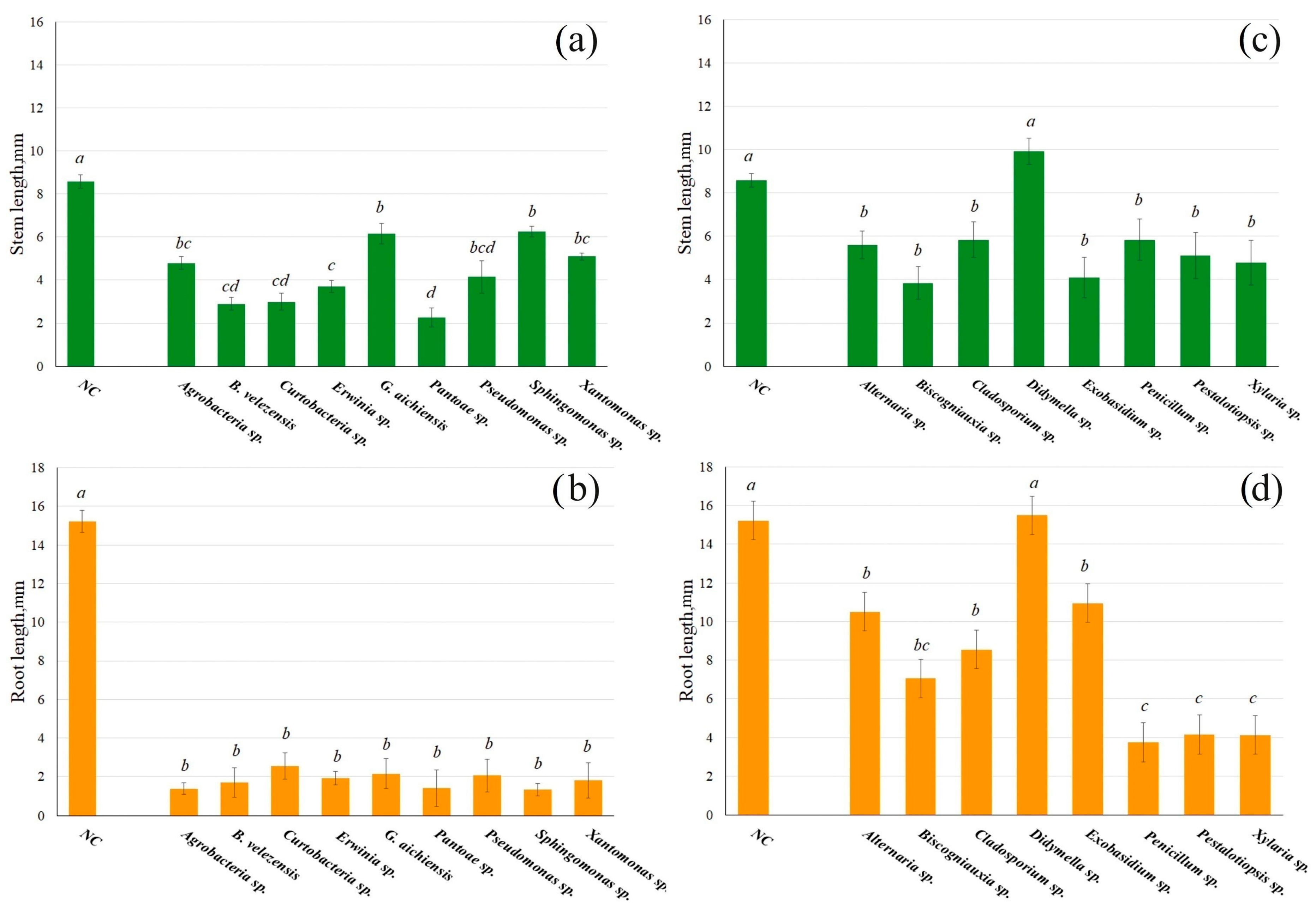
The grapevine endophytes were distributed onto the surface of Petri dishes containing MS medium, where sterile Arabidopsis seeds were then placed. After 7 days of inoculation of A. thaliana seeds with grapevine endophytic bacteria, a significant delay in seedling development was observed, affecting both shoot (stem) and root growth. The Arabidopsis root appears 2–3 days after exposure to a nutrient medium under normal conditions. In the case of seeds treated with grapevine endophytes, the root appeared on 4–6 days. The length of the stem was 1.1–2.9 times smaller when germinated with endophytic bacteria, and the root was 2.1–11.1 times smaller compared to control plants (Figure 1a,b). Seedling development was most strongly inhibited by bacteria of the genus Pantoea.
The inhibition of A. thaliana seedling development, inoculated with endophytic grapevine fungi, was not as strong as compared to seed germination in the presence of endophytic bacteria (Figure 1c,d). In general, the stem was 1.5–2.3 times smaller and the root 1.4–4.5 times smaller. An exception was the treatment with Didymella sp., which did not significantly change the size of A. thaliana seedlings (Figure 1c,d).
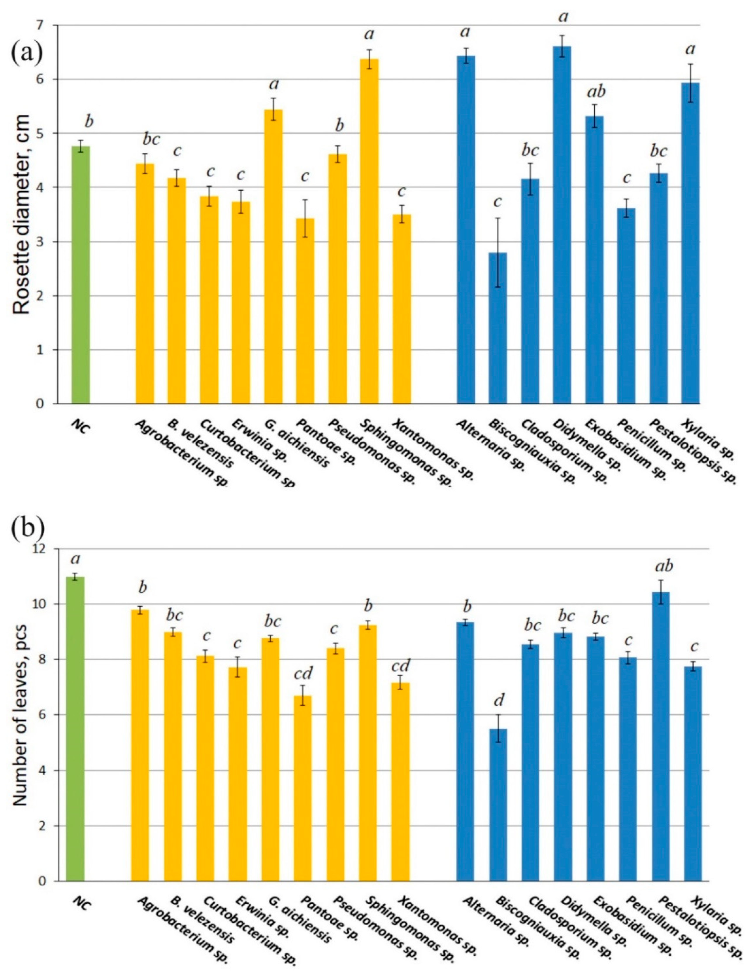
After inoculation with endophytes on a MS medium (contains sucrose as a carbon source) in Petri dishes, Arabidopsis seedlings were planted in non-sterile, commercially available, rich soil and cultivated in a climate chamber for 30 days. When A. thaliana plants were further cultured, different effects on rosette diameter, number of leaves, and fresh weight accumulation were observed. The diameters of the rosettes of 1-month-old A. thaliana plants grown in the presence of the endophytic bacteria Gordonia aichiensis and Sphingomonas sp. and the fungi Didymella spp., Exobasidium sp., and Xylaria sp. were 1.2–1.4 times larger than those of the control plants (Figure 2a). The diameter of rosettes of A. thaliana plants grown in the presence of endophytic bacteria Bacillus velezensis, Curtobacterium sp., Erwinia sp., Pantoea sp., Pseudomonas sp., and Xanthomonas sp., and fungi Biscogniauxia sp. and Penicillium sp., was significantly smaller compared to the control plants. The number of leaves of 1-month-old plants grown in the presence of wild grapevine endophytes was significantly reduced, except for plants germinated together with the fungus Pestalotiopsis sp. (Figure 2b).
Next, three endophytic bacterial isolates (B. velezensis, G. aichiensis, and Sphingomonas sp.) and three fungal isolates (Xylaria sp., Didymella sp. and Exobasidium sp.) were selected based on their strongest positive effects on the rosette diameter and leaf number. Their impact on the accumulation of fresh weight of Arabidopsis plants was then investigated in detail.
As a result of re-isolation of endophytes from leaves, stems and roots of 7-day-old A. thaliana seedlings, it was found that tissues contained bacteria B. velezensis, G. aichiensis and Sphingomonas sp. in the amounts of 330 × 103, 270 × 103 and 300 × 103 CFU/g of wet weight, respectively. The CFU numbers of Xylaria sp., Didymella sp. and Exobasidium sp. were 20–50 × 103 CFU/g of wet weight.
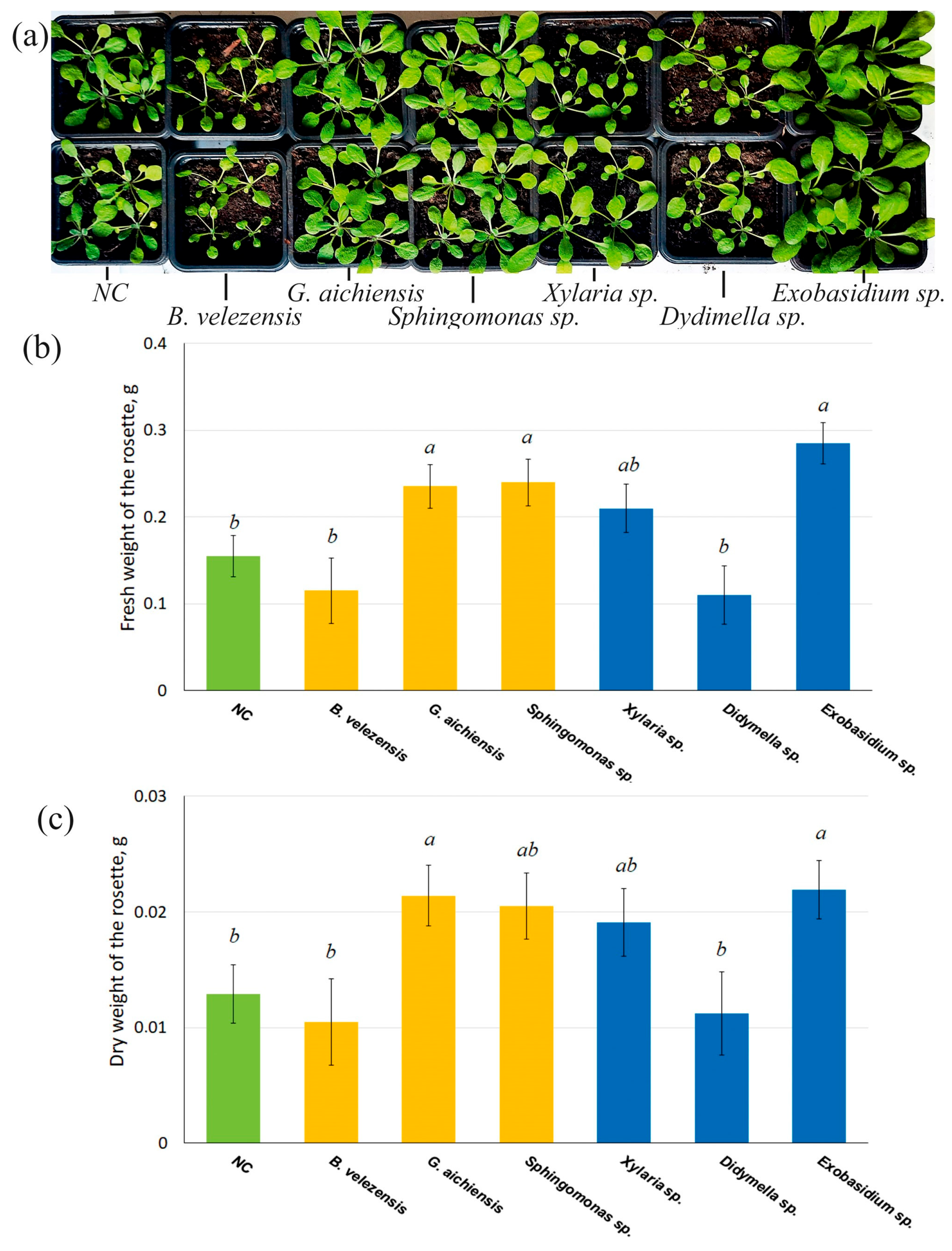
Treatment with G. aichiensis, Sphingomonas sp., and Exobasidium sp. significantly increased the accumulation of fresh and dry weight in Arabidopsis rosettes by 1.5–1.8 times (Figure 3), compared to untreated controls. Thus, it was these endophytic microorganisms that had the best effect. The remaining isolates had no significant effect on the accumulation of fresh weight in Arabidopsis plants (Figure 3).
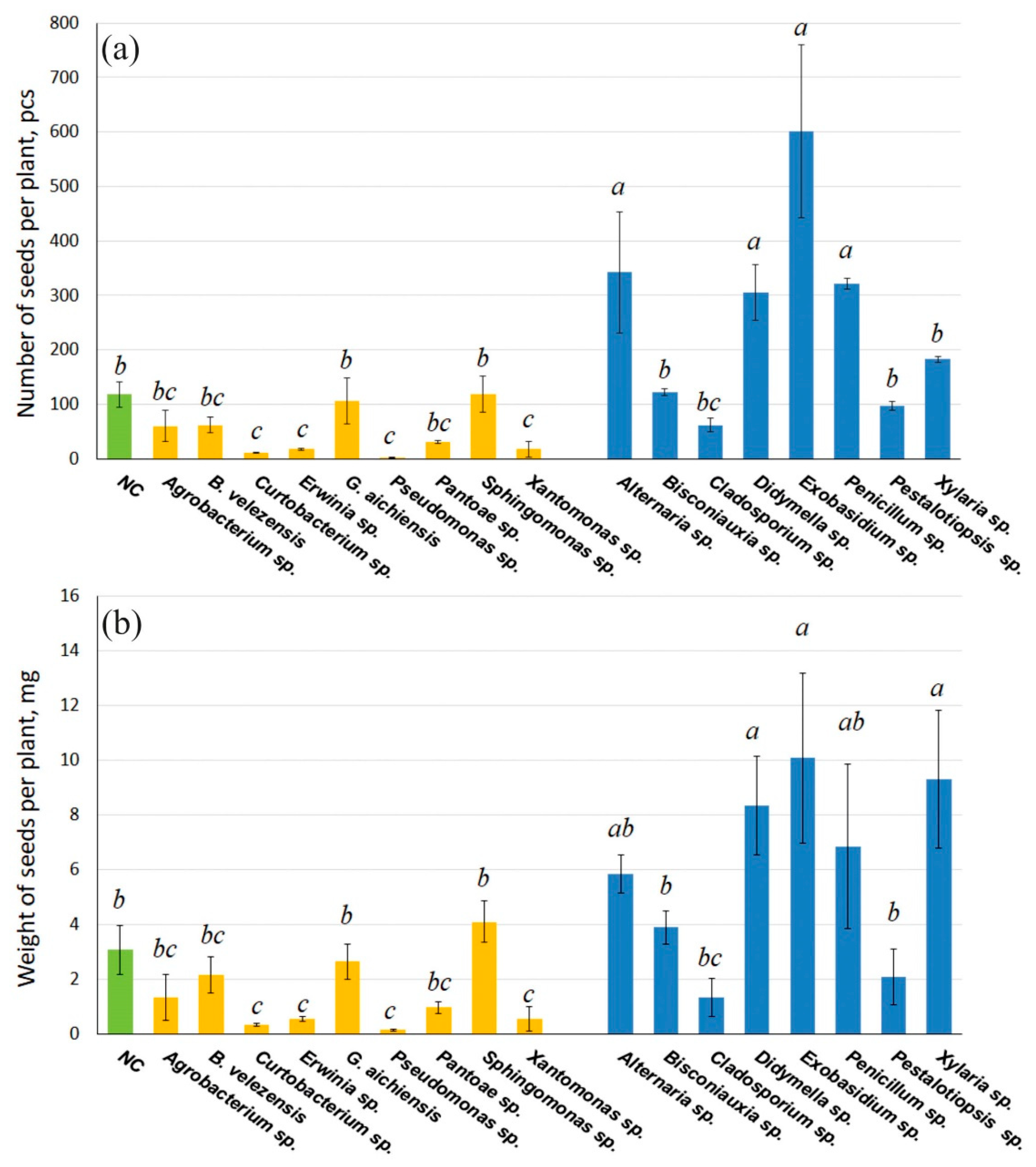
After the formation and maturation of A. thaliana pods, seeds were collected and counted from each group of plants grown in the presence of selected endophytic bacteria and fungi. Wild-type A. thaliana plants grown without exposure to V. amurensis-derived endophytic microorganisms produced an average of 100 seeds per plant, weighing 3 mg (Figure 4). Seed production was significantly reduced by 6.5–38-fold when A. thaliana was inoculated with the grape endophytic bacteria Erwinia sp., Pantoea sp., Pseudomonas sp. and Xanthomonas sp. The minimum number of seeds was observed when germinating together with endophytic bacteria of the genus Pseudomonas and amounted to three seeds per plant. The bacteria Agrobacterium sp., B. velezensis, G.aichiensis, and Sphingomonas sp. did not significantly affect the production of Arabidopsis seeds (Figure 4).
Inoculation of A. thaliana seeds with endophytic fungi of wild grape V. amurensis did not reduce seed production and weight. In addition, inoculation of seeds in the presence of the fungi Xylaria sp., Didymella sp., Exobasidium sp., and Penicillium sp. significantly increased seed production and weight by 2.5–5 times. The highest seed production was observed in plants germinated together with Exobasidium sp. (Figure 4). Thus, the fungus Exobasidium sp. enhances the size and yield of Arabidopsis more effectively than other grapevine endophytes studied.

3.3. Gene Expression of Phytohormone Metabolism Genes in Arabidopsis Plants After Inoculation with V. amurensis Endophytic Microorganisms

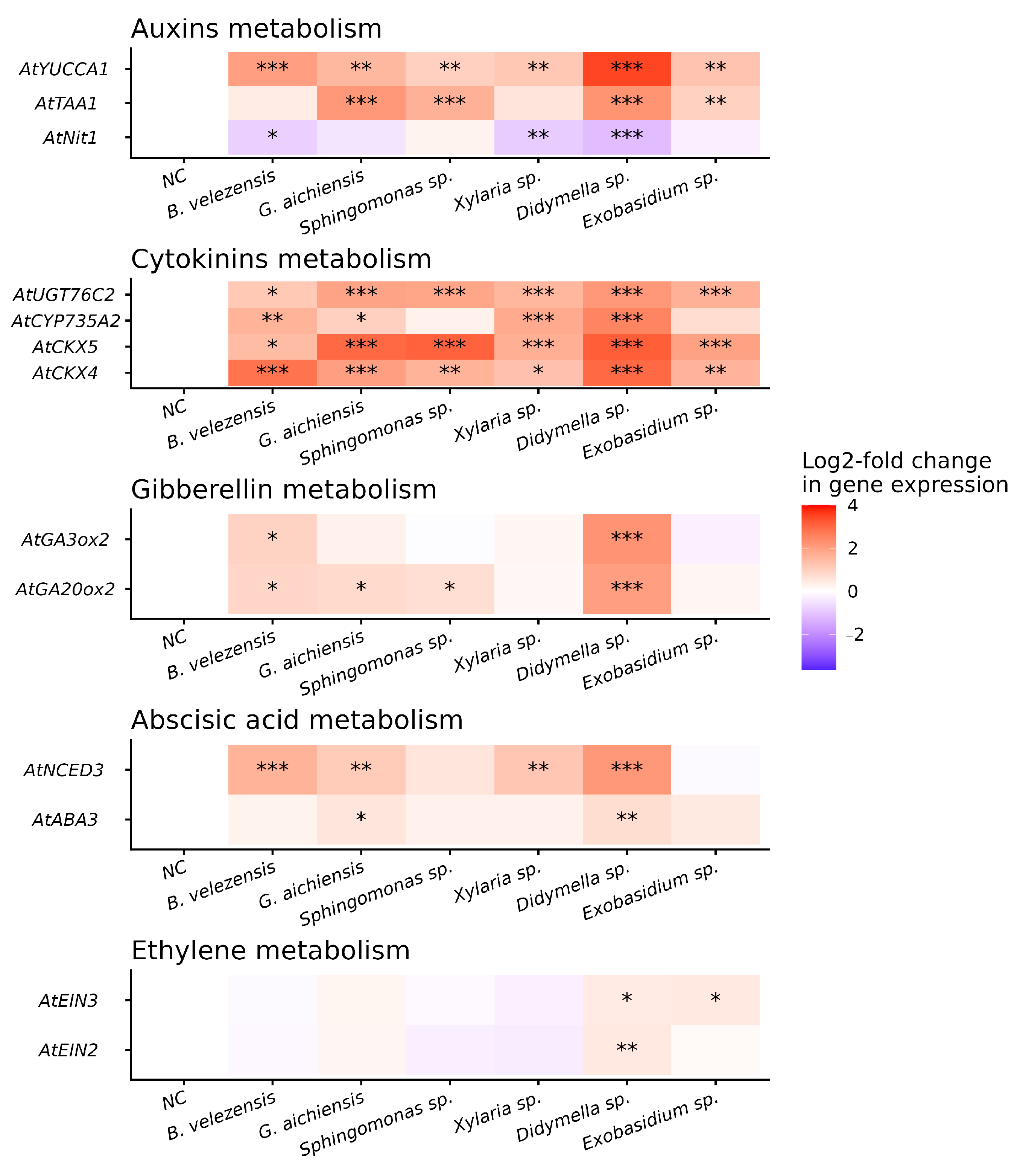
In order to explain the effects of grapevine endophytes on plant growth, we analyze the expression level of A. thaliana genes encoding phytohormone biosynthesis in leaves, stems and roots of 7-day-old seedlings. The expression of seedlings inoculated with bacteria and fungi was analyzed, which demonstrated an increase in plant size and biomass by 1 month, particularly after inoculation with endophytic bacteria B. velezensis, G. aichiensis, and Sphingomonas sp. and fungi Xylaria sp., Didymella sp., and Exobasidium sp. (Figure 5; Table 2). The qPCR analysis revealed that the level of gene transcription of auxin (AtTAA1 and AtYUCCA1) and cytokinin (AtCKX4, 5, AtCYP735A2, and AtUGT76C2) metabolism considerably increased in all Arabidopsis 7-day-old seedlings inoculated with grapevine endophytes (Figure 5). AtTAA1 and AtYUCCA1 catalyze the first and second stages of auxin synthesis (IAA) via indole-3-pyruvate (IPA). AtCKX4 and AtUGT76C2 are responsible for regulating the content of endogenous CKs. AtCYP735A2 is the enzyme that catalyzes the synthesis of CKs (Table 2).
The expression of the AtNIT1 gene, which is an enzyme that converts indole-3-acetonitrile to IAA, was significantly reduced compared to the control in all A. thaliana seedlings inoculated with endophytes. Additionally, the expression of genes involved in ABA (AtNCED3, which is an enzyme that catalyzes the rate-limiting step of ABA biosynthesis) and GAs (AtGA2ox2, which catalyzes oxidation at a late stage of GA biosynthesis) metabolism was significantly upregulated; a significant increase in all genes of these groups was observed when A. thaliana seeds were inoculated with Didymella sp. (Figure 5). Furthermore, genes AtEIN2 and AtEIN3, which play key roles in ethylene signaling, showed significantly higher expression in Arabidopsis seedlings inoculated with Didymella sp. or Exobasidium sp. (Figure 5).
We performed the Pearson correlation analysis between the expression levels of genes involved in the phytohormone metabolism and the diameter and mass of 1-month-old Arabidopsis rosettes (Supplementary Table S1). Only correlations with an absolute Pearson coefficient (r) greater than 0.5 or less than −0.5 were considered significant. The analysis revealed a selective moderate positive correlation between the expression of genes involved in cytokinin and auxin metabolism and plant size. Specifically, expression of the AtNIT1 was positively correlated with increased rosette mass (r = 0.591, p = 0.001), while expression of AtTAA1, AtCKX5, and AtUGT76C2 showed positive correlations with increased Arabidopsis rosette diameter (r = 0.654, p = 0.002; 0.766, p = 0.003; and 0.769, p = 0.007) (Supplementary Table S1). In contrast, negative correlations were observed between Arabidopsis biomass accumulation and the expression of AtNCED3 (r = −0.715, p = 0.003) involved in ABA metabolism (Supplementary Table S1).

4. Discussion

Endophytic microbes enhance plant resistance to both abiotic and biotic stresses [72,73] and can influence seed germination and seedling vigor, with significant implications for plant health and ecosystem dynamics [74]. Despite this potential, studies examining the specific effects of individual endophytic microorganisms on agricultural crops remain limited [75].
The medicinal plants, which include wild grapevine V. amurensis, could be a promising source for isolating plant-beneficial endophytes that can be used to enhance plant growth and protect plants from soil-borne pathogens [31,32,48]. This paper analyzes the influence of the most frequently encountered endophytic bacteria and fungi of wild grapevine on the growth characteristics of the model plant A. thaliana. For 7-day-old Arabidopsis seedlings inoculated with endophytic microorganisms from V. amurensis, there was a significant inhibition of development, except for seeds inoculated with Didymella sp. Significant increases in plant size were observed in one-month-old seedlings inoculated with the endophytic bacteria G. aichiensis and Sphingomonas sp., and the fungi Xylaria sp., Didymella sp., and Exobasidium sp. Thus, the effect of endophytes on A. thaliana plants is contingent upon the specific endophyte species and its strategy of interaction with the host. Initially, seedlings exposed to high microbial concentrations appear to activate canonical defense responses, such as upregulation of genes involved in ABA metabolism, a hormone typically accumulated under stress conditions [76], or delayed seed development may stem from microbial metabolites [77] or germination-inhibiting enzymes [78]. Subsequently, a selective interaction dynamic emerges between the plant and its microbial colonizers, leading to a functional dichotomy: a subset of endophytes act as plant growth promoters, while others behave as conditional pathogens, as evidenced by their negative impact on biomass accumulation and seed production.
Endophytes that have a positive effect on plant size likely accelerate plant development not only through direct synthesis of phytohormones but also by triggering a cascade of host plant hormone biosynthesis gene expression [17,18]. Our results reveal a significant upregulation of genes associated with auxin and CK biosynthesis in A. thaliana seedlings colonized by grapevine endophytes. A moderate, though statistically non-significant, trend toward increased expression of ABA- and GA-related genes was also observed. Activation of the genes involved in the metabolism of these important plant hormones is likely to lead to an increase in the diameter and weight of the Arabidopsis plants [10,11,79]. Pearson’s correlation analysis revealed a selective positive relationship between the expression of genes involved in CK and auxin biosynthesis and biomass accumulation and plant diameter in Arabidopsis rosettes. There was a significant decrease in AtNIT1 expression in all Arabidopsis seedlings inoculated with grapevine endophytes, which had a positive effect on plant size. It is known that AtNIT1 homologs regulate cell proliferation and differentiation [80] and participate in glucosinolate catabolism [63], which is important for the development of seedlings. Also, the AtNIT2, AtNIT3, and AtNIT4 genes were distinctly induced in A. thaliana leaves by P. syringae pv. tomato infection [64]. The observed significant downregulation of AtNIT1 expression may result from transcriptional repression mediated by the competitive activation of alternative nitrilase isoforms, such as AtNIT2, AtNIT3, and AtNIT4, in response to colonization by grapevine-associated endophytes. This isoform switching likely alters substrate partitioning within the nitrilase metabolic network, thereby reducing the availability of common substrates (e.g., indole-3-acetonitrile) for AtNIT1, which may further reinforce its transcriptional suppression through feedback mechanisms. A negative correlation was found between Arabidopsis biomass accumulation and the expression of genes involved in GA and ABA metabolism. Also, there could be inhibitory mechanisms outside of hormonal changes, too. It is possible that low biomass accumulation in plants inoculated with B. velezensis and Didymella sp. is due to activation of Arabidopsis protective responses, which eventually leads to a slowdown of active growth and development. It is worth noting that changes in the expression of genes involved in plant hormone metabolism were detected in 7-day-old seedlings, but the growth-stimulating effect manifested later. This delay likely reflects the complex regulation of the hormonal system during plant development: gene expression changes represent an early signaling event, whereas alterations in phytohormone levels require additional time for biosynthesis, transport, and signaling cascade activation [81]. Thus, the expression of plant hormone metabolism genes is usually measured in the early stages of plant development, because after a certain level of phytohormones is accumulated, their expression changes significantly. Therefore, we can assume that endophytic bacteria and fungi of the grapevine V. amurensis stimulate the growth of Arabidopsis plants by activating different genes involved in phytohormone biosynthesis.
The results demonstrated that the inoculation with most endophytic bacteria significantly reduced the total seed yield of A. thaliana seeds. However, this trend was not observed in strains of G. aichiensis and Sphingomonas sp., which had no significant effect on seed production. While most of the tested endophytic fungi did not significantly affect the seed yield of A. thaliana plants, the Xylaria sp., Didymella sp. and Exobasidium sp. increased seed production by 2.5- to 5-fold. This pronounced enhancement correlated with upregulated expression of the ethylene signaling genes AtEIN2 and AtEIN3 in A. thaliana plants inoculated with Didymella sp. and Exobasidium sp. AtEIN2 and AtEIN3 play distinct but complementary roles in the ET signaling pathway, both being critical for regulating plant growth, development, and stress responses [68,69]. AtEIN2 serves as a central component in the ethylene signaling cascade, which is involved in both the initial and sustained phases of ET-mediated growth inhibition [82]. AtEIN3, a key transcription factor, receives signals relayed by the EIN2-mediated pathway and activates transcriptional reprogramming of ethylene-responsive genes, thereby amplifying the ethylene signal [83].
The observed upregulation of these genes suggests a potential mechanistic link between fungal inoculation, enhanced ethylene signaling, and increased seed yield. However, whether this upregulation directly causes the observed phenotypic effect—or is a correlative response—remains to be determined. Further functional studies are needed to establish causality and elucidate the precise molecular mechanisms underlying this phenomenon.
In addition to activating genes that encode enzymes for hormone metabolism in Arabidopsis, plant growth and yield can also occur due to the production of metabolites by endophytes. We have demonstrated that bacteria B. velezensis AMR25 and G. aichiensis P6PL2 possessed genes for the production of phytohormones (auxins and CKs) and an increased bioavailability of nutrients such as nitrogen, phosphorus, potassium, and sulfur [32,42]. In the future, it is planned to conduct whole-genome sequencing of wild grapevine endophytic microorganisms (Sphingomonas sp., Xylaria sp., Didymella sp., and Exobasidium sp.) in order to identify the molecular mechanisms that stimulate the active growth of the host plant.

5. Conclusions

The inoculation of dominant endophytic bacteria and fungi from wild grapevine V. amurensis exerted differential effects on plant size and seed germination in A. thaliana, in part correlating with transcriptional changes in individual genes of hormone biosynthetic pathways. Therefore, a very important aspect in the introduction of endophytic microorganisms into agriculture involves not only the verification of endophyte protective properties (e.g., antipathogen properties or effects on abiotic stress tolerance) but also assessing their impact on crop growth parameters and yield potential.

Supplementary Materials

The following are available online at https://www.mdpi.com/article/10.3390/life16040566/s1, Supplementary Table S1. The correlation analysis of phytohormone metabolism gene expression with Arabidopsis rosette mass and diameter; Supplementary Material S1. Characteristics of bacteria and fungi based on 16S rRNA (bacteria) or ITS1 (fungi) gene sequences used in experiments isolated from the Vitis amurensis grapevine microbiome; Supplementary Figure S1. Arabidopsis seed germination with endophytic grapevine bacteria Vitis amurensis; Supplementary Figure S2. Arabidopsis seed germination with endophytic grapevine fungi Vitis amurensis; Supplementary Figure S3. Measurement of root length and stem height of Arabidopsis seedlings germinated with Vitis amurensis grapevine endophytes; Supplementary Figure S4. Measurement of root length and stem height of Arabidopsis seedlings germinated with Vitis amurensis grapevine endophytes.

Author Contributions

O.A.A. and K.V.K. performed research design, data analysis, interpretation, paper preparation, and experimental process. A.A.A. and N.N.N. performed isolation DNA of microorganisms, PCR, and sequencing analysis. A.A.A. and N.N.N. performed isolation RNA and qRT-PCRs. All authors have read and agreed to the published version of the manuscript.

Funding

The research was supported by a grant from the Russian Science Foundation (grant number 22–74–10001-П, https://rscf.ru/en/project/22-74-10001-П/), (accessed on 21 January 2026).

Institutional Review Board Statement

Not applicable.

Informed Consent Statement

Not applicable.

Data Availability Statement

The data presented in this study are available within the article and Supplementary Materials.

Acknowledgments

Access to the article publisher sites for data analysis was provided by the Ministry of Science and Higher Education of the Russian Federation (theme number 124012200181-4).

Conflicts of Interest

The authors declare no conflicts of interest.

Abbreviations

The following abbreviations are used in this manuscript:
ACC deaminase1-aminocyclopropane-1-carboxylate deaminase
CKscytokinins
GAsgibberellins
ABAabscisic acid
ETethylene

References

  1. Fan, D.; Schwinghamer, T.; Smith, D.L. Isolation and Diversity of Culturable Rhizobacteria Associated with Economically Important Crops and Uncultivated Plants in Québec, Canada. Syst. Appl. Microbiol. 2018, 41, 629–640. [Google Scholar] [CrossRef]
  2. Vandana, U.K.; Rajkumari, J.; Singha, L.P.; Satish, L.; Alavilli, H.; Sudheer, P.D.V.N.; Chauhan, S.; Ratnala, R.; Satturu, V.; Mazumder, P.B.; et al. The Endophytic Microbiome as a Hotspot of Synergistic Interactions, with Prospects of Plant Growth Promotion. Biology 2021, 10, 101. [Google Scholar] [CrossRef]
  3. Lugtenberg, B.J.J.; Caradus, J.R.; Johnson, L.J. Fungal Endophytes for Sustainable Crop Production. FEMS Microbiol. Ecol. 2016, 92, fiw194. [Google Scholar] [CrossRef] [PubMed]
  4. Lata, R.; Chowdhury, S.; Gond, S.K.; White, J.F. Induction of Abiotic Stress Tolerance in Plants by Endophytic Microbes. Lett. Appl. Microbiol. 2018, 66, 268–276. [Google Scholar] [CrossRef]
  5. van Loon, L.C. Plant Responses to Plant Growth-Promoting Rhizobacteria. Eur. J. Plant Pathol. 2007, 119, 243–254. [Google Scholar] [CrossRef]
  6. Eid, A.M.; Fouda, A.; Abdel-Rahman, M.A.; Salem, S.S.; Elsaied, A.; Oelmüller, R.; Hijri, M.; Bhowmik, A.; Elkelish, A.; Hassan, S.E.-D. Harnessing Bacterial Endophytes for Promotion of Plant Growth and Biotechnological Applications: An Overview. Plants 2021, 10, 935. [Google Scholar] [CrossRef]
  7. Glick, B.R. Plant Growth-Promoting Bacteria: Mechanisms and Applications. Scientifica 2012, 2012, 963401. [Google Scholar] [CrossRef]
  8. Jiang, L.; Seo, J.; Peng, Y.; Jeon, D.; Park, S.J.; Kim, C.Y.; Kim, P.I.; Kim, C.H.; Lee, J.H.; Lee, J. Genome Insights into the Plant Growth-Promoting Bacterium Saccharibacillus brassicae ATSA2T. AMB Expr. 2023, 13, 9. [Google Scholar] [CrossRef] [PubMed]
  9. Arnao, M.B.; Hernández-Ruiz, J. Melatonin and Its Relationship to Plant Hormones. Ann. Bot. 2018, 121, 195–207. [Google Scholar] [CrossRef] [PubMed]
  10. Lata, H.; Li, X.C.; Silva, B.; Moraes, R.M.; Halda-Alija, L. Identification of IAA-Producing Endophytic Bacteria from Micropropagated Echinacea Plants Using 16S rRNA Sequencing. Plant Cell Tiss. Organ. Cult. 2006, 85, 353–359. [Google Scholar] [CrossRef]
  11. Bhutani, N.; Maheshwari, R.; Negi, M.; Suneja, P. Optimization of IAA Production by Endophytic Bacillus Spp. from Vigna Radiata for Their Potential Use as Plant Growth Promoters. Isr. J. Plant Sci. 2018, 65, 83–96. [Google Scholar] [CrossRef]
  12. Kieber, J.J.; Schaller, G.E. Cytokinin Signaling in Plant Development. Development 2018, 145, dev149344. [Google Scholar] [CrossRef]
  13. Rong, Z.-Y.; Lei, A.-Q.; Wu, Q.-S.; Srivastava, A.K.; Hashem, A.; Abd_Allah, E.F.; Kuča, K.; Yang, T. Serendipita indica Promotes P Acquisition and Growth in Tea Seedlings under P Deficit Conditions by Increasing Cytokinins and Indoleacetic Acid and Phosphate Transporter Gene Expression. Front. Plant Sci. 2023, 14, 1146182. [Google Scholar] [CrossRef]
  14. Gupta, K.; Wani, S.H.; Razzaq, A.; Skalicky, M.; Samantara, K.; Gupta, S.; Pandita, D.; Goel, S.; Grewal, S.; Hejnak, V.; et al. Abscisic Acid: Role in Fruit Development and Ripening. Front. Plant Sci. 2022, 13, 817500. [Google Scholar] [CrossRef]
  15. Forchetti, G.; Masciarelli, O.; Alemano, S.; Alvarez, D.; Abdala, G. Endophytic Bacteria in Sunflower (Helianthus annuus L.): Isolation, Characterization, and Production of Jasmonates and Abscisic Acid in Culture Medium. Appl. Microbiol. Biotechnol. 2007, 76, 1145–1152. [Google Scholar] [CrossRef]
  16. Waqas, M.; Khan, A.L.; Kamran, M.; Hamayun, M.; Kang, S.-M.; Kim, Y.-H.; Lee, I.-J. Endophytic Fungi Produce Gibberellins and Indoleacetic Acid and Promotes Host-Plant Growth during Stress. Molecules 2012, 17, 10754–10773. [Google Scholar] [CrossRef]
  17. Khare, E.; Mishra, J.; Arora, N.K. Multifaceted Interactions Between Endophytes and Plant: Developments and Prospects. Front. Microbiol. 2018, 9, 2732. [Google Scholar] [CrossRef]
  18. Wu, F.-L.; Li, Y.; Tian, W.; Sun, Y.; Chen, F.; Zhang, Y.; Zhai, Y.; Zhang, J.; Su, H.; Wang, L. A Novel Dark Septate Fungal Endophyte Positively Affected Blueberry Growth and Changed the Expression of Plant Genes Involved in Phytohormone and Flavonoid Biosynthesis. Tree Physiol. 2020, 40, 1080–1094. [Google Scholar] [CrossRef] [PubMed]
  19. Li, Z.; Wen, W.; Qin, M.; He, Y.; Xu, D.; Li, L. Biosynthetic Mechanisms of Secondary Metabolites Promoted by the Interaction Between Endophytes and Plant Hosts. Front. Microbiol. 2022, 13, 928967. [Google Scholar] [CrossRef] [PubMed]
  20. Rodríguez-Blanco, A.; Sicardi, M.; Frioni, L. Plant Genotype and Nitrogen Fertilization Effects on Abundance and Diversity of Diazotrophic Bacteria Associated with Maize (Zea mays L.). Biol. Fertil. Soils 2015, 51, 391–402. [Google Scholar] [CrossRef]
  21. Ding, T.; Melcher, U. Influences of Plant Species, Season and Location on Leaf Endophytic Bacterial Communities of Non-Cultivated Plants. PLoS ONE 2016, 11, e0150895. [Google Scholar] [CrossRef]
  22. Bacon, C.W.; Glenn, A.E.; Yates, I.E. Fusarium verticillioides: Managing the Endophytic Association with Maize for Reduced Fumonisins Accumulation. Toxin Rev. 2008, 27, 411–446. [Google Scholar] [CrossRef]
  23. Kloepper, J.W.; McInroy, J.A.; Liu, K.; Hu, C.-H. Symptoms of Fern Distortion Syndrome Resulting from Inoculation with Opportunistic Endophytic Fluorescent Pseudomonas spp. PLoS ONE 2013, 8, e58531. [Google Scholar] [CrossRef]
  24. Hardoim, P.R.; van Overbeek, L.S.; Berg, G.; Pirttilä, A.M.; Compant, S.; Campisano, A.; Döring, M.; Sessitsch, A. The Hidden World within Plants: Ecological and Evolutionary Considerations for Defining Functioning of Microbial Endophytes. Microbiol. Mol. Biol. Rev. 2015, 79, 293–320. [Google Scholar] [CrossRef]
  25. Meinert, L. Fine Wine and Terroir—The Geoscience Perspective; Geological Association of Canada: St. John’s, NL, Canada, 2006; ISBN 978-1-897095-21-8. [Google Scholar]
  26. Compant, S.; Reiter, B.; Sessitsch, A.; Nowak, J.; Clément, C.; Ait Barka, E. Endophytic Colonization of Vitis vinifera L. by Plant Growth-Promoting Bacterium Burkholderia sp. Strain PsJN. Appl. Environ. Microbiol. 2005, 71, 1685–1693. [Google Scholar] [CrossRef] [PubMed]
  27. Mantzoukas, S.; Lagogiannis, I.; Mpousia, D.; Ntoukas, A.; Karmakolia, K.; Eliopoulos, P.A.; Poulas, K. Beauveria bassiana Endophytic Strain as Plant Growth Promoter: The Case of the Grape Vine Vitis vinifera. J. Fungi 2021, 7, 142. [Google Scholar] [CrossRef] [PubMed]
  28. Csótó, A.; Kovács, C.; Pál, K.; Nagy, A.; Peles, F.; Fekete, E.; Karaffa, L.; Kubicek, C.P.; Sándor, E. The Biocontrol Potential of Endophytic Trichoderma Fungi Isolated from Hungarian Grapevines, Part II, Grapevine Stimulation. Pathogens 2023, 12, 2. [Google Scholar] [CrossRef]
  29. Dubrovina, A.S.; Kiselev, K.V. Regulation of Stilbene Biosynthesis in Plants. Planta 2017, 246, 597–623. [Google Scholar] [CrossRef]
  30. Chen, Q.; Diao, L.; Song, H.; Zhu, X. Vitis amurensis Rupr: A Review of Chemistry and Pharmacology. Phytomedicine 2018, 49, 111–122. [Google Scholar] [CrossRef]
  31. Aleynova, O.A.; Nityagovsky, N.N.; Dubrovina, A.S.; Kiselev, K.V. The Biodiversity of Grapevine Bacterial Endophytes of Vitis amurensis Rupr. Plants 2022, 11, 1128. [Google Scholar] [CrossRef]
  32. Aleynova, O.A.; Nityagovsky, N.N.; Suprun, A.R.; Ananev, A.A.; Dubrovina, A.S.; Kiselev, K.V. The Diversity of Fungal Endophytes from Wild Grape Vitis amurensis Rupr. Plants 2022, 11, 2897. [Google Scholar] [CrossRef]
  33. Pacifico, D.; Squartini, A.; Crucitti, D.; Barizza, E.; Lo Schiavo, F.; Muresu, R.; Carimi, F.; Zottini, M. The Role of the Endophytic Microbiome in the Grapevine Response to Environmental Triggers. Front. Plant Sci. 2019, 10, 1256. [Google Scholar] [CrossRef] [PubMed]
  34. Huang, X.; Zeng, Z.; Chen, Z.; Tong, X.; Jiang, J.; He, C.; Xiang, T. Deciphering the Potential of a Plant Growth Promoting Endophyte Rhizobium sp. WYJ-E13, and Functional Annotation of the Genes Involved in the Metabolic Pathway. Front. Microbiol. 2022, 13, 1035167. [Google Scholar] [CrossRef] [PubMed]
  35. Verdonck, L.; Mergaert, J.; Rijckaert, C.; Swings, J.; Kersters, K.; De Ley, J. Genus Erwinia: Numerical Analysis of Phenotypic Features. Int. J. Syst. Evol. Microbiol. 1987, 37, 4–18. [Google Scholar] [CrossRef]
  36. Lòpez-Fernàndez, S.; Sonego, P.; Moretto, M.; Pancher, M.; Engelen, K.; Pertot, I.; Campisano, A. Whole-Genome Comparative Analysis of Virulence Genes Unveils Similarities and Differences between Endophytes and Other Symbiotic Bacteria. Front. Microbiol. 2015, 6, 419. [Google Scholar] [CrossRef]
  37. Ramírez-Bahena, M.-H.; Salazar, S.; Cuesta, M.J.; Tejedor, C.; Igual, J.-M.; Fernández-Pascual, M.; Peix, Á. Erwinia endophytica sp. nov., Isolated from Potato (Solanum tuberosum L.) Stems. Int. J. Syst. Evol. Microbiol. 2016, 66, 975–981. [Google Scholar] [CrossRef]
  38. Daranagama, D.A.; Hyde, K.D.; Sir, E.B.; Thambugala, K.M.; Tian, Q.; Samarakoon, M.C.; McKenzie, E.H.C.; Jayasiri, S.C.; Tibpromma, S.; Bhat, J.D.; et al. Towards a Natural Classification and Backbone Tree for Graphostromataceae, Hypoxylaceae, Lopadostomataceae and Xylariaceae. Fungal Divers. 2018, 88, 1–165. [Google Scholar] [CrossRef]
  39. Zhao, H.; Liu, Y.; Zhang, M.; Chen, G.; Hu, D.; Guo, L.; Zhao, Z.; Zhi, H.; Gao, H. Two New Diterpenoids from Biscogniauxia sp. and Their Activities. Front. Chem. 2021, 9, 749272. [Google Scholar] [CrossRef]
  40. Han, L.; Zheng, W.; He, Z.; Qian, S.; Ma, X.; Kang, J. Endophytic Fungus Biscogniauxia petrensis Produces Antibacterial Substances. PeerJ 2023, 11, e15461. [Google Scholar] [CrossRef]
  41. Hamayun, M.; Afzal Khan, S.; Ahmad, N.; Tang, D.-S.; Kang, S.-M.; Na, C.-I.; Sohn, E.-Y.; Hwang, Y.-H.; Shin, D.-H.; Lee, B.-H.; et al. Cladosporium sphaerospermum as a New Plant Growth-Promoting Endophyte from the Roots of Glycine max (L.) Merr. World J. Microbiol. Biotechnol. 2009, 25, 627–632. [Google Scholar] [CrossRef]
  42. Yang, N.; Zhang, W.; Wang, D.; Cao, D.; Cao, Y.; He, W.; Lin, Z.; Chen, X.; Ye, G.; Chen, Z.; et al. A Novel Endophytic Fungus Strain of Cladosporium: Its Identification, Genomic Analysis, and Effects on Plant Growth. Front. Microbiol. 2023, 14, 1287582. [Google Scholar] [CrossRef] [PubMed]
  43. Watanabe, K.; Motohashi, K.; Ono, Y. Description of Pestalotiopsis pallidotheae: A New Species from Japan. Mycoscience 2010, 51, 182–188. [Google Scholar] [CrossRef]
  44. Maharachchikumbura, S.S.N.; Guo, L.-D.; Cai, L.; Chukeatirote, E.; Wu, W.P.; Sun, X.; Crous, P.W.; Bhat, D.J.; McKenzie, E.H.C.; Bahkali, A.H.; et al. A Multi-Locus Backbone Tree for Pestalotiopsis, with a Polyphasic Characterization of 14 New Species. Fungal Divers. 2012, 56, 95–129. [Google Scholar] [CrossRef]
  45. Wu, C.; Wang, Y.; Yang, Y. Pestalotiopsis Diversity: Species, Dispositions, Secondary Metabolites, and Bioactivities. Molecules 2022, 27, 8088. [Google Scholar] [CrossRef]
  46. Macías-Rubalcava, M.L.; Sánchez-Fernández, R.E. Secondary Metabolites of Endophytic Xylaria Species with Potential Applications in Medicine and Agriculture. World J. Microbiol. Biotechnol. 2016, 33, 15. [Google Scholar] [CrossRef]
  47. Liu, X.; Dong, M.; Chen, X.; Jiang, M.; Lv, X.; Zhou, J. Antimicrobial Activity of an Endophytic Xylaria sp. YX-28 and Identification of Its Antimicrobial Compound 7-Amino-4-Methylcoumarin. Appl. Microbiol. Biotechnol. 2008, 78, 241–247. [Google Scholar] [CrossRef]
  48. Aleynova, O.A.; Suprun, A.R.; Nityagovsky, N.N.; Dubrovina, A.S.; Kiselev, K.V. The Influence of the Grapevine Bacterial and Fungal Endophytes on Biomass Accumulation and Stilbene Production by the In Vitro Cultivated Cells of Vitis amurensis Rupr. Plants 2021, 10, 1276. [Google Scholar] [CrossRef] [PubMed]
  49. Kiselev, K.V.; Ageenko, N.V.; Kurilenko, V.V. Involvement of the Cell-Specific Pigment Genes Pks and Sult in Bacterial Defense Response of Sea Urchins Strongylocentrotus intermedius. Dis. Aquat. Org. 2013, 103, 121–132. [Google Scholar] [CrossRef] [PubMed]
  50. Lane, D.J. 16S/23S rRNA Sequencing. In Nucleic Acid Techniques in Bacterial Systematics; Stackebrandt, E., Goodfellow, M., Eds.; John Wiley and Sons: New York, NY, USA, 1991; pp. 115–175. [Google Scholar]
  51. Altschul, S.F.; Gish, W.; Miller, W.; Myers, E.W.; Lipman, D.J. Basic Local Alignment Search Tool. J. Mol. Biol. 1990, 215, 403–410. [Google Scholar] [CrossRef]
  52. Sorokan, A.; Cherepanova, E.; Burkhanova, G.; Veselova, S.; Rumyantsev, S.; Alekseev, V.; Mardanshin, I.; Sarvarova, E.; Khairullin, R.; Benkovskaya, G.; et al. Endophytic Bacillus spp. as a Prospective Biological Tool for Control of Viral Diseases and Non-Vector Leptinotarsa decemlineata Say. in Solanum tuberosum L. Front. Microbiol. 2020, 11, 569457. [Google Scholar] [CrossRef]
  53. Kiselev, K.V.; Suprun, A.R.; Aleynova, O.A.; Ogneva, Z.V.; Kalachev, A.V.; Dubrovina, A.S. External dsRNA Downregulates Anthocyanin Biosynthesis-Related Genes and Affects Anthocyanin Accumulation in Arabidopsis thaliana. Int. J. Mol. Sci. 2021, 22, 6749. [Google Scholar] [CrossRef]
  54. Aleynova, O.A.; Suprun, A.R.; Ananev, A.A.; Nityagovsky, N.N.; Ogneva, Z.V.; Dubrovina, A.S.; Kiselev, K.V. Effect of Calmodulin-like Gene (CML) Overexpression on Stilbene Biosynthesis in Cell Cultures of Vitis amurensis Rupr. Plants 2022, 11, 171. [Google Scholar] [CrossRef]
  55. Lehmann, T.; Janowitz, T.; Sánchez-Parra, B.; Alonso, M.-M.P.; Trompetter, I.; Piotrowski, M.; Pollmann, S. Arabidopsis NITRILASE 1 Contributes to the Regulation of Root Growth and Development through Modulation of Auxin Biosynthesis in Seedlings. Front. Plant Sci. 2017, 8, 36. [Google Scholar] [CrossRef]
  56. Sato, A.; Soeno, K.; Kikuchi, R.; Narukawa-Nara, M.; Yamazaki, C.; Kakei, Y.; Nakamura, A.; Shimada, Y. Indole-3-Pyruvic Acid Regulates TAA1 Activity, Which Plays a Key Role in Coordinating the Two Steps of Auxin Biosynthesis. Proc. Natl. Acad. Sci. USA 2022, 119, e2203633119. [Google Scholar] [CrossRef]
  57. Bartrina, I.; Otto, E.; Strnad, M.; Werner, T.; Schmülling, T. Cytokinin Regulates the Activity of Reproductive Meristems, Flower Organ Size, Ovule Formation, and Thus Seed Yield in Arabidopsis thaliana. Plant Cell 2011, 23, 69–80. [Google Scholar] [CrossRef]
  58. Takei, K.; Yamaya, T.; Sakakibara, H. Arabidopsis CYP735A1 and CYP735A2 Encode Cytokinin Hydroxylases That Catalyze the Biosynthesis of Trans-Zeatin*. J. Biol. Chem. 2004, 279, 41866–41872. [Google Scholar] [CrossRef]
  59. Wang, J.; Ma, X.-M.; Kojima, M.; Sakakibara, H.; Hou, B.-K. N-Glucosyltransferase UGT76C2 Is Involved in Cytokinin Homeostasis and Cytokinin Response in Arabidopsis thaliana. Plant Cell Physiol. 2011, 52, 2200–2213. [Google Scholar] [CrossRef]
  60. Curaba, J.; Moritz, T.; Blervaque, R.; Parcy, F.; Raz, V.; Herzog, M.; Vachon, G. AtGA3ox2, a Key Gene Responsible for Bioactive Gibberellin Biosynthesis, Is Regulated during Embryogenesis by LEAFY COTYLEDON2 and FUSCA3 in Arabidopsis. Plant Physiol. 2004, 136, 3660–3669. [Google Scholar] [CrossRef] [PubMed]
  61. Plackett, A.R.G.; Powers, S.J.; Fernandez-Garcia, N.; Urbanova, T.; Takebayashi, Y.; Seo, M.; Jikumaru, Y.; Benlloch, R.; Nilsson, O.; Ruiz-Rivero, O.; et al. Analysis of the Developmental Roles of the Arabidopsis Gibberellin 20-Oxidases Demonstrates That GA20ox1, -2, and -3 Are the Dominant Paralogs. Plant Cell 2012, 24, 941–960. [Google Scholar] [CrossRef] [PubMed]
  62. Behnam, B.; Iuchi, S.; Fujita, M.; Fujita, Y.; Takasaki, H.; Osakabe, Y.; Yamaguchi-Shinozaki, K.; Kobayashi, M.; Shinozaki, K. Characterization of the Promoter Region of an Arabidopsis Gene for 9-Cis-Epoxycarotenoid Dioxygenase Involved in Dehydration-Inducible Transcription. DNA Res. 2013, 20, 315–324. [Google Scholar] [CrossRef] [PubMed]
  63. Janowitz, T.; Trompetter, I.; Piotrowski, M. Evolution of Nitrilases in Glucosinolate-Containing Plants. Phytochemistry 2009, 70, 1680–1686. [Google Scholar] [CrossRef]
  64. Choi, D.S.; Lim, C.W.; Hwang, B.K. Proteomics and Functional Analyses of Arabidopsis nitrilases Involved in the Defense Response to Microbial Pathogens. Planta 2016, 244, 449–465. [Google Scholar] [CrossRef]
  65. Livak, K.J.; Schmittgen, T.D. Analysis of Relative Gene Expression Data Using Real-Time Quantitative PCR and the 2−ΔΔCT Method. Methods 2001, 25, 402–408. [Google Scholar] [CrossRef]
  66. Aleynova, O.A.; Kiselev, K.V.; Suprun, A.R.; Ananev, A.A.; Dubrovina, A.S. Involvement of the Calmodulin-like Protein Gene VaCML92 in Grapevine Abiotic Stress Response and Stilbene Production. Int. J. Mol. Sci. 2023, 24, 15827. [Google Scholar] [CrossRef] [PubMed]
  67. Aleynova, O.A.; Ogneva, Z.V.; Suprun, A.R.; Ananev, A.A.; Nityagovsky, N.N.; Beresh, A.A.; Dubrovina, A.S.; Kiselev, K.V. The Effect of External Treatment of Arabidopsis thaliana with Plant-Derived Stilbene Compounds on Plant Resistance to Abiotic Stresses. Plants 2024, 13, 184. [Google Scholar] [CrossRef]
  68. Binder, B.M.; Mortimore, L.A.; Stepanova, A.N.; Ecker, J.R.; Bleecker, A.B. Short-Term Growth Responses to Ethylene in Arabidopsis Seedlings Are EIN3/EIL1 Independent. Plant Physiol. 2004, 136, 2921–2927. [Google Scholar] [CrossRef] [PubMed]
  69. Binder, B.M. Ethylene Signaling in Plants. J. Biol. Chem. 2020, 295, 7710–7725. [Google Scholar] [CrossRef] [PubMed]
  70. Ananev, A.A.; Ogneva, Z.V.; Nityagovsky, N.N.; Suprun, A.R.; Kiselev, K.V.; Aleynova, O.A. Whole Genome Sequencing of Bacillus velezensis AMR25, an Effective Antagonist Strain against Plant Pathogens. Microorganisms 2024, 12, 1533. [Google Scholar] [CrossRef]
  71. Ananev, A.A.; Aleynova, O.A.; Nityagovsky, N.N.; Suprun, A.R.; Ogneva, Z.V.; Kiselev, K.V. Whole Genome of Gordonia aichiensis P6PL2 Associated with Vitis amurensis That Stimulates Plant Growth. Horticulturae 2025, 11, 735. [Google Scholar] [CrossRef]
  72. Fontana, D.C.; de Paula, S.; Torres, A.G.; de Souza, V.H.M.; Pascholati, S.F.; Schmidt, D.; Dourado Neto, D. Endophytic Fungi: Biological Control and Induced Resistance to Phytopathogens and Abiotic Stresses. Pathogens 2021, 10, 570. [Google Scholar] [CrossRef]
  73. Verma, A.; Shameem, N.; Jatav, H.S.; Sathyanarayana, E.; Parray, J.A.; Poczai, P.; Sayyed, R.Z. Fungal Endophytes to Combat Biotic and Abiotic Stresses for Climate-Smart and Sustainable Agriculture. Front. Plant Sci. 2022, 13, 953836. [Google Scholar] [CrossRef]
  74. Bergmann, G.E.; Leveau, J.H.J. A Metacommunity Ecology Approach to Understanding Microbial Community Assembly in Developing Plant Seeds. Front. Microbiol. 2022, 13, 877519. [Google Scholar] [CrossRef]
  75. Shurigin, V.; Alimov, J.; Davranov, K.; Gulyamova, T.; Egamberdieva, D. The Diversity of Bacterial Endophytes from Iris pseudacorus L. and Their Plant Beneficial Traits. Curr. Res. Microb. Sci. 2022, 3, 100133. [Google Scholar] [CrossRef]
  76. Cao, F.Y.; Yoshioka, K.; Desveaux, D. The Roles of ABA in Plant–Pathogen Interactions. J. Plant Res. 2011, 124, 489–499. [Google Scholar] [CrossRef]
  77. Chahtane, H.; Füller, T.N.; Allard, P.-M.; Marcourt, L.; Queiroz, E.F.; Shanmugabalaji, V.; Falquet, J.; Wolfender, J.-L.; Lopez-Molina, L. The Plant Pathogen Pseudomonas aeruginosa Triggers a DELLA-Dependent Seed Germination Arrest in Arabidopsis. eLife 2018, 7, e37082. [Google Scholar] [CrossRef]
  78. Han, Y.; Georgii, E.; Priego-Cubero, S.; Wurm, C.J.; Hüther, P.; Huber, G.; Koller, R.; Becker, C.; Durner, J.; Lindermayr, C. Arabidopsis Histone Deacetylase HD2A and HD2B Regulate Seed Dormancy by Repressing DELAY OF GERMINATION 1. Front. Plant Sci. 2023, 14, 1124899. [Google Scholar] [CrossRef] [PubMed]
  79. Jayakumar, A.; Kumar, V.P.; Joseph, M.; Nair, I.C.; Remakanthan, A.; Radhakrishnan, E.K. 3—Plant Growth-Promoting Mechanisms of Endophytes. In Microbial Endophytes; Kumar, A., Radhakrishnan, E.K., Eds.; Woodhead Publishing: Cambridge, UK, 2020; pp. 57–74. ISBN 978-0-12-819654-0. [Google Scholar]
  80. Doskočilová, A.; Kohoutová, L.; Volc, J.; Kourová, H.; Benada, O.; Chumová, J.; Plíhal, O.; Petrovská, B.; Halada, P.; Bögre, L.; et al. NITRILASE1 Regulates the Exit from Proliferation, Genome Stability and Plant Development. New Phytol. 2013, 198, 685–698. [Google Scholar] [CrossRef]
  81. Michael, T.P.; Breton, G.; Hazen, S.P.; Priest, H.; Mockler, T.C.; Kay, S.A.; Chory, J. A Morning-Specific Phytohormone Gene Expression Program Underlying Rhythmic Plant Growth. PLoS Biol. 2008, 6, e225. [Google Scholar] [CrossRef]
  82. Yin, C.-C.; Zhao, H.; Ma, B.; Chen, S.-Y.; Zhang, J.-S. Diverse Roles of Ethylene in Regulating Agronomic Traits in Rice. Front. Plant Sci. 2017, 8, 1676. [Google Scholar] [CrossRef] [PubMed]
  83. Zhao, H.; Yin, C.-C.; Ma, B.; Chen, S.-Y.; Zhang, J.-S. Ethylene Signaling in Rice and Arabidopsis: New Regulators and Mechanisms. J. Integr. Plant Biol. 2021, 63, 102–125. [Google Scholar] [CrossRef]
Figure 1. The stem (a,c) (green color) and root (b,d) (orange color) lengths of 7-day-old Arabidopsis thaliana seedlings were measured after being grown on a ½ Murashige and Skoog (MS) nutrient medium with endophytic bacteria (a,b) and fungi (c,d) from wild grapevine Vitis amurensis distributed on the surface of the MS medium. The negative control (NC)—uninoculated A. thaliana seedlings. Data represent mean ± SE from five independent experiments with twenty technical replicates. Mean values that are followed by the same letter did not differ according to the ANOVA with Tukey post-test. The accepted significance level was p < 0.05.
Life 16 00566 g001
Figure 2. The rosette diameter (a) and leaf number (b) of 1-month-old Arabidopsis thaliana plants growing in the pots with soil after inoculation with endophytic bacteria (yellow color) and fungi (blue color). The negative control (NC)—uninoculated A. thaliana plants. Data represent mean ± SE from five independent experiments with twenty technical replicates. Mean values that are followed by the same letter did not differ according to the ANOVA with Tukey post-test. The accepted significance level was p < 0.05.
Life 16 00566 g002
Figure 3. The aerial growth (a), fresh (b) and dry (c) weight accumulation of 1-month-old Arabidopsis thaliana plants after inoculation with endophytic bacteria (yellow) and fungi (blue). The negative control (NC, green)—uninoculated Arabidopsis plants. Data represent mean ± SE from five independent experiments with twenty technical replicates. Mean values that are followed by the same letter did not differ according to the ANOVA with Tukey post-test. The accepted significance level was p < 0.05.
Life 16 00566 g003
Figure 4. The number (a) and weight (b) of seeds per one Arabidopsis thaliana plant after inoculation with endophytic bacteria (yellow) and fungi (blue) from wild grapevine Vitis amurensis. The negative control (NC, green)—uninoculated Arabidopsis plants. Data represent mean ± SE from five independent experiments with twenty technical replicates. Mean values that are followed by the same letter did not differ according to the ANOVA with Tukey post-test. The accepted significance level was p < 0.05.
Life 16 00566 g004
Figure 5. Heatmap of the phytohormone metabolism genes AtNIT1, AtTAA1, AtYUCCA1, AtCKX4, 5, AtCYP735A2, AtUGT76C2, AtGA3ox2, AtGA2ox2, AtNCED3, AtABA3, and AtEIN2; 3 expression levels in Arabidopsis thaliana 7-day-old seedlings after inoculation with endophytic bacteria and fungi from wild grapevine Vitis amurensis. The negative control (NC)—uninoculated Arabidopsis plants. Data represent mean ± SE from two independent experiments. *, **, ***—significantly different from the values of gene expression in A. thaliana 7-day-old seedling plant under the control conditions (Wt) at p ≤ 0.05, 0.01 and 0.001 according to Student’s t-test.
Life 16 00566 g005
Table 1. Primers for real-time quantitative PCR (qRT-PCR) of the expression of some Arabidopsis thaliana genes involved in the regulation of phytohormone metabolism.
#Gene (GeneBank)Phytohormone Gene FunctionsPrimersReferences
1Nitrilase 1, AtNIT1 (NM_180680.3)Auxin metabolismConverts indole-3-acetonitrile (IAN) into the major plant growth hormone, indole-3-acetic acid (IAA).5′GGC GTT CAT AAC GAA GAA GGG CGT G,
5′TTC CTT CTC TAT GGC TCC CAT TAC C
[55]
2Trypthophan amonotransferase of Arabidopsis 1, AtTAA1 (NM_105724.3)Auxin metabolismTAA1 belongs to TAA1-Related (TAA1/TAR) family of Trp aminotransferases. It catalyzes the first stage of auxin synthesis (IAA) by transferring the Trp to an alpha-keto acid, and to generate IPyA and another amino acid like L-glutamate.5′AAC GCT GCG ACG GAG GAT CG,
5′CGT GGA CGG CGG CTT GAC AA
[56]
3Flavin monooxygenases, AtYUCCA1 (XP_002869265.2)Auxin metabolismParticipates in the second step of auxin synthesis (IAA) by converting IPyA into IAA via an oxygen and NADPH-dependent reaction.5′TCC GCA TCG CTC CAA GGT TC,
5′GGA AGT ATG GAT CTG CGT TCT CAC C
[56]
4Cytokinin oxidase 4, AtCKX4 (NM_001341977)Cytokinins metabolismCKX is responsible for regulating the content of endogenous CKs by oxidative side chain removal. This enzyme catalyzes the catabolism of specific CKs to inactive products that lack the N6-unsaturated side chain.5′TGG GTG GAT GTT CTG AAG GCG,
5′ACG TTA CTA ATC TGA GGG CCG T;
[57]
5Cytokinin oxidase 5, AtCKX5 (NM_106199.5)Cytokinins metabolismCKX is responsible for regulating the content of endogenous CKs by oxidative side chain removal. This enzyme catalyzes the catabolism of specific CKs to inactive products that lack the N6-unsaturated side chain5′GAG CCA TTG GCC GTG CTT CA,
5′AAC CAC CAC ACC GTT CCT CCC
[57]
6Cytochrome P450 monooxygenase, AtCYP735A2 (NM_105381.5)Cytokinins metabolismEncode cytokinin hydroxylases that catalyze the biosynthesis of trans-zeatin.5′CTA AAC CCC GTC TCC TCA CC,
5′CTC TTC CCA TAT TGT TTG GAC C
[58]
7Cytokinin N-glucosyltransferase, AtUGT76C2 (NM_120668.4)Cytokinins metabolismCytokinin glycosyltransferase, which is involved in the regulation of CK homeostasis in plants. 5′CCA TTA CCG TGA TCC ACA CG,
5′CAC GAA ACG GAG ACT CAG CG
[59]
8Gibberellin 3 beta-hydroxylase, AtGA3ox2 (NM_106683.2)Gibberellin metabolismResponsible for converting inactive GAs into biologically active ones.5′CTG CCG CTC ATC GAC CTC,
5′AGC ATG GCC CAC AAG AGT G
[60]
9Gibberellin 20-oxidase, AtGA20ox2 (NM_124560.4)Gibberellin metabolismCatalyze consecutive steps of oxidation in the late part of the GA biosynthetic pathway.5′AGA AAC CTT CCA TTG ACA TTC CA,
5′AGA GAT CGA TGA ACG GGA CG
[61]
109-cis-epoxycarotenoid dioxygenase, AtNCED3 (NM_112304.3)Abscisic acid metabolismThe enzyme catalyzes the rate-limiting step of ABA biosynthesis.5′AGCTAACCCACTTCACGAGC,
5′CCAATTGACGTTCCTGAAC
[62]
11Molybdenum cofactor sulfurase, AtABA3 (NM_001332230)Abscisic acid metabolismParticipating in the activation of aldehyde oxidase and xanthine dehydrogenase. These enzymes are involved in ABA biosynthesis and in purine degradation.5′TCACATCATTGGGCGGTTGT,
5′AGATCTTTCCCTTTACTCTC
[62]
12Ethylene-insensitive 2 transmembrane protein, AtEIN2 (AF141202)Ethylene metabolismA protein that is involved in the ET signaling pathway in plants. It is an integral membrane protein located in the endoplasmic reticulum.5′GAGAGTCGGCCTGAGCTTTG,
5′GTGGCTCGCTGGAATCTGA
[68]
13Ethylene-insensitive 3 transcription factor, AtEIN3 (NM_112968)Ethylene metabolismA key transcription factor in the ET signaling pathway in plants.5′ACAGTAGCGGCAACAGGTTC,
5′TTGCTGCTTCTGCTGCATTC
[68,69]
Table 2. The identity of the bacterial and fungal isolates obtained from the Vitis amurensis microbiome, based on the analysis of 16S rRNA and ITS1 gene sequences for bacteria and fungi. The resulting nucleotide sequences were generated using the Staden Package software 1.6-r. The similarity of the collected nucleotide sequences was assessed using the NCBI BLAST tool (http://blast.ncbi.nlm.nih.gov/; accessed on 21 January 2026), employing the nucleotide-nucleotide BLAST algorithm v.2.17.0.
Used GeneGenus and Sequence IDThe Close Species and Sequence IDPercent Identity
116S rRNAAgrobacterium (MZ424738)Agrobacterium rubi (MN752429.1) [48]99.17%
2Whole genomeBacillus velezensisBacillus velezensis (CP140115.1) [70]100%
316S rRNACurtobacterium (MZ424740) Curtobacterium flaccumfaciens
(AJ310414.1) [31]
100%
416S rRNAErwinia (MZ424741)Erwinia billingiae (KM408608.1) [48]100%
5Whole genomeGordonia aichiensisGordonia aichiensis
(BioProject PRJNA1267753) [71]
100%
616S rRNAPantoea (MZ424742)Pantoea agglomerans (MT605813.1) [48]99.75%
716S rRNAPseudomonas (MZ424743)Pseudomonas alkylphenolica (MN813762.1) [48]99.89%
816S rRNASphingomonas (PX909750)Sphingomonas aerolata (CP098762)99%
916S rRNAXanthomonas (MZ424744) Xanthomonas campestris (MN108237.1) [48]99.13%
10ITS1Biscogniauxia (MZ427923)Biscogniauxia maritima (MN341558.1) [48]100%
11ITS1Cladosporium (MZ427924)Cladosporium perangustum (MT645918.1) [48]100%
12ITS1Didymella (MZ427926)Didymella pinodella (KX869956.1) [48]100%
13ITS1Exobasidium (PX916210)Exobasidium japonicum (EU692773.1)96%
14ITS1Penicillium (PX916211)Penicillium brevicompactum (MW018697.1)96%
15ITS1Pestalotiopsis (PX916212)Pestalotiopsis biciliate (PP146582.1)99%
16ITS1Xylaria (PX920275)Xylaria flabelliformis (PQ632332.1)100%
Disclaimer/Publisher’s Note: The statements, opinions and data contained in all publications are solely those of the individual author(s) and contributor(s) and not of MDPI and/or the editor(s). MDPI and/or the editor(s) disclaim responsibility for any injury to people or property resulting from any ideas, methods, instructions or products referred to in the content.