Isolation of Human Osteal Macrophages

 , ,
, ,
Abstract
1. Introduction
2. Experimental Design
2.1. Isolation Procedure of Primary Human Osteomac
2.2. Characterization of Isolated Primary Human Osteomacs
3. Materials and Equipment
3.1. Materials
3.2. Bone Material, Ethics Approval, and Patient Information
3.3. Step-by-Step Protocol for the Isolation of Osteomacs
- Wash Medium: Dulbecco’s Modified Eagle Medium (DMEM), high-glucose without phenol red (Gibco® by Life TechnologiesTM, Carlsbad, CA, USA) with 1% Penicillin/Streptomycin (Pen/Strep)
- Wash Buffer: phosphate-buffered solution (PBS) with 1% Pen/Strep and 1% fetal bovine serum (FBS) by PAN-Biotech GmbH (Aidenbach, Germany)
- Collagenase II (2.5 mg/mL) and Dispase (2.5 mg/mL) in Wash Medium
- MACS buffer: PBS with 0.5% Bovine Serum Albumine (BSA) and 2 mM EDTA (pH = 7.2)
- OM Medium: alpha-Minimum Essential Medium Eagle (α-MEM) with 10% FBS, 1% Pen/Strep and 10 ng/mL macrophage-stimulating factor (M-CSF)
- OB Medium: DMEM with 10% FBS, 1% Pen/Strep, 1% HEPES
- (1)
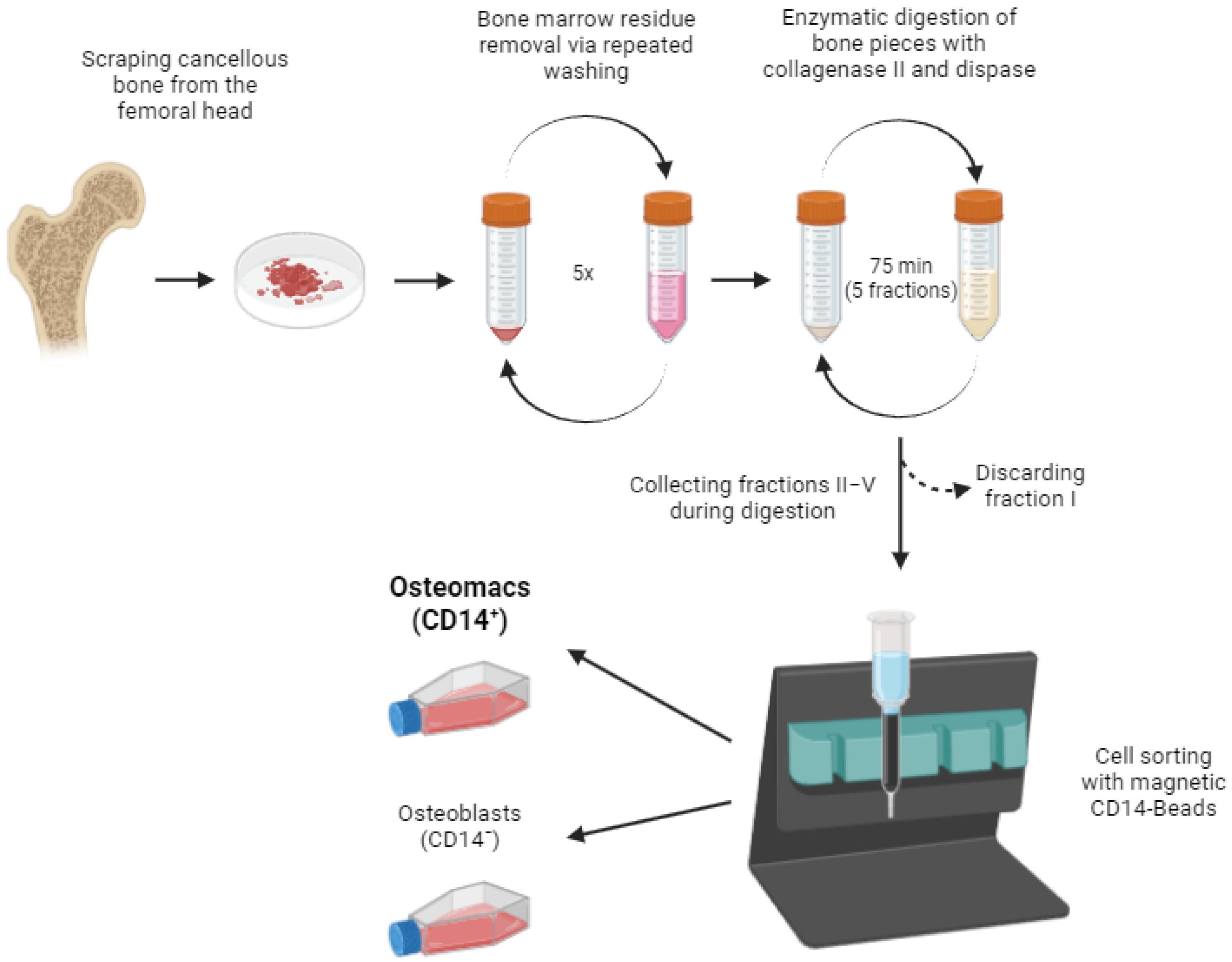
- The cancellous bone from human femoral heads was scraped out and the bone pieces were transferred into a 50 mL Falcon tube. In a representative isolation, the retrieved tissue mass was approximately 5 mg, but may vary between donors due to differences in tissue firmness and ease of retrieval. Supplementary Tables S1–S3 report the cell yields and donor characteristics for each isolation.
- Bone marrow removal (for optional use for bone marrow-derived macrophage (BMDM) or bone morrow-derived mesenchymal stem cells (BMSC) isolation):
- (2)
- The bone pieces were rinsed with 25 mL Wash Buffer and vigorously shaken. The supernatant was discarded.
- (3)
- 20 mL of Wash Medium was added and the falcon was placed on a tube roller for 10 min at 37 °C. The supernatant was discarded.
- (4)
- 20 mL of Wash Medium was added and rotated again for 10 min (37 °C). The supernatant was discarded.
- (5)
- Steps 3 and 4 were repeated.
- Release of osteoblasts and osteomacs from bone matrix by collagenase digestion:
- (6)
- 8 mL Collagenase II/Dispase solution was added to the bone pieces and incubated on a tube roller for 15 min at 37 °C. The supernatant (fraction I) was discarded.
- (7)
- Step 6 was repeated four times, each time using a fresh Collagenase II/Dispase solution. After digestion, the supernatant of each fraction was centrifuged at 400× g for 5 min to remove the enzymes. The cell pellet was resuspended in OB Medium and the cells from fractions II–V were pooled.
- (8)
- The pooled cell suspension was filtered through a 70 µm cell strainer.
- Isolation of Osteomacs by MACS®, based on the protocol by Miltenyi Biotec (Bergisch Gladbach, Germany):
- (9)
- Cells were centrifuged at 400× g for 5 min and transferred to a 2 mL reaction tube.
- (10)
- Cells were centrifuged again and resuspended in 80–160 µL MACS-Puffer (PBS with 0.5% BSA and 2 mM EDTA, pH = 7.2) depending on the cell number. 20–40 µL CD14-Beads (Miltenyi Biotec) were added and incubated for 15 min at 4 °C.
- (11)
- An MS column was prepared by placing it into the magnetic field of a MiniMACSTM Separator and rinsing it with 500 µL MACS buffer.
- (12)
- The cell suspension was carefully applied into the column and washed three times with 500 µL MACS buffer. CD14-negative osteoblasts were eluted during the washing steps.
- (13)
- The column was removed from the magnet and the CD14-positive Osteomacs were eluted by rinsing with 1 mL of MACS buffer.
- (14)
- After centrifugation (400× g, 5 min), the osteomacs were resuspended in OM Medium and seeded into a T75 cell culture flask. The OM-depleted osteoblast culture was resuspended in OB Medium and seeded into a T175 cell culture flask. The cell cultures were maintained in a humidified chamber at 37 °C with 5% CO2 with medium changes twice per week.
3.4. Fluorescence-Activated Cell Sorting
3.5. Immunfluorescence Staining
3.6. Toluidine and Phalloidin Staining
4. Expected Results
Supplementary Materials
Author Contributions
Funding
Institutional Review Board Statement
Informed Consent Statement
Data Availability Statement
Conflicts of Interest
Abbreviations
| α-MEM | alpha-Minimum Essential Medium Eagle |
| BMDM | Bone Marrow Derived Macrophages |
| BSA | Bovine Serum Albumine |
| FACS | Fluorescence Activated Cell Sorting |
| M-CSF | Macrophage Stimulating Factor |
| MACS | Magnetic Activated Cell Sorting |
| NGS | Normal Goat Serum |
| OM | Osteomacs |
| PFA | Paraformaldehyde |
| Pen/Strep | 1% Penicillin/Streptomycin |
| PBS | Phosphate-Buffered Saline |
References
- Arron, J.R.; Choi, Y. Bone versus immune system. Nature 2000, 408, 535–536. [Google Scholar] [CrossRef] [PubMed]
- Chang, M.K.; Raggatt, L.-J.; Alexander, K.A.; Kuliwaba, J.S.; Fazzalari, N.L.; Schroder, K.; Maylin, E.R.; Ripoll, V.M.; Hume, D.A.; Pettit, A.R. Osteal tissue macrophages are intercalated throughout human and mouse bone lining tissues and regulate osteoblast function in vitro and in vivo. J. Immunol. 2008, 181, 1232–1244. [Google Scholar] [CrossRef] [PubMed]
- Mills, C.D.; Kincaid, K.; Alt, J.M.; Heilman, M.J.; Hill, A.M. M-1/M-2 macrophages and the Th1/Th2 paradigm. J. Immunol. 2000, 164, 6166–6173. [Google Scholar] [CrossRef] [PubMed]
- Wang, Y.; Wang, Q.; Xu, Q.; Li, J.; Zhao, F. Single-cell RNA sequencing analysis dissected the osteo-immunology microenvironment and revealed key regulators in osteoporosis. Int. Immunopharmacol. 2022, 113, 109302. [Google Scholar] [CrossRef] [PubMed]
- Yan, Q.; Liu, H.; Zhu, R.; Zhang, Z. Contribution of macrophage polarization in bone metabolism: A literature review. Cytokine 2024, 184, 156768. [Google Scholar] [CrossRef] [PubMed]
- Mohamad, S.F.; Kacena, M.A. Isolation of Murine Neonatal and Adult Osteomacs to Examine Their Role in the Hematopoietic Niche. Methods Mol. Biol. 2024, 2939, 151–160. [Google Scholar]
- Jenkins, S.J.; Ruckerl, D.; Cook, P.C.; Jones, L.H.; Finkelman, F.D.; van Rooijen, N.; MacDonald, A.S.; Allen, J.E. Local macrophage proliferation, rather than recruitment from the blood, is a signature of TH2 inflammation. Science 2011, 332, 1284–1288. [Google Scholar] [CrossRef] [PubMed]
- Pelegrin, P.; Surprenant, A. Dynamics of macrophage polarization reveal new mechanism to inhibit IL-1beta release through pyrophosphates. EMBO J. 2009, 28, 2114–2127. [Google Scholar] [CrossRef] [PubMed]
- Vereyken, E.J.; Heijnen, P.D.; Baron, W.; de Vries, E.H.; Dijkstra, C.D.; Teunissen, C.E. Classically and alternatively activated bone marrow derived macrophages differ in cytoskeletal functions and migration towards specific CNS cell types. J. Neuroinflamm. 2011, 8, 58. [Google Scholar] [CrossRef] [PubMed]
- Rostam, H.M.; Reynolds, P.M.; Alexander, M.R.; Gadegaard, N.; Ghaemmaghami, A.M. Image based Machine Learning for identification of macrophage subsets. Sci. Rep. 2017, 7, 3521. [Google Scholar] [CrossRef] [PubMed]
- Ronzier, E.; Laurenson, A.J.; Manickam, R.; Liu, S.; Saintilma, I.M.; Schrock, D.C.; Hammer, J.A.; Rotty, J.D. The Actin Cytoskeleton Responds to Inflammatory Cues and Alters Macrophage Activation. Cells 2022, 11, 1806. [Google Scholar] [CrossRef] [PubMed]
- El Kasmi, K.C.; Qualls, J.E.; Pesce, J.T.; Smith, A.M.; Thompson, R.W.; Henao-Tamayo, M.; Basaraba, R.J.; König, T.; Schleicher, U.; Koo, M.-S.; et al. Toll-like receptor–induced arginase 1 in macrophages thwarts effective immunity against intracellular pathogens. Nat. Immunol. 2008, 9, 1399–1406. [Google Scholar] [CrossRef] [PubMed]
- Davis, M.J.; Tsang, T.M.; Qiu, Y.; Dayrit, J.K.; Freij, J.B.; Huffnagle, G.B.; Olszewski, M.A. Macrophage M1/M2 polarization dynamically adapts to changes in cytokine microenvironments in Cryptococcus neoformans infection. mBio 2013, 4, e00264–13. [Google Scholar] [CrossRef] [PubMed]



| Antibody/Conjugate | Reference Number | Company | Dilution |
|---|---|---|---|
| CD14-PE | 130-110-519 | Miltenyi Biotec | 1/50 |
| CD45-APC | 340910 | Miltenyi Biotec | 1/15 |
| ALP-Alexa Fluor 488 | NB110-3638AF488 | Novus Biologicals (Littleton, CO, USA) | 1/15 |
| Antibody (Clone) | Host/Target Species | Reference Number | Company | Dilution |
|---|---|---|---|---|
| CD14 (1H5D8) | mouse anti-human | ABIN1724922 | Antibodies-online GmbH (Aachen, Germany) | 1/500 |
| CD45 (2B11) | mouse anti-human | sc-20056 | Santa Cruz Biotechnology (Dallas, TX, USA) | 1/50 |
| CD68 (KP1) | mouse anti-human | sc-20060 | Santa Cruz Biotechnology | 1/50 |
| CD209 (UW60.1) | mouse anti-human | AM26379PU-N | OriGene Technologies, Inc. (Rockville, MD, USA) | 1/50 |
| CD86 | rabbit anti-human | bs-1035R | Bioss Antibodies (Woburn, MA, USA) | 1/100 |
| CD163 | rabbit anti-human | bs-23127R | Bioss Antibodies | 1/100 |
Disclaimer/Publisher’s Note: The statements, opinions and data contained in all publications are solely those of the individual author(s) and contributor(s) and not of MDPI and/or the editor(s). MDPI and/or the editor(s) disclaim responsibility for any injury to people or property resulting from any ideas, methods, instructions or products referred to in the content. |
© 2026 by the authors. Licensee MDPI, Basel, Switzerland. This article is an open access article distributed under the terms and conditions of the Creative Commons Attribution (CC BY) license.
Share and Cite
Bousch, J.F.; Lichtenberg, S.; Schnitker, M.; Schlösser, J.; Suschek, C.V.; Maus, U.; Beyersdorf, C. Isolation of Human Osteal Macrophages. Life 2026, 16, 376. https://doi.org/10.3390/life16030376
Bousch JF, Lichtenberg S, Schnitker M, Schlösser J, Suschek CV, Maus U, Beyersdorf C. Isolation of Human Osteal Macrophages. Life. 2026; 16(3):376. https://doi.org/10.3390/life16030376
Chicago/Turabian StyleBousch, Juliana Franziska, Stefanie Lichtenberg, Matthis Schnitker, Jenny Schlösser, Christoph Viktor Suschek, Uwe Maus, and Christoph Beyersdorf. 2026. "Isolation of Human Osteal Macrophages" Life 16, no. 3: 376. https://doi.org/10.3390/life16030376
APA StyleBousch, J. F., Lichtenberg, S., Schnitker, M., Schlösser, J., Suschek, C. V., Maus, U., & Beyersdorf, C. (2026). Isolation of Human Osteal Macrophages. Life, 16(3), 376. https://doi.org/10.3390/life16030376