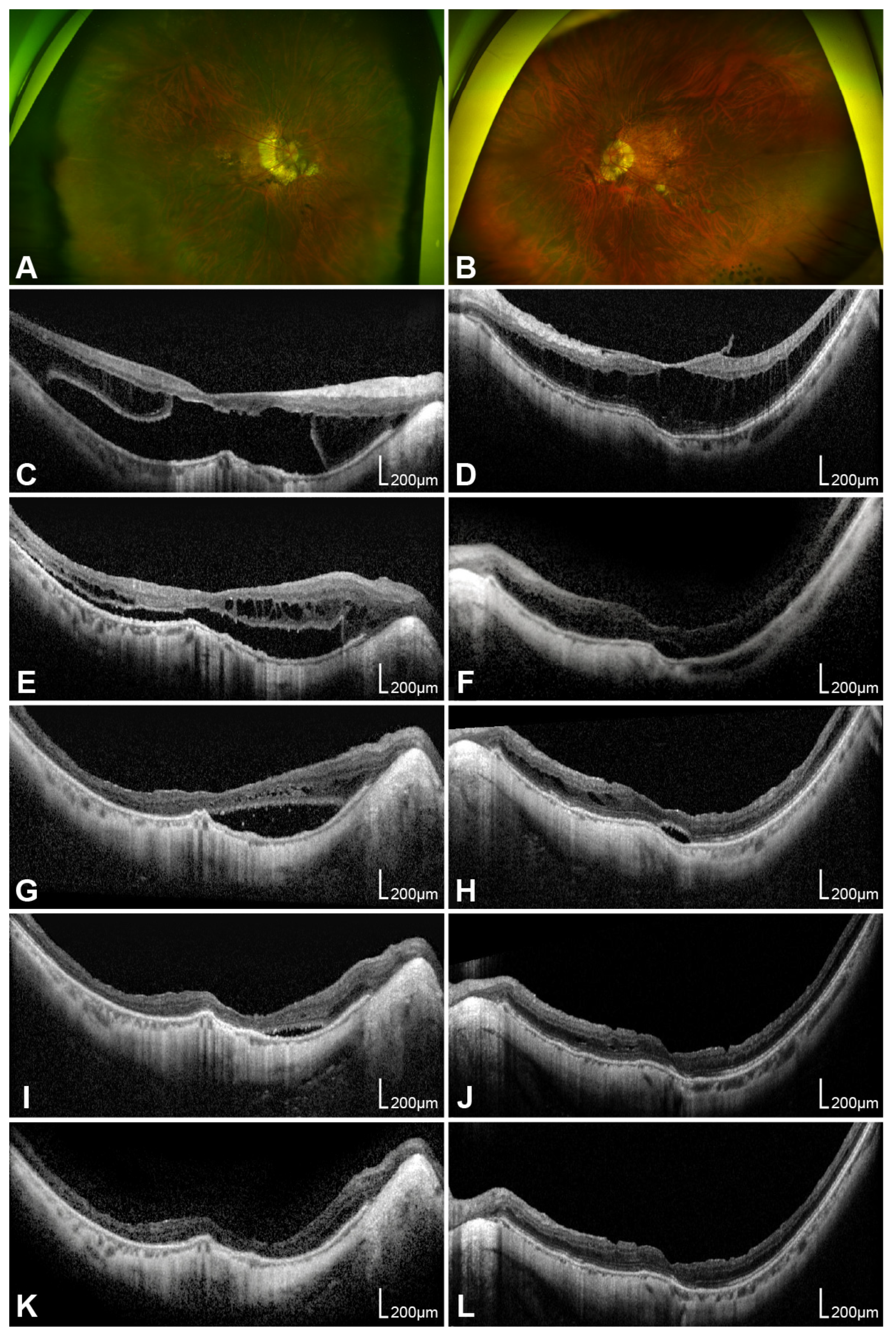
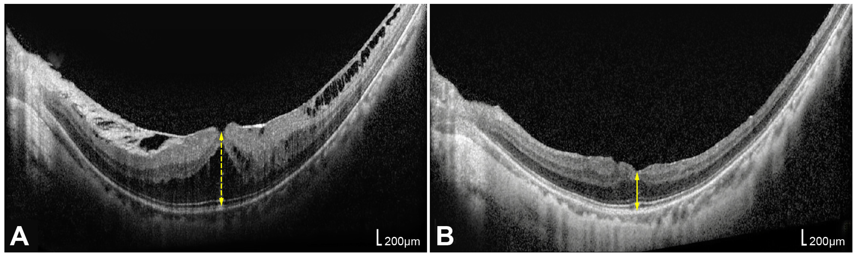
Preoperative Parameters Associated with Vitrectomy Outcomes in Myopic Traction Maculopathy Without a Full-Thickness Macular Hole

Abstract
1. Introduction
2. Materials and Methods
2.1. Ophthalmic Examinations
2.2. Classification of Myopic Maculopathy
2.3. Surgical Procedures
2.4. Statistical Analyses
3. Results
3.1. Baseline Characteristics
3.2. Visual and Anatomical Outcomes
3.3. Prognostic Factors
3.4. Influence of Combined Cataract Surgery on Outcomes
4. Discussion
4.1. Importance and Safety of ILM Peeling
4.2. Prognostic Factors for Functional and Anatomical Outcomes
4.3. Implications of Combined Cataract Surgery for Visual Outcomes
4.4. Optimal Timing for Surgery
4.5. Limitations and Further Research Directions
5. Conclusions
Supplementary Materials
Author Contributions
Funding
Institutional Review Board Statement
Informed Consent Statement
Data Availability Statement
Acknowledgments
Conflicts of Interest
Abbreviations
| SE | Spherical equivalent |
| D | Diopters |
| AXL | Axial length |
| PM | Pathologic myopia |
| MTM | Myopic traction maculopathy |
| RD | Retinal detachment |
| FD | Foveal detachment |
| LMH | Lamellar macular hole |
| FTMH | Full-thickness macular hole |
| MHRD | Macular hole-related retinal detachment |
| PPV | Pars plana vitrectomy |
| ILM | Internal limiting membrane |
| OCT | Optical coherence tomography |
| VMT | Vitreomacular traction |
| ERM | Epiretinal membrane |
| BCVA | Best-corrected visual acuity |
| logMAR | Logarithm of the minimum angle of resolution |
| FH | Foveal height |
| RPE | Retinal pigment epithelium |
| CFT | Central foveal thickness |
| SF6 | Sulfur hexafluoride |
| C3F8 | Perfluoropropane |
| EZ | Ellipsoid zone |
| MAM | Myopic atrophy maculopathy |
| MNM | Myopic neovascular maculopathy |
| PVD | Posterior vitreous detachment |
| CI | Confidence interval |
Appendix A

| Postoperative BCVA (logMAR) | Postoperative CFT (μm) | |||||||
|---|---|---|---|---|---|---|---|---|
| B | p a | 95% CI | p b | B | p a | 95% CI | p b | |
| Preoperative BCVA | 0.291 | 0.005 * | 0.097 to 0.485 | 0.345 | 5.232 | 0.820 | −41.330 to 51.794 | 0.945 |
| Axial length | 0.034 | 0.420 | −0.051 to 0.120 | 0.717 | −19.397 | 0.016 * | −34.867 to −3.928 | 0.396 |
| Foveal height | 0.001 | 0.002 * | <0.001 to 0.001 | 0.660 | 0.065 | 0.211 | −0.039 to 0.169 | 0.795 |
| MAM grade | 0.046 | 0.568 | −0.116 to 0.208 | 0.998 | −25.955 | 0.076 | −54.798 to 2.888 | 0.300 |
| MTM grade | 0.101 | 0.195 | −0.055 to 0.258 | 0.830 | −18.415 | 0.239 | −49.790 to 12.961 | 0.984 |
| ATN total grade | 0.03 | 0.456 | −0.051 to 0.110 | 0.869 | −11.530 | 0.137 | −26.947 to 3.887 | 0.934 |
| Foveal detachment | 0.430 | 0.063 | −0.025 to 0.886 | 0.215 | −11.629 | 0.797 | −103.537 to 80.278 | 0.443 |
| EZ disruption | 0.203 | 0.117 | −0.054 to 0.461 | 0.660 | −34.437 | 0.177 | −85.319 to 16.446 | 0.655 |
References
- Foster, P.J.; Jiang, Y. Epidemiology of myopia. Eye 2014, 28, 202–208. [Google Scholar] [CrossRef] [Scilit]
- Holden, B.A.; Fricke, T.R.; Wilson, D.A.; Jong, M.; Naidoo, K.S.; Sankaridurg, P.; Wong, T.Y.; Naduvilath, T.J.; Resnikoff, S. Global prevalence of myopia and high myopia and temporal trends from 2000 through 2050. Ophthalmology 2016, 123, 1036–1042. [Google Scholar] [CrossRef] [Scilit]
- Flitcroft, D.I.; He, M.; Jonas, J.B.; Jong, M.; Naidoo, K.; Ohno-Matsui, K.; Rahi, J.; Resnikoff, S.; Vitale, S.; Yannuzzi, L. IMI–defining and classifying myopia: A proposed set of standards for clinical and epidemiologic studies. Investig. Ophthalmol. Vis. Sci. 2019, 60, M20–M30. [Google Scholar] [CrossRef] [Scilit]
- Frisina, R.; Gius, I.; Palmieri, M.; Finzi, A.; Tozzi, L.; Parolini, B. Myopic traction maculopathy: Diagnostic and management strategies. Clin. Ophthalmol. 2020, 14, 3699–3708. [Google Scholar] [CrossRef] [Scilit]
- Ruiz-Medrano, J.; Montero, J.A.; Flores-Moreno, I.; Arias, L.; García-Layana, A.; Ruiz-Moreno, J.M. Myopic maculopathy: Current status and proposal for a new classification and grading system (ATN). Prog. Retin. Eye Res. 2019, 69, 80–115. [Google Scholar] [CrossRef] [Scilit] [PubMed]
- Cedrone, C.; Nucci, C.; Scuderi, G.; Ricci, F.; Cerulli, A.; Culasso, F. Prevalence of blindness and low vision in an Italian population: A comparison with other European studies. Eye 2006, 20, 661–667. [Google Scholar] [CrossRef] [Scilit] [PubMed]
- Ohno-Matsui, K.; Kawasaki, R.; Jonas, J.B.; Cheung, C.M.; Saw, S.M.; Verhoeven, V.J.; Klaver, C.C.; Moriyama, M.; Shinohara, K.; Kawasaki, Y.; et al. International photographic classification and grading system for myopic maculopathy. Am. J. Ophthalmol. 2015, 159, 877–883.e7. [Google Scholar] [CrossRef] [Scilit]
- Panozzo, G.; Mercanti, A. Optical coherence tomography findings in myopic traction maculopathy. Arch. Ophthalmol. 2004, 122, 1455–1460. [Google Scholar] [CrossRef] [Scilit] [PubMed]
- Parolini, B.; Palmieri, M.; Finzi, A.; Besozzi, G.; Frisina, R. Myopic traction maculopathy: A new perspective on classification and management. Asia-Pac. J. Ophthalmol. 2021, 10, 49–59. [Google Scholar] [CrossRef] [Scilit]
- Shimada, N.; Tanaka, Y.; Tokoro, T.; Ohno-Matsui, K. Natural course of myopic traction maculopathy and factors associated with progression or resolution. Am. J. Ophthalmol. 2013, 156, 948–957.e1. [Google Scholar] [CrossRef] [Scilit]
- Gaucher, D.; Haouchine, B.; Tadayoni, R.; Massin, P.; Erginay, A.; Benhamou, N.; Gaudric, A. Long-term follow-up of high myopic foveoschisis: Natural course and surgical outcome. Am. J. Ophthalmol. 2007, 143, 455–462. [Google Scholar] [CrossRef] [Scilit]
- Ikuno, Y.; Sayanagi, K.; Soga, K.; Oshima, Y.; Ohji, M.; Tano, Y. Foveal anatomical status and surgical results in vitrectomy for myopic foveoschisis. Jpn. J. Ophthalmol. 2008, 52, 269–276. [Google Scholar] [CrossRef] [Scilit]
- Feng, J.; Yu, J.; Chen, Q.; Zhou, H.; Chen, F.; Wang, W.; Xu, X.; Fan, Y. Long-term surgical outcomes and prognostic factors of foveal detachment in pathologic myopia: Based on the ATN classification. BMC Ophthalmol. 2022, 22, 175. [Google Scholar] [CrossRef] [Scilit]
- Snyder, A.A.; Thompson, F.B. A simplified technique for surgical treatment of degenerative myopia. Am. J. Ophthalmol. 1972, 74, 273–277. [Google Scholar] [CrossRef] [Scilit]
- Shevelev, M. Operation against high myopia and scleralectasia with aid of the transplantation of fascia lata on thinned sclera. Russ. Oftalmol. J. 1930, 11, 107–110. [Google Scholar]
- Schepens, C.L.; Okamura, I.D.; Brockhurst, R.J. The scleral buckling procedures. I. Surgical techniques and management. AMA Arch. Ophthalmol. 1957, 58, 797–811. [Google Scholar] [CrossRef] [Scilit]
- Shimada, N.; Ohno-Matsui, K.; Yoshida, T.; Sugamoto, Y.; Tokoro, T.; Mochizuki, M. Progression from macular retinoschisis to retinal detachment in highly myopic eyes is associated with outer lamellar hole formation. Br. J. Ophthalmol. 2008, 92, 762–764. [Google Scholar] [CrossRef] [Scilit] [PubMed]
- Borley, W.E.; Snyder, A.A. Surgical treatment of degenerative myopia; the combined lamellar scleral resection with scleral reinforcement using donor eye. Trans. Pac. Coast Otoophthalmol. Soc. Annu. Meet. 1958, 39, 275–291. [Google Scholar] [PubMed]
- Curtin, B.J. Scleral support of the posterior sclera. II. Clinical results. Am. J. Ophthalmol. 1961, 52, 853–862. [Google Scholar] [CrossRef] [Scilit]
- Momose, A. Surgical treatment of myopia--with special references to posterior scleral support operation and radial keratotomy. Indian J. Ophthalmol. 1983, 31, 759–767. [Google Scholar]
- Gonvers, M.; Machemer, R. A new approach to treating retinal detachment with macular hole. Am. J. Ophthalmol. 1982, 94, 468–472. [Google Scholar] [CrossRef] [Scilit]
- Ho, T.C.; Yang, C.M.; Huang, J.S.; Yang, C.H.; Yeh, P.T.; Chen, T.C.; Ho, A.; Chen, M.S. Long-term outcome of foveolar internal limiting membrane nonpeeling for myopic traction maculopathy. Retina 2014, 34, 1833–1840. [Google Scholar] [CrossRef] [Scilit] [PubMed]
- Shimada, N.; Sugamoto, Y.; Ogawa, M.; Takase, H.; Ohno-Matsui, K. Fovea-sparing internal limiting membrane peeling for myopic traction maculopathy. Am. J. Ophthalmol. 2012, 154, 693–701. [Google Scholar] [CrossRef] [Scilit]
- Kobayashi, H.; Kishi, S. Vitreous surgery for highly myopic eyes with foveal detachment and retinoschisis. Ophthalmology 2003, 110, 1702–1707. [Google Scholar] [CrossRef] [Scilit]
- Kanda, S.; Uemura, A.; Sakamoto, Y.; Kita, H. Vitrectomy with internal limiting membrane peeling for macular retinoschisis and retinal detachment without macular hole in highly myopic eyes. Am. J. Ophthalmol. 2003, 136, 177–180. [Google Scholar] [CrossRef] [Scilit]
- Chen, L.; Wei, Y.; Zhou, X.; Zhang, Z.; Chi, W.; Gong, L.; Jiang, X.; Zhang, S. Morphologic, biomechanical, and compositional features of the internal limiting membrane in pathologic myopic foveoschisis. Investig. Ophthalmol. Vis. Sci. 2018, 59, 5569–5578. [Google Scholar] [CrossRef] [Scilit] [PubMed]
- Zhang, Z.; Wei, Y.; Jiang, X.; Zhang, S. Pars plana vitrectomy and wide internal limiting membrane peeling with perfluoropropane tamponade for highly myopic foveoschisis-associated macular hole. Retina 2017, 37, 274–282. [Google Scholar] [CrossRef] [Scilit] [PubMed]
- Kim, C.Y.; Kim, M.S.; Kim, K.L.; Woo, S.J.; Park, K.H. Prognostic factors related with surgical outcome of vitrectomy in myopic traction maculopathy. Korean J. Ophthalmol. 2020, 34, 67–75. [Google Scholar] [CrossRef] [Scilit]
- Shin, J.Y.; Yu, H.G. Visual prognosis and spectral-domain optical coherence tomography findings of myopic foveoschisis surgery using 25-gauge transconjunctival sutureless vitrectomy. Retina 2012, 32, 486–492. [Google Scholar] [CrossRef] [Scilit]
- Hattori, K.; Kataoka, K.; Takeuchi, J.; Ito, Y.; Terasaki, H. Predictive factors of surgical outcomes in vitrectomy for myopic traction maculopathy. Retina 2018, 38, S23–S30. [Google Scholar] [CrossRef] [Scilit]
- Kumagai, K.; Furukawa, M.; Ogino, N.; Larson, E. Factors correlated with postoperative visual acuity after vitrectomy and internal limiting membrane peeling for myopic foveoschisis. Retina 2010, 30, 874–880. [Google Scholar] [CrossRef] [Scilit]
- Lee, K.S.; Lee, J.S.; Koh, H.J. Surgical outcomes of myopic traction maculopathy according to the international photographic classification for myopic maculopathy. Retina 2020, 40, 1492–1499. [Google Scholar] [CrossRef] [Scilit]
- Li, S.; Li, T.; Wang, X.; Cai, X.; Lu, B.; Chen, Y.; Liu, C.; Wu, Q. Natural course of myopic traction maculopathy and factors influencing progression and visual acuity. BMC Ophthalmol. 2021, 21, 347. [Google Scholar] [CrossRef] [Scilit]
- Shinohara, K.; Tanaka, N.; Jonas, J.B.; Shimada, N.; Moriyama, M.; Yoshida, T.; Ohno-Matsui, K. Ultrawide-field OCT to investigate relationships between myopic macular retinoschisis and posterior staphyloma. Ophthalmology 2018, 125, 1575–1586. [Google Scholar] [CrossRef] [Scilit] [PubMed]
- Carrero, J.L. Incomplete posterior vitreous detachment: Prevalence and clinical relevance. Am. J. Ophthalmol. 2012, 153, 497–503. [Google Scholar] [CrossRef] [Scilit]
- Falkner-Radler, C.I.; Glittenberg, C.; Hagen, S.; Benesch, T.; Binder, S. Spectral-domain optical coherence tomography for monitoring epiretinal membrane surgery. Ophthalmology 2010, 117, 798–805. [Google Scholar] [CrossRef] [Scilit]
- Mitamura, Y.; Hirano, K.; Baba, T.; Yamamoto, S. Correlation of visual recovery with presence of photoreceptor inner/outer segment junction in optical coherence images after epiretinal membrane surgery. Br. J. Ophthalmol. 2009, 93, 171–175. [Google Scholar] [CrossRef] [Scilit]
- Fujimoto, S.; Ikuno, Y.; Nishida, K. Postoperative optical coherence tomographic appearance and relation to visual acuity after vitrectomy for myopic foveoschisis. Am. J. Ophthalmol. 2013, 156, 968–973.e1. [Google Scholar] [CrossRef] [Scilit] [PubMed]
- Inoue, M.; Morita, S.; Watanabe, Y.; Kaneko, T.; Yamane, S.; Kobayashi, S.; Arakawa, A.; Kadonosono, K. Inner segment/outer segment junction assessed by spectral-domain optical coherence tomography in patients with idiopathic epiretinal membrane. Am. J. Ophthalmol. 2010, 150, 834–839. [Google Scholar] [CrossRef] [Scilit] [PubMed]
- Meng, N.; Xia, L.; Gong, Y.; Shi, C.; Zhao, Z. Correlation between anatomical and functional outcomes in patients with idiopathic epiretinal membrane after vitrectomy. Discov. Med. 2023, 35, 975–981. [Google Scholar] [CrossRef] [Scilit]

| Baseline Parameters | Values |
|---|---|
| Age (years) | 62.4 ± 11.2 (39–75) |
| Sex, male:female, n (%) | 5:28 (15.2%:84.8%) |
| BCVA (logMAR) | 0.74 ± 0.52 |
| SE (diopter) | −12.54 ± 5.28 (n = 17) |
| Refractive Surgery history, n (%) | 9 (27.3%) |
| Axial length (mm) | 28.48 ± 1.40 |
| Lens status, n (%) | |
| Cataract | 25 (75.8%) |
| No cataract | 1 (3.0%) |
| Pseudophakia | 7 (21.2%) |
| Preoperative OCT parameters | |
| Foveal height(μm) | 408.7 ± 211.3 |
| ERM, n (%) | 27 (81.8%) |
| VMT, n (%) | 17 (51.5%) |
| Foveal detachment, n (%) | 7 (21.2%) |
| EZ disruption, n (%) | 8 (24.2%) |
| ATN classification, n (%) | |
| MAM grade 0:1:2:3:4 | 1:21:6:5:0 (3.0%:63.6%:18.2%:15.2%:0%) |
| MTM grade 0:1:2:3:4:5 | 0:16:10:7:0:0 (0%:48.5%:30.3%:21.2%:0%:0%) |
| MNM grade 0:1:2a:2s | 30:1:0:2 (90.9%:3.0%:0%:6.1%) |
| Operational parameters (%) | |
| Combined Phacoemulsification, n (%) | 23 (69.7%) |
| Gas, SF6: C3F8, n (%) | 2:31 (6.1%:93.9%) |
| Preoperative | Postoperative (12 Months) | Change | p Value | |
|---|---|---|---|---|
| BCVA (logMAR) | 0.744 ± 0.52 | 0.299 ± 0.30 | −0.445 ± 0.41 | <0.001 |
| Foveal thickness (μm) | 408.7 ± 211.3 (FH) | 211.2 ± 58.5 (CFT) | −201.4 ± 216.0 | <0.001 |
| Postop BCVA (logMAR) | Postop CFT (μm) | ||||
|---|---|---|---|---|---|
| r | p value a | r | p value a | ||
| Age (years) | 0.409 | 0.020 * | −0.365 | 0.040 * | |
| Preop BCVA (logMAR) | 0.528 | 0.002 * | −0.276 | 0.126 | |
| SE (diopter) | −0.187 | 0.304 | 0.194 | 0.286 | |
| Axial length (mm) | 0.283 | 0.117 | −0.494 | 0.004 * | |
| Foveal height (μm) | 0.380 | 0.032 * | −0.060 | 0.743 | |
| MAM grade | 0.378 | 0.033 * | −0.528 | 0.002 * | |
| MTM grade | 0.409 | 0.020 * | −0.368 | 0.038 * | |
| MNM grade | 0.081 | 0.659 | −0.200 | 0.272 | |
| ATN total grade | 0.385 | 0.030 * | −0.425 | 0.015 * | |
| Mean ± SD | p value b | Mean ± SD | p value b | ||
| Gender | M (n = 5) | 0.564 ± 0.433 | 277.6 ± 53.0 | ||
| F (n = 27) | 0.254 ± 0.257 | 0.078 | 198.9 ± 52.7 | 0.011 * | |
| Lens status | Cataract (n = 25) | 0.287 ± 0.325 | 215.1 ± 56.7 | ||
| No cataract or Pseudophakia (n = 7) | 0.358 ± 0.230 | 0.310 | 197.3 ± 71.6 | 0.274 | |
| Phacoemulsification | Yes (n = 23) | 0.281 ± 0.336 | 214.7 ± 59.2 | ||
| No (n = 9) | 0.357 ± 0.211 | 0.218 | 202.3 ± 62.8 | 0.379 | |
| ERM | Present (n = 27) | 0.297 ± 0.315 | 215.0 ± 61.2 | ||
| Absent (n = 5) | 0.330 ± 0.274 | 0.693 | 191.0 ± 49.5 | 0.392 | |
| VMT | Present (n = 17) | 0.381 ± 0.339 | 212.9 ± 59.5 | ||
| Absent (n = 15) | 0.213 ± 0.242 | 0.112 | 209.3 ± 61.4 | 0.880 | |
| Foveal detachment | Present (n = 7) | 0.528 ± 0.373 | 168.6 ± 68.5 | ||
| Absent (n = 25) | 0.239 ± 0.257 | 0.030 * | 223.2 ± 52.1 | 0.036 * | |
| EZ disruption | Present (n = 8) | 0.489 ± 0.362 | 174.8 ± 65.8 | ||
| Absent (n = 24) | 0.240 ± 0.263 | 0.047 * | 223.4 ± 53.2 | 0.045 * | |
| Gas tamponade | SF6 (n = 2) | 0.049 ± 0.069 | 218.5 ± 2.1 | ||
| C3F8 (n = 30) | 0.319 ± 0.307 | 0.180 | 210.7 ± 61.4 | 1.000 | |
| Postoperative BCVA (logMAR) | Postoperative CFT (μm) | |||||
|---|---|---|---|---|---|---|
| B | p Value | 95% CI | B | p Value | 95% CI | |
| Preop BCVA (logMAR) | 0.356 | <0.001 * | 0.189 to 0.524 | −12.588 | 0.540 | −54.036 to 28.860 |
| Axial length (mm) | 0.058 | 0.137 | −0.019 to 0.135 | −20.616 | 0.004 * | −34.193 to −7.038 |
| Foveal height (μm) | 0.001 | <0.001 * | <0.001 to 0.001 | 0.025 | 0.616 | −0.077 to 0.127 |
| MAM grade | 0.102 | 0.138 | −0.035 to 0.239 | −37.588 | 0.003 * | −61.453 to −13.723 |
| MTM grade | 0.152 | 0.022 * | 0.024 to 0.280 | −26.139 | 0.045 * | −51.610 to −0.668 |
| ATN total grade | 0.057 | 0.063 | −0.003 to 0.118 | −14.392 | 0.015 * | −25.721 to −3.062 |
| Foveal detachment | 0.289 | 0.024 * | 0.041 to 0.537 | −54.589 | 0.029 * | −103.266 to −5.912 |
| EZ disruption | 0.249 | 0.043 * | 0.008 to 0.490 | −48.625 | 0.043 * | −95.620 to −1.630 |
| Postoperative BCVA (logMAR) | Postoperative CFT (μm) | |||||
|---|---|---|---|---|---|---|
| B | p Value | 95% CI | B | p Value | 95% CI | |
| Preop BCVA (logMAR) | 0.317 | 0.002 * | 0.132 to 0.502 | 5.694 | 0.792 | −38.033 to 49.421 |
| Axial length (mm) | 0.029 | 0.458 | −0.051 to 0.110 | −17.366 | 0.021 * | −31.974 to −2.757 |
| Foveal height (μm) | 0.001 | <0.001 * | <0.001 to 0.001 | 0.062 | 0.213 | −0.037 to 0.161 |
| MAM grade | 0.046 | 0.525 | −0.100 to 0.192 | −31.943 | 0.019 * | −58.352 to −5.533 |
| MTM grade | 0.109 | 0.118 | −0.029 to 0.246 | −18.278 | 0.187 | −45.913 to 9.356 |
| ATN total grade | 0.033 | 0.310 | −0.033 to 0.099 | −11.181 | 0.081 | −23.815 to 1.452 |
| Foveal detachment | 0.202 | 0.137 | −0.068 to 0.472 | −39.865 | 0.139 | −93.389 to 13.659 |
| EZ disruption | 0.186 | 0.126 | −0.055 to 0.428 | −37.830 | 0.116 | −85.577 to 9.918 |
| Phaco-Vitrectomy Group | Vitrectomy-Only Group | p Value | |
|---|---|---|---|
| Age | 63.0 ± 9.6 | 58.3 ± 10.1 | 0.200 a |
| Preop BCVA (logMAR) | 0.843 ± 0.575 | 0.531 ± 0.326 | 0.175 a |
| Axial length (mm) | 28.39 ± 1.31 | 28.54 ± 1.76 | 0.900 a |
| Preop Foveal height (μm) | 409.1 ± 229.0 | 421.6 ± 194.3 | 0.900 a |
| ERM, n (present:absent) | 20:3 | 7:2 | 0.604 b |
| VMT, n (present:absent) | 12:11 | 5:4 | 1 b |
| FD, n (present:absent) | 4:19 | 3:6 | 0.370 b |
| EZ disruption, n (present:absent) | 4:19 | 4:5 | 0.176 b |
| MAM grade | 1.39 ± 0.78 | 1.56 ± 0.88 | 0.691 a |
| MTM grade | 1.74 ± 0.75 | 1.67 ± 1.0 | 0.648 a |
| MNM grade | 0.13 ± 0.46 | 0.22 ± 0.67 | 0.836 a |
| ATN total grade | 3.26 ± 1.63 | 3.44 ± 2.19 | 0.807 a |
| BCVA (logMAR) | Retinal Thickness (μm) | |||||||
|---|---|---|---|---|---|---|---|---|
| Preop BCVA | Postop BCVA | BCVA Change | p a | Preop FH (μm) | Postop CFT (μm) | Thickness Change | p a | |
| Phaco-vitrectomy | 0.84 ± 0.57 | 0.28 ± 0.34 | −0.56 ± 0.42 | <0.001 | 409.1 ± 229.0 | 214.7 ± 59.2 | −194.4 ± 228.6 | <0.001 |
| Vitrectomy-only | 0.53 ± 0.33 | 0.36 ± 0.21 | −0.17 ± 0.25 | 0.092 | 421.6 ± 194.3 | 202.3 ± 62.8 | −219.2 ± 205.8 | 0.019 |
| p value b | 0.017 | 0.615 | ||||||
Disclaimer/Publisher’s Note: The statements, opinions and data contained in all publications are solely those of the individual author(s) and contributor(s) and not of MDPI and/or the editor(s). MDPI and/or the editor(s) disclaim responsibility for any injury to people or property resulting from any ideas, methods, instructions or products referred to in the content. |
© 2026 by the authors. Licensee MDPI, Basel, Switzerland. This article is an open access article distributed under the terms and conditions of the Creative Commons Attribution (CC BY) license.
Share and Cite
Lee, S.K.; Yeo, S.; Chung, Y.-R.; Kim, H.R.; Song, J.H. Preoperative Parameters Associated with Vitrectomy Outcomes in Myopic Traction Maculopathy Without a Full-Thickness Macular Hole. Life 2026, 16, 356. https://doi.org/10.3390/life16020356
Lee SK, Yeo S, Chung Y-R, Kim HR, Song JH. Preoperative Parameters Associated with Vitrectomy Outcomes in Myopic Traction Maculopathy Without a Full-Thickness Macular Hole. Life. 2026; 16(2):356. https://doi.org/10.3390/life16020356
Chicago/Turabian StyleLee, Su Kyung, Suji Yeo, Yoo-Ri Chung, Hae Rang Kim, and Ji Hun Song. 2026. "Preoperative Parameters Associated with Vitrectomy Outcomes in Myopic Traction Maculopathy Without a Full-Thickness Macular Hole" Life 16, no. 2: 356. https://doi.org/10.3390/life16020356
APA StyleLee, S. K., Yeo, S., Chung, Y.-R., Kim, H. R., & Song, J. H. (2026). Preoperative Parameters Associated with Vitrectomy Outcomes in Myopic Traction Maculopathy Without a Full-Thickness Macular Hole. Life, 16(2), 356. https://doi.org/10.3390/life16020356