Article

The Human Penile Fibro-Vascular Assembly Requires the Integrity of Ten Fibro-Ligaments

1
Microsurgical Potency Reconstruction and Research Center, Puli Christian Hospital, No. 1, Tieshan Rd., Puli, Nantou 54546, Taiwan
2
Graduate Institute of Clinical Medicine, College of Medicine, National Taiwan University, Taipei 100225, Taiwan
3
Microsurgical Potency Reconstruction Center, Shu-Tien Urology Ophthalmology Clinic, The Yin Shu-Tien Medical Foundation, Taipei 10662, Taiwan
4
Educational Resources Section, China Medical University Hospital, Taichung 40402, Taiwan
5
Department of Anatomy, China Medical University Hospital, Taichung 40402, Taiwan
*
Author to whom correspondence should be addressed.
Life 2025, 15(9), 1492; https://doi.org/10.3390/life15091492
Submission received: 12 June 2025 / Revised: 4 August 2025 / Accepted: 16 September 2025 / Published: 22 September 2025

Abstract

Introduction: Penile fibro-ligaments have been extensively studied for many centuries; however, there is, unfortunately, a lack of thorough understanding. We aimed to bridge the research gap between anatomy and surgical reconstruction. Methods: We excluded cadaveric penises, already dissected and disassembled by medical students, that had damaged the corpora cavernosa (CC) or corpus spongiosum (CS). However, penises were included if both the ischiocavernosus and bulbospongiosus muscles were undamaged. A total of 8 out of 52 penises were meticulously examined. Our dissection findings were supplemented with 101 cadaveric photos, 255 penile vascular surgeries, 11 CT imaging scans, and 8 MRI imaging scans. The combined understanding was reprocessed with radiographic imaging, and patients underwent penile surgeries, notably eight hypospadias surgeries, and eight penile elongation surgeries were performed elsewhere. Results: Bilaterally, the penile CC is primarily anchored to the pelvic wall through the cavernosal ligament (CL = 2), while the CS is connected to the urinary sphincter. The suspensory, fundiform, and arcuate pubic ligament (two anatomically and one functionally) assist in stabilizing and holding the penile shaft to the pelvic wall. Distally, a distal ligament (DL = 1) and spongiosal ligament (SL = 1) extend the CC and CS to the glans penis and frenulum, ensuring urethral patency when necessary. The CC is encircled by a bi-layered tunica consisting of a 360° inner circular and a 300° outer longitudinal tunica. The ischiocavernosus muscle wraps around the penile crus and envelops the CL, connecting to the ischial tuberosity. The CS is partially surrounded by the bulbospongiosus muscle proximally and receives the SL distally. The entire penis interconnects with the skeletal muscle of the urogenital diaphragm. Conclusion: The physiological integrity of the human penis relies on ten anatomically and six functionally fibro-muscular ligaments.

1. Introduction

Practitioner Points

1.
As a male potency reconstructive practitioner, maintaining the integrity of the penile fibro-vascular assembly is second to none. The human penis is the ultimate coevolutionary nature as an intromittent organ. The penile fibro-vascular assembly relies on a combination to ensure its physiological integrity.
2.
The penile fibrous, anatomical ten and functionally six, play a structural role in supporting and stabilizing the various components of the penis; these ligaments include the arcuate pubic ligament, cavernosal ligaments, distal ligament, fundiform ligament, spongiosal ligament, and suspensory ligament.
3.
Given the importance of these ligaments in maintaining penile sexual health, caution should be taken when considering procedures such as ligamentolysis, which involves the release or modification of these ligaments; preserving the integrity and functionality of the ligaments is essential for ensuring optimal physiological and functional outcomes in the penile fibro-vascular assembly.
It is widely accepted that evolution plays a significant role in shaping the development of animal species on our planet [1]. Among these, humans are considered the most advanced and have dominated the Earth for three thousand centuries [2]. An intriguing aspect of human evolution is the coevolution of male genitalia, resulting in the unique intriguing micro-architectures of the penis. This unique structure enables piston-like mechanical interaction with the female vagina during copulation [3]. Such interaction is crucial for successful reproduction and the transmission of genetic material from one generation to the next, especially before the advent of in vitro fertilization (IVF) [4]. Recent reviews have provided substantial evidence for the evolution of genital structures, highlighting the coevolution of male and female genitalia in vertebrates [5,6]. Ongoing research endeavors to comprehend the mechanics of copulatory interaction, which necessitates a functional erectile system [7,8].
The human body is remarkable, equipped with a bony skull that safeguards the brain. This skull is connected to the body’s trunk through the cervical spine, which is essentially enhanced by fibrillar ligaments. The skeletal structure supports the body and enables unique movements, such as shaking the head, made possible by the atlas bone [9]. The body-supportive framework, formed by combining bony structures and fibro-muscular elements [10]. The body is articulated, with limbs serving the purposes of living and locomotion. The body core is centrally positioned on each level of the extremity, while the body elements are outside the body. The upper and lower limbs contain multiple joints at various levels, each associated with ten fingers and toes, as well as numerous ligaments [9].
Interestingly, the human penis is a fibrovascular assembly that has recently evolved as an independent compartment rather than an extension of the cardiovascular system [11]. It lacks bones and relies instead on centrally located hydraulic sinusoids to provide bony rigidity when needed [12]. As a protruding member of the extremity family, the human penis stands apart from others by lacking articulation and a central core supporter; however, it still adheres to the general design principle observed throughout the body, where a rigid supporter is typically centrally located in the case of the penis, this role is fulfilled by the functional sinusoids found in the CC and CS. Despite these differences, the penile design follows the same principle, albeit without the presence of bones. Therefore, the boneless penile CC can serve as an ideal milieu for applying Pascal’s law to the entire human body, given that the CC chamber remains free from veno-occlusive dysfunction (VOD) [13].
Furthermore, according to Pascal’s law, applied pressure remains constant within an enclosed space [14]. This implies that the penile structure requires collagen bundles, essential ingredients of bone, with the strength of steel to meet the requirements [15]. Not surprisingly, the CC can withstand intracorporeal pressure of 1800–2000 mmHg without rupturing [16]. To ensure this resilience, the outer shell of the CC, known as the bi-layered tunica albuginea (TA), particularly its outer coat, must be tightly stretched and exceptionally tough [17]. TA comprises multiple collagen bundles that run parallel to the skeletal structure, similar to the muscular system of the regular skeletal and muscular systems; categorically, it contains numerous fibrous ligaments.
A ligament is a fibrous connective tissue that connects bones or organs to other bones or organs [18]. As a result, ligaments are commonly found in various internal organs, including the fetal remnant, the peritoneal, and the falciform ligaments. Round ligaments are present in the urogenital system, where ligaments such as the inguinal, testicular, lacunar, fundiform, and suspensory ligaments are present [19,20]. Thus, the concept of the ligament is widely accepted in the field of penile fibro-vascular anatomy [21]. In our study, we explored the human penile fibro-vascular assembly using multiple-planar approaches; this exploration aimed to elucidate the last remaining independent vascular component in the human body [22]. However, due to the scarcity of detailed descriptions of the human penile fibro-ligaments in the medical literature, we were inspired to study the penile fibromuscular ligaments to understand their role in maintaining sexual health.

2. Materials and Methods

The Institute Review Board of China Medical University approved the study. The penises used in the study were already dissected, and the intromittent pendulous portion was disassembled into 2.5 cm cross-section blocks by medical students [23,24]. Penises were excluded from the study if both sides of the corpora cavernosa (CC) and corpus spongiosum (CS) were damaged or if a median sagittal resection had already been performed on the blocks. However, they were included in the study if a unilateral side of the CC remained undamaged with an intact Colle’s fascia. As a result, 15 out of 46 cadaveric penises were meticulously examined.
The distal blocks of the penises were prepared as median, 2.5 mm, and 5.0 mm sagittal sections from the median to the lateral. These sections were then examined microscopically, with analysis of the cut surfaces (Figure 1A,B). Additionally, clinical patients underwent hemodynamic cavernosograms by injecting 10 mL of contrast medium via a # 19 scalp needle to obtain intracavernous images (Figure 1C,D) [25]. Furthermore, dissection findings were correlated with 101 cadaveric photos and various diagnostic imaging techniques, including 255 sets of dual cavernosograpy from patients who underwent penile vascular surgery. Additionally, analysis was performed on 11 CT imaging scans (Ingenia 3T), 8 MRI (3T Philips MRI) imaging scans (Figure 2 and Figure 3), and five sets of spongiosographyy. Moreover, an information analysis was conducted on eight men with erectile dysfunction (ED) who had standard erectile capabilities before undergoing ligamentolysis elsewhere.
This analysis focused on dual cavernosography and emphasizing the venous vasculature related to erection (Figure 4). Additionally, eight patients with hypospadias were questioned to substantiate the role of the ejaculation bolus that might be related to the SL guarding meatal patency.

3. Results

Table 1 summarizes the ten human penile fibro-muscular ligaments. The human penis is a uniquely protruding organ composed of multiple fascial layers that surround the three cylinders of erectile sinusoids. It consists of the glans penis, the paired corpora cavernosa (CC) enveloped by the ischiocavernosus muscles, and the corpus spongiosum belted by the bulbospongiosus muscle. The CC is surrounded by a thin cavernosal membrane (Figure 1B). It is then anchored to the body trunk through the bilateral cavernosal ligament (CL = 2), while the CS contains the urethra connected to the urinary sphincter.
In the pubic region, proceeding from the median to lateral and from extra to intra-pelvis, there is the suspensory ligament (anatomically 2, functionally 1), the fundiform ligaments (anatomically 2, functionally 1), and the arcuate pubic ligament (anatomically 2, functionally 1), These ligaments sequentially stabilize and hold the penile shaft to the body trunk. Distally, there is a distal ligament (DL = 1), an os analog, and a spongiosal ligament (SL = 1). These ligaments project the CC and CS, respectively, to ensure the patency of the urethral meatus when necessary.
This physiological function is likely evidenced by the absence of hypospadias in the eight patients included in this study. Moreover, the SL is arrayed in a gillnetting manner with the primary 180° across-street position to DL (Figure 1A,B), which ensures the patency for ejaculate squirting. Finally, the CC is encircled by a bi-layered fibrous tunica model with 360° inner circular tunica albuginea and 300° outer longitudinal tunica albuginea, within which fibro-muscular tissue is expressed. The skeletal type includes bilateral CL, anchoring the penis to the pelvic wall at the ischial tuberosity. The CC sinusoids are 8.0–1.5 mm, evidenced in various imaging (Figure 1, Figure 2 and Figure 3) distally. Proximally, the bilateral ischiocavernosus sockets the CC, 300° outer longitudinal tunica, and DL in the glans penis, bulbospongiosus partially surrounds the CS and forms the ventral thickening of the outer tunica at the 5 and 7 O’clock positions, respectively, and a spongiosal ligament that anchors the CS to the frenulum. Proximally, the entire penis intertwines with the skeletal muscle of the urogenital diaphragm. The smooth component makes an exclusively hydraulic milieu, such as the penile artery, venous structure, intracavernosal pillars, and three specific sinusoids.
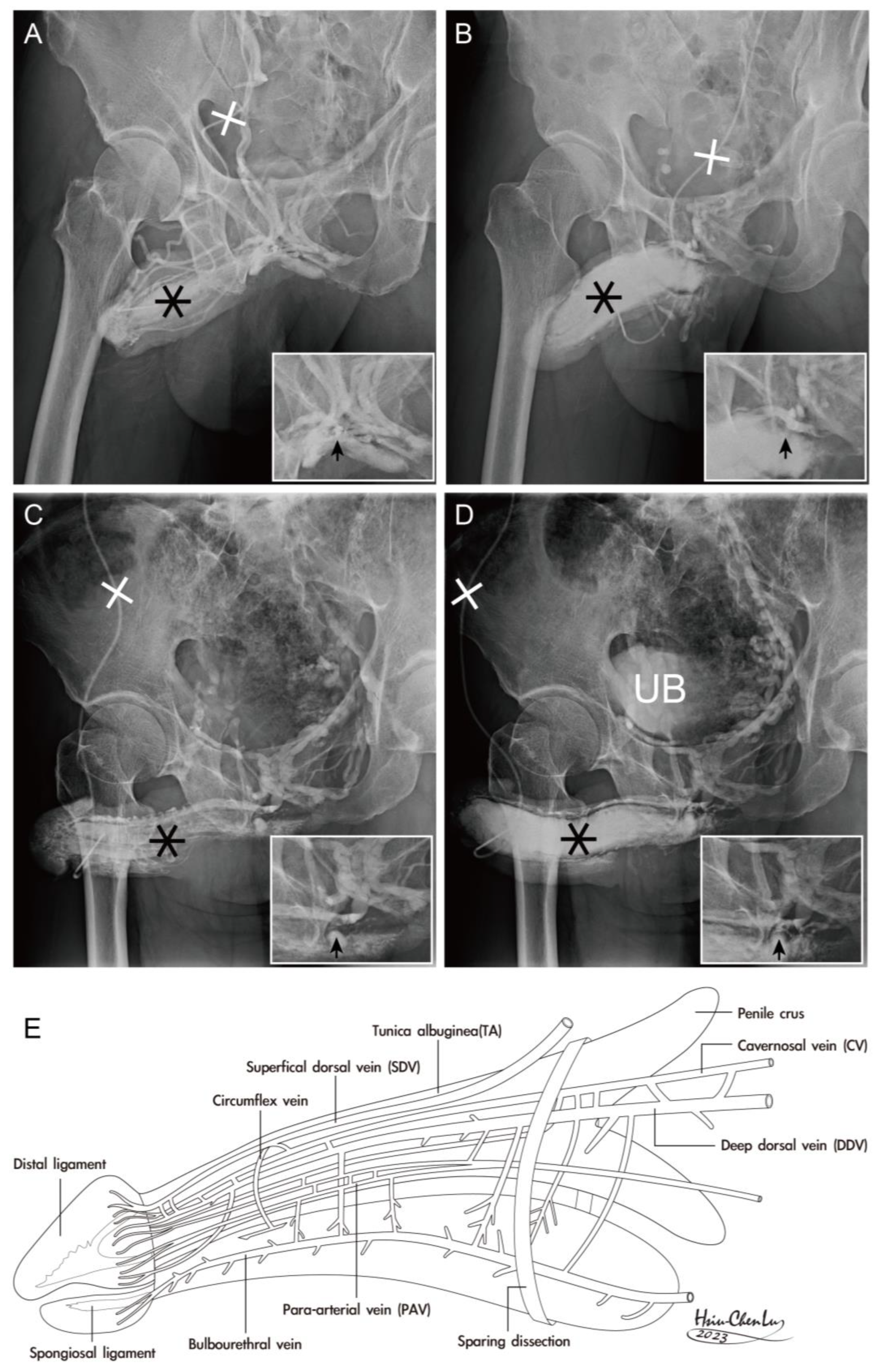
The sparing of either suspensory or fundiform ligament is challenging in penile ligamentolysis lengthening (Figure 4), as evidenced by the uneven lineation of the entire dorsal aspect of the CC and the loss of fibro-muscular banding that limits the erection-related veins. In addition, skeletal components encase the smooth part, which also serves as both a blood supply and drainage conduit in the paramount role of the integral hydraulic assembly. Consequently, a lateral aspect of the median-sagittal and para-medial sagittal section and a ventral view of the entire human penis is illustrated (Figure 5).

4. Discussion

In most quadriceps, the glans penis is supported by an os penis, which aids intromission and facilitates mating [26]. However, humans have lost the os penis, although there is an analog structure in histology [27]. Therefore, the human penis relies on the hydraulic CC to provide necessary rigidity during sexual activity. Thus, the ultimate hydraulic corpora cavernosa can meet the necessity of bony rigidity when required because a rigid intromittent human penis plays an essential role in intromission sex. Human penile erectile function is a delicately hydraulic event in which a seamless interplay of vascular, hormonal, psychological, neurological, pharmacological, sinusoidal, and metabolic contributors is involved [28]. The healthy vascular factor is essential in the sinusoidal function in the CC, CS, and the glans penis, independently. Physiologically, a penile, rigid erection ensues if and only if penile arterial, sinusoidal, and venous factors exist, while the corporal-veno-occlusive mechanism works vigorously. Thus, the integrity of penile fibro-vascular apparatus is a parallel prerequisite in erectile function corresponding with a cotton-soft flaccid arterial supply just being 2–3 mL/min, and bone-rigid erection the arterial supply being surged 30-fold to 60–80 mL/min for erection purposes [29], implying, upon activation, blood flow to the CC sinusoids will reach 240–320 mL/min if the penile arteries function bilaterally [30].
Many researchers are proud of the ultimate primordial human owning hydraulic erectile sinusoids gifted with magician-like erection rigidity for stroking the glans against the vaginal wall of a female counterpart. Otherwise, a penile bone is awkward in locomotion, for we erect human beings; therefore, humans must thank the creature for giving us a soft fibro-skeleton penis that can transform into a bony rigidity compartment, when necessary. There must be lots of coevolutionary fibrous ligaments inside and outside the penile fibro-vascular assembly, like a wooden house negating a nail. Whether the organ is healthy depends upon its ten ligamentous integrities. Interestingly, these ten numbers are inadvertently the same as the number of fingers and toes.
Although the human penis is generously extruded, its anatomy seems too simple-looking to conduct further study in the medical literature thus far [31]. The conventional human penile anatomy, a presumed illustration by sagacious Du Vinci in the medieval time, has been consistent for 550 years, albeit incorrectly, describing the tunica albuginea as a single circumferential layer with a uniform thickness as well as strength, and the glans penis exclusively comprises the sinusoid [32]. Also, there is just one deep dorsal vein sandwiched by a pair of dorsal arteries for erection-related drainage between the buck’s fascia and the tunica albuginea [33]. Without this strong DL, the glans would be too weak to bear the buckling pressure generated during coitus. And cannot interpret many physiological phenomena, including integral erection strength transmitting to the glans tip, meatal patency for ejaculation, Glans nodding secondary to the intracavernous pressure surge during ejaculation rhythm, and the pull-back force against the glans penis during anal constriction. The human penis parallels the structure of the human body, where the skeletal fibro-muscular wall encompasses those visceral organs, mainly composed of smooth muscles [34]. It is a boneless organ suspended from the prepubic region in front and strongly socketing to the pubic ramus via the tenacious CL at the ischial tuberosity. The organ leans upon the suspensory ligament, an extension of the linea alba. An erect penis is analogous to an athletic diver without upper extremities standing on a springboard, ready to dive. Thus, the glans penis corresponds to the head, the penile shaft corresponds to the body trunk, and the penile crura correspond to the legs.
The de novo penile fibro-vascular assembly involves the presence of the bi-layered tunica albuginea (tA), which plays a crucial role in the erection process [35]. The TA comprises an inner circular layer (360°) and an outer longitudinal coat (300°), serving as the fundamental structure for penile morphology and protecting the penile prosthesis [36]. Despite the significant volume difference between the body and the penis, the principle of having a fibro-muscular outer wall encasing the smooth component remains the same [37]. This implies that functional compartments composed of smooth muscles require protection from a more robust skeletal fibro-muscular outer shell. The inspiration for relevant research and studies often comes from patients’ inquiries during daily medical practice and virtual anatomical dissections, which reveal newly discovered penile venous anatomy concerning the penile fibroskeleton [38]. An analogy can be drawn to how an umbrella protects against rain, as a stick (representing the distal ligament) is necessary to function effectively.
While advanced technologies, such as MRI and artificial intelligence, have revolutionized medical research [39], the contribution of cadaveric dissection remains rudimentary but essential. Some researchers argue that the most advanced imaging methods, like CT-penile imaging [40] and MRI-penile imaging [41], are the best tools for demonstrating the penile fibro-vascular assembly. However, it is intriguing that specific critical penile structures remained elusive until recent research efforts, which were inspired by patient questions and involved multiplanar collaborations, including traditional cadaveric dissection and conventional cavernosography. Traditional dissection remains the primary method for revealing the intricate lattice array of the spongiosal ligament (SL), which is often scarce in patients with hypospadias. Combining the distal ligament, mimicking two poles, is crucial in ensuring urethral meatus potency (Figure 1).
Similarly, the conventional anatomy dissection method is indispensable for penile erection-related venous vasculature. This study combines various techniques, including cadaveric dissection, X-ray imaging, CT scanning, and MRI imaging, to comprehensively explore different aspects of penile anatomy. While innovation is important, it can be done without completely abandoning conventional methods in favor of the most advanced facilities. Traditional and advanced approaches have unique contributions, and a virtual anatomic dissection is an invaluable tool for understanding complex anatomical structures. Ultimately, the truth and value of medical practice are determined by practical application and patient outcomes.
CT and MRI can help visualize the deep-seated fibrous ligaments in the penile anatomy [42]. These ligaments, including the suspensory ligament, fundiform ligament, and arcuate pubic ligament, support the penis in the prepubic region. While advanced imaging technology allows researchers to observe these ligaments more efficiently, they can be challenging to identify during penile lengthening procedures such as ligamentolysis [43,44]. Those ligament lysis procedures are not considered viable in modern expertise. Researchers have encountered difficulties in preserving the banding ability of fibro-ligaments during these procedures, leading to compromised support for erection-related veins and subsequent veno-occlusive dysfunction (VOD). However, the sample size is small (eight cases), and severe VOD was observed in this series of patients who underwent penile lengthening. In male potency reconstruction, it is crucial to recognize the integral relationship between the architecture of skeletal and smooth muscles. This understanding extends to various approaches for penile erection restoration and morphology reconstruction [45], emphasizing the importance of considering both structural and functional aspects of the penis [46].

5. Conclusions

Maintaining the integrity of the penile fibro-vascular assembly is crucial due to its coevolutionary nature as an intromittent organ. The penile fibro-vascular assembly relies on a combination to ensure its physiological integrity. The anatomical ligaments play a structural role in supporting and stabilizing the various components of the penis; these ligaments include the cavernosal ligaments, suspensory ligament, fundiform ligament, arcuate pubic ligament, distal ligament, and spongiosal ligament. Given the importance of these ligaments in maintaining penile health and function, caution should indeed be exercised when considering procedures such as ligamentolysis, which involve the release or modification of these ligaments. Preserving the integrity and functionality of the ligaments is crucial for ensuring optimal physiological and functional outcomes in the penile fibro-vascular assembly.

Author Contributions

H.-S.C.: Identifying, recognizing, collecting cadavers with formalin-immersed dissection, writing, and editing the manuscripts. C.-W.F.: Assisting various reconstructive strategies, data acquisition, analysis, editing the entire article, and helping to spread this innovation for educational purposes. G.-L.H.: Researched design, cadavers with formalin-immersed dissection and clinical reconstructive surgeries and wrote the article. M.-H.T.: Fresh and defrosted cadavers were provided, debates were held in teams, and the medical students and related departments in the Medical School of China Medical University were educated. R.W.M.T.: Conducting CT/MRI penile imaging scanning, discussion, and debate, along with the preparation of this manuscript. C.-Y.H.: Assuring reconstructive strategies, data acquisition and analysis, and editing the entire article, and helping to spread this innovation for educational purposes. H.-C.L.: Prepare all necessary photos, illustrations, and educational materials for medical professionals. J.S.C.C.: Contributed substantially to discussions of the content, reviewed and edited the entire manuscript before submission. G.-L.H., H.-S.C. and C.-W.F. designed the study; all authors conducted the study; H.-S.C., R.W.M.T., and C.-Y.H. collected and analyzed the data. G.-L.H., H.-C.L., M.-H.T. and J.S.C.C. participated in drafting the manuscript, and all authors contributed to critical revision of the manuscript for important intellectual content. All authors gave final approval of the version to be published. All authors participated fully in the work, took public responsibility for appropriate portions of the content, and agreed to be accountable for all aspects of the work in ensuring that questions related to the accuracy or completeness of any part of the work were appropriately investigated and resolved. All authors have read and agreed to the published version of the manuscript.

Funding

The authors received no financial support for the research, authorship, or publication of this article. For this work, the authors claim neither funding nor commercial interest.

Institutional Review Board Statement

This study was approved by the Ethics Committee of China Medical University Hospital (CMUH112-ERC1-077, date: 2 May 2023) and was conducted in accordance with the principles outlined in the Declaration of Helsinki. All eligible participants signed an informed consent form preoperatively as per official rules under the Ministry of Health and Welfare.

Informed Consent Statement

Informed consent was obtained from all subjects involved in the study, a practice routinely employed in clinical settings. The necessity of obtaining next-of-kin informed consent was deemed inapplicable in cadaveric dissection, as medical students had already received it before dissection.

Data Availability Statement

The original contributions presented in this study are included in the article. Further inquiries can be directed to the corresponding author.

Acknowledgments

We express our gratitude to Chih-Chung Lu at National Taiwan University, Hsiu-Chen Lu at China Medical University, Ying-Hui Chen, and Kuo-Wei Cheng for their invaluable contributions in preparing the illustrations and photographs for this manuscript.

Conflicts of Interest

The authors declare no conflicts of interest. None of the contributing authors has any conflict of interest, including specific commercial interests, relationships, and affiliations relevant to the subject matter or materials discussed in the manuscript.

References

  1. Hosken, D.J.; Stockley, P. Sexual selection and genital evolution. Trends Ecol. Evol. 2004, 82, 87–93. [Google Scholar] [CrossRef]
  2. Hublin, J.J.; Ben-Ncer, A.; Bailey, S.E.; Freidline, S.E.; Neubauer, S.; Skinner, M.M.; Bergmann, I.; Le Cabec, A.; Benazzi, S.; Harvati-Papatheodorou, K.; et al. New fossils from Jebel Irhoud, Morocco and the pan-African origin of Homo sapiens. Nature 2017, 546, 289–292. [Google Scholar] [CrossRef]
  3. Brennan, P.L. Studying Genital Coevolution to Understand Intromittent Organ Morphology. Integr. Comp. Biol. 2016, 56, 669–681. [Google Scholar] [CrossRef] [PubMed]
  4. Menken, M.F.; Rock, J. In Vitro Fertilization and Cleavage of Human Ovarian Eggs. Am. J. Obstet. Gynecol. 1948, 55, 440–452. [Google Scholar] [CrossRef]
  5. Simmons, L.W. Sexual selection and genital evolution. Austral Entomol. 2014, 53, 1–17. [Google Scholar] [CrossRef]
  6. Kelly, D.A. Intromittent Organ Morphology and Biomechanics: Defining the Physical Challenges of Copulation. Integr. Comp. Biol. 2016, 56, 705–714. [Google Scholar] [CrossRef] [PubMed]
  7. Dean, R.C.; Lue, T.F. Physiology of penile erection and pathophysiology of erectile dysfunction. Urol. Clin. N. Am. 2005, 32, 379–395. [Google Scholar] [CrossRef]
  8. Wierzbicki, A.S.; Jackson, G. NO problem: Arterial and venous endothelial function and erectile dysfunction. Eur. Urol. 2011, 59, 956–958. [Google Scholar] [CrossRef] [PubMed]
  9. Bettany-Saltikov, J.; Turnbull, D.; Ng, S.Y.; Webb, R. Management of Spinal Deformities and Evidence of Treatment Effectiveness. Open Orthop. J. 2017, 11, 1521–1547. [Google Scholar] [CrossRef]
  10. Emico, T.J.; Lonner, B.S.; Moulton, A.W. Clinically Relevant Spinal anatomy. In Surgical Management of Spinal Deformities E-Book; Elsevier: Amsterdam, The Netherlands, 2009; pp. 13–43. [Google Scholar]
  11. Hsu, G.L.; Chang, H.C.; Molodysky, E.; Hsu, C.Y.; Tsai, M.H.; Yin, J.H.; Chen, M.T. A detailed analysis of the penile fibro-vascular assembly. J. Sex. Med. 2025, 22, 225–234. [Google Scholar] [CrossRef]
  12. Gratzke, C.; Angulo, J.; Chitaley, K.; Dai, Y.-T.; Kim, N.N.; Paick, J.-S.; Simonsen, U.; Ückert, S.; Wespes, E.; Andersson, K.E.; et al. Anatomy, physiology, and pathophysiology of erectile dysfunction. J. Sex. Med. 2010, 7, 445–475. [Google Scholar] [CrossRef]
  13. Hsu, G.L. The hypothesis of human penile anatomy, erection hemodynamic and their clinical applications. Asian J. Androl. 2006, 8, 225–234. [Google Scholar] [CrossRef] [PubMed]
  14. Halliday, D. Pascal’s principle, fluids. In Fundamentals of Physics; Halliday, D., Resnick, R., Walker, J., Eds.; Wiley: New York, NY, USA, 1997; pp. 355–356. [Google Scholar]
  15. Hsu, G.L.; Brock, G.; von Heyden, B.; Lue, T.F. The three-dimensional structure of the human tunica albuginea: Anatomical and ultrastructural level. Int. J. Impot. Res. 1992, 4, 117–129. [Google Scholar]
  16. Bitsch, M.; Kromann-Andersen, B.; Schou, J.; Sjøntoft, E. The elasticity and the tensile strength of tunica albuginea of the corpora cavernosa. J. Urol. 1990, 143, 642–645. [Google Scholar] [CrossRef] [PubMed]
  17. Hsu, G.L.; Brock, G.; Martinez-Pineiro, L.; von Heyden, B.; Lue, T.F.; Tanagho, E.A. Anatomy and strength of the tunica albuginea: Its relevance to penile prosthesis extrusion. J. Urol. 1994, 151, 1205–1208. [Google Scholar] [CrossRef]
  18. Cunningham, D.J. Cunningham’s Text-Book of Anatomy, 5th ed.; Oxford Press: Oxford, UK, 1918; p. 1593. [Google Scholar]
  19. Hsu, G.L.; Brock, G.; von Heyden, B.; Nunes, L.; Lue, T.; Tanagho, E. The distribution of elastic fibrous elements within the human penis. Br. J. Urol. 1994, 73, 566–571. [Google Scholar] [CrossRef]
  20. Hinman, F., Jr. Penis and male urethra. In Atlas of Urological Anatomy; WB Saunders: Philadelphia, PA, USA, 1993. [Google Scholar]
  21. Smith, T.C.; Hechtel, L. Erectile dysfunction and the baculum. Evol. Med. Public Health 2019, 2019, 147–148. [Google Scholar] [CrossRef]
  22. Hsu, G.L.; Chang, H.C.; Molodysky, E.; Hsu, C.Y.; Tsai, M.H.; Yin, J.H.; Chen, M.T. The penile fibro-vascular assembly is the last remaining independent vascular compartment to be elucidated in the entire human body. J. Sex. Med. 2022, in press. [Google Scholar]
  23. Loreto, C.; Garaffa, G.; Dijnovic, R.; Barbagli, G.; Villa, M.; Sansalone, S. Penile disassembly: Anatomical surgical steps. BJU Int. 2013, 112, 1035–1045. [Google Scholar] [CrossRef]
  24. Toledo-Pereyra, L.H. De Humani Corporis Fabrica surgical revolution. J. Investig. Surg. 2008, 21, 232–236. [Google Scholar] [CrossRef] [PubMed]
  25. Hsu, G.L.; Hill, J.W.; Chen, H.S.; Huang, S.-J. Novel pilot films providing indispensable information in pharmaco-cavernosography. Transl. Androl. Urol. 2014, 4, 398–405. [Google Scholar]
  26. Schultz, N.G.; Lough-Stevens, M.; Abreu, E.; Orr, T.; Dean, M.D. The baculum was gained and lost multiple times during mammalian evolution. Integr. Comp. Biol. 2016, 56, 644–656. [Google Scholar] [CrossRef] [PubMed]
  27. Hsu, G.L.; Lin, C.W.; Hsieh, C.H.; Hsieh, J.; Chen, S.; Kuo, T.; Ling, P.; Huang, H.; Wang, C.; Tseng, G. Distal ligament in human glans: A comparative study of penile architecture. J. Androl. 2005, 26, 624–628. [Google Scholar] [CrossRef] [PubMed]
  28. Hsu, G.L.; Hung, Y.P.; Tsai, M.H.; Hsieh, C.; Chen, H.; Molodysky, E.; Huynh, C.C.; Yu, H. Penile veins are the principal component in erectile rigidity: A study of penile venous stripping on defrosted human cadavers. J. Androl. 2012, 33, 1176–1185. [Google Scholar] [CrossRef]
  29. André, G.I.; Firman, R.C.; Simmons, L.W. The coevolution of male and female genitalia in a mammal: A quantitative genetic insight. Evolution 2020, 74, 1558–1567. [Google Scholar] [CrossRef] [PubMed]
  30. Hsu, G.L.; Chiang, I.N. Penile Vascular Surgery for Erectile Dysfunction. In Encyclopedia of Reproduction, 3rd ed.; Skinn: Los Angeles, CA, USA, 2026; accepted, in press. [Google Scholar]
  31. Eardley, I.; Sethia, K. Anatomy and Physiology of Erection. In Erectile Dysfunction, Current Investigation and Management; Mosby: London, UK, 2003; pp. 7–23. [Google Scholar]
  32. Ducasse, E.; Speziale, F.; Baste, J.C.; Midy, D. Vascular knowledge in medieval times was the turning point for the humanistic trend. Eur. J. Vasc. Endovasc. Surg. 2006, 31, 600–608. [Google Scholar] [CrossRef]
  33. Keele, K.D. Leonardo da Vinci’s Influence on Renaissance Anatomy. Med. Hist. 1964, 8, 360–370. [Google Scholar] [CrossRef]
  34. Sam, P.; LaGrange, C.A. Anatomy, Abdomen and Pelvis, Penis; StatPearls Publishing: Treasure Island, FL, USA, 2022. [Google Scholar]
  35. Hsieh, C.H.; Huang, Y.P.; Tsai, M.H.; Chen, H.S.; Huang, P.C.; Lin, C.W.; Hsu, G.L. Tunical Outer Layer Plays an Essential Role in Penile veno-occlusive Mechanism Evidenced from Electrocautery Effects to the Corpora Cavernosa in Defrosted Human Cadavers. Urology 2015, 86, 1129–1136. [Google Scholar] [CrossRef]
  36. Hsu, G.L.; Chen, H.S.; Huang, S.J. Does tunica anatomy matter in penile implants? Transl. Androl. Urol. 2014, 4, 406–412. [Google Scholar]
  37. Hsieh, C.H.; Liu, S.P.; Hsu, G.L.; Chen, H.S.; Molodysky, E.; Chen, Y.H.; Yu, H.J. Advances in our understanding of mammalian penile evolution, human penile anatomy and human erection physiology: Clinical implications for physicians and surgeons. Med. Sci. Monit. 2012, 18, RA118–RA125. [Google Scholar]
  38. Hsu, G.L.; Liu, S.P. Penis Structure. In Encyclopedia of Reproduction; Skinner, M.K., Ed.; Elsevier: Amsterdam, The Netherlands, 2018; Volume 1, pp. 357–366. [Google Scholar] [CrossRef]
  39. Chen, X.; Wu, Y.; Tao, L.; Yan, Y.; Pang, J.; Zhang, S.; Li, S. Visualization of Penile Suspensory Ligamentous System Based on Visible Human Data Sets. Med. Sci. Monit. 2017, 23, 2436–2444. [Google Scholar] [CrossRef]
  40. Ghafoori, M. CT Cavernosography: A New Method for Evaluating venous Incompetence in Impotent Patients. Indian J. Radiol. 2010, 7, e78769. [Google Scholar]
  41. Kurbatov, D.G.; Kuznetsky, Y.Y.; Kitaev, S.V.; Brusensky, V.A. Magnetic resonance imaging as a potential tool for objective visualization of venous leakage in patients with veno-occlusive erectile dysfunction. Int. J. Impot. Res. 2008, 20, 192–198. [Google Scholar] [CrossRef] [PubMed]
  42. Hoznek, A.; Rahmouni, A.; Abbou, C.; Delmas, V.; Colombel, M. The suspensory ligament of the penis: An anatomic and radiologic description. Surg. Radiol. Anat. 1998, 20, 413–417. [Google Scholar] [CrossRef] [PubMed]
  43. Li, C.Y.; Agrawal, V.; Minhas, S.; Ralph, D.J. The penile suspensory ligament: Abnormalities and repair. BJU Int. 2007, 99, 117–120. [Google Scholar] [CrossRef] [PubMed]
  44. Spyropoulos, E.; Christoforidis, C.; Borousas, D.; Mavrikos, S.; Bourounis, M.; Athanasiadis, S. Augmentation phalloplasty surgery for penile dysmorphophobia in young adults: Considerations regarding patient selection, outcome evaluation and techniques applied. Eur. Urol. 2005, 48, 121–128. [Google Scholar] [CrossRef]
  45. Hsu, G.L.; Molodysky, E.; Liu, S.P.; Chang, H.C.; Hsieh, C.H.; Hsu, C.Y. Reconstructive surgery for idealizing penile shape and erectile functional restoration on patients with penile dysmorphology and erectile dysfunction. Arab J. Urol. 2013, 11, 375–383. [Google Scholar] [CrossRef]
  46. Brant, W.O.; Bella, A.I.; Lue, T.F. Anatomy of erectile function. In Textbook of Erectile Dysfunction, 2nd ed.; Carson, C.C., III, Kirby, R.S., Goldstein, I., Wyllie, M.G., Eds.; Informa Health Care: St Heler, UK, 2008; pp. 25–27. [Google Scholar]
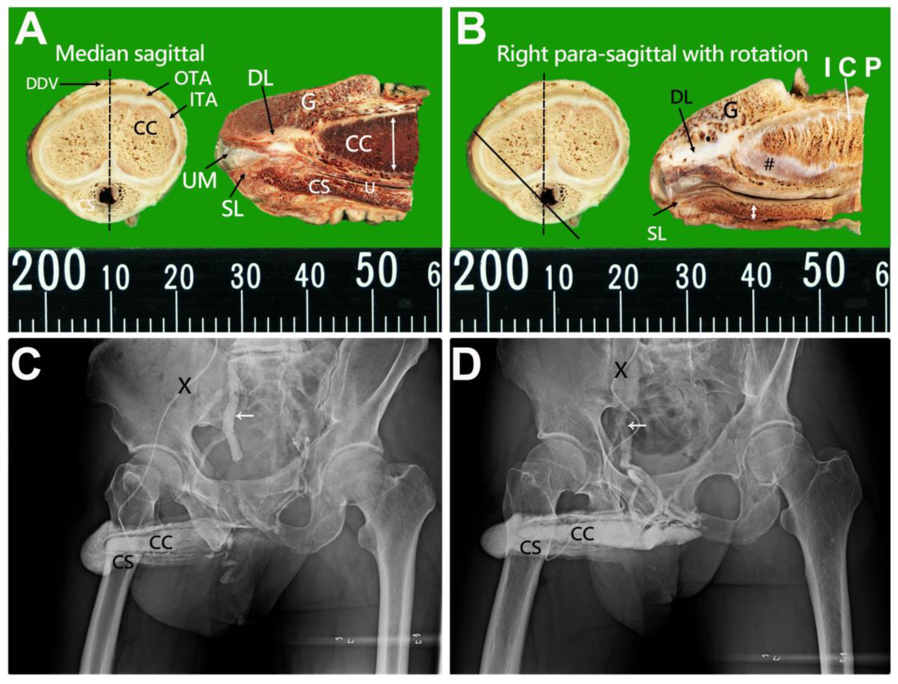
Figure 1. Photos of the distal penis and erection-related veins were demonstrated in cavernosograms. (A) A Median-sagittal section (black dotted line) of the human penis along the deep dorsal vein (DDV). It shows a bi-layered model of the outer tunica albuginea (OTA) and inner tunica albuginea (ITA), corpora cavernosa (CC) with distal ligament (DL) within the glans penis (G), corpus spongiosum (CS) housing the urethra (U) and spongiosal ligament (SL). Note that the urethral meatus and the tunica thickness are much thicker dorsally than the ventral portion (double-headed arrow). (B) A right para-sagittal section presents a similar scenario with DL, SL a gillnetting array, G, and intracavernosal pillars (ICP). Note the thickness of the spongiosal tunica (double-headed arrow) being identically thin, and a cavernosal membrane (Pound) is seen. (C) Cavernosogram was performed via a 19-gauge scalp needle (black cross) inserted in the corpora cavernosa (CC) communication to CS and G; subsequently, the contrast medium drains to the internal iliac vein (arrow). The drainage of blood is tremendous. (D) A similar scenario was demonstrated. Note that the primary drainage venous system is the right iliac vein (arrow).
Life 15 01492 g001
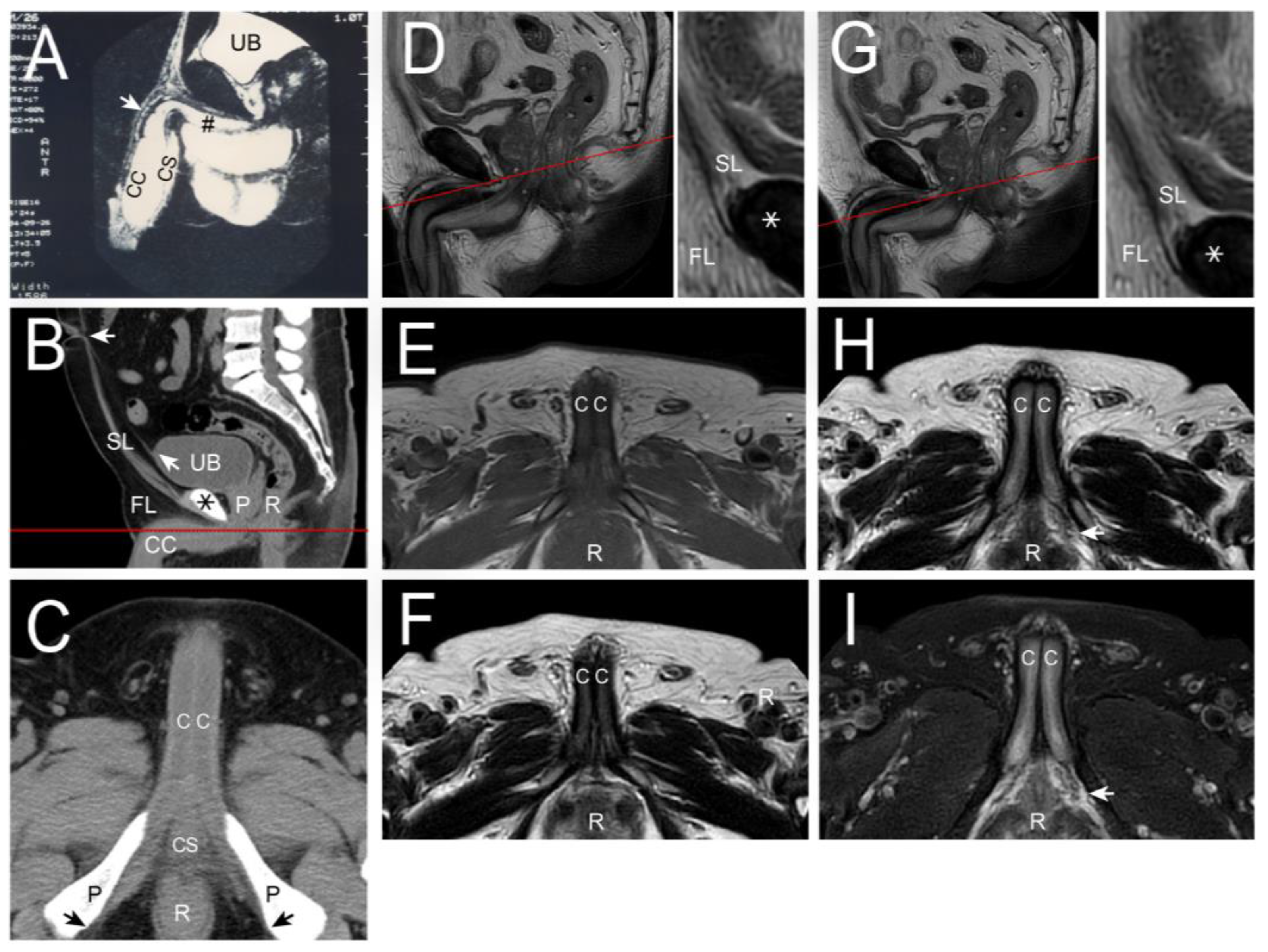
Figure 2. The human penile fibro-vascular assembly obtained from CT and MRI imaging. (A) In the CT imaging, a median sagittal section was made on a 28-year-old man with crural hypoplasia while the penile crus (pound) was petite, whose erection-related veins showed merely the proximal portion (arrow), otherwise masked by sinusoidal opacity. In addition, organoids were demonstrated, for the urinary bladder (UB), corpus cavernosum (CC), and corpus spongiosum (CS). (B) A median sagittal section of the pelvis was conducted to present the symphysis pubis (asterisk), the extension of the urinary bladder (UB) with the median umbilical ligament (oblique arrow), the prostate gland (P), the rectum (R), the suspensory ligament along the linear alba to the umbilicus (left-directed arrow). The section line, red color, was made below the symphysis pubis, resulting in the image of the following one. (C) Given the penis in erection status, it showed the penile corpora cavernosa (CC), (CS), the pubic bones (P), and the rectum (R) ischiocavernosus with cavernosal ligament socketed to the ischial tuberosity (arrow). (D) In MRI imaging, a cross-section was made below the symphysis pubis, red line, for comparison with CT imaging, again demonstrating the suspensory ligament (SL inserted right), fundiform ligament (FL, inserted right) and pubic bone (asterisk). For comparison, imaging was conducted in the T1 mode shown in (E) and T2 mode presented in (F) where the corpora cavernosa (CC) and the rectum (R) are well demonstrated, respectively. Note the right R points the right side. The erection-related veins could be well identified in the T2 mode. (G), however, not intriguing sufficiently. A cross-section was made below the symphysis pubis 2 mm lower, red line, and the mode was conducted in the T2 (H) and 2-FS (I), respectively. Note all the CC, R, FL, and SL mean the same. Overall, the T2 mode was preferred as its enhancing the fibrous tissue of the CC in this study.
Life 15 01492 g002
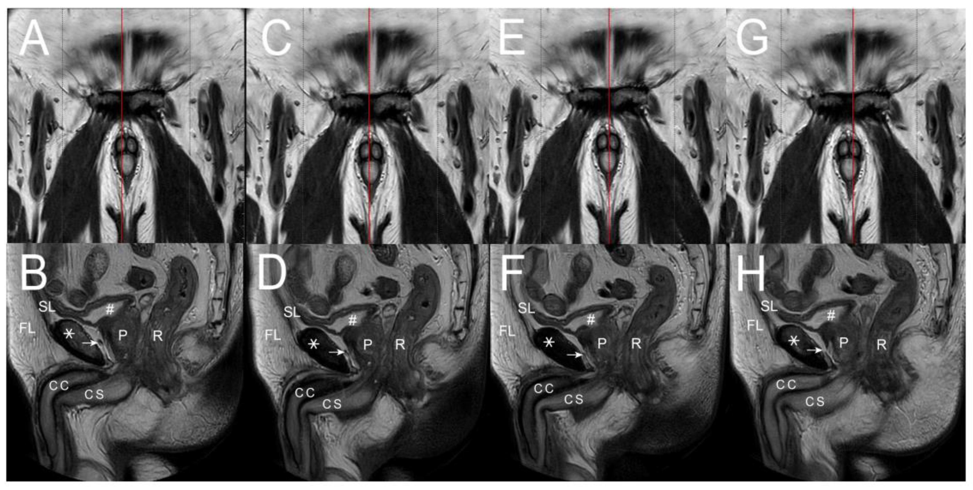
Figure 3. In the MRI T2 imaging study. (A) A 2 mm right para-sagittal section, red line, was made. (B) It demonstrated the symphysis pubis (asterisk), corpora cavernosa (CC), corpus spongiosum (CS), fundiform ligament (FL), suspensory ligament (SL), urinary bladder (pound), prostate gland (P), and rectum (R). Note the arcuate pubic ligament (arrow) scattered along the pubic bone (asterisk) to join the SL. Meanwhile, the erection-related veins, like the periprostatic ones, could be well seen. (C) The section was performed 1 mm medial to the film (A), resulting in the following imaging. (D) The deep dorsal vein climbs over the cavernosal vein along the corpora cavernosa (CC). A similar scenario was shown. (E) A median sagittal section, red line, was made to obtain similar methods in (F). (G) A 1 mm left para-sagittal section, red line, was further conducted to obtain the scenario of the above structures (H), which were unable to tell the bi-layered model of the tunica albuginea, DL, and SL identified in Figure 1A,B. Overall, the detailed three-dimensional architectures might be exclusively disclosed by the macroscopical dissection of the human cadaveric penises.
Life 15 01492 g003
Figure 4. Physiologically, penile integrity is compromised after ligamentolysis for penile elongation elsewhere. (A) A 52-year-old patient sustained severe early detumescence and insufficient rigidity secondary to a penile ligamentolysis for penile lengthening elsewhere, even though his preoperative erection was gratifying. A cavernosogram is performed via a 19-gauge scalp needle (white cross) inserted into the corpora cavernosa (star)—subsequently, the contrast medium drains into the systemic circulation. The alignment integrity could not lash the erection-related veins (inserted, arrow). (B) A similar scenario was well demonstrated in a 47-year-old patient. (C) A 57-year-old patient sustained severe erectile dysfunction after two episodes of ligamentolysis for penile elongation internationally. The erection-related veins are distributed in a seaweed pattern. (D) These unlashed erection veins could not be limited after prostaglandin-E1 intracavernous injection, the erection rigidity is challenging to induce artificially. (E) An illustration was made to explain the lashing effect of the functional integrity by those coevolutionary fibromuscular ligaments (circle belt), such as the arcuate pubic, suspensory, and fundiform ligaments. Is the ligamentolysis, therefore, sustainable?
Life 15 01492 g004
Figure 5. Schematic illustration of a three-dimensional view of the human penis. (A) Lateral aspect. The corpora cavernosa are held by the fundiform ligament from the pubic periosteum to the Colle’s fascia, adhered to the tunica albuginea. The glans penis caps it, mimicking a helmet for safety’s sake, and is buttered by a distal ligament. The bulbospongiosus muscle wraps the corpus spongiosum. The muscle fibers are primarily oblique, and distal threads are bundled, forming the thicker ventral thickening of the outer tunica at the 5 and 7 O’clock positions, respectively. The corpora cavernosa are surrounded by the tunica albuginea, a bi-layered model (an inner circular 360° and 300° outer longitudinal layer with multiple sublayers). The more distal and numerous intracavernosal pillars continue the inner circular layer. The corpus cavernosum is socketed in the ischiocavernosus muscle with the muscle fiber aligned in the longitudinal direction, scattered around 1 to 11 O’clock, respectively. (B) Medial aspect. The distal ligament is continued from the collagen bundles of the outer longitudinal layer of the tunica albuginea. It is an inelastic fibrous structure that supports the glans penis, mimicking a tree trunk of a Christmas tree. The incomplete septum is dorsally fenestrated. The corpus spongiosum contains the urethra. Note the fundiform ligament; the distal ligament is across-street with a spongiosal ligament. (C) Ventral aspect. The three-dimensional structure of the coevolutionary human penis is evident. The ischiocavernosus muscle is paired and sucked at the ischial tuberosity with the cavernosal ligament, then intertwined with the urogenital diaphragm. Each muscle boots its ipsilateral penile crus. Could you note the arrangement of the cavernosal ligament?
Life 15 01492 g005
Table 1. Ten skeletal fibrous ligaments in the human penis based on de novo penile fibro-vascular assembly.
NoTerminologyOriginInsertionNumber of LigamentsConnecting Structures
Ligament/T Ligament/Tendon Anatomical Functional
1CavernosalIschial tuberosityDorsal aspect of outer Tunica21Ischium and corpora cavernosa
2SuspensoryBuck’s fasciaPubic symphysis21Buck’s fascia∼Pubic symphysis
3Fundiform Lateral pubic boneLateral pubic bone21Lateral pubic bone∼Penile base
4Arcuate pubic ligament
Distal (os analog)
Buck’s fascia
Corpora cavernosa
Medial surface of pubic ramus
Glans penis
2
1
1
1
Glans penis and corpora cavernosa
Gans penis and corpora cavernosa
5SpongiosalCorpus spongiosumFrenulum11Frenulum and corpus spongiosum
Total 106
Disclaimer/Publisher’s Note: The statements, opinions and data contained in all publications are solely those of the individual author(s) and contributor(s) and not of MDPI and/or the editor(s). MDPI and/or the editor(s) disclaim responsibility for any injury to people or property resulting from any ideas, methods, instructions or products referred to in the content.