Systematic Review

Refractive Outcomes in Keratoconus Patients Following Toric Lens Implantation: A Systematic Review and Single-Group Meta-Analysis

1
Torbay Hospital, Torbay and South Devon NHS Foundation Trust, Torquay TQ2 7AA, UK
2
Royal Eye Infirmary, University Hospitals Plymouth NHS Trust, Plymouth PL6 5ZF, UK
3
Faculty of Health and Life Sciences, University of Exeter Medical School, Exeter EX1 2LU, UK
4
West of England Eye Unit, Royal Devon University Healthcare NHS Foundation Trust, Exeter EX2 5DW, UK
*
Author to whom correspondence should be addressed.
Life 2025, 15(9), 1362; https://doi.org/10.3390/life15091362
Submission received: 26 July 2025 / Revised: 23 August 2025 / Accepted: 24 August 2025 / Published: 27 August 2025
(This article belongs to the Special Issue Vision Science and Optometry: 2nd Edition)

Abstract

This systematic review and meta-analysis evaluated refractive outcomes, particularly astigmatic correction, in keratoconus following toric intraocular lens (tIOL) implantation. A systematic search identified eligible studies reporting pre- and postoperative refractive cylinder, spherical equivalent (SE), uncorrected distance visual acuity (UDVA), and corrected distance visual acuity (CDVA). Eight studies, comprising 135 eyes, were included. Outcomes were pooled using a random-effects model with restricted maximum likelihood as the estimator for tau2. Methodological quality was assessed using the MINORS tool for non-comparative studies and the JBI checklist for case series. Postoperative refractive cylinder and SE improved by 2.28 dioptres (95% CI, 1.60–2.96) and 4.17 dioptres (95% CI, 2.32–6.01), respectively. UDVA and CDVA also improved substantially, with pooled gains of 0.87 logMAR (95% CI, 0.71–1.03) and 0.19 logMAR (95% CI, 0.12–0.26), respectively. Most tIOL rotations did not exceed 10 degrees, with only one case requiring realignment surgery. Complications were infrequent and mostly minor. tIOL implantation is effective in reducing astigmatism and improving vision in stable keratoconus patients. However, limitations in vector analysis and methodology heterogeneity underscore the need for standardised reporting to optimise outcomes.

1. Introduction

Keratoconus (KC) is a bilateral, asymmetric, progressive ectatic condition characterised by central or paracentral corneal stromal thinning. The resultant apical protrusion of the cornea leads to decreased visual acuity due to induced myopia, astigmatism, and higher-order aberrations (HOAs) [1,2,3,4,5]. HOAs are complex refractive errors arising from imperfect focusing of light through the optical system, which cannot be corrected with conventional spherocylindrical lenses [6].
Toric intraocular lens (tIOL) implantation is an established option to neutralise regular corneal astigmatism during cataracts, refractive lens exchange (RLE), phakic IOL, or supplementary IOL surgery, improving uncorrected distance visual acuity (UDVA) [7,8,9,10,11,12,13]. tIOLs have been reported as a feasible option for eyes with stable KC and repeatable astigmatism, with several case reports demonstrating promising results in select patients [14,15,16]. A recent meta-analysis examined spherical equivalent (SE) outcomes after tIOL implantation in KC; however, it did not evaluate astigmatic outcomes, which are of particular interest when implanting such prostheses [17]. Predominantly coma-type HOAs in KC affect manifest astigmatism [18,19,20,21,22,23], and optical quality is in turn influenced by complex interactions between coma and astigmatism [24,25,26]. These factors create practical uncertainties in selecting the cylinder magnitude and the axis for tIOL implantation in irregular corneas.
To our knowledge, this is the first systematic review with a meta-analysis reporting the astigmatic outcomes of tIOLs in KC. This review aims to examine the available evidence and investigate refractive outcomes in terms of lower-order wavefront aberrations in KC patients following monofocal tIOL implantation.

2. Methods

2.1. Search Strategy

This systematic review with a meta-analysis was conducted in accordance with the Cochrane Handbook and reported using the PRISMA 2020 guidelines [27]. A systematic literature search was performed on electronic databases, including PubMed/MEDLINE, Embase Ovid, and Google Scholar, using relevant keywords, phrases, and medical subject heading terms. The full search strategy is provided in the Supplementary Material. Clinical trial registries, including the Cochrane Central Register of Controlled Trials (CENTRAL) and the ISRCTN registry, were also searched. The literature from inception until 27 April 2023 was reviewed, with an updated search performed on 31 December 2024. Keywords used in the literature search included the following: (keratoconus OR keratoconic) AND (phacoemulsification OR cataract surgery OR cataract operation OR intraocular lens). The study protocol was registered in the International Prospective Register of Systematic Reviews (PROSPERO 2023, https://www.crd.york.ac.uk/prospero/, accessed on 28 April 2023: registration number, CRD42023422070).

2.2. Study Eligibility Criteria

All prospective and retrospective studies meeting the following inclusion criteria were considered: (1) articles published in English; (2) studies of the following designs: randomised controlled trial, case–control, cohort, or case series; (3) patients aged 18 or above with stable, non-progressing KC, including those with prior corneal cross-linking, who received a monofocal toric lens implant via refractive lens exchange or cataract extraction by phacoemulsification; (4) studies reporting outcomes relevant to this review. Exclusion criteria were (1) patients with stage 4 keratoconus according to the Krumeich classification or central corneal scarring; (2) patients with other ocular comorbidities such as glaucoma, maculopathy, retinopathy, or ocular surface inflammation; (3) patients with previous refractive or corneal surgery, excluding corneal cross-linking; (4) combined procedures such as phacoemulsification with keratoplasty, intrastromal corneal ring segments, or corneal cross-linking therapy; (5) conference abstracts or posters.

2.3. Screening and Data Extraction

Duplicate articles from the database search were removed using the web version of reference management software EndNote. The remaining studies were screened independently by two authors (T.G.T., K.O.K.) using the systematic review screening software Rayyan QCRI. Titles and abstracts were initially screened, followed by full-text review for study inclusion. Disagreements were resolved through consultation with a third author (H.W.R.).
Outcome measures were independently extracted by two authors (T.G.T., K.O.K.) using a pre-specified template to capture study characteristics (author, publication year, country, study design), sample characteristics (size, sex, age at presentation), baseline parameters (spherical equivalent [SE], refractive cylinder, uncorrected distance visual acuity [UDVA], and corrected distance visual acuity [CDVA]), and outcomes (SE, refractive cylinder, UDVA, CDVA, complications, intraocular lens rotation, and length of follow-up). Extracted data were then discussed and double-checked to ensure completeness and accuracy. Two attempts via email were made to contact the corresponding author of any study that met the inclusion criteria but had missing data. Studies with insufficient responses were excluded to preserve statistical rigour.

2.4. Quality Assessment

Quality assessment of non-comparative studies was performed using the Methodological Index for Non-Randomized Studies (MINORS) tool, which assesses studies across domains such as patient selection, outcome measures, outcome reporting, and follow-up duration [28]. Each study was assigned a total score out of 16 based on an 8-item checklist, with scores of ≤8 indicating poor methodological quality, 9–14 indicating moderate quality, and ≥15 indicating good quality. All non-comparative studies in this review were deemed to be of moderate methodological quality (see Supplementary Table S1). The methodological quality of case series was assessed using the JBI critical appraisal checklist for case series, which comprises 10 questions addressing factors such as complete patient inclusion, outcome reporting, and the appropriateness of statistical analysis (see Supplementary Table S2) [29]. Exclusion of studies based on quality assessment was not attempted due to the limited available literature at the time of this review.

2.5. Outcome Measures and Statistical Analysis

The primary outcomes were the mean difference in refractive cylinder, SE, UDVA, and CDVA following tIOL implantation. These were calculated using preoperative and postoperative means and standard deviations (SDs) reported in the included studies. Given the anticipated between-study heterogeneity, pooled outcomes were calculated using a random-effects model with restricted maximum likelihood (REML) [30] as the estimator for tau2. Secondary outcomes included postoperative tIOL rotation and complications. Publication bias was planned to be assessed via funnel plot asymmetry using Egger’s test. However, this was not performed due to the small number of studies, which would render the test underpowered to distinguish chance from true asymmetry [31]. All analyses were performed using the “meta-for” and “effectsize” packages in R (version 4.4.1).

3. Results

3.1. The Literature Search

A total of 941 records were identified from the initial and rerun searches of electronic databases, including PubMed and Embase. After removing 353 duplicates, 588 records remained for title and abstract screening, of which 42 were selected for full-text evaluation. Forty-one of these full-text reports were successfully retrieved. Following full-text screening, eight original studies were included in this review (Table 1) [32,33,34,35,36,37,38,39].

3.2. Primary Outcomes

A total of 135 eyes from 102 patients were included across the eight studies. One patient (one eye) in the retrospective study by Jaimes et al. had a dense cataract, preventing measurement of preoperative refraction and visual acuity, and was therefore excluded from the meta-analysis [36]. Preoperatively, the mean refractive cylinder was 3.38 ± 2.11 dioptres (D), while the mean residual refractive cylinder postoperatively was 1.30 ± 1.17 D. The pooled mean difference (MD) in the refractive cylinder was a reduction of 2.28 D (95% CI, 1.60 to 2.96) (Figure 1).
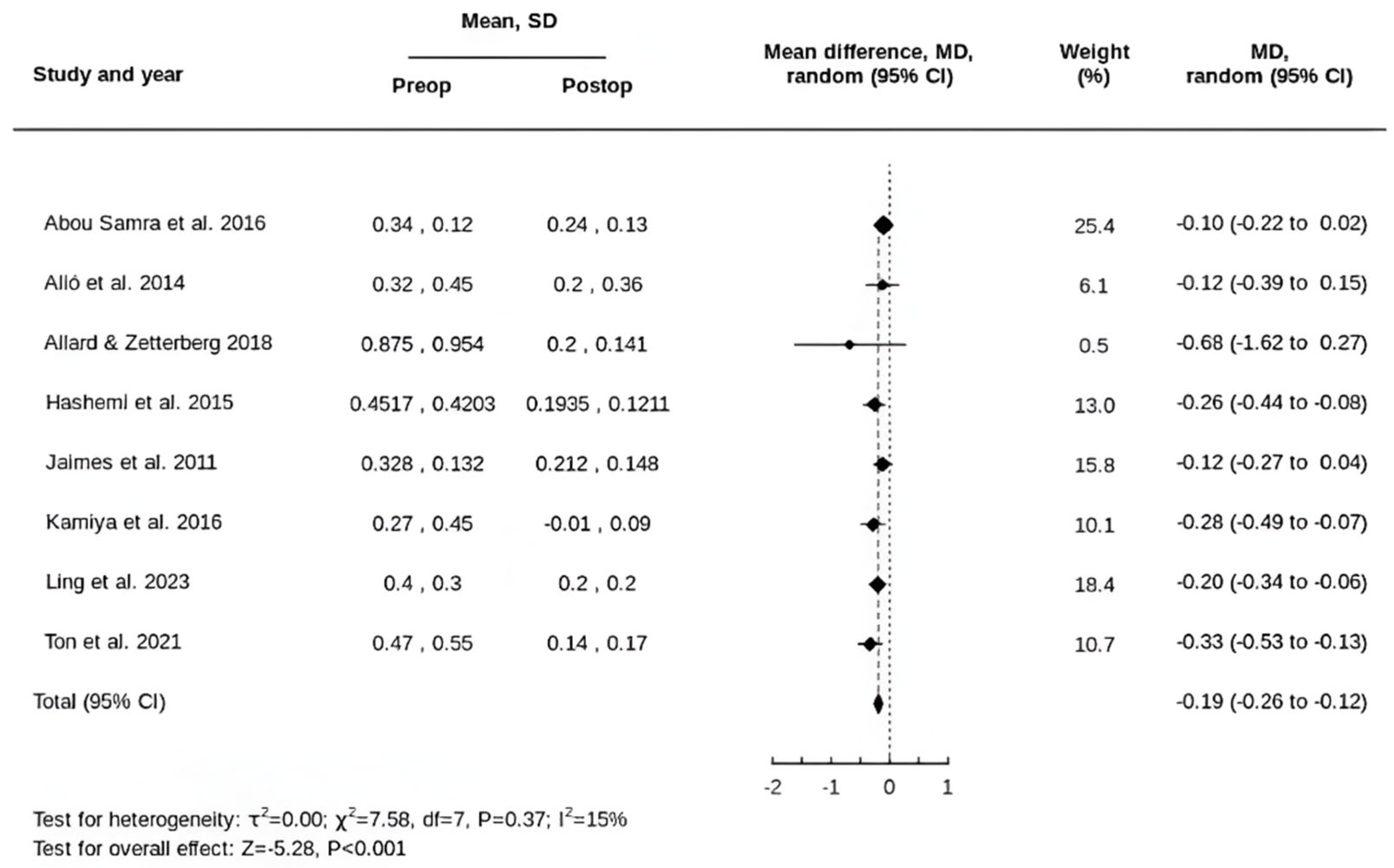
For SE, the preoperative mean was −4.67 ± 5.08 D, and the postoperative mean was −0.58 ± 1.17 D. The pooled MD in SE was a reduction of 4.17 D (95% CI, 2.32 to 6.01 D) (Figure 2).
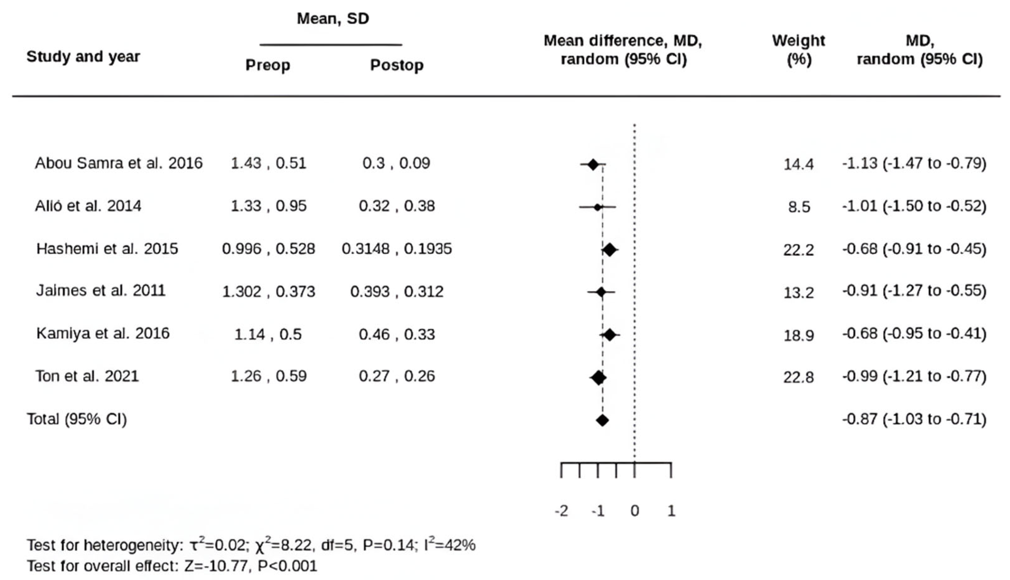
In terms of UDVA, six of the eight included studies provided sufficient data, comprising 107 eyes from 67 patients. The preoperative mean was 1.21 ± 0.62 logMAR, and the postoperative mean was 0.33 ± 0.28 logMAR. The pooled MD in UDVA following tIOL implantation was a gain of 0.87 logMAR (95% CI, 0.71 to 1.03) (Figure 3).
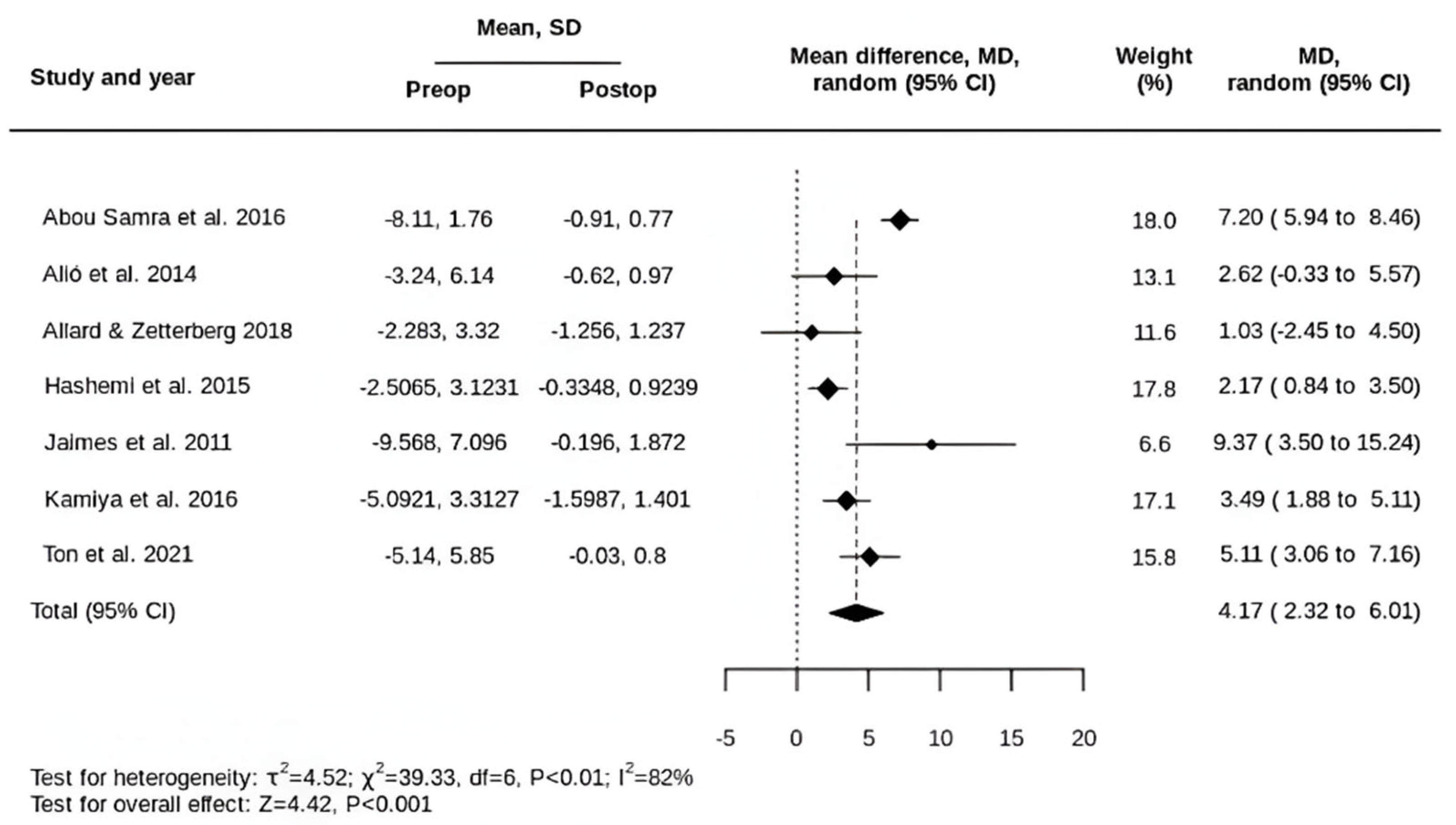
Following tIOL implantation, the mean CDVA improved from 0.40 ± 0.45 logMAR preoperatively to 0.16 ± 0.20 logMAR postoperatively, corresponding to a pooled mean difference of 0.19 logMAR (95% CI, 0.12 to 0.26) (Figure 4).

3.3. Secondary Outcomes

Only four of the eight included studies reported postoperative tIOL rotation. Allard & Zetterberg reported 8 degrees of tIOL rotation in one eye, the most significant in their case series [34]. Hashemi et al. reported mean tIOL rotations of 2.50 ± 1.18 degrees (range, 1–5), 2.50 ± 1.51 degrees (range, 0–6), and 2.67 ± 1.15 degrees (range, 2–6) in the mild, moderate, and severe keratoconus groups, respectively [35]. Jaimes et al. documented three cases requiring tIOL realignment: one due to haptic luxation, one due to tIOL misalignment, and one due to tIOL rotation [36]. Kamiya et al. reported a mean tIOL rotation of 5.2 ± 2.6 degrees (range, 1–10) [37].
Amongst the eight studies, three reported postoperative complications. Both Allard & Zetterberg and Kamiya et al. noted a single case of postoperative cystoid macular oedema (CMO) in their cohorts [34,37]. Jaimes et al. reported two cases of wound leak, with one requiring a re-suturing and the other managed with a bandage contact lens [36].

4. Discussion

Stable KC is a prerequisite for tIOL implantation. Only three of the included studies specified criteria for defining non-progression of KC preoperatively. These criteria were inconsistent and were based on a combination of stability of keratometry, refraction, and pachymetry over a 6- to 12-month period [33,35,36]. In the study by Abou Samra et al., the cohort had progressive KC and underwent a two-stage procedure, consisting of corneal cross-linking (CXL) followed by tIOL implantation six months later [32]. Although the refractive outcomes in this study appeared favourable, the sample size was too small to draw generalisable conclusions.
All the studies in this meta-analysis reported a reduction in mean SE following tIOL implantation. However, this gives no indication of the effectiveness of astigmatism correction. In this review, our primary area of interest was cylindrical outcomes, as SE outcomes have been described in previous studies [17,40,41]. All eight included studies reported a reduction in mean refractive astigmatism in keratoconic eyes following tIOL implantation.
Preoperatively, the mean astigmatism was 3.38 ± 2.11 dioptres (D), which is relatively low for a cohort of keratoconic eyes. Some included studies excluded patients with more severe KC based on the Amsler–Krumeich classification system, which incorporates keratometry, optical pachymetry, refraction and biomicroscopy [42,43]. In studies that included patients with more severe KC, the cohort was skewed towards mild or moderate cases. An important source of heterogeneity in this review arises from the refraction method. Five out of eight studies reported manifest refraction, with Abou Samra et al. including at least one cycloplegic refraction during follow-up, without further specification [32,35,36,37,38]. In the series by Allard & Zetterberg, two of the four patients were assessed with autorefraction, and two studies did not specify the refraction method used [34,36,39]. A prior study by Soeters et al., comprising 90 keratoconic eyes of 61 patients, concluded that autorefraction is unreliable in KC, with greater discrepancies observed in steeper corneas [44].
It is worth noting that two cases reported by Kamiya et al. had high residual astigmatism, partly due to the unavailability of higher-powered tIOLs in the study country at the time [37]. Additionally, a frequently cited study by Nanavaty et al. was not included in this review because one of the twelve eyes had a prior Intacs corneal implant, and there was insufficient information to separate its data from the rest. This study similarly reported a significant reduction in astigmatism magnitude, from 3.00 ± 1.00 D to 0.70 ± 0.80 D [45]. Although studies show that astigmatism less than 0.5 D does not degrade visual acuity [46,47], reporting only postoperative mean values does not distinguish patients who achieve <0.5 D of residual astigmatism from those who do not. Therefore, Holladay et al. recommended reporting the percentages of patients within specific residual astigmatism intervals [48]. Some studies in this review reported only the postoperative mean astigmatism.
Astigmatism is defined by both magnitude and axis. Evaluating postoperative residual astigmatism in terms of its magnitude is useful, given its correlation with UDVA regardless of axis change [46,47]. However, assessing change in astigmatism magnitude alone is insufficient to determine the effectiveness of an astigmatism correction. Alió et al. reported a vector analysis using the Alpins method in their study [33]. Kamiya et al. presented postoperative refractive outcomes using power vector analysis as described by Thibos and Horner [37]. Power vectors represent spherocylindrical refraction geometrically in three independent dioptric components, permitting a more detailed description of postoperative refractive changes [49]. Other studies reported a simple subtraction or scalar analysis, comparing preoperative and postoperative astigmatism in terms of magnitude alone.
The Alpins method evaluates the effectiveness of astigmatism treatment by examining the relationships between three fundamental vectors, namely, target-induced astigmatism (TIA), surgically induced astigmatism (SIA), and difference vector (DV) [49,50]. TIA represents the intended astigmatic treatment of an intervention, in both magnitude and axis. SIA is the vectorial difference between preoperative and postoperative astigmatism, which can be corneal or refractive. DV is the residual astigmatic correction required to achieve the TIA. DV is an absolute measure of success and equals zero when SIA exactly matches TIA. The correction index (CI), defined as the ratio of SIA to TIA, indicates overcorrection when greater than one and undercorrection when less than one [50,51]. Apart from Alió et al. [33], the studies in this review did not report TIA, precluding vector analysis. Since stock tIOLs are available only in fixed dioptric steps, TIA cannot simply be assumed to achieve a postoperative zero-dioptre target. Without vector analysis using TIA as a reference, residual astigmatism alone provides insufficient information to determine under- or overcorrection.
In the Alpins method, aggregate analysis of multiple eyes using double-angle vector diagrams (DAVDs) permits calculation of systematic errors, such as undercorrection, overcorrection, or misalignment, which are reported as geometric means [50]. This visualisation can guide adjustments to improve postoperative refractive outcomes. Of the eight included studies, only Ling et al. reported preoperative and postoperative astigmatism using a DAVD [38]. Aggregate data can also be presented using surgical polar vector diagrams, which halve the astigmatism meridian and axis values used in DAVD calculations [50]. To standardise outcome reporting, The Journal of Refractive Surgery (JRS) provides standard graphs illustrating the proportions of eyes with under- or overcorrection of astigmatism in both magnitude and axis [52]. None of the studies in this review adhered to the JRS standard for reporting astigmatic outcomes.
The cornea contributes approximately three quarters of the eye’s refractive power [1], making accurate keratometry essential for optimising preoperative tIOL calculations. In KC, the orthogonally asymmetric cornea with a displaced optical centre produces coma-like aberrations [18,19,20,21,22], which clinically contribute to manifest astigmatism [23]. Keratometry can be challenging in KC due to corneal irregularities, and associated corneal HOAs may explain the lower predictability of postoperative refractive outcomes in severe KC [17,40,41,53,54]. The location of corneal ectasia also affects refractive predictability, with more central ectasias having a greater impact on keratometry accuracy along the visual axis [33]. Multiple corneal imaging devices are currently available, including Placido-based systems and corneal tomography techniques such as scanning slit, Scheimpflug, and optical coherence tomography (OCT)-based devices. Placido-based devices provide corneal topography for mapping the anterior corneal surface, whereas corneal tomography techniques generate three-dimensional maps, including posterior elevation, which is particularly relevant in KC [55,56]. Studies report varying levels of inter-device agreement, with lower device repeatability observed in more advanced KC [18,57,58].
Most studies in this review employed multiple devices to derive keratometric data, and some applied different methods for different patients, representing another important source of heterogeneity. None of the studies used newer devices such as the Sirius (Costruzione Strumenti Oftalmici, CSO), which combines a rotating Scheimpflug camera and a small-angle Placido-disk topographer, or the MS-39 (CSO), which integrates an anterior-segment OCT with a Placido-disk topographer. Both the Sirius and MS-39 had demonstrated good repeatability of keratometry in keratoconic eyes [59,60].
A linear relation between refractive astigmatism (RA) and keratometric astigmatism (KA) was first proposed by Émile Javal in 1890 and subsequently refined by others [61,62,63]. Historically, the disparity between the two astigmatic values was attributed to lenticular astigmatism (LA) [64]. However, studies have shown that this discrepancy persists even in pseudophakic eyes [65,66]. Koch et al. suggested that posterior corneal astigmatism (PCA) accounts for the difference between RA and KA [64,67]. Conventional or simulated keratometry (Sim-K) estimates KA using the measured anterior corneal curvature and a standard keratometric index (1.3375), assuming a fixed ratio between anterior and posterior corneal curvatures [22,68]. In KC, this ratio is not constant, leading to inaccurate postoperative refractive outcomes [53,55,69,70,71]. Two case series, comprising 137 and 161 eyes with KC, respectively, reported that PCA accounts for approximately one fifth of total corneal astigmatism (TCA), highlighting its clinical relevance [72,73].
Incorporation of PCA into tIOL calculations was first introduced using Koch’s Baylor nomogram [64]. Subsequently, Holladay and George devised a formula accounting for total surgically induced astigmatism (SIA) to improve postoperative astigmatism outcomes [74]. Other methods, including the Barrett Toric Calculator, Kane Toric formula, and Naeser–Savini formula, incorporate all three vectors (keratometry, PCA, and SIA) to determine tIOL toricity and axis [75,76,77]. Application of the Abulafia–Koch (AK) regression formula has been reported to further improve postoperative astigmatic outcomes by enabling more accurate TCA estimation [78]. The Barrett Toric Calculator can use either predicted or measured PCA, with studies showing comparable postoperative results between the two, as well as with the Kane Toric formula and AK regression formula [79,80]. To date, no study has evaluated the predictability of astigmatic outcomes in KC comparing the different tIOL formulae.
Apart from Alió et al., all included studies used tIOL calculators provided by the respective manufacturers, with the AcrySof Toric IOL calculator being the most frequently cited. It was unclear whether the Barrett Toric or the Holladay Total SIA Toric Calculator within the AcySof Toric IOL calculator was used in these studies. Additionally, the studies did not specify whether predicted or measured PCA was used in preoperative calculations. Intraoperative aberrometry, another method of tIOL calculation, was not utilised in any of the studies [81].
Another important vector in the preoperative analysis of tIOL is SIACornea, which represents the change in corneal astigmatism resulting from corneal incisions [82]. Abulafia et al. recommended using the term SIACornea to differentiate it from the astigmatic change induced by a tIOL, SIAIOL, or by the surgery as a whole, SIATotal [83]. None of the included studies specified their method of calculating SIACornea, a variable influenced by multiple factors including corneal incision location [83]. The varied corneal incision sizes and locations across studies represent important sources of heterogeneity in this review. Corneal incision sizes ranged from 1.0 mm to 3.2 mm. Febbraro et al. reported that 3.2 mm incisions induce greater astigmatic effects than 1.8 mm or 2.2 mm incisions [84]. Similar findings were reported by Yu et al. for 3.0 mm incisions versus 1.8 mm or 2.0 mm incisions [85].
In the included studies, some surgeries used incisions at the steep meridian [32,33], others used temporal incisions [35,37], and some did not specify the incision location [34,36,38,39]. This is relevant because corneal incisions along the steep meridian induce greater astigmatism reduction compared with off-axis incisions [50]. Furthermore, the studies did not specify whether mean or centroid SIACornea values were used for tIOL calculations. The centroid value accounts for both magnitude and axis and is usually smaller than the mean, which reflects magnitude alone, and is considered more accurate for tIOL planning [83,86].
Overall, both UDVA and CDVA improved following tIOL implantation. The effect of tIOL implantation on CDVA is smaller than on UDVA. CDVA changes are less relevant when assessing the effectiveness of refractive surgery, as they would predominantly be the result of cataract removal [87]. Improvements in UDVA can be further attributed to the correction of sphere and cylinder with tIOL. In some studies, such as that by Abou Samra et al. and Jaimes et al., all but one KC patient underwent clear lens exchange [32,36]. In other words, it would be reasonable to postulate that the suboptimal visual quality of these patients was primarily due to optical aberrations rather than media opacity, assuming an absence of ocular comorbidities. Thus, one would expect such refractive surgeries to lead to greater improvements in UDVA than in CDVA postoperatively.
Increased magnitudes of HOAs are negatively correlated with corrected visual acuity [88]. Whilst lenticular opacities contribute to overall ocular HOAs, those in KC are mainly due to the air–tear interface at the anterior corneal surface [89,90,91]. Modern phacoemulsification surgery itself generally does not alter keratometric properties in a way that is beneficial in reducing corneal HOAs. In fact, corneal HOAs can increase due to corneal incisions during cataract surgery [88,92]. This review focuses on tIOLs, which cannot fully correct the aberrations present in keratoconic corneas. In contrast, customised aberration-correcting IOLs have been shown to reduce HOAs in eyes with non-progressive keratoconus [93]. Future studies comparing the refractive outcomes of tIOLs and customised IOLs in KC may provide further insights.
Four studies reported postoperative tIOL rotation. All used either AcrySof IQ tIOLs or Acrysof toric SN60TT IOLs, except for two cases in the series by Allard & Zetterberg [34,35,36,37]. In a randomised–controlled trial, Holland et al. demonstrated good rotational stability of AcrySof tIOLs. At one year, the authors reported a mean rotation of less than 4 degrees, with a range between 0 and 20 degrees [94]. Apart from one case of tIOL rotation requiring realignment surgery described by Jaimes et al. [36], no study reported any tIOL rotation of more than 10 degrees. Tognetto et al. concluded that tIOL rotation of greater than 10 degrees is significant for image quality loss [95]. This is consistent with the findings of Filipe et al. [96], who concluded that tIOL rotation of less than 10 degrees still provides satisfactory astigmatism correction. KC is associated with longer axial lengths on average, mainly due to longer posterior segments [97]. In turn, axial length is positively correlated with tIOL rotation [98]. However, no current studies have demonstrated an increased risk of tIOL rotation in keratoconic eyes.
The follow-up period in the included studies ranged from 1 to 31 months, including the case series by Ling et al., which reported an average follow-up of over 2 years. Five of the eight included studies had a follow-up of one year or less, highlighting a lack of long-term outcomes for tIOL implantation in KC. Whilst most cases in the included studies involved mild or moderate KC, different severity grading systems were used. The Amsler–Krumeich classification was cited in three studies [32,35,36]. Other staging systems referenced include the steepest K-value classification proposed by Thebpatiphat et al. [69] and a Pentacam-based system described by Hashemi et al. [99]. At present, no global consensus exists on KC classification [100], making subgroup analysis by severity unfeasible in this meta-analysis. Going forward, establishing a new consensus would improve the evidence base and better guide management.
In this review, forme fruste KC cases were excluded due to the lack of unified diagnostic criteria [101]. Another limitation of this review is the small number of eligible studies, reflecting the paucity of published data on this subject. Furthermore, all studies had small sample sizes, limiting statistical power. Finally, it is worth pointing out again that some studies did not specify their refraction method, and one case series partially reported refractive outcomes based on autorefraction, which is unreliable in KC [34]. This represents a notable weakness of the current literature.

5. Conclusions

tIOL implantation in KC appears to be a safe intervention for reducing astigmatism, based on a small number of heterogeneous studies. However, the true effectiveness of tIOLs in KC remains unclear due to the lack of vector analysis of astigmatic outcomes in the published literature. Moving forward, adopting standardised terminology, vector analysis methodology, and consistent reporting of astigmatic outcomes would help advance understanding and improve the effectiveness of tIOL implantation in KC.

Supplementary Materials

The following supporting information can be downloaded at https://www.mdpi.com/article/10.3390/life15091362/s1, Figure S1: PRISMA flow diagram; Table S1: MINORS risk of bias tool; Table S2: JBI critical appraisal checklist for case series.

Author Contributions

Conceptualisation, T.G.T., K.O. and H.W.R.; methodology, T.G.T., K.O. and H.W.R.; project administration, T.G.T., K.O. and H.W.R.; data curation, T.G.T. and K.O.; formal analysis, visualisation, T.G.T.; writing—original draft, T.G.T.; writing—review and editing, T.G.T. and H.W.R.; supervision, H.W.R. All authors have read and agreed to the published version of the manuscript.

Funding

This research received no external funding.

Institutional Review Board Statement

Not applicable.

Informed Consent Statement

Not applicable.

Data Availability Statement

Data available upon written request to the corresponding author.

Conflicts of Interest

There are no financial conflicts of interest to disclose.

Abbreviations

The following abbreviations are used in this manuscript:
KCKeratoconus
tIOLToric intraocular lens
SESpherical equivalent
UDVAUncorrected distance vision
CDVACorrected distance vision
REMLRestricted maximum likelihood
HOAsHigher-order aberrations
RLERefractive lens exchange
SDStandard deviation
DDioptre
MDMean difference
CMOCystoid macular oedema
CXLCorneal cross-linking
TIATarget induced astigmatism
SIASurgically induced astigmatism
DVDifference vector
CICorrection index
DAVDDouble-angle vector diagram
OCTOptical coherence tomography
RARefractive astigmatism
KAKeratometric astigmatism
LALenticular astigmatism
PCAPosterior corneal astigmatism
TCATotal corneal astigmatism
IOLIntraocular lens

References

  1. Salmon, J.F. Kanski’s Clinical Ophthalmology: A Systematic Approach, 9th ed.; Elsevier Limited: Amsterdam, The Netherlands, 2000. [Google Scholar]
  2. Santodomingo-Rubido, J.; Carracedo, G.; Suzaki, A.; Villa-Collar, C.; Vincent, S.J.; Wolffsohn, J.S. Keratoconus: An updated review. Cont. Lens Anterior Eye 2022, 45, 101559. [Google Scholar] [CrossRef]
  3. Wagner, H.; Barr, J.T.; Zadnik, K. Collaborative Longitudinal Evaluation of Keratoconus (CLEK) Study: Methods and findings to date. Cont. Lens Anterior Eye 2007, 30, 223–232. [Google Scholar] [CrossRef] [PubMed]
  4. Sideroudi, H.; Labiris, G.; Georgantzoglou, K.; Ntonti, P.; Siganos, C.; Kozobolis, V. Fourier analysis algorithm for the posterior corneal keratometric data: Clinical usefulness in keratoconus. Ophthalmic Physiol. Opt. 2017, 37, 460–466. [Google Scholar] [CrossRef]
  5. Oshika, T.; Tanabe, T.; Tomidokoro, A.; Amano, S. Progression of keratoconus assessed by fourier analysis of videokeratography data. Ophthalmology 2002, 109, 339–342. [Google Scholar] [CrossRef]
  6. Hashemi, H.; Khabazkhoob, M.; Jafarzadehpur, E.; Yekta, A.; Emamian, M.H.; Shariati, M.; Fotouhi, A. Higher order aberrations in a normal adult population. J. Curr. Ophthalmol. 2016, 27, 115–124. [Google Scholar] [CrossRef] [PubMed]
  7. Kessel, L.; Andresen, J.; Tendal, B.; Erngaard, D.; Flesner, P.; Hjortdal, J. Toric Intraocular Lenses in the Correction of Astigmatism During Cataract Surgery: A Systematic Review and Meta-analysis. Ophthalmology 2016, 123, 275–286. [Google Scholar] [CrossRef] [PubMed]
  8. Güell, J.L.; Vázquez, M.; Malecaze, F.; Manero, F.; Gris, O.; Velasco, F.; Hulin, H.; Pujol, J. Artisan toric phakic intraocular lens for the correction of high astigmatism. Am. J. Ophthalmol. 2003, 136, 442–447. [Google Scholar] [CrossRef]
  9. Alió, J.L.; Mulet, M.E.; Gutiérrez, R.; Galal, A. Artisan toric phakic intraocular lens for correction of astigmatism. J. Refract. Surg. 2005, 21, 324–331. [Google Scholar] [CrossRef]
  10. Visser, N.; Beckers, H.J.; Bauer, N.J.; Gast, S.T.; Zijlmans, B.L.; Berenschot, T.T.; Webers, C.A.; Nuijts, R.M. Toric vs aspherical control intraocular lenses in patients with cataract and corneal astigmatism: A randomized clinical trial. JAMA Ophthalmol. 2014, 132, 1462–1468. [Google Scholar] [CrossRef]
  11. Ruíz-Mesa, R.; Carrasco-Sánchez, D.; Díaz-Alvarez, S.B.; Ruíz-Mateos, M.A.; Ferrer-Blasco, T.; Montés-Micó, R. Refractive lens exchange with foldable toric intraocular lens. Am. J. Ophthalmol. 2009, 147, 990–996.e1. [Google Scholar] [CrossRef]
  12. McLintock, C.A.; McKelvie, J.; Niyazmand, H.; Apel, A.J. Outcomes of a Toric Monofocal Piggyback Intraocular Lens for Residual Astigmatic Refractive Error in Pseudophakic Eyes. Curr. Eye Res. 2022, 47, 443–449. [Google Scholar] [CrossRef] [PubMed]
  13. Royo, M.; Jiménez, Á.; Martínez-Alberquilla, I.; Alfonso, J.F. Eight-year follow-up of Artiflex and Artiflex Toric phakic intraocular lens. Eur. J. Ophthalmol. 2022, 32, 2051–2058. [Google Scholar] [CrossRef] [PubMed]
  14. Sauder, G.; Jonas, J.B. Treatment of Keratoconus by Toric Foldable Intraocular Lenses. Eur. J. Ophthalmol. 2003, 13, 577–579. [Google Scholar] [CrossRef]
  15. Visser, N.; Gast, S.T.; Bauer, N.J.; Nuijts, R.M. Cataract surgery with toric intraocular lens implantation in keratoconus: A case report. Cornea 2011, 30, 720–723. [Google Scholar] [CrossRef]
  16. Zvornicanin, J.; Cabric, E.; Jusufovic, V.; Musanovic, Z.; Zvornicanin, E. Use of the toric intraocular lens for keratoconus treatment. Acta Inform. Med. 2014, 22, 139–141. [Google Scholar] [CrossRef] [PubMed]
  17. Yahalomi, T.; Achiron, A.; Hecht, I.; Arnon, R.; Levinger, E.; Pikkel, J.; Tuuminen, R. Refractive Outcomes of Non-Toric and Toric Intraocular Lenses in Mild, Moderate and Advanced Keratoconus: A Systematic Review and Meta-Analysis. J. Clin. Med. 2022, 11, 2456. [Google Scholar] [CrossRef] [PubMed]
  18. Hashemi, H.; Yekta, A.; Khabazkhoob, M. Effect of keratoconus grades on repeatability of keratometry readings: Comparison of 5 devices. J. Cataract. Refract. Surg. 2015, 41, 1065–1072. [Google Scholar] [CrossRef]
  19. Feizi, S.; Einollahi, B.; Raminkhoo, A.; Salehirad, S. Correlation between Corneal Topographic Indices and Higher-Order Aberrations in Keratoconus. J. Ophthalmic Vis. Res. 2013, 8, 113–118. [Google Scholar]
  20. Maeda, N.; Fujikado, T.; Kuroda, T.; Mihashi, T.; Hirohara, Y.; Nishida, K.; Watanabe, H.; Tano, Y. Wavefront aberrations measured with Hartmann-Shack sensor in patients with keratoconus. Ophthalmology 2002, 109, 1996–2003. [Google Scholar] [CrossRef]
  21. Pantanelli, S.; MacRae, S.; Jeong, T.M.; Yoon, G. Characterizing the wave aberration in eyes with keratoconus or penetrating keratoplasty using a high-dynamic range wavefront sensor. Ophthalmology 2007, 114, 2013–2021. [Google Scholar] [CrossRef]
  22. Zhou, W.; Stojanovic, A.; Utheim, T.P. Assessment of refractive astigmatism and simulated therapeutic refractive surgery strategies in coma-like-aberrations-dominant corneal optics. Eye Vis. 2016, 3, 13. [Google Scholar] [CrossRef] [PubMed]
  23. Wei, R.H.; Lim, L.; Chan, W.K.; Tan, D.T. Higher Order Ocular Aberrations in Eyes With Myopia in a Chinese Population. J. Refract. Surg. 2006, 22, 695–702. [Google Scholar] [CrossRef]
  24. Zhou, W.; Stojanovic, F.; Reinstein, D.Z.; Archer, T.J.; Chen, X.; Feng, Y.; Stojanovic, A. Coma Influence on Manifest Astigmatism in Coma-Dominant Irregular Corneal Optics. J. Refract. Surg. 2021, 37, 274–282. [Google Scholar] [CrossRef]
  25. de Gracia, P.; Dorronsoro, C.; Gambra, E.; Marin, G.; Hernández, M.; Marcos, S. Combining coma with astigmatism can improve retinal image over astigmatism alone. Vis. Res. 2010, 50, 2008–2014. [Google Scholar] [CrossRef]
  26. de Gracia, P.; Dorronsoro, C.; Gambra, E.; Marin, G.; Hernández, M.; Marcos, S. Visual acuity under combined astigmatism and coma: Optical and neural adaptation effects. J. Vis. 2011, 11, 5. [Google Scholar] [CrossRef] [PubMed]
  27. Page, M.J.; McKenzie, J.E.; Bossuyt, P.M.; Boutron, I.; Hoffmann, T.C.; Mulrow, C.D.; Shamseer, L.; Tetzlaff, J.M.; Akl, E.A.; Brennan, S.E.; et al. The PRISMA 2020 statement: An updated guideline for reporting systematic reviews. BMJ 2021, 372, n71. [Google Scholar] [CrossRef]
  28. Slim, K.; Nini, E.; Forestier, D.; Kwiatkowski, F.; Panis, Y.; Chipponi, J. Methodological index for non-randomized studies (minors): Development and validation of a new instrument. ANZ J. Surg. 2003, 73, 712–716. [Google Scholar] [CrossRef]
  29. Munn, Z.; Barker, T.H.; Moola, S.; Tufanaru, C.; Stern, C.; McArthur, A.; Stephenson, M.; Aromataris, E. Methodological quality of case series studies: An introduction to the JBI critical appraisal tool. JBI Evid. Synth. 2020, 18, 2127–2133. [Google Scholar] [CrossRef]
  30. Viechtbauer, W. Bias and Efficiency of Meta-Analytic Variance Estimators in the Random-Effects Model. J. Educ. Behav. Stat. 2005, 30, 261–293. [Google Scholar] [CrossRef]
  31. Sterne, J.A.; Sutton, A.J.; Ioannidis, J.P.; Terrin, N.; Jones, D.R.; Lau, J.; Carpenter, J.; Rücker, G.; Harbord, R.M.; Schmid, C.H.; et al. Recommendations for examining and interpreting funnel plot asymmetry in meta-analyses of randomised controlled trials. BMJ 2011, 343, d4002. [Google Scholar] [CrossRef] [PubMed]
  32. Abou Samra, W.A.; Awad, E.A.; El Kannishy, A.H. Objective and Subjective Outcome of Clear Lensectomy With Toric IOL Implantation After Corneal Collagen Cross-Linking in Selected Cases of Keratoconus. Eye Contact Lens 2018, 44 (Suppl. 1), S87–S91. [Google Scholar] [CrossRef]
  33. Alió, J.L.; Peña-García, P.; Abdulla Guliyeva, F.; Soria, F.A.; Zein, G.; Abu-Mustafa, S.K. MICS with toric intraocular lenses in keratoconus: Outcomes and predictability analysis of postoperative refraction. Br. J. Ophthalmol. 2014, 98, 365–370. [Google Scholar] [CrossRef] [PubMed]
  34. Allard, K.; Zetterberg, M. Implantation of toric intraocular lenses in patients with cataract and keratoconus: A case series. Int. Med. Case Rep. J. 2018, 11, 185–191. [Google Scholar] [CrossRef]
  35. Hashemi, H.; Heidarian, S.; Seyedian, M.A.; Yekta, A.; Khabazkhoob, M. Evaluation of the Results of Using Toric IOL in the Cataract Surgery of Keratoconus Patients. Eye Contact Lens 2015, 41, 354–358. [Google Scholar] [CrossRef] [PubMed]
  36. Jaimes, M.; Xacur-García, F.; Alvarez-Melloni, D.; Graue-Hernández, E.O.; Ramirez-Luquín, T.; Navas, A. Refractive lens exchange with toric intraocular lenses in keratoconus. J. Refract. Surg. 2011, 27, 658–664. [Google Scholar] [CrossRef]
  37. Kamiya, K.; Shimizu, K.; Miyake, T. Changes in astigmatism and corneal higher-order aberrations after phacoemulsification with toric intraocular lens implantation for mild keratoconus with cataract. Jpn. J. Ophthalmol. 2016, 60, 302–308. [Google Scholar] [CrossRef]
  38. Ling, J.Y.M.; Qiao, G.; Iovieno, A.; Yeung, S.N. Visual Outcomes of Cataract Surgery in Patients With Keratoconus Using Toric and Non-toric Lenses. J. Refract. Surg. 2023, 39, 319–325. [Google Scholar] [CrossRef] [PubMed]
  39. Ton, Y.; Barrett, G.D.; Kleinmann, G.; Levy, A.; Assia, E.I. Toric intraocular lens power calculation in cataract patients with keratoconus. J. Cataract. Refract. Surg. 2021, 47, 1389–1397. [Google Scholar] [CrossRef]
  40. Kamiya, K.; Iijima, K.; Nobuyuki, S.; Mori, Y.; Miyata, K.; Yamaguchi, T.; Shimazaki, J.; Watanabe, S.; Maeda, N. Predictability of Intraocular Lens Power Calculation for Cataract with Keratoconus: A Multicenter Study. Sci. Rep. 2018, 8, 1312. [Google Scholar] [CrossRef]
  41. Savini, G.; Abbate, R.; Hoffer, K.J.; Mularoni, A.; Imburgia, A.; Avoni, L.; D’Eliseo, D.; Schiano-Lomoriello, D. Intraocular lens power calculation in eyes with keratoconus. J. Cataract. Refract. Surg. 2019, 45, 576–581. [Google Scholar] [CrossRef]
  42. Amsler, M. Le kératôcne fruste au Javal. Ophtalmologica 1938, 96, 77–83. [Google Scholar] [CrossRef]
  43. Krumeich, J.H.; Daniel, J.; Knülle, A. Live-epikeratophakia for keratoconus. J. Cataract. Refract. Surg. 1998, 24, 456–463. [Google Scholar] [CrossRef] [PubMed]
  44. Soeters, N.; Muijzer, M.B.; Molenaar, J.; Godefrooij, D.A.; Wisse, R.P.L. Autorefraction Versus Manifest Refraction in Patients with Keratoconus. J. Refract. Surg. 2018, 34, 30–34. [Google Scholar] [CrossRef]
  45. Nanavaty, M.A.; Lake, D.B.; Daya, S.M. Outcomes of pseudophakic toric intraocular lens implantation in keratoconic eyes with cataract. J. Refract. Surg. 2012, 28, 884–890. [Google Scholar] [CrossRef]
  46. Schallhorn, S.C.; Hettinger, K.A.; Pelouskova, M.; Teenan, D.; Venter, J.A.; Hannan, S.J.; Schallhorn, J.M. Effect of residual astigmatism on uncorrected visual acuity and patient satisfaction in pseudophakic patients. J. Cataract. Refract. Surg. 2021, 47, 991–998. [Google Scholar] [CrossRef]
  47. Villegas, E.A.; Alcón, E.; Artal, P. Minimum amount of astigmatism that should be corrected. J. Cataract. Refract. Surg. 2014, 40, 13–19. [Google Scholar] [CrossRef]
  48. Holladay, J.T.; Wilcox, R.R.; Koch, D.D.; Wang, L. Astigmatism analysis and reporting of surgically induced astigmatism and prediction error. J. Cataract. Refract. Surg. 2022, 48, 799–812. [Google Scholar] [CrossRef] [PubMed]
  49. Thibos, L.N.; Horner, D. Power vector analysis of the optical outcome of refractive surgery. J. Cataract. Refract. Surg. 2001, 27, 80–85. [Google Scholar] [CrossRef]
  50. Alpins, N. Astigmatism analysis by the Alpins method. J. Cataract. Refract. Surg. 2001, 27, 31–49. [Google Scholar] [CrossRef] [PubMed]
  51. Alpins, N. A new method of analyzing vectors for changes in astigmatism. J. Cataract. Refract. Surg. 1993, 19, 524–533. [Google Scholar] [CrossRef] [PubMed]
  52. Reinstein, D.Z.; Archer, T.J.; Randleman, J.B. JRS standard for reporting astigmatism outcomes of refractive surgery. J Refract Surg. 2014, 30, 654–659. [Google Scholar] [CrossRef]
  53. Watson, M.P.; Anand, S.; Bhogal, M.; Gore, D.; Moriyama, A.; Pullum, K.; Hau, S.; Tuft, S.J. Cataract surgery outcome in eyes with keratoconus. Br. J. Ophthalmol. 2014, 98, 361–364. [Google Scholar] [CrossRef]
  54. Kane, J.X.; Connell, B.; Yip, H.; McAlister, J.C.; Beckingsale, P.; Snibson, G.R.; Chan, E. Accuracy of Intraocular Lens Power Formulas Modified for Patients with Keratoconus. Ophthalmology 2020, 127, 1037–1042. [Google Scholar] [CrossRef] [PubMed]
  55. Dharwadkar, S.; Nayak, B.K. Corneal topography and tomography. J. Clin. Ophthalmol. Res. 2015, 3, 45–62. [Google Scholar] [CrossRef] [PubMed]
  56. Kanclerz, P.; Khoramnia, R.; Wang, X. Current Developments in Corneal Topography and Tomography. Diagnostics 2021, 11, 1466. [Google Scholar] [CrossRef]
  57. Jin, A.; Han, X.; Zhang, J.; Qiu, X.; Zhang, Y.; Qu, B.; Tan, X.; Luo, L. Agreement of Total Keratometry and Posterior Keratometry Among IOLMaster 700, CASIA2, and Pentacam. Transl. Vis. Sci. Technol. 2023, 12, 13. [Google Scholar] [CrossRef]
  58. Crawford, A.Z.; Patel, D.V.; McGhee, C.N. Comparison and repeatability of keratometric and corneal power measurements obtained by Orbscan II, Pentacam, and Galilei corneal tomography systems. Am. J. Ophthalmol. 2013, 156, 53–60. [Google Scholar] [CrossRef]
  59. Savini, G.; Barboni, P.; Carbonelli, M.; Hoffer, K.J. Repeatability of automatic measurements by a new Scheimpflug camera combined with Placido topography. J. Cataract. Refract. Surg. 2011, 37, 1809–1816. [Google Scholar] [CrossRef] [PubMed]
  60. Schiano-Lomoriello, D.; Bono, V.; Abicca, I.; Savini, G. Repeatability of anterior segment measurements by optical coherence tomography combined with Placido disk corneal topography in eyes with keratoconus. Sci. Rep. 2020, 10, 1124. [Google Scholar] [CrossRef]
  61. Javal, É. Mémoires d’Ophtalmométrie: Annotés et Précédés d’une Introduction; G. Masson: Paris, France, 1890; p. 131. [Google Scholar]
  62. Grosvenor, T.; Quintero, S.; Perrigin, D.M. Predicting refractive astigmatism: A suggested simplification of Javal’s rule. Am. J. Optom. Physiol. Opt. 1988, 65, 292–297. [Google Scholar] [CrossRef]
  63. Salvador-Roger, R.; Albarrán-Diego, C.; Garzón, N.; García-Montero, M.; Muñoz, G.; Micó, V.; Esteve-Taboada, J.J. Revisiting Javal’s rule: A fresh and improved power vector approach according to age. Graefes Arch. Clin. Exp. Ophthalmol. 2024, 262, 249–260. [Google Scholar] [CrossRef]
  64. Koch, D.D.; Jenkins, R.B.; Weikert, M.P.; Yeu, E.; Wang, L. Correcting astigmatism with toric intraocular lenses: Effect of posterior corneal astigmatism. J. Cataract. Refract. Surg. 2013, 39, 1803–1809. [Google Scholar] [CrossRef]
  65. Teus, M.A.; Arruabarrena, C.; Hernández-Verdejo, J.L.; Sales-Sanz, A.; Sales-Sanz, M. Correlation between keratometric and refractive astigmatism in pseudophakic eyes. J. Cataract. Refract. Surg. 2010, 36, 1671–1675. [Google Scholar] [CrossRef]
  66. Athukorala, S.; Kansara, N.; Lehman, E.; Pantanelli, S.M. Correlation Between Keratometric and Refractive Astigmatism in Pseudophakes. Clin. Ophthalmol. 2021, 15, 3909–3913. [Google Scholar] [CrossRef]
  67. Koch, D.D.; Ali, S.F.; Weikert, M.P.; Shirayama, M.; Jenkins, R.; Wang, L. Contribution of posterior corneal astigmatism to total corneal astigmatism. J. Cataract. Refract. Surg. 2012, 38, 2080–2087. [Google Scholar] [CrossRef]
  68. Kim, J.; Whang, W.J.; Kim, H.S. Analysis of total corneal astigmatism with a rotating Scheimpflug camera in keratoconus. BMC Ophthalmol. 2020, 20, 475. [Google Scholar] [CrossRef]
  69. Thebpatiphat, N.; Hammersmith, K.M.; Rapuano, C.J.; Ayres, B.D.; Cohen, E.J. Cataract surgery in keratoconus. Eye Contact Lens 2007, 33, 244–246. [Google Scholar] [CrossRef] [PubMed]
  70. Tomidokoro, A.; Oshika, T.; Amano, S.; Higaki, S.; Maeda, N.; Miyata, K. Changes in anterior and posterior corneal curvatures in keratoconus. Ophthalmology 2000, 107, 1328–1332. [Google Scholar] [CrossRef] [PubMed]
  71. Piñero, D.P.; Alió, J.L.; Alesón, A.; Escaf Vergara, M.; Miranda, M. Corneal volume, pachymetry, and correlation of anterior and posterior corneal shape in subclinical and different stages of clinical keratoconus. J. Cataract. Refract. Surg. 2010, 36, 814–825. [Google Scholar] [CrossRef] [PubMed]
  72. Kamiya, K.; Shimizu, K.; Igarashi, A.; Miyake, T. Assessment of Anterior, Posterior, and Total Central Corneal Astigmatism in Eyes With Keratoconus. Am. J. Ophthalmol. 2015, 160, 851–857.e1. [Google Scholar] [CrossRef]
  73. Aslani, F.; Khorrami-Nejad, M.; Aghazadeh Amiri, M.; Hashemian, H.; Askarizadeh, F.; Khosravi, B. Characteristics of Posterior Corneal Astigmatism in Different Stages of Keratoconus. J. Ophthalmic Vis. Res. 2018, 13, 3–9. [Google Scholar] [CrossRef] [PubMed]
  74. Holladay, T.; Pettit, G. Improving toric intraocular lens calculations using total surgically induced astigmatism for a 2.5 mm temporal incision. J. Cataract. Refract. Surg. 2019, 45, 272–283. [Google Scholar] [CrossRef]
  75. Kane, J.X. Kane Formula. In Intraocular Lens Calculations. Essentials in Ophthalmology; Aramberri, J., Hoffer, K.J., Olsen, T., Savini, G., Shammas, H.J., Eds.; Springer: Cham, Switzerland, 2024. [Google Scholar] [CrossRef]
  76. Savini, G.; Næser, K.; Schiano-Lomoriello, D.; Ducoli, P. Optimized keratometry and total corneal astigmatism for toric intraocular lens calculation. J. Cataract. Refract. Surg. 2017, 43, 1140–1148. [Google Scholar] [CrossRef]
  77. Available online: http://www.ascrs.org/barrett-toric-calculator (accessed on 22 July 2024).
  78. Abulafia, A.; Koch, D.D.; Wang, L.; Hill, W.E.; Assia, E.I.; Franchina, M.; Barrett, G.D. New regression formula for toric intraocular lens calculations. J. Cataract. Refract. Surg. 2016, 42, 663–671. [Google Scholar] [CrossRef]
  79. Skrzypecki, J.; Sanghvi Patel, M.; Suh, L.H. Performance of the Barrett Toric Calculator with and without measurements of posterior corneal curvature. Eye 2019, 33, 1762–1767. [Google Scholar] [CrossRef]
  80. Reitblat, O.; Barnir, M.; Qassoom, A.; Levy, A.; Assia, E.I.; Kleinmann, G. Comparison of the Barrett toric calculator using measured and predicted posterior corneal astigmatism and the Kane and Abulafia-Koch calculators. J. Cataract. Refract. Surg. 2023, 49, 704–710. [Google Scholar] [CrossRef] [PubMed]
  81. Ianchulev, T.; Salz, J.; Hoffer, K.; Albini, T.; Hsu, H.; Labree, L. Intraoperative optical refractive biometry for intraocular lens power estimation without axial length and keratometry measurements. J. Cataract. Refract. Surg. 2005, 31, 1530–1536. [Google Scholar] [CrossRef] [PubMed]
  82. Langenbucher, A.; Szentmáry, N.; Cayless, A.; Casaza, M.; Weisensee, J.; Hoffmann, P.; Wendelstein, J. Surgically Induced Astigmatism after Cataract Surgery—A Vector Analysis. Curr. Eye Res. 2022, 47, 1279–1287. [Google Scholar] [CrossRef]
  83. Abulafia, A.; Koch, D.D.; Holladay, J.T.; Wang, L.; Hill, W. Pursuing perfection in intraocular lens calculations: IV. Rethinking astigmatism analysis for intraocular lens-based surgery: Suggested terminology, analysis, and standards for outcome reports. J. Cataract. Refract. Surg. 2018, 44, 1169–1174. [Google Scholar] [CrossRef]
  84. Febbraro, J.L.; Wang, L.; Borasio, E.; Richiardi, L.; Khan, H.N.; Saad, A.; Gatinel, D.; Koch, D.D. Astigmatic equivalence of 2.2-mm and 1.8-mm superior clear corneal cataract incision. Graefes. Arch. Clin. Exp. Ophthalmol. 2015, 253, 261–265. [Google Scholar] [CrossRef]
  85. Yu, Y.B.; Zhu, Y.N.; Wang, W.; Zhang, Y.D.; Yu, Y.H.; Yao, K. A comparable study of clinical and optical outcomes after 1.8, 2.0 mm microcoaxial and 3.0 mm coaxial cataract surgery. Int. J. Ophthalmol. 2016, 9, 399–405. [Google Scholar] [CrossRef] [PubMed]
  86. Kamiya, K.; Iijima, K.; Ando, W.; Shoji, N. Comparison of Mean and Centroid of Surgically Induced Astigmatism After Standard Cataract Surgery. Front. Med. 2021, 8, 670337. [Google Scholar] [CrossRef]
  87. Reinstein, D.Z.; Archer, T.J.; Srinivasan, S.; Mamalis, N.; Kohnen, T.; Dupps, W.; Randleman, B.J. Standard for reporting refractive outcomes of intraocular lens–based refractive surgery. J. Cataract. Refract. Surg. 2017, 43, 435–439. [Google Scholar] [CrossRef] [PubMed]
  88. Sabesan, R.; Yoon, G. Visual performance after correcting higher order aberrations in keratoconic eyes. J. Vis. 2009, 9, 6. [Google Scholar] [CrossRef][Green Version]
  89. Rocha, K.M.; Nosé, W.; Bottós, K.; Bottós, J.; Morimoto, L.; Soriano, E. Higher-order aberrations of age-related cataract. J. Cataract. Refract. Surg. 2007, 33, 1442–1446. [Google Scholar] [CrossRef]
  90. Kosaki, R.; Maeda, N.; Bessho, K.; Hori, Y.; Nishida, K.; Suzaki, A.; Hirohara, Y.; Mihashi, T.; Fujikado, T.; Tano, Y. Magnitude and orientation of Zernike terms in patients with keratoconus. Investig. Ophthalmol. Vis. Sci. 2007, 48, 3062–3068. [Google Scholar] [CrossRef]
  91. van den Berg, R.M.; van den Berg, A.B.; Maia, R.K.; Fetrin de Barros, M.; Dodhia, M.B.S.; Shahid, M.B.A.; Klyce, S.D. Prediction of the small aperture intraocular lens on visual acuity in patients with keratoconus. J. Cataract. Refract. Surg. 2024, 50, 930–935. [Google Scholar] [CrossRef]
  92. Guirao, A.; Tejedor, J.; Artal, P. Corneal aberrations before and after small-incision cataract surgery. Investig. Ophthalmol. Vis. Sci. 2004, 45, 4312–4319. [Google Scholar] [CrossRef]
  93. Schröder, S.; Eppig, T.; Liu, W.; Schrecker, J.; Langenbucher, A. Keratoconic eyes with stable corneal tomography could benefit more from custom intraocular lens design than normal eyes. Sci. Rep. 2019, 9, 3479. [Google Scholar] [CrossRef] [PubMed]
  94. Holland, E.; Lane, S.; Horn, J.D.; Ernest, P.; Arleo, R.; Miller, K.M. The AcrySof Toric intraocular lens in subjects with cataracts and corneal astigmatism: A randomized, subject-masked, parallel-group, 1-year study. Ophthalmology 2010, 117, 2104–2111. [Google Scholar] [CrossRef]
  95. Tognetto, D.; Perrotta, A.A.; Bauci, F.; Rinaldi, S.; Antonuccio, M.; Pellegrino, F.A.; Fenu, G.; Stamatelatos, G.; Alpins, N. Quality of images with toric intraocular lenses. J. Cataract. Refract. Surg. 2018, 44, 376–381. [Google Scholar] [CrossRef]
  96. Felipe, A.; Artigas, J.M.; Díez-Ajenjo, A.; García-Domene, C.; Alcocer, P. Residual astigmatism produced by toric intraocular lens rotation. J. Cataract. Refract. Surg. 2011, 37, 1895–1901. [Google Scholar] [CrossRef] [PubMed]
  97. Ernst, B.J.; Hsu, H.Y. Keratoconus association with axial myopia: A prospective biometric study. Eye Contact Lens 2011, 37, 2–5. [Google Scholar] [CrossRef]
  98. Zhu, X.; He, W.; Zhang, K.; Lu, Y. Factors influencing 1-year rotational stability of AcrySof Toric intraocular lenses. Br. J. Ophthalmol. 2016, 100, 263–268. [Google Scholar] [CrossRef] [PubMed]
  99. Hashemi, H.; Beiranvand, A.; Yekta, A.; Maleki, A.; Yazdani, N.; Khabazkhoob, M. Pentacam top indices for diagnosing subclinical and definite keratoconus. J. Curr. Ophthalmol. 2016, 28, 21–26. [Google Scholar] [CrossRef] [PubMed]
  100. Gomes, J.A.; Tan, D.; Rapuano, C.J.; Belin, M.W.; Ambrósio, R., Jr.; Guell, J.L.; Malecaze, F.; Nishida, K.; Sangwan, V.S.; Group of Panelists for the Global Delphi Panel of Keratoconus and Ectatic Diseases. Global consensus on keratoconus and ectatic diseases. Cornea 2015, 34, 359–369. [Google Scholar] [CrossRef]
  101. Henriquez, M.A.; Hadid, M.; Izquierdo, L., Jr. A Systematic Review of Subclinical Keratoconus and Forme Fruste Keratoconus. J. Refract. Surg. 2020, 36, 270–279. [Google Scholar] [CrossRef]
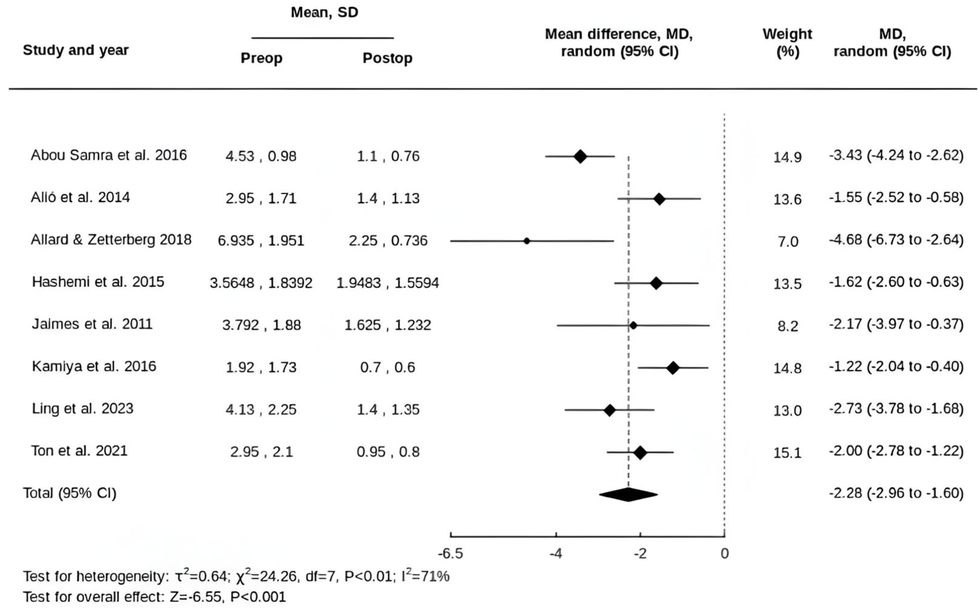
Figure 1. Forest plot of the MD in refractive cylinder following tIOL implantation [32,33,34,35,36,37,38,39].
Life 15 01362 g001
Figure 2. Forest plot of the MD in SE following tIOL implantation [32,33,34,35,36,37,39].
Life 15 01362 g002
Figure 3. Forest plot of the MD in UDVA following tIOL implantation [32,33,34,35,36,39].
Life 15 01362 g003
Figure 4. Forest plot of the MD in CDVA following tIOL implantation [32,33,34,35,36,37,38,39].
Life 15 01362 g004
Table 1. Summary characteristics of included studies.
StudyStudy DesignCountryFollow-UpPreoperative AstigmatismSample SizetIOL Model
Abou Samra WA et al., 2016 [32]Prospective; CXL then tIOL after 6/12Egypt12 monthsMean: 4.53 ± 0.98 D6 patients (9 eyes)TECNIS toric IOL
Alió JL et al., 2014 [33]RetrospectiveSpain3–15 monthsMean: −2.95 ± 1.71 D10 patients (17 eyes)Acrysof IQ toric IOL
Allard K et al., 2018 [34]Prospective case seriesSweden1–7 monthsMean: 6.94 ± 1.95 D4 patients (4 eyes)AcrySof IQ toric SN6AT IOL (patient 1 and 2), AT Torbi 709M(P) IOL (patient 3 and 4)
Hashemi H et al., 2015 [35]Case seriesIran3 monthsMean: 3.56 ± 1.84 D17 patients (23 eyes)AcrySof toric SN60TT IOL
Jaimes M et al., 2011 [36]RetrospectiveMexico3–31 monthsMean: 3.79 ± 1.88 D6 patients (7 eyes)AcrySof toric SN60TT IOL
Kamiya K et al., 2015 [37]ProspectiveJapan3 monthsMean: 1.92 ± 1.73 D19 patients (19 eyes)AcrySof IQ toric SN6AT IOL
Ling JYM et al., 2023 [38]Case seriesCanadaAvg ≥ 2 yearsMean: 4.13 ± 2.25 D17 patients (24 eyes)Multiple models; unspecified
Ton Y et al., 2021 [39]Case seriesIsrael1 monthMean: 2.95 ± 2.10 D23 patients (32 eyes)AcrySof toric SA6AT IOL, TECNIS toric IOL (ZCT series), Medicontur Bi-Flex toric IOL, Rayner T-flex toric IOL
Disclaimer/Publisher’s Note: The statements, opinions and data contained in all publications are solely those of the individual author(s) and contributor(s) and not of MDPI and/or the editor(s). MDPI and/or the editor(s) disclaim responsibility for any injury to people or property resulting from any ideas, methods, instructions or products referred to in the content.