Article

Microarray Analysis of Differentially Expressed Genes in Peripheral Blood of Postpartum Women with Gestational Diabetes Mellitus and Type 2 Diabetes

1
Medical Laboratory Sciences, Faculty of Applied Medical Sciences, King Abdulaziz University, Jeddah 21589, Saudi Arabia
2
Regenerative Medicine Unit, King Fahad Medical Research Centre, King Abdulaziz University, Jeddah 21589, Saudi Arabia
Life 2025, 15(8), 1270; https://doi.org/10.3390/life15081270
Submission received: 9 June 2025 / Revised: 18 July 2025 / Accepted: 8 August 2025 / Published: 11 August 2025
(This article belongs to the Section Genetics and Genomics)

Abstract

The etiology of women with gestational diabetes mellitus (GDM) and a greater risk of developing type 2 diabetes (T2D) after delivery remains unknown. This study aimed to investigate the global gene expression in four postpartum women with previous GDM (pGDM), three with T2D, and three with a history of normoglycemic pregnancy (controls). Total RNA was extracted from whole blood between March and May 2020. Global mRNA expression was determined using an Affymetrix Human Gene 2.0 ST Array. The expression of the selected focused genes was validated by RT-PCR. The microarray revealed 140 transcripts (p < 0.05, fold change cut-off ≥ 2) in patients with pGDM compared to controls. We identified 583 gene-altered transcripts between patients with T2D and controls. Interestingly, 60 transcripts had genes shared by pGDM or T2D versus the controls. The selected upregulated genes involved in inflammatory response, glycosylation, and death-like domains, according to the functional network analysis of pGDM (TNFAIP6, PDK3) and T2D (MMP9 and CARD6), showed similar trends to those obtained via microarray. Thus, these differentially expressed genes and their corresponding network and pathway analyses in women with pGDM and T2D offer valuable insights into the possible biological mechanisms of the progression of GDM to T2D.

1. Introduction

The etiology of diabetes is complicated and multifactorial, and alterations in gene transcripts may play a role in the pathogenesis of this metabolic disease. Type 2 diabetes (T2D), the most common type of diabetes, is caused by decreased insulin sensitivity and resistance [1]. Gestational diabetes mellitus (GDM) is the most common metabolic disturbance in pregnancy, occurs due to impaired glucose tolerance, and usually disappears following delivery [2]; however, up to 35% of cases progress to T2D within the first two months following delivery [3]. This is supported by long-term follow-up studies showing that women with previous GDM (pGDM) are at a high risk of developing T2D and associated cardiovascular diseases (CVDs) later in life [4,5]. The mechanisms of progression to T2D are not thoroughly understood and could be attributed to risk factors such as family history, obesity, and the need for insulin treatment [6,7]. Women with a history of GDM develop insulin resistance, dyslipidemia, and hypertension, which can lead to T2D and CVD [8,9]. Moreover, a recent study by Van et al. showed that notable inflammation and elevated triglyceride levels occurred shortly after delivery and were more pronounced in women with progression to overt T2D [10].
The pathophysiology of GDM is similar to that of T2D. GDM develops in pregnant women during late pregnancy as a result of an increase in insulin resistance and reduced insulin sensitivity compared to those in non-pregnant women [11]. This increase in insulin resistance leading to GDM is due to pancreatic beta cell insufficiency [12].
Several lines of evidence support the involvement of genetic components in the progression of GDM to T2D. A previous study showed a 2.3-fold higher risk of GDM in women with any parental history of diabetes compared to women with non-diabetic parents [13]. The risk of GDM increased 8.4-fold in women with diabetic siblings than in women without diabetic siblings [13].
The concept of glycemic memory or metabolic memory, which occurs when human cells are exposed to sustained elevated blood glucose levels, has recently received increasing attention. Sustained exposure to hyperglycemia can epigenetically alter gene expression profiles, and this effect persists even after therapeutic hyperglycemic control is therapeutically achieved [14]. However, the underlying mechanisms are not fully understood. Interestingly, it has been shown that oxidative-stress-induced metabolic memory is responsible for diabetes progressing to complications such as retinopathy and CVDs [15,16].
Changes in gene transcript expression in women with pGDM may also be implicated in the development of T2D [17]. In our previous study, we showed alterations in the antioxidant enzymes superoxide dismutase and sirtuin transcripts and protein in women with both pGDM and T2D, which may be related to oxidative stress [17]. To the best of our knowledge, there is no known global gene expression profile in the blood of women with pGDM. Therefore, the aim of this study was to identify changes in gene expression in peripheral blood at one day postpartum in women with GDM and T2D compared to non-diabetic women (controls) using microarray analysis and to validate the selected genes via real-time polymerase chain reaction (RT-PCR).

2. Materials and Methods

2.1. Samples

Peripheral whole blood samples were collected one day postpartum from four women with pGDM (pGDM group), three healthy women (control group), and three women with T2D (T2D group) after a full-term pregnancy, normal delivery, and matched age between March and May 2020. One day postpartum is the typical amount of time postpartum pregnant mothers are allowed to stay at the hospital after normal delivery. The subjects in the control group were non-smokers, did not have a history of T2D, and did not have hypertension. Individuals in the T2D group received insulin along with additional medications, including metformin. Mothers with GDM or T2D were selected based on the recommendations of the American Diabetes Association [18]. Mothers with GDM had no other complications, such as preeclampsia or hypertension, and they were on standardized diets. All participants were informed about the objectives of the research; informed written consent was obtained before inclusion; and the study was performed in accordance with the Declaration of Helsinki and approved by the Ethics Committee of the Research and Studies Department, Directorate of Health Affairs, Jeddah (IRB registration number: H-02-J-002; approved on 29 January 2020). The donors’ physical examination results, such as weight, height, and BMI, were recorded. Biochemical analyses were performed using the patients’ medical records.

2.2. RNA Extraction and Microarray Processing

Total RNA (250 ng/sample) was isolated from 2.5 mL of whole blood using the PAXgene Blood RNA System (Qiagen, Manchester, UK) following the manufacturer’s instructions. The RNA concentration and purity were measured spectrophotometrically using a NanoDrop spectrophotometer (Nano-Drop Technologies, Wilmington, DE, USA). The quality of RNA was additionally confirmed through analysis with an Agilent bioanalyzer (Agilent Technologies, Boeblingen, Germany) prior to the hybridization of the microarray chip. The GeneChipTM Whole Transcript PLUS kit (Affymetrix, Santa Clara, CA, USA) was used to screen for gene expression in patients with pGDM and T2D according to the manufacturer’s instructions. Extracted RNA was amplified and transcribed into cRNAs. The obtained cRNAs were then hybridized to Human Gene 2.0 ST arrays (Affymetrix, Santa Clara, CA, USA), which measured the signals for >30,000 coding transcripts and >11,000 long intergenic non-coding transcripts. The Affymetrix GeneChip Hybridization, Wash, and Stain Kit was used for hybridization. Sample hybridization to the GeneChip microarray (performed in triplicate) and image scanning were performed according to the manufacturer’s instructions.

2.3. Microarray Enrichment Analysis

Microarray expression data were imported into Bioconductor version 3.7. The data were normalized to minimize the effects of systematic non-biological variations. The analysis was focused on genes with a fold change ≥ 2 and p-value < 0.05 in pGDM and T2D relative to the controls. For subsequent functional clustering of the differentially expressed genes, they were enriched into the Gene Ontology (GO) terms cellular component, biological process, and molecular function using the GO chart feature of the Database for Annotation, Visualization, and Integrated Discovery (DAVID), and the results were ranked based on enrichment scores.
Data sets that include gene identifiers along with their corresponding expression values (fold change) from the microarray analysis were uploaded into the IPA software (build version 463341M, Ingenuity® Systems, www.ingenuity.com, accessed on 20 March 2018). Each gene identifier was matched to its corresponding gene entity in the Ingenuity Pathways Knowledge Base. We utilized data sources from Ingenuity expert insights and employed the ‘Core Analysis’ function to interpret the data in terms of biological processes, pathways, and networks. Differentially expressed gene identifiers were established as value parameters for analysis, revealing the relationship between alterations in gene expression and associated changes in biofunctions under the following categories: Molecular and Cellular Functions, Physiological System Development and Function, and Disease and Disorders. Genes that were differentially expressed with a p-value of less than 0.05 were overlaid onto global molecular networks created from the information available in the IPA knowledge base. These networks were then algorithmically generated based on their connectivity. The networks were ‘named’ according to their most prevalent functional group(s) that were identified. Canonical pathway (CP) analysis pinpointed function-specific genes that were significantly present within the networks.

2.4. Validation of Selected Genes Using RT-PCR

Total RNA (1 µg) was used to synthesize cDNA using an ImProm-II Reverse Transcription System kit (Promega, Madison, WI, UK), following the manufacturer’s protocol. Primers for the target genes were designed using the Primer3Primer software version 6.2.0. β-actin was used to normalize mRNA levels. Sequences of the target primers and β-actin are listed in Table 1.
Quantitative RT-PCR was performed in duplicate using a QuantiTect SYBR Green PCR kit (Qiagen, Manchester, UK) on an iCycler iQ Real-Time PCR Detection System (Applied Biosystems, Cheshire, UK) according to the manufacturer’s instructions. Reactions were run according to the following profile: 10 min denaturing at 95 °C; and 40 cycles of denaturing at 95 °C for 15 s, annealing at 63 °C for 10 s, and extension at 72 °C for 20 s. Data analysis was performed using the Rest 2009 software version 2.0.13 [19].

2.5. Statistical Analysis

Unpaired data were analyzed using Student’s t-test. The results are presented as mean ± SEM. Differences were considered statistically significant at p < 0.05.

3. Results

The basic clinical characteristics of the study participants are presented in Table 2.
Random plasma glucose levels were significantly higher in women with T2D than in the controls (p = 0.01). However, the other parameters did not differ among the pGDM, T2D, and control groups.
The isolated RNA purity was assessed using a 260/280 ratio and was approximately 2.0. mRNA analyses were performed by comparing the two groups with the controls, i.e., pGDM versus controls and T2D versus controls. We identified 140 transcripts that were differentially expressed between the pGDM and control groups (p-value < 0.05, fold change cut-off ≥ 2). A total of 106 genes were upregulated, and 34 genes were downregulated in the pGDM group compared to the controls, as indicated by the heatmap (Figure 1, Table 3).
The most upregulated genes in pGDM included maltase-glucoamylase 2, while the most significantly downregulated genes in pGDM were small nucleolar RNA C/D box 1B. Functional annotation cluster analysis was performed using DAVID, which showed that these 140 differentially expressed genes were involved in the immunoglobulin complex, glycosylation, and proteolysis (Table 4).
Additionally, an examination of the genes mentioned earlier (140 gene transcripts) uncovered four notable genetic networks (p < 0.05, enrichment score ≥ 2) in pGDM using IPA, as detailed in Table 5.
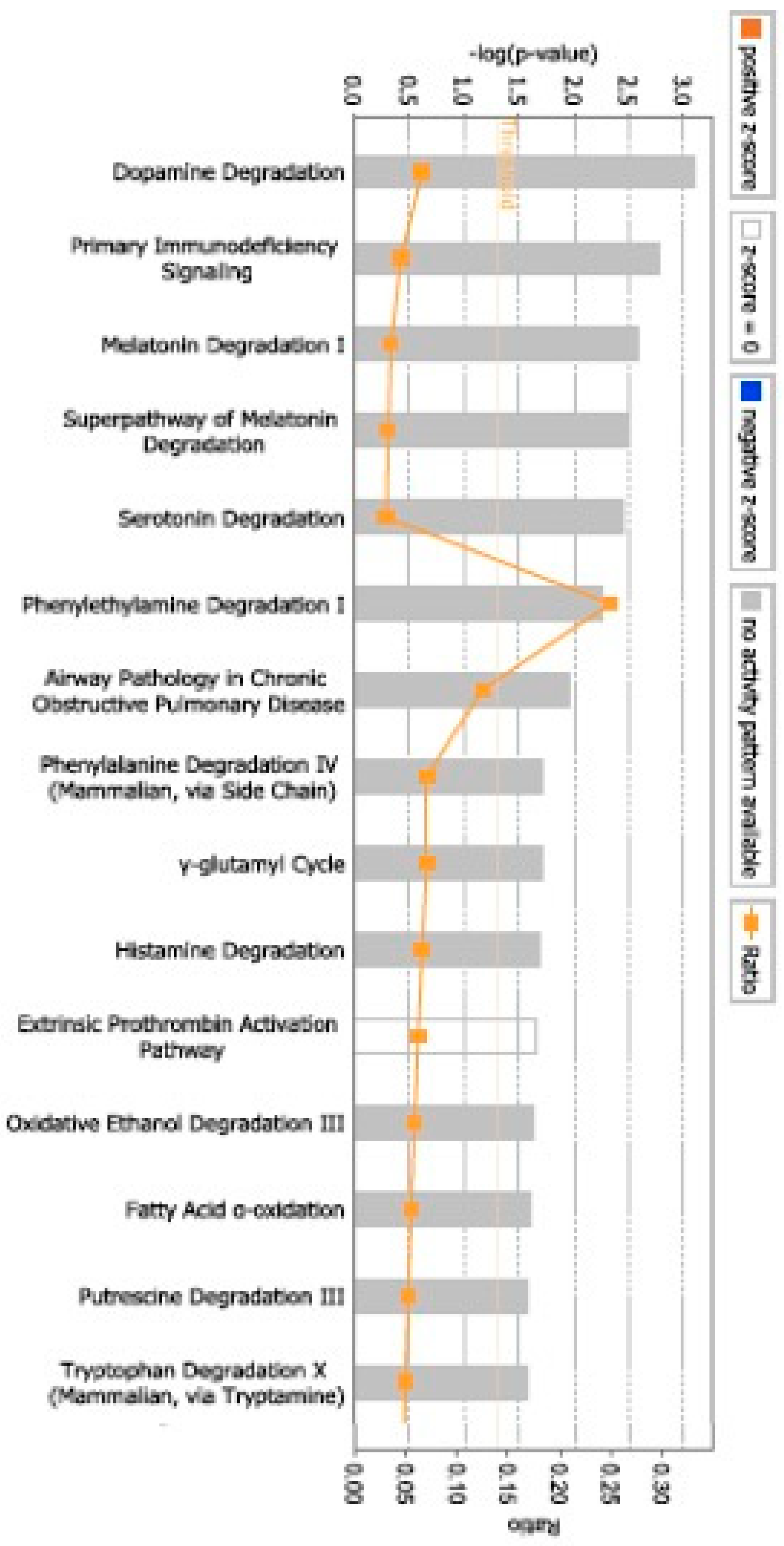
The highest-scoring networks indicated associations with diseases and disorders that mainly comprised (number of molecules) Cancer (13), Cardiovascular Disease (5), Connective Tissue Disorders (2), Dermatological Disease and Conditions (6), and Developmental Disorders (2). Genes within the Molecular and Cellular Functions network included Drug Metabolism (3), Carbohydrate Metabolism (1), Cell-to-Cell Signaling and Interaction (6), Cellular Assembly and Organization (5), and Energy Production (1). In the Physiological System Development and Functions network, the significant functions were Embryonic Development (4), Organ Development (4), Organ Morphology (1), Organismal Development (5), and Organismal Functions (1). Our IPA analysis indicates that multiple significant CPs are impacted by pGDM, i.e., Dopamine Degradation (Figure 2).
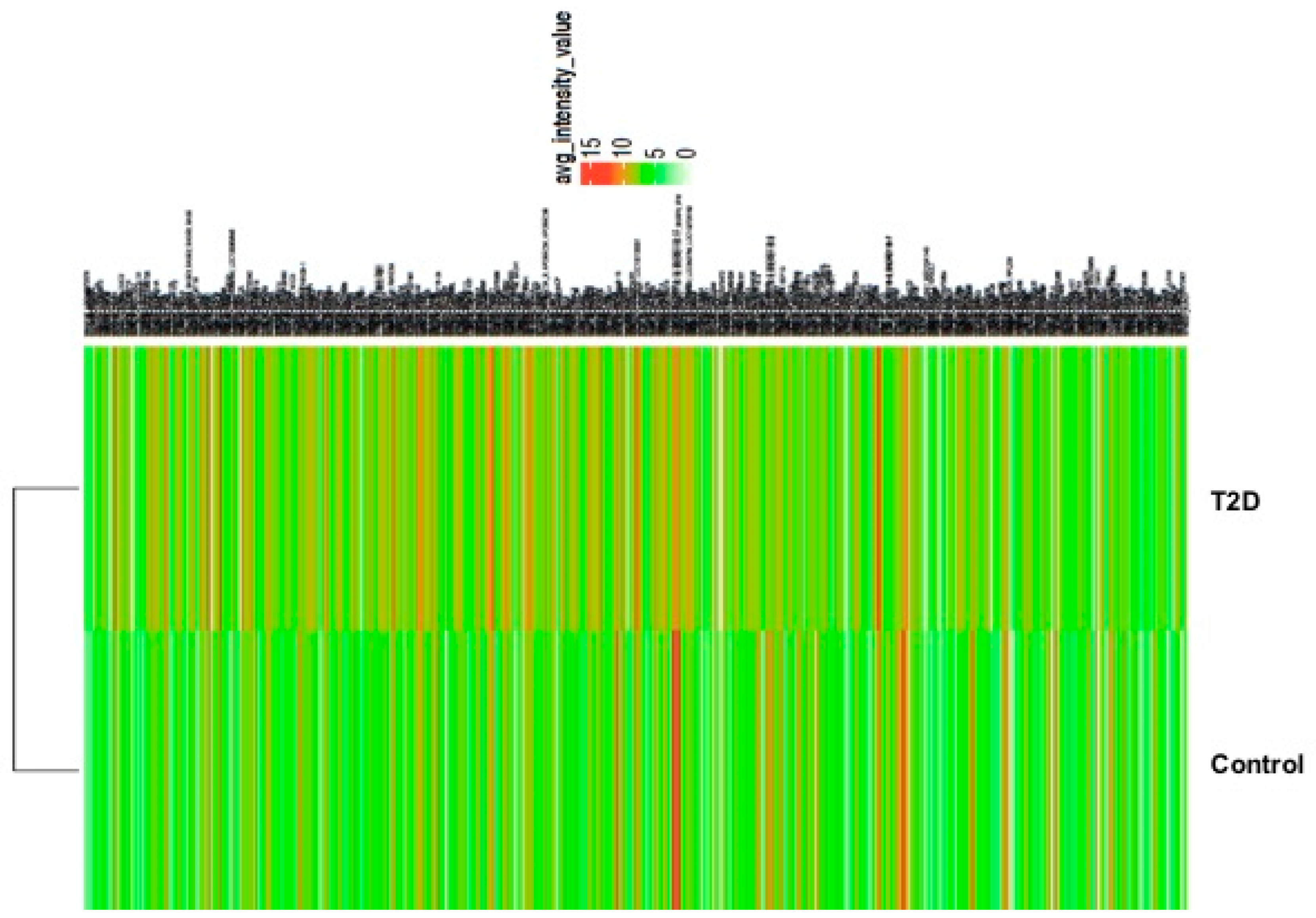
In patients with T2D, 583 gene transcripts were significantly altered in comparison with controls (p < 0.05). Of these, 490 showed an increase, and 93 displayed a decrease in transcript levels, as depicted by the heatmap (Figure 3, Table 6).
The most upregulated genes in T2D included transmembrane and tetratricopeptide repeat containing 1 (TMTC1). The most significantly downregulated genes were interferon-induced protein with tetratricopeptide repeat 1B (IFIT1B). The functional annotation cluster showed that these 583 differentially expressed genes were involved in immune response, glycosylation, and death-like domains (Table 7).
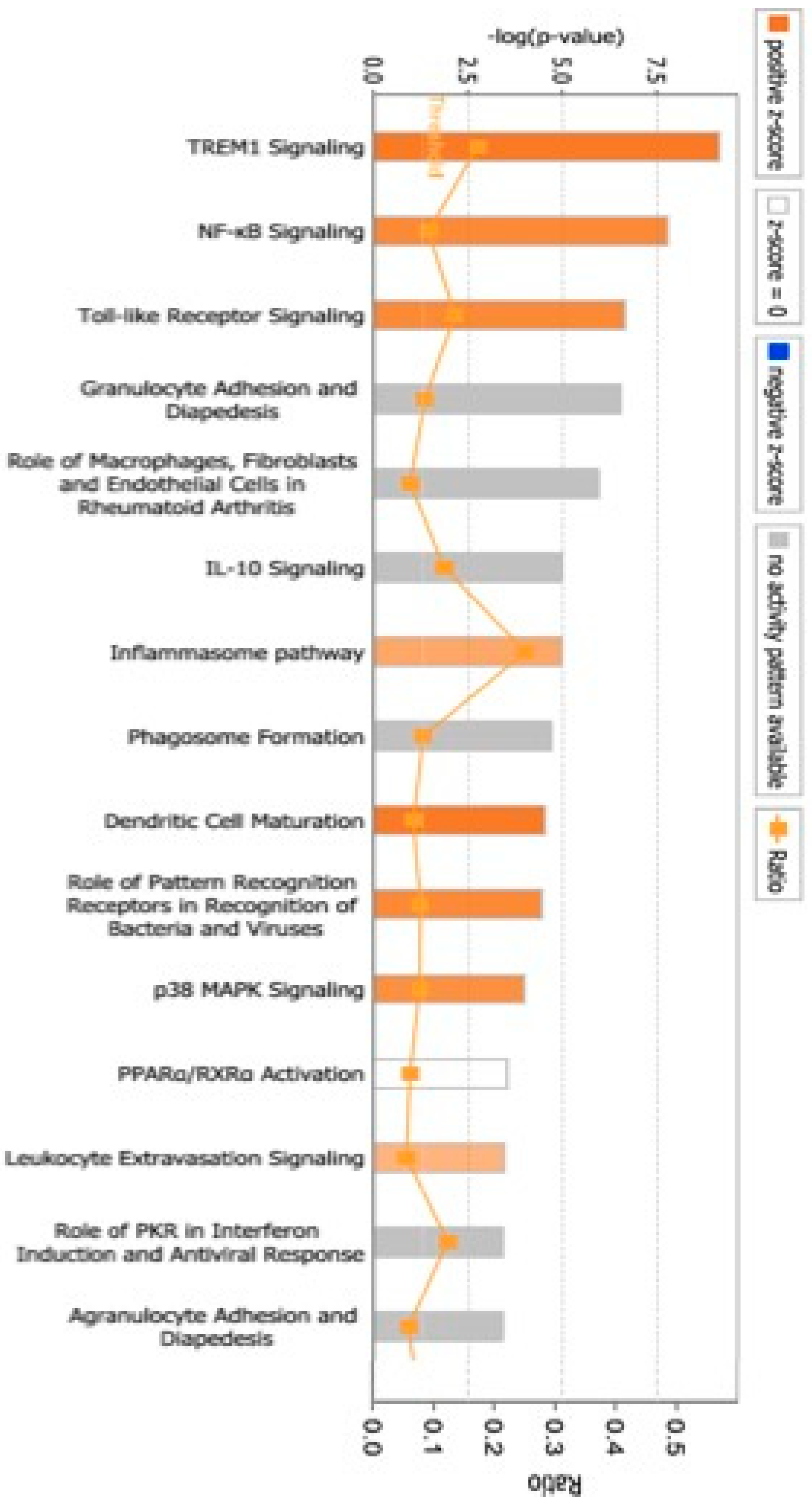
Furthermore, a review of the previously mentioned genes (583 gene transcripts) revealed five significant genetic networks (p < 0.05, score ≥ 23) in T2D through the use of IPA, as outlined in Table 8. The networks with the highest scores suggested associations with diseases and disorders that primarily included (number of molecules) Connective Tissue Disorders (66), Immunological Disease (120), Inflammatory Disease (90), Inflammatory Response (95), and Organismal Injury and Abnormalities (282). Genes located within the Molecular and Cellular Functions network included Cell Death and Survival (103), Cell-To-Cell Signaling and Interaction (72), Free Radical Scavenging (20), Cellular Movement (68), and Post-Translational Modification (30). In the Physiological System Development and Functions network, the important functions were Hematological System Development and Function (51), Immune Cell Trafficking (38), Digestive System Development and Function (14), Hepatic System Development and Function (14), and Organ Development (19). Our IPA analysis reveals that several important CPs are affected by T2D, specifically TREM1 Signaling (Figure 4).
Furthermore, 60 differentially expressed transcripts (p < 0.05, fold change cut-off ≥ 2) that were shared by pGDM and T2D are presented in Figure 5.
According to the functional network analyses (DAVID and IPA), we focused on the upregulated genes involved in inflammatory response, glycosylation, and death-like domains, as these could play a role in the pathogenesis of diabetes and its associated complications (Table 4, Table 5, Table 7, and Table 8). Combining the differentially expressed gene lists of pGDM versus controls (Table 3) and the gene clusters in Table 4 and Table 5, we extracted two key genes (TNFAIP6 and PDK3) for forward RT-PCR validation. Similarly, MMP9 and CARD6 were selected when we compared the differentially expressed gene lists of patients with T2D versus controls, as shown in Table 6, Table 7 and Table 8. For each of the four genes (TNFAIP6, PDK3, MMP9, and CARD6), the mRNA levels showed trends consistent with those observed in the microarray analysis. The expression of TNFAIP6 and PDK3 increased significantly, approximately 7- (p = 0.0001) and 2-fold (p = 0.0001), respectively, in the pGDM group compared to the controls (Figure 3). The mRNA levels of MMP9 and CARD6 were significantly upregulated, approximately 10.1- (p = 0.0001) and 40-fold (p = 0.0001), respectively, in patients with T2D compared to the controls (Figure 6).

4. Discussion

Women with pGDM are at a higher risk of developing T2D later in life [20], and the fundamental pathophysiological mechanisms that contribute to this remain incompletely understood. The role of inflammation in the onset of GDM has become a central focus in recent research [21]. Modified glycosylation patterns could act as potential biomarkers of the pathophysiology related to diabetes and could be used for therapeutic targeting [22]. Apoptosis plays an important role in the pathogenesis of diabetes, as hyperglycemia induces reactive oxygen species (ROS). This leads to considerable cellular damage and to a point of no return in apoptosis when insufficient cytoprotective and ROS scavenging molecules, such as catalase and superoxide dismutase, are available [23]. In this study, our microarray results revealed 140 differentially expressed transcripts in pGDM compared to the controls. A total of 583 differentially expressed transcripts were identified in patients with T2D compared to the controls. There were 60 shared genes between T2D versus controls and GDM versus controls, and this could imply possible disease progression to T2D. Of these differentially expressed genes, PDK3 and TNFAIP6 in pGDM and MMP9 and CARD6 in T2D were selected, as they are involved in inflammatory response, glycosylation, and death-like domains based on DAVID and IPA function analyses, which may be involved in the pathological progression to T2D. RT-PCR validation showed that these genes were upregulated, which confirmed the microarray results.
In glucose metabolism, an irreversible mitochondrial oxidative decarboxylation reaction of pyruvate to acetyl-CoA is catalyzed by the action of both enzymes’ pyruvate dehydrogenase kinases (PDKs) 1–4 and pyruvate dehydrogenase phosphatases 1–2 [24]. Our results are the first to show that PDK3 expression is increased in women with pGDM compared to the controls. To the best of our knowledge, no study has investigated PDK3 levels in patients with GDM or diabetes. However, upregulation of PDK4 and PDK1 expression has been linked to obesity [25] and diabetes [26], respectively. Notably, because PDK3 is shared between pGDM and T2D, it could serve as a biomarker for diabetes development.
TNFAIP6 or TSG6 is a secretory protein produced by inflammatory cells in response to inflammatory stimuli, such as TNF [27]. It acts as an anti-inflammatory agent by upregulating cytokines, such as IL-4 and IL-10, and preventing the migration of neutrophils [28]. Our previous study showed that proatherogenic inflammatory cytokines increased in GDM-derived fetal endothelial cells compared to those in control non-diabetic fetal endothelial cells after several passages of culture under normal glucose conditions, supporting the concept of glycemic memory or programming [29,30]. Subclinical inflammation has been observed in women with a history of GDM [31], which could at least be partially explained by the observed increase in TSG6 in patients with pGDM compared with the controls. However, this hypothesis warrants further investigation. Many studies have reported an association of subclinical inflammatory markers with increased levels of IL-6, C-reactive protein, plasminogen activator inhibitor-1, and the future development of T2D and CVD [32,33]. Interestingly, IPA determined that TNFAIP6 is a molecule involved in the Carbohydrate Metabolism as well as Cardiovascular Disease networks. To our knowledge, no study has investigated the expression of TSG6 in patients with GDM or diabetes, and the overexpression of TSG6 suggests its possible role in the later development of diabetes.
MMPs are zinc-dependent endopeptidases consisting of 25 members with similar structures and functions that are genetically different [34]. This family exerts catalytic activity against various extracellular matrix components, including proteoglycans, laminins, and collagens [35]. Under normal conditions, their expression levels and activity are low, but they increase under pathological conditions, such as CVD [36]. We found an increase in MMP9 levels in the T2D group compared with those in the control group. Consistent with previous studies, MMP9 was overexpressed in patients with diabetes [37] and streptozotocin-induced diabetic rats [38]. In addition, the in vitro exposure of endothelial cells to elevated glucose levels causes an increase in MMP9 [39]. Increased ROS and nitric oxide synthase have been shown to enhance the activity of MMP9 in placental tissue from patients with GDM [40]. We did not test the reduced expression of MMP8 in T2D using RT-PCR to rule out errors. Therefore, the mechanisms behind the increased MMP9 expression with concurrent reduction in MMP8 expression in women with T2D are unclear, and this merits further investigation. These results suggest that in the diabetes context, high glucose, oxidative stress, and insulin resistance could modify the expression and activity of MMPs, and these could serve as risk markers for diabetes.
Apoptosis is a biochemical process that leads to specific morphological changes and eventual cell death. The pro-apoptotic protein CARD6 has been suggested to trigger nuclear factor kappa beta (NFκB) activation and regulates the function of RIP kinase family members [41]. An oxidative-stress-induced inflammatory secretion such as NF-κB has been shown to play a role in the development of insulin resistance and diabetes [42]. In the current study, CARD6 mRNA was overexpressed in the T2D group compared to the controls and appeared to be shared between the pGDM and T2D groups, suggesting its role in the pathogenesis of diabetes. Consistent with another study, CARD6 expression was increased in retinal cells isolated from streptozotocin-induced diabetic rats and in retinal pigment epithelium cells, and it was involved in cell apoptosis and oxidative stress [43,44]. To the best of our knowledge, no study has investigated the expression of CARD6 in GDM or pGDM. Further study is required to characterize the role of CARD6-induced NF-κB in GDM and pGDM.
The main limitation of this study is its small sample size due to budget constraints. Despite this, our results are supported by strong statistical results and highlight altered gene transcripts that could be used as biomarkers for diabetes, and these results are strengthened through multiple experiments. Similar studies in the literature include few samples [45,46]. To the best of our knowledge, there are no studies regarding global gene expression in pGDM to explore the possible biomarkers of T2D progression. Thus, our study can be considered a pilot for future research with larger numbers of samples and higher budgets to validate our results and protect postpartum mothers with GDM from progression to T2D. We also did not have information regarding the ethnicity and lifestyle of the individuals, which may influence gene expression. We did not directly test the impact of insulin and metformin treatment in T2D patients. However, this is worthy of further investigation. The functional validation (e.g., knockdown, pathway modulation) of the selected gene transcripts merits future investigation.

5. Conclusions

This study provides insights into differentially expressed genes and their associated biological networks and pathways in pGDM and T2D. The potential genetic biomarkers we obtained (PDK3, NFAIP6, MMP9, CARD6) could be implicated in the pathogenesis of GDM and progression to T2D. It is essential to validate these biomarkers on larger and more diverse populations to evaluate their predictive value; afterward, they could be utilized to screen GDM individuals to predict T2D following GDM pregnancies. The integration of data from multiple studies is necessary to identify the target biomarkers associated with pGDM progression.

Funding

Funding information is not available.

Institutional Review Board Statement

The study was conducted in accordance with the Declaration of Helsinki and approved by the Ethics Committee of the Research and Studies Department, Directorate of Health Affairs, Jeddah (IRB registration number: H-02-J-002; approved on 29 January 2020).

Informed Consent Statement

Written informed consent was obtained from all subjects involved in the study.

Data Availability Statement

All data generated or analyzed during this study are included in the published article.

Conflicts of Interest

The authors declare no conflicts of interest.

References

  1. Młynarska, E.; Czarnik, W.; Dzieża, N.; Jędraszak, W.; Majchrowicz, G.; Prusinowski, F.; Stabrawa, M.; Rysz, J.; Franczyk, B. Type 2 diabetes mellitus: New pathogenetic mechanisms, treatment and the most important complications. Int. J. Mol. Sci. 2025, 26, 1094. [Google Scholar] [CrossRef]
  2. Immanuel, J.; Cheung, N.W.; Mohajeri, M.; Simmons, D.J.; Hague, W.M.; Teede, H.; Nolan, C.J.; Peek, M.J.; Flack, J.R.; McLean, M.; et al. Association between glycemia, glycemic variability, and pregnancy complications in early GDM. Diabetes Care 2025, 48, 285–291. [Google Scholar] [CrossRef] [PubMed]
  3. Farahmand, M.; Rahmati, M.; Azizi, F.; Ramezani Tehrani, F. Lactation duration and lifetime progression to metabolic syndrome in women according to their history of gestational diabetes: A prospective longitudinal community-based cohort study. J. Transl. Med. 2023, 21, 177. [Google Scholar] [CrossRef] [PubMed]
  4. Bellamy, L.; Casas, J.P.; Hingorani, A.D.; Williams, D. Type 2 diabetes mellitus after gestational diabetes: A systematic review and meta-analysis. Lancet 2009, 373, 1773–1779. [Google Scholar] [CrossRef]
  5. Gunderson, E.P.; Chiang, V.; Pletcher, M.J.; Jacobs, D.R., Jr.; Quesenberry, C.P., Jr.; Sidney, S.; Lewis, C.E. History of gestational diabetes mellitus and future risk of atherosclerosis in mid-life: The coronary artery risk development in young adults study. J. Am. Heart Assoc. 2014, 3, e000490. [Google Scholar] [CrossRef]
  6. Rayanagoudar, G.; Hashi, A.A.; Zamora, J.; Khan, K.S.; Hitman, G.A.; Thangaratinam, S. Quantification of the type 2 diabetes risk in women with gestational diabetes: A systematic review and meta-analysis of 95,750 women. Diabetologia 2016, 59, 1403–1411. [Google Scholar] [CrossRef] [PubMed]
  7. Baptiste-Roberts, K.; Barone, B.B.; Gary, T.L.; Golden, S.H.; Wilson, L.M.; Bass, E.B.; Nicholson, W.K. Risk factors for type 2 diabetes among women with gestational diabetes: A systematic review. Am. J. Med. 2009, 122, 207–214.e204. [Google Scholar] [CrossRef]
  8. Göbl, C.S.; Bozkurt, L.; Yarragudi, R.; Prikoszovich, T.; Tura, A.; Pacini, G.; Koppensteiner, R.; Kautzky-Willer, A. Biomarkers of endothelial dysfunction in relation to impaired carbohydrate metabolism following pregnancy with gestational diabetes mellitus. Cardiovasc. Diabetol. 2014, 13, 138. [Google Scholar] [CrossRef]
  9. Vrachnis, N.; Augoulea, A.; Iliodromiti, Z.; Lambrinoudaki, I.; Sifakis, S.; Creatsas, G. Previous gestational diabetes mellitus and markers of cardiovascular risk. Int. J. Endocrinol. 2012, 2012, 458610. [Google Scholar] [CrossRef]
  10. Van, J.A.; Luo, Y.; Danska, J.S.; Dai, F.; Alexeeff, S.E.; Gunderson, E.P.; Rost, H.; Wheeler, M.B. Postpartum defects in inflammatory response after gestational diabetes precede progression to type 2 diabetes: A nested case-control study within the SWIFT study. Metabolism 2023, 149, 155695. [Google Scholar] [CrossRef]
  11. Bassaw, B.; Fletcher, H.; Rattray, C.; McIntyre, G.; Sarkharkar, V.; Sankat, S.; Sirjusingh, A.; Chinnia, J. Screening for gestational diabetes mellitus: A Caribbean perspective. J. Obstet. Gynaecol. 2018, 38, 1035–1038. [Google Scholar] [CrossRef] [PubMed]
  12. Buchanan, T.A. Pancreatic B-cell defects in gestational diabetes: Implications for the pathogenesis and prevention of type 2 diabetes. J. Clin. Endocrinol. Metab. 2001, 86, 989–993. [Google Scholar] [CrossRef]
  13. Williams, M.A.; Qiu, C.; Dempsey, J.C.; Luthy, D.A. Familial aggregation of type 2 diabetes and chronic hypertension in women with gestational diabetes mellitus. J. Reprod. Med. 2003, 48, 955–962. [Google Scholar]
  14. Lee, C.; An, D.; Park, J. Hyperglycemic memory in metabolism and cancer. Horm. Mol. Biol. Clin. Investig. 2016, 26, 77–85. [Google Scholar] [CrossRef]
  15. Wang, Z.; Zhao, H.; Guan, W.; Kang, X.; Tai, X.; Shen, Y. Metabolic memory in mitochondrial oxidative damage triggers diabetic retinopathy. BMC Ophthalmol. 2018, 18, 258. [Google Scholar] [CrossRef]
  16. Keating, S.T.; Plutzky, J.; El-Osta, A. Epigenetic changes in diabetes and cardiovascular risk. Circ. Res. 2016, 118, 1706–1722. [Google Scholar] [CrossRef]
  17. Sultan, S.; Alzahrani, N.; Al-Sakkaf, K. The postpartum effect of maternal diabetes on the circulating levels of sirtuins and superoxide dismutase. FEBS Open Bio. 2018, 8, 256–263. [Google Scholar] [CrossRef] [PubMed]
  18. Metzger, B.E.; Gabbe, S.G.; Persson, B.; Lowe, L.P.; Dyer, A.R.; Oats, J.J.; Buchanan, T.A. International association of diabetes and pregnancy study groups recommendations on the diagnosis and classification of hyperglycemia in pregnancy: Response to Weinert. Diabetes Care 2010, 33, e98. [Google Scholar] [CrossRef]
  19. Pfaffl, M.W.; Horgan, G.W.; Dempfle, L. Relative expression software tool (REST©) for group-wise comparison and statistical analysis of relative expression results in real-time PCR. Nucleic Acids Res. 2002, 30, e36. [Google Scholar] [CrossRef]
  20. Zhao, M.; Yao, Z.; Zhang, Y.; Ma, L.; Pang, W.; Ma, S.; Xu, Y.; Wei, L. Predictive value of machine learning for the progression of gestational diabetes mellitus to type 2 diabetes: A systematic review and meta-analysis. BMC Med. Inform. Decis. Mak. 2025, 25, 18. [Google Scholar] [CrossRef]
  21. Wang, F.; Zhao, Y.; Xing, J.; Gong, L. Avicularin as a Promising Therapeutic Agent for Gestational Diabetes Mellitus: Modulation of Inflammatory, Oxidative Stress, and Apoptotic Pathways. In Doklady Biochemistry and Biophysics; Springer: Berlin/Heidelberg, Germany, 2025; pp. 206–220. [Google Scholar]
  22. Elguoshy, A.; Yamamoto, K.; Hirao, Y.; Yanagita, K.; Uchimoto, T.; Kamimura, T.; Takazawa, T.; Yamamoto, T. Machine Learning-Driven Glycoproteomic Profiling Identifies Novel Diabetes-Associated Glycosylation Biomarkers. medRxiv 2025. [Google Scholar] [CrossRef]
  23. Liu, H.; Yao, Q.; Wang, X.; Xie, H.; Yang, C.; Gao, H.; Xie, C. The research progress of crosstalk mechanism of autophagy and apoptosis in diabetic vascular endothelial injury. Biomed. Pharmacother. 2024, 170, 116072. [Google Scholar] [CrossRef]
  24. Sheng, N.; Zhang, Z.; Zheng, H.; Ma, C.; Li, M.; Wang, Z.; Wang, L.; Jiang, J.; Zhang, J. Scutellarin rescued mitochondrial damage through ameliorating mitochondrial glucose oxidation via the Pdk-Pdc axis. Adv. Sci. 2023, 10, 2303584. [Google Scholar] [CrossRef] [PubMed]
  25. Zhang, S.; Hulver, M.W.; McMillan, R.P.; Cline, M.A.; Gilbert, E.R. The pivotal role of pyruvate dehydrogenase kinases in metabolic flexibility. Nutr. Metab. 2014, 11, 10. [Google Scholar] [CrossRef]
  26. Yao, Y.; Shi, J.; Zhang, C.; Gao, W.; Huang, N.; Liu, Y.; Yan, W.; Han, Y.; Zhou, W.; Kong, L. Pyruvate dehydrogenase kinase 1 protects against neuronal injury and memory loss in mouse models of diabetes. Cell Death Dis. 2023, 14, 722. [Google Scholar] [CrossRef]
  27. Huang, P.; Sun, R.; Xu, C.; Jiang, Z.; Zuo, M.; Li, Y.; Liu, R.; Gong, P.; Han, Y.; Fang, J.; et al. Glucocorticoid activates STAT3 and NF-κB synergistically with inflammatory cytokines to enhance the anti-inflammatory factor TSG6 expression in mesenchymal stem/stromal cells. Cell Death Dis. 2024, 15, 70. [Google Scholar] [CrossRef]
  28. Dyer, D.P.; Salanga, C.L.; Johns, S.C.; Valdambrini, E.; Fuster, M.M.; Milner, C.M.; Day, A.J.; Handel, T.M. The anti-inflammatory protein TSG-6 regulates chemokine function by inhibiting chemokine/glycosaminoglycan interactions. J. Biol. Chem. 2016, 291, 12627–12640. [Google Scholar] [CrossRef]
  29. Sultan, S. Aberrant expression of proatherogenic cytokines and growth factors in human umbilical vein endothelial cells from newborns of type 2 diabetic women. SAGE Open Med. 2021, 9, 20503121211026832. [Google Scholar] [CrossRef]
  30. Sultan, S.A.; Liu, W.; Peng, Y.; Roberts, W.; Whitelaw, D.; Graham, A.M. The role of maternal gestational diabetes in inducing fetal endothelial dysfunction. J. Cell. Physiol. 2015, 230, 2695–2705. [Google Scholar] [CrossRef] [PubMed]
  31. Heitritter, S.M.; Solomon, C.G.; Mitchell, G.F.; Skali-Ounis, N.; Seely, E.W. Subclinical inflammation and vascular dysfunction in women with previous gestational diabetes mellitus. J. Clin. Endocrinol. Metab. 2005, 90, 3983–3988. [Google Scholar] [CrossRef]
  32. Ridker, P.M.; Hennekens, C.H.; Buring, J.E.; Rifai, N. C-reactive protein and other markers of inflammation in the prediction of cardiovascular disease in women. N. Engl. J. Med. 2000, 342, 836–843. [Google Scholar] [CrossRef] [PubMed]
  33. Festa, A.; D’Agostino, R., Jr.; Howard, G.; Mykkänen, L.; Tracy, R.P.; Haffner, S.M. Chronic subclinical inflammation as part of the insulin resistance syndrome: The Insulin Resistance Atherosclerosis Study (IRAS). Circulation 2000, 102, 42–47. [Google Scholar] [CrossRef]
  34. Bai, C.; Yang, W.; Qi, G.; Yang, L.; Wu, Q.; Peng, J.; Wang, N.; Liu, T. Causal association between matrix metalloproteinases and diabetic neuropathy: A two-sample Mendelian randomization study. Front. Endocrinol. 2025, 15, 1429121. [Google Scholar] [CrossRef]
  35. Liu, Z.; Yu, C.; Li, Z.; Wang, X.; Shang, D.; Dong, W. Matrix metalloproteinase-triggered self-assembling peptides for biomedical applications. J. Mater. Chem. B. 2025, 13, 8298–8334. [Google Scholar] [CrossRef]
  36. Tanase, D.M.; Valasciuc, E.; Anton, I.B.; Gosav, E.M.; Dima, N.; Cucu, A.I.; Costea, C.F.; Floria, D.E.; Hurjui, L.L.; Tarniceriu, C.C.; et al. Matrix Metalloproteinases: Pathophysiologic Implications and Potential Therapeutic Targets in Cardiovascular Disease. Biomolecules 2025, 15, 598. [Google Scholar] [CrossRef] [PubMed]
  37. Liu, B.; Su, L.; Loo, S.J.; Gao, Y.; Khin, E.; Kong, X.; Dalan, R.; Su, X.; Lee, K.O.; Ma, J.; et al. Matrix metallopeptidase 9 contributes to the beginning of plaque and is a potential biomarker for the early identification of atherosclerosis in asymptomatic patients with diabetes. Front. Endocrinol. 2024, 15, 1369369. [Google Scholar] [CrossRef]
  38. Silva, F.S.; Bortolin, R.H.; Araújo, D.N.; Marques, D.E.; Lima, J.P.M.; Rezende, A.A.; Vieira, W.H.; Silva, N.B.; Medeiros, K.C.; Ackermann, P.W.; et al. Exercise training ameliorates matrix metalloproteinases 2 and 9 messenger RNA expression and mitigates adverse left ventricular remodeling in streptozotocin-induced diabetic rats. Cardiovasc. Pathol. 2017, 29, 37–44. [Google Scholar] [CrossRef]
  39. Death, A.K.; Fisher, E.J.; McGrath, K.C.; Yue, D.K. High glucose alters matrix metalloproteinase expression in two key vascular cells: Potential impact on atherosclerosis in diabetes. Atherosclerosis 2003, 168, 263–269. [Google Scholar] [CrossRef]
  40. Pustovrh, C.; Jawerbaum, A.; Sinner, D.; Pesaresi, M.; Baier, M.; Micone, P.; Gimeno, M.; Gonzalez, E.T. Membrane-type matrix metalloproteinase-9 activity in placental tissue from patients with pre-existing and gestational diabetes mellitus. Reprod. Fertil. Dev. 2000, 12, 269–275. [Google Scholar] [CrossRef] [PubMed]
  41. Dufner, A.; Pownall, S.; Mak, T.W. Caspase recruitment domain protein 6 is a microtubule-interacting protein that positively modulates NF-κB activation. Proc. Natl. Acad. Sci. USA 2006, 103, 988–993. [Google Scholar] [CrossRef] [PubMed]
  42. Ho, E.; Quan, N.; Tsai, Y.H.; Lai, W.; Bray, T.M. Dietary zinc supplementation inhibits NFκB activation and protects against chemically induced diabetes in CD1 mice. Exp. Biol. Med. 2001, 226, 103–111. [Google Scholar] [CrossRef] [PubMed]
  43. Fan, X.; Yang, Y.; Wu, G.; Kong, Y.; Zhang, Y.; Zha, X. Circ-CARD6 inhibits oxidative stress-induced apoptosis and autophagy in ARPE-19 cells via the miR-29b-3p/PRDX6/PI3K/Akt axis. Exp. Eye Res. 2024, 238, 109690. [Google Scholar] [CrossRef] [PubMed]
  44. Kowluru, R.A.; Chan, P.S. Metabolic memory in diabetes–from in vitro oddity to in vivo problem: Role of Apoptosis. Brain Res. Bull. 2010, 81, 297–302. [Google Scholar] [CrossRef] [PubMed]
  45. Baelde, H.J.; Eikmans, M.; Doran, P.P.; Lappin, D.W.; de Heer, E.; Bruijn, J.A. Gene expression profiling in glomeruli from human kidneys with diabetic nephropathy. Am. J. Kidney Dis. 2004, 43, 636–650. [Google Scholar] [CrossRef]
  46. Ma, R.; Deng, X.L.; Aleteng, Q.Q.G.; Li, L.; Zhu, J. Genome-Wide Transcriptome Analysis in Type 2 Diabetes Patients Treated by Sitagliptin. Diabetes Metab. Syndr. Obes. 2022, 15, 1761–1770. [Google Scholar] [CrossRef]
Figure 1. Heatmap and hierarchical cluster analysis of differentially expressed genes. The heatmap shows the relative induction or repression of genes in gestational diabetes mellitus (GDM) and controls. Green indicates higher expression; red indicates reduced mRNA expression; light green indicates no changes.
Life 15 01270 g001
Figure 2. Canonical pathways (CPs) associated with pGDM subjects. CPs that were identified as the most statistically significant in the IPA core analysis are presented in accordance with their p-values (−Log). The threshold line represents a p-value of 0.05.
Life 15 01270 g002
Figure 3. Heatmap and hierarchical cluster analysis of differentially expressed genes and validation of indicated selected mRNA by qRT-PCR. The heatmap shows the relative induction or repression of genes in T2D and controls. Green color indicates higher expression; red color indicates reduced expression of mRNA; light green indicates no changes.
Life 15 01270 g003
Figure 4. CPs associated with T2D subjects. CPs that were identified as the most statistically significant in the IPA core analysis are presented in accordance with their p-values (−Log). The threshold line represents a p-value of 0.05.
Life 15 01270 g004
Figure 5. Venn diagrams showing the total number of shared genes between pGDM or T2D (A and B, respectively) and controls (N). Sixty differentially expressed transcripts were shared by pGDM and T2D (AB), of which seventeen are displayed along with their gene symbols and fold changes (p < 0.05, fold change cut-off ≥ 2).
Life 15 01270 g005
Figure 6. Validation of the indicated selected mRNAs by qRT-PCR. Total RNA isolated from peripheral blood mononuclear cells of (A) GDM and (B) T2D and the indicated selected target genes tested using qRT-PCR. The results are depicted as fold change over a housekeeping gene (b-actin). The results are expressed as mean ± SEM (n = 3–4). *** p < 0.0001 versus controls.
Life 15 01270 g006
Table 1. Primer sequences.
PrimersSequences
PDK3Forward:
5′-GAGCAATCCCAGCAGTGAAC-3′
Reverse:
5′-ATAACTGTGATGCCACGCTC-3′
TNFAIP6Forward:
5′-GCTGGATGGATGGCTAAGGG-3′
Reverse:
5′-CCTTTGCGTGTGGGTTGTAG-3′
MMP9Forward:
5′-GGTGATTGACGACGCCTTTG-3′
Reverse:
5′-GGACCACAACTCGTCATCGT-3′
CARD6Forward:
5′-CGAGAGTACTCCCTCAGAGAT-3′
Reverse:
5′-GCCCCCATAGATTGAGGAGG-3′
β-actinForward:
5′-AGCGGGAAATCGTGCGTGAC-3′
Reverse:
5′-CGGACTCGTCATACTCCTGCT-3′
Table 2. Donors’ clinical characteristics.
Mothers’ Status Controls (n = 3)pGDM (n = 4)T2D (n = 3)P (pGDM vs. Controls/T2D vs. Controls)
Age (years)28.6 ± 0.930 ± 131.7 ± 1.60.29/0.2
BMI28.8 ± 330 ± 1.331.3 ± 3.90.37/0.32
HbA1c (%)
RPG (mM)
2h OGTT
-
4 ± 0.3
-
6.1 ± 0.1
5.3 ± 0.9
9.9 ± 1
6.3 ± 0.5
5.5 ± 0.2
-
-
0.28/0.01 *
-
RPG, random plasma glucose; BMI, body mass index; HbA1c, glycosylated hemoglobin; OGTT, oral glucose tolerance test. Data expressed as mean ± SEM. * p-value < 0.05.
Table 3. List of selected differentially upregulated and downregulated genes in previous GDM (pGDM) versus controls.
Gene SymbolFold Changep-ValueGene Name
MGAM24.70.0343maltase-glucoamylase 2 (putative)
LOC1053725784.367.38 × 10−7uncharacterized LOC105372578
TRGV54.240.0414T-cell receptor gamma variable 5
LOC1005076393.940.0147uncharacterized LOC100507639
TRDJ13.860.0019T-cell receptor delta joining 1
TRDJ43.570.0095T-cell receptor delta joining 4
CST73.558.22 × 10−5cystatin F (leukocystatin)
CD1773.540.0129CD177 molecule
LOC1027233733.350.0051uncharacterized LOC102723373
HCG263.120.0326HLA complex group 26 (non-protein-coding)
LINC010612.960.0353long intergenic non-protein-coding RNA 1061
RNU6-59P2.940.0004RNA, U6 small nuclear 59, pseudogene
KIAA13242.860.0025KIAA1324
ANPEP2.750.0059alanyl (membrane) aminopeptidase
TRDJ22.710.0493T-cell receptor delta joining 2
RNU5B-12.640.0031RNA, U5B small nuclear 1
SULT1B12.60.0237sulfotransferase family 1B member 1
CEP170P12.570.0024centrosomal protein 170 kDa pseudogene 1
TNFAIP62.510.0088tumor necrosis factor, alpha-induced protein 6
BACH1-IT22.460.002BACH1 intronic transcript 2
MIR548K2.450.0159microRNA 548k
PSMD5-AS12.410.0203PSMD5 antisense RNA 1 (head-to-head)
DYSF2.380.025dysferlin
LOC1053798182.370.0199uncharacterized LOC105379818
F52.360.0333coagulation factor V (proaccelerin, labile factor)
CRISPLD22.354.47 × 10−5cysteine-rich secretory protein LCCL domain containing 2
C3orf622.330.0123chromosome 3 open reading frame 62
DDX12P; DDX112.30.0015DEAD/H (Asp-Glu-Ala-Asp/His) box polypeptide 12, pseudogene; DEAD/H (Asp-Glu-Ala-Asp/His) box helicase 11
CCHCR12.240.0014coiled-coil alpha-helical rod protein 1
TRDC2.230.0432T-cell receptor delta constant
ALDH22.210.0432aldehyde dehydrogenase 2 family (mitochondrial)
SPATA12.180.0386spermatogenesis associated 1
CARD62.170.044caspase recruitment domain family, member 6
CCDC144CP2.170.0111coiled-coil domain containing 144C, pseudogene
BMS1P42.160.0027BMS1 ribosome biogenesis factor pseudogene 4
FAM120A2.150.0068family with sequence similarity 120A
SDHAP2; LINC009692.140.0361succinate dehydrogenase complex subunit A, flavoprotein pseudogene 2; long intergenic non-protein-coding RNA 969
MIR12992.110.0403microRNA 1299
PDCD62.080.0014programmed cell death 6
PDK32.060.0028pyruvate dehydrogenase kinase, isozyme 3
RAB3D2.060.0037RAB3D, member of RAS oncogene family
FAM111B2.020.0195family with sequence similarity 111, member B
IGLL5; IGLV1-3620.0024immunoglobulin lambda-like polypeptide 5; immunoglobulin lambda variable 1-36
XGY2; XG20.0009Xg pseudogene, Y-linked 2; Xg blood group
CYP2J220.0017cytochrome P450, family 2, subfamily J, polypeptide 2
SNORD14A−2.010.0154small nucleolar RNA, C/D box 14A
MIR4279−2.130.046microRNA 4279
MIR3137−2.170.0059microRNA 3137
HBD−2.180.0494hemoglobin, delta
MIR514A1−2.180.0399microRNA 514a-1
IGHD−2.20.016immunoglobulin heavy constant delta
MMP8−2.230.0204matrix metallopeptidase 8
LOC101928794−2.360.0249uncharacterized LOC101928794
SNORD1B−2.480.0488small nucleolar RNA, C/D box 1B
Table 4. DAVID mapping of differentially expressed genes in pGDM versus controls.
Gene List Enrichment Score *Cluster
IGHD, TRDC, IGLL5, HBD2.4GO:0042571 immunoglobulin complex, circulating
TNFAIP6, F5, CRISPLD2, CST7, CD177, IGHD, TREML5P, MMP8, KIAA1324, ANPEP0.9GO:0005615 extracellular space, glycosylation site: N-linked (GlcNAc), glycoprotein
F5, MMP8, ANPEP, PDCD60.8GO:0006508 proteolysis
The functional annotation cluster was obtained using DAVID version 6.8. * The enrichment score is calculated in the log scale of groups of p-values in corresponding annotation clusters. The clusters shown here are those with enrichment scores > 0.5.
Table 5. Networks with high scores identified through Ingenuity® Pathway Analysis (IPA) in pGDM: The four most impacted networks.
Network FunctionsEnrichment Score *Gene in Network
Cellular Movement, Hematological System Development and Function, Immune Cell Trafficking17ANPEP, CYP2J2, CD177, IGHD, TNFAIP6, DYSF, IGLL1, IGLL5
Cell Cycle, Gene Expression, Cellular Development17mir-506, PDCD6, PDK3
Cancer, Cell Cycle, Cellular Assembly and Organization2ANPEP, mir-506, mir-548, CD177, CST7, PDK3
Cell-To-Cell Signaling and Interaction, Cancer, Gastrointestinal Disease2DYSF, F5, CD177, ANPEP, CYP2J2, PDCD6 IGHD, TNFAIP6
ALDH2, C3orf62, mir-506
Hereditary Disorder, Neurological Disease, Organismal Injury and Abnormalities2TNFAIP6, ANPEP, F5, HBD, MMP8, ALDH2, PDK3
* The enrichment score is produced by utilizing a p-value of less than 0.05.
Table 6. List of selected differentially upregulated and downregulated genes in T2D versus controls.
Gene SymbolFold Changep-ValueGene Name
TMTC16.750.0129transmembrane and tetratricopeptide repeat containing 1
TRDJ45.250.0049T-cell receptor delta joining 4
CLEC12A4.740.0225C-type lectin domain family 12, member A
DYSF4.650.0013dysferlin
MT1L; MT1M4.40.0012metallothionein 1L (gene/pseudogene)
MGAM24.380.0455maltase-glucoamylase 2 (putative)
CLEC12B4.220.0174C-type lectin domain family 12, member B
ANPEP4.130.0008alanyl (membrane) aminopeptidase
ALPL4.010.0018alkaline phosphatase, liver/bone/kidney
LOC1027242313.950.0088uncharacterized LOC102724231
FCGR1A3.930.0289Fc fragment of IgG, high-affinity Ia, receptor (CD64)
CST73.820.0004cystatin F (leukocystatin)
IL3RA3.580.0007interleukin 3 receptor, alpha (low-affinity)
BMX3.250.0027BMX non-receptor tyrosine kinase
ADGRG33.240.0174adhesion G-protein-coupled receptor G3
ICAM13.230.0072intercellular adhesion molecule 1
MIR39393.190.0242microRNA 3939
TLR53.030.0319Toll-like receptor 5
HAUS430.0004HAUS augmin-like complex subunit 4
CASP52.990.0063caspase 5
PIK3AP12.950.0056phosphoinositide-3-kinase adaptor protein 1
KREMEN12.950.0104kringle containing transmembrane protein 1
HCG262.940.0259HLA complex group 26 (non-protein-coding)
CARD172.910.0109caspase recruitment domain family, member 17
TECPR22.890.0094tectonin beta-propeller repeat containing 2
SSH12.830.0033slingshot protein phosphatase 1
IL3RA2.820.0005interleukin 3 receptor, alpha (low-affinity)
TREML22.820.0069triggering receptor expressed on myeloid cell-like 2
NQO22.80.0244NAD(P)H dehydrogenase, quinone 2
IL1B2.650.0209interleukin 1 beta
CEP192.630.0002centrosomal protein 19kDa
ADGRE12.630.002adhesion G-protein-coupled receptor E1
SEMA4A2.630.0178sema domain, immunoglobulin domain (Ig)
MEFV2.620.0012Mediterranean fever
SLC25A442.620.013solute carrier family 25, member 44
LINC001732.610.001long intergenic non-protein-coding RNA 173
BCL62.610.0068B-cell CLL/lymphoma 6
SLC26A82.610.0072solute carrier family 26 (anion exchanger), member 8
ALDH22.610.009aldehyde dehydrogenase 2 family (mitochondrial)
ORAI22.590.0163ORAI calcium-release-activated calcium modulator 2
NR6A12.580.0013nuclear receptor subfamily 6, group A, member 1
CHSY12.580.0224chondroitin sulfate synthase 1
CARD62.570.0057caspase recruitment domain family, member 6
LINC012722.030.0062long intergenic non-protein-coding RNA 1272
SLC6A62.030.0096solute carrier family 6 (neurotransmitter transporter)
MMP92.030.0129matrix metallopeptidase 9
MIR133A1; MIR133A1HG−2.010.0193microRNA 133a-1; MIR133A1 host gene (non-protein-coding)
SNORD9−2.050.0025small nucleolar RNA, C/D box 9
HK1−2.070.0181hexokinase 1
TRAJ28−2.070.0419T-cell receptor alpha joining 28
CUL4A−2.080.0021cullin 4A
GYPA−4.360.0114glycophorin A
MMP8−4.720.0214matrix metallopeptidase 8
AHSP−5.30.0366alpha hemoglobin stabilizing protein
IFIT1B−6.90.0017interferon-induced protein with tetratricopeptide repeats 1B
Table 7. DAVID mapping of differentially expressed genes in T2D versus controls.
Gene ListEnrichment Score *GO ID/Cluster
ADGRE1, FFAR2, HCK, MSRB1, TLR5, TLR8, LILRB2, IFIT2, NLRC4, MYD88, TNFSF13B, MEFV, FCGR1A, LILRA4, BCL6, FCGR2A, CLEC4D, EIF2AK2, APOBEC3A_B, TBKBP1, AKIRIN2, SEMA4A3.7GO:0045087 innate immune response
CGB1, IER3, OR1A1, NDST1, MMP9, TREML5P, MMP8, GYPA, SIRPB2, CXCR2, ANPEP, TLR5, FCRL6, TLR8, CANT1, SMPDL3A, ITPRIP, HPSE, LTB4R, LILRA4, CSF3R, SEMA3C, SERPINA1, CLEC4D3.2GO:0005886 plasma membrane, glycosylation site: N-linked (GlcNAc), glycoprotein
CASP5, TNFRSF1A, IRAK3, NLRC4, MYD88, MEFV, CARD17, NLRP12, CARD6, CASP1
BCL3, BCL6
3.1IPR011029 death-like domains
The functional annotation cluster was obtained using DAVID version 6.8. * The enrichment score is calculated in the log scale of groups of p-values in corresponding annotation clusters. The clusters shown here had enrichment scores > 0.5.
Table 8. Networks with high scores identified through IPA in T2D: The five most impacted networks.
Network FunctionsEnrichment Score *Genes in Network
Inflammatory Disease, Inflammatory Response, Cellular Movement39MMP8, MMP9, ICAM1, IL1B, IL3RA, IMPDH1, MGAM, mir-21, mir-368
Infectious Diseases, Inflammatory Response, Organismal Injury and Abnormalities35BASP1, BMX, BPGM, CACNA1E, CANT1, CARD6, CCR1, CD274, CPD, CSF3R, CSTA, DGAT2, DYSF, ELL, FCAR
Cellular Movement, Hematological System31C5AR2, CCR1, CXCR2,
DEFA1, MMP9, PLAUR, SEMA4A, SERPINA1, TIMP2, TNFRSF1A
Cell Death and Survival, Cancer29BCL6, CD274, FCGR3B, CARD6, ICAM1, IL1B, IL1RN, MYD88, NFIL3, NFKBIA, SIGLEC9, TNFRSF1A, TNFSF13B, MMP9
Connective Tissue Disorders, Immunological Disease23MMP25, MMP8, MMP9, NFKBIA, SAMD9L, TIMP2, TLE3, TLR8, TNFRSF1A, TNFSF13B, TRIM21, ZFP36, ZNF281
* The enrichment score was produced by utilizing a p-value of less than 0.05.
Disclaimer/Publisher’s Note: The statements, opinions and data contained in all publications are solely those of the individual author(s) and contributor(s) and not of MDPI and/or the editor(s). MDPI and/or the editor(s) disclaim responsibility for any injury to people or property resulting from any ideas, methods, instructions or products referred to in the content.