Article

Phlogacanthus pulcherrimus Leaf Extract as a Functional Feed Additive: Influences on Growth Indices, Bacterial Challenge Survival, and Expression of Immune-, Growth-, and Antioxidant-Related Genes in Labeo chrysophekadion (Bleeker, 1849)

by
Sontaya Sookying
1,
Panitnart Auputinan
2,
Dutrudi Panprommin
3 and
Paiboon Panase
3,4,*
1
Division of Pharmacy and Technology, Department of Pharmaceutical Care, School of Pharmaceutical Sciences, University of Phayao, Mueang, Phayao 56000, Thailand
2
Division of Biotechnology, School of Agriculture and Natural Resources, University of Phayao, Mueang, Phayao 56000, Thailand
3
Division of Fisheries, School of Agriculture and Natural Resources, University of Phayao, Mueang, Phayao 56000, Thailand
4
Unit of Excellence “Physiology and Sustainable Production of Terrestrial and Aquatic Animals”, Division of Fisheries, School of Agriculture and Natural Resources, University of Phayao, Mueang, Phayao 56000, Thailand
*
Author to whom correspondence should be addressed.
Life 2025, 15(8), 1220; https://doi.org/10.3390/life15081220 (registering DOI)
Submission received: 12 June 2025 / Revised: 23 July 2025 / Accepted: 29 July 2025 / Published: 1 August 2025
(This article belongs to the Special Issue Nutrition–Physiology Interactions in Aquatic Species)

Abstract

This research examined the impact of dietary supplementation with Phlogacanthus pulcherrimus extract (PPE) on the growth, disease resistance, and expression of immune-, growth-, and antioxidant-related genes in Labeo chrysophekadion. Over 150 days, 90 fish from each group were fed diets with 0 (control), 0.25, 0.50, or 0.75 g/kg of PPE. Phytochemical analysis revealed phenolics (96.00 mg GAE/g), flavonoids (17.55 mg QE/g), anthraquinones, and triterpenoids, along with moderate antioxidant activity (IC50 = 1314.08 μg/mL). One-way ANOVA of growth indices, including weight gain, specific growth rate, feed conversion ratio, and survival rate, revealed no significant differences (p > 0.05); however, PPE supplementation significantly enhanced immune and antioxidant gene expression. IL-1β was significantly (p < 0.05) upregulated at all doses, with the highest expression observed at 0.50 g/kg, showing a fivefold increase compared to the control. In addition, the highest relative expressions of IGF-1 and CAT were found at 0.75 g/kg, with 4.5-fold and 3.5-fold increases compared to the control, respectively. PPE at 0.75 g/kg decreased the cumulative mortality rate (CMR) by 20% compared to the control group, which had a CMR of 50% following exposure to Aeromonas hydrophila. PPE acted as an effective immunostimulant and antioxidant, supporting reduced antibiotic reliance in aquaculture.

1. Introduction

Black sharkminnow (Labeo chrysophekadion (Bleeker, 1849)) (family Cyprinidae) is a freshwater fish commonly found in both stagnant and flowing waters across Southeast Asia. Its natural distribution includes the Mekong and Chao Phraya basins, the Malay Peninsula, Sumatra, Java, and Borneo [1]. This species is highly valued for consumption due to its large body size, substantial flesh content, and palatable taste [2]. It is recognized as an economically important aquatic species in several Asian countries, including Thailand, Laos, Vietnam, and Cambodia. Nonetheless, the natural populations of L. chrysophekadion are currently declining, primarily because of environmental degradation that adversely affects their reproductive capabilities and habitat quality. Although breeding technologies under controlled conditions for this species have been successfully developed [3], the market demand for black sharkminnow remains high. Apart from being consumed as food, it is also popular as an ornamental fish [2,4], contributing to its relatively high market price. However, the only available export data for this species from Thailand pertains to its status as an ornamental fish, with statistics recorded from 2018 to 2020. During this period, approximately 60,000 individuals were exported, with an estimated value of around THB 335,557 (equivalent to approximately USD 10,000) [5].
Phlogacanthus pulcherrimus T. Anderson (family Acanthaceae), commonly known as “Dee Pla Kang” in Thai, is a widely distributed medicinal and culinary plant, particularly in northern and northeastern Thailand. In Thai traditional medicine, it has been used as a general tonic, diuretic [6], and appetite stimulant [6,7]. P. pulcherrimus is commonly found in the wild and is also cultivated in home gardens in some regions [7]. Remarkably, reports show a 100% survival rate when wild plants are transplanted for cultivation in the greenhouse condition [8], suggesting that P. pulcherrimus is a native, non-toxic, easily cultivated food plant with no known adverse effects on humans or the environment.
Beyond its traditional uses, recent studies have revealed additional pharmacological activities of P. pulcherrimus. Boontha et al. [9] reported that ethanol extracts of the leaves exhibit anticancer activity against MCF-7 breast cancer cells through multiple mechanisms. Further research by Kheawchaum et al. [10] identified diterpenoid lactone glycosides and phenolic glycosides from P. pulcherrimus with cytotoxic activity against various cancer cell lines, including T-lymphoblast (MOLT-3), hepatocellular carcinoma (HepG2), and cervical cancer (HeLa). Notably, only one out of twenty compounds showed low-level toxicity against normal MRC-5 cells [10]. The safety of P. pulcherrimus extracts in normal Vero cells was confirmed by Athipornchai et al. [11].
The plant also exhibits strong inhibitory activity against alpha-glucosidase, a key enzyme in carbohydrate digestion that influences blood glucose levels and reflects physiological stress. The leaf extracts of P. pulcherrimus significantly outperformed the standard drug acarbose in α-glucosidase inhibition assays, as confirmed by several studies [11,12,13]. In addition to its pharmacological properties, the nutritional composition of P. pulcherrimus has also been analyzed. Chaichana et al. reported that the plant is rich in minerals—particularly calcium, iron, magnesium, and zinc—and contains protein, fat, carbohydrates, and dietary fiber at levels of 22.5%, 4.2%, 41.1%, and 16.3%, respectively [14]. The high magnesium and zinc content may contribute to its antioxidant and biological activities, complementing its phytochemical profile, including flavonoids and phenolic compounds.
The application of herbal extracts as dietary supplements in aquaculture has garnered increasing scientific attention due to their potential benefits, including antimicrobial and antifungal activities, immune system enhancement, stress alleviation, improved growth performance, decreased dependence on chemical treatments, and alignment with consumer preferences and ongoing research innovations [15,16,17]. Given the high market demand for L. chrysophekadion both as a food source and as an ornamental fish amidst the declining wild populations and reproductive challenges, the pharmacological potential, non-toxicity, and ease of cultivation of P. pulcherrimus is of scientific interest.
Based on the literature review, there are no prior reports on the use of this plant extract in L. chrysophekadion. Consequently, this study aims to investigate the effects of ethanol leaf extract of P. pulcherrimus on growth performance and the expression of key genes, including interleukin-1β (IL-1β), insulin-like growth factor 1 (IGF-1), and catalase (CAT) of black sharkminnow. These genes were selected due to their fundamental roles in immune response, growth regulation, and oxidative stress defense, respectively.

2. Materials and Methods

2.1. Ethical Considerations and Animal Use Authorization

All experimental procedures were approved by the Committee of Institutional Animal Care, University of Phayao, Thailand (approval ID: 1-010-67; date of approval: 1 August 2024), and were performed in compliance with the institutional guidelines. Authorization to use laboratory animals was granted to the researcher, Paiboon Panase, under license No. Ul-01217-2558 by the National Research Council of Thailand.

2.2. Plant Materials and Chemicals

Fresh leaves of P. pulcherrimus were collected from natural areas in Phayao Province and purchased from a reliable local supplier. A voucher specimen was prepared by pressing and drying the plant material, which was subsequently identified by a botanical taxonomist. The specimen was deposited at the Queen Sirikit Botanic Garden (QBG) Herbarium, with voucher number 149,658.
All chemicals used in the experiments were of analytical grade. Ethanol (99.9%) and methanol (99.9%) were obtained from RCI Labscan, Dublin, Ireland. Aluminum chloride hexahydrate (95%) and sodium carbonate anhydrous (99.5%) were sourced from KemAus, Sydney, Australia. Sodium hydroxide (99%) was acquired from RCI Labscan, Ireland. Gallic acid (98%) was supplied by AK Scientific, Union City, CA, USA. Quercetin dihydrate (98%) and L-ascorbic acid (99.7%) originated from Sisco Research Laboratories, Mumbai, India. Folin–Ciocalteu’s phenol reagent was purchased from Loba Chemie, Mumbai, India, and 2,2-diphenyl-1-picrylhydrazyl (DPPH) was supplied by Sigma-Aldrich, Waltham, MA, USA. Reagents for phytochemical screening tests were reagents of analytical grade and were supported by the School of Pharmaceutical Sciences, University of Phayao.

2.3. Preparation of P. pulcherrimus Extract

Fresh leaves of P. pulcherrimus at the intermediate growth stage (neither too young nor too mature), exhibiting intact morphology and free from diseases and insect damage [13], were thoroughly washed and dried in a hot-air oven at a temperature not exceeding 45 °C. The dried leaves were ground using a grinder and sieved through a 40-mesh sieve to obtain a powder with a particle size of 400 μm. A total of 500 g of dried leaf powder was weighed and macerated with 95% ethanol at a ratio of 1:4 (w/v). The mixture was continuously agitated using a shaker at 100 rpm for 72 h. Upon completion of the extraction period, the mixture was filtered through Whatman no. 4 filter paper with a pore size of 20–25 μm, in combination with a Buchner funnel and suction flask, and concentrated to dryness using a rotary evaporator. The resulting PPE was stored in a tightly sealed, light-protected container at a temperature not exceeding 8 °C until further use [18].

2.4. Phytochemical Screening of PPE

In this investigation, the phytochemical profile of PPE was examined using standardized protocols, as outlined by Pant et al., Hartanti and Cahyani, and Shaikh and Patil [19,20,21]. The presence of alkaloids was assessed using Dragendorff’s and Valser’s reagents. To evaluate steroidal and terpenoid components, the Liebermann–Burchard test was employed. Molisch and Keller–Killiani assays were used to detect carbohydrates and deoxy sugars, respectively, while α,β-unsaturated five-membered lactone rings were identified via the Kedde test. Cyanogenic glycosides were screened using the sodium picrate technique, and the frothing method was utilized to detect saponins. Ferric chloride reagent was applied to identify phenolic compounds, whereas flavonoids were confirmed using Shinoda’s test. Tannins and condensed tannins were indicated by reactions with gelatin solution and bromine water, respectively. The sodium hydroxide paper method was used for detecting coumarins, and anthraquinones were determined through a modified Borntrager’s procedure. Lastly, the presence of anthocyanins was established via acid–base indicator testing.

2.5. Determination of Total Phenolic Content in PPE

The total phenolic content of the extract was quantified using the Folin–Ciocalteu colorimetric method, as adapted with minor modifications from the procedure described by Ref. [22]. Gallic acid served as the reference standard, with a calibration curve prepared over the concentration range of 3.125–200 μg/mL. Both gallic acid and PPE stock and working solutions were prepared using 80% methanol as the solvent. For the assay, 20 μL of either the standard or the extract was dispensed into a 96-well microplate. Subsequently, 50 μL of 10% Folin–Ciocalteu reagent (diluted in water) was added. After gentle mixing, the mixture was incubated in the dark for 5 min. Then, 80 μL of 7.5% sodium carbonate solution was introduced into each well. The reaction was allowed to proceed in the dark for 30 min before measuring absorbance at 760 nm. The calibration curve generated from gallic acid standards followed the linear equation y = 0.0044x + 0.0041, with a coefficient of determination (R2) of 0.9958. The phenolic content was expressed as milligrams of gallic acid equivalents per gram of extract (mg GAE/g extract). All measurements were performed in triplicate to ensure accuracy and reproducibility.

2.6. Determination of Total Flavonoid Content in PPE

The total flavonoid content was assessed using a modified aluminum chloride colorimetric assay based on the method described by Ref. [22]. Quercetin was employed as the standard compound, and both quercetin and PPE solutions were prepared in 80% methanol. A calibration curve was established using quercetin concentrations ranging from 3.125 to 200 μg/mL. For the assay, 20 μL of each standard or test sample was dispensed into a 96-well microplate, followed by the addition of 50 μL of 2% aqueous aluminum chloride. After a 5 min incubation in the dark, 50 μL of 50 mM sodium hydroxide solution was added. The mixture was then further incubated in the dark for 30 min, and the absorbance was recorded at 415 nm. Flavonoid content was quantified based on the quercetin calibration curve (y = 0.0037x + 0.0011, R2 = 0.9999) and expressed as milligrams of quercetin equivalents per gram of extract (mg QE/g extract). All analyses were performed in triplicate to ensure data reliability.

2.7. Determination of Antioxidant Capacity of PPE

The antioxidant capacity of PPE was evaluated using the DPPH radical scavenging assay, with slight modifications from the method of Ref. [23]. Stock solutions of DPPH (0.1 mM) and ascorbic acid (2000 μg/mL) were prepared in methanol, while working solutions of ascorbic acid and PPE were diluted with 80% methanol. In a 96-well plate, 100 μL of sample or standard was mixed with 100 μL of DPPH solution. After shaking and incubating in the dark for 30 min, absorbance was measured at 517 nm. Radical scavenging activity was calculated as % inhibition = [(Ablank − Asample)/Ablank] × 100 and plotted against concentration to determine the IC50 value, where Ablank and Asample are the absorption of the blank and sample, respectively. All measurements were performed in triplicate.

2.8. Experimental Fish and Acclimatization Conditions

The protocols for this research involved procuring juvenile L. chrysophekadion from a local aquaculture facility in Phayao province, Thailand. All the fish were transferred to a net cage (5 m × 3 m × 2 m) situated in the earthen ponds and acclimatized for four weeks under a natural photo period. Throughout the acclimatization phase, the monitored water quality metrics were as follows: The temperature was maintained at 28.6 ± 3.18 °C, the dissolved oxygen (DO) content was 6.50 ± 1.8 mg/L, and the pH was 7.8 ± 1.52 (multi-probes, HORIBA, U50 series, Kyoto, Kapan). During the acclimatization phase, the fish were fed twice daily (8 a.m. and 5 p.m.) with commercial fish feed at a rate of 4% of their body weight per day (40% crude protein); at this stage, their diet did not include any PPE.

2.9. Preparation of a PPE-Supplemented Diet

During the investigation, 2 mm-diameter commercial fish feed pellets (Hi-grade 9961, CPF Co., Ltd., Bangkok, Thailand) were utilized, including 40% crude protein, 4% fat, 12% moisture, and 4% fiber. Commercial fish feed comprises fish meal, soybean meal, maize, broken rice, vitamins, and minerals. The PPE was dissolved in 100 mL of distilled water. The feed was categorized into four groups: a control group (0.00) that did not receive PPE (T1) and three experimental groups treated with varying concentrations of 0.25 (T2), 0.50 (T3), and 0.75 (T4) g/kg, respectively. The substance was meticulously blended using a pan coating machine with an air blower (CM/Thai, CMCA-10 model, Pharmaceutical and Medical Supply Co., Ltd., Samut Sakhon, Thailand). Subsequently, the pellets from all four groups were covered with a 4% agar solution at a rate of 10 mL/kg and then air-dried once more [24]. Subsequently, the combined fish meal quantities were placed individually in sterile containers at ambient temperature. This approach was executed weekly, after which the blended diet was promptly ingested by the fish.

2.10. Experimental Design

Following acclimatization, healthy fish with an average body weight of 0.89 ± 0.52 g were allocated to 12 net cages (1 m × 2 m × 0.8 m; mesh size, 2.5 mm2) arranged in four triplicate groups. The stocking density was 30 fish per net cage (19 fish per m2), and all groups were assigned to the same earthen pond. All groups administered a diet tailored to their specifications at 4% of their body weight twice daily for a duration of 150 days. Despite all net cages being situated in the same earthen pond, water quality was assessed, revealing the following parameters: water temperature ranged from 28.5 to 30.2 °C, DO levels were between 6.5 and 7.3 mg/L, pH values varied from 7.15 to 8.52, and total dissolved solids were between 0.21 and 0.52 g/L.

2.11. Determination of Growth Performance

All the fish in each net cage were weighed at 15-day intervals to adjust feed volume, and growth parameters were then estimated with the results being put in a report at 30-day intervals. Growth indices such as weight gain (WG), average daily gain (ADG), specific growth rate (SGR), feed conversion rate (FCR), protein efficiency ratio (PER), and survival rate (SR) were determined using the following equations [25]:
WG = final weight (g) − initial weight (g)
ADG = [{final weight (g) − initial weight (g)}/experimental days]
SGR = [{ln final weight (g) − ln initial weight (g)}/experimental days] × 100
FCR = total feed fed (g) / weight gain (g)
PER = WG (g)/crude protein fed (g)
SR = [number of survival fish/initial number of fish] × 100

2.12. Pathogenic Challenge Test

Aeromonas hydrophila strain DMST 21250 was procured from the Department of Medical Sciences, Ministry of Public Health, Thailand. To obtain a fresh, actively growing culture, the strain was cultured aerobically in Tryptic Soy Broth (TSB) at 37 °C for 18 to 24 h. Physiological saline was used to dilute the bacterial suspension to a turbidity equivalent to the 0.5 McFarland standard. This turbidity matched roughly 1.0 × 10⁸ CFU/mL, as confirmed by serial dilution and colony enumeration. A new inoculum was prepared daily before injection and kept on ice to maintain bacteria viability. The virulence and pathogenicity were assessed using LD50 prior to the challenge test on L. chrysophekadion, revealing a concentration of 108 CFU/mL. To investigate the bacterial resistance of L. chrysophekadion to A. hydrophila, 60 fish (20 per replication) from each group were utilized. A simple random sampling method was employed by using a handheld fish net to scoop fish from the tank until 20 individuals were obtained. Following the 150-day feeding trial, the four groups administered an intraperitoneal injection of 0.1 mL containing 108 CFU/mL, whereas the negative control group received 0.1 mL of physiological saline (0.85%). The mortality assessment parameters for the bacterial challenge involved recording the death of fish in each group daily for 7 days after inoculation. The cumulative mortality rate (%) was then calculated and presented in the form of a line graph.

2.13. Gene Expressions

2.13.1. Primers

Since the nucleotide sequences of the IL-1β, IGF-1, CAT, and β-actin genes of L. chrysophekadion are not available in public databases, primers for RT-qPCR amplification were used based on homologous gene sequences from the closely related species Labeo rohita (Table 1). To verify the identity of the target genes, two amplified sequence samples from each gene were subjected to bidirectional sequencing at ATGC Co., Ltd., Khlong Luang, Thailand. The forward and reverse sequences were aligned and assembled using the Clustal Omega program version 1.2.4 [26]. Subsequently, the consensus sequences were subjected to similar analysis using the BLASTn program [27].

2.13.2. RT-qPCR Analysis

A total of six fish were randomly sampled from each treatment group (T1–T4) for gene expression analysis. Simple random sampling was conducted by using a handheld fish net to scoop two fish at a time from the tank. Following anesthesia using MS222 (Sigma, St. Louis, MO, USA) solution at a concentration of 0.2 g/L, liver tissues were collected and preserved in TRIzol reagent (Molecular Research Center, Cincinnati, OH, USA) at −20 °C until RNA extraction. One microgram of the extracted total RNA was used for first-strand cDNA synthesis using the iScript™ Select cDNA Synthesis Kit (Bio-Rad, Hercules, CA, USA), following the manufacturer’s protocol. RT-qPCR reactions were conducted in a final volume of 20 μL, comprising 10 μL of 2× master mix (THUNDERBIRD™ SYBR® qPCR Mix, TOYOBO, Osaka, Japan), 0.3 μL of 10 μM of each primer, and 2 μL of first-strand cDNA (10 ng/μL). The amplification protocol included an initial denaturation at 95 °C for 1 min, followed by 40 cycles of denaturation at 95 °C for 15 s; annealing at 60 °C for 15 s for the IL-1β, IGF-1, and β-actin genes, and 57 °C for 15 s for the CAT gene; and extension at 72 °C for 10 s. A melting curve analysis was subsequently performed from 55 °C to 95 °C, with a ramp rate of 0.3 °C/s. Each sample was subjected to triplicate reactions for analysis.
The relative expression levels of the three target genes, including IL-1β, IGF-1, and CAT, were determined using the 2−ΔΔCT method, following Ref. [31]. To account for variations in total RNA input, the threshold cycle difference (ΔCT) between each target gene and the reference gene β-actin was calculated for each reaction, using β-actin as the internal control.

2.14. Statistical Analysis

Statistical analyses were conducted utilizing SPSS version 25.0 (IBM Corporation, Armonk, NY, USA). The level of significance was established at p < 0.05. Differences among groups were analyzed using one-way ANOVA, with Tukey’s post hoc test applied for multiple comparisons. Normality of data was checked with the Shapiro–Wilk test, and the homogeneity of variances was assessed using Levene’s test. All data were expressed as mean ± standard deviation (SD).

3. Results

3.1. Phytochemical Profiles of PPE

Although several constituents were not detected, anthraquinones, phenolics, triterpenoids, and carbohydrates were successfully identified using standard phytochemical reagents (Table 2).

3.2. Total Phenolic Content, Total Flavonoid Content, and Antioxidant Capacity of PPE

The ethanolic extract of P. pulcherimus leaves contained a total phenolic content of 96.00 mg gallic acid equivalents per gram of extract. The total flavonoid content was 17.55 mg quercetin equivalents per gram of extract. The antioxidant activity, assessed via DPPH radical scavenging assay, revealed that the concentration of the test sample that produces the 50% inhibition (IC50) value of the PPE was 1314.08 μg/mL, which is approximately 175 times less potent than that of ascorbic acid (IC50 = 7.53 μg/mL). These results are presented in Table 3.

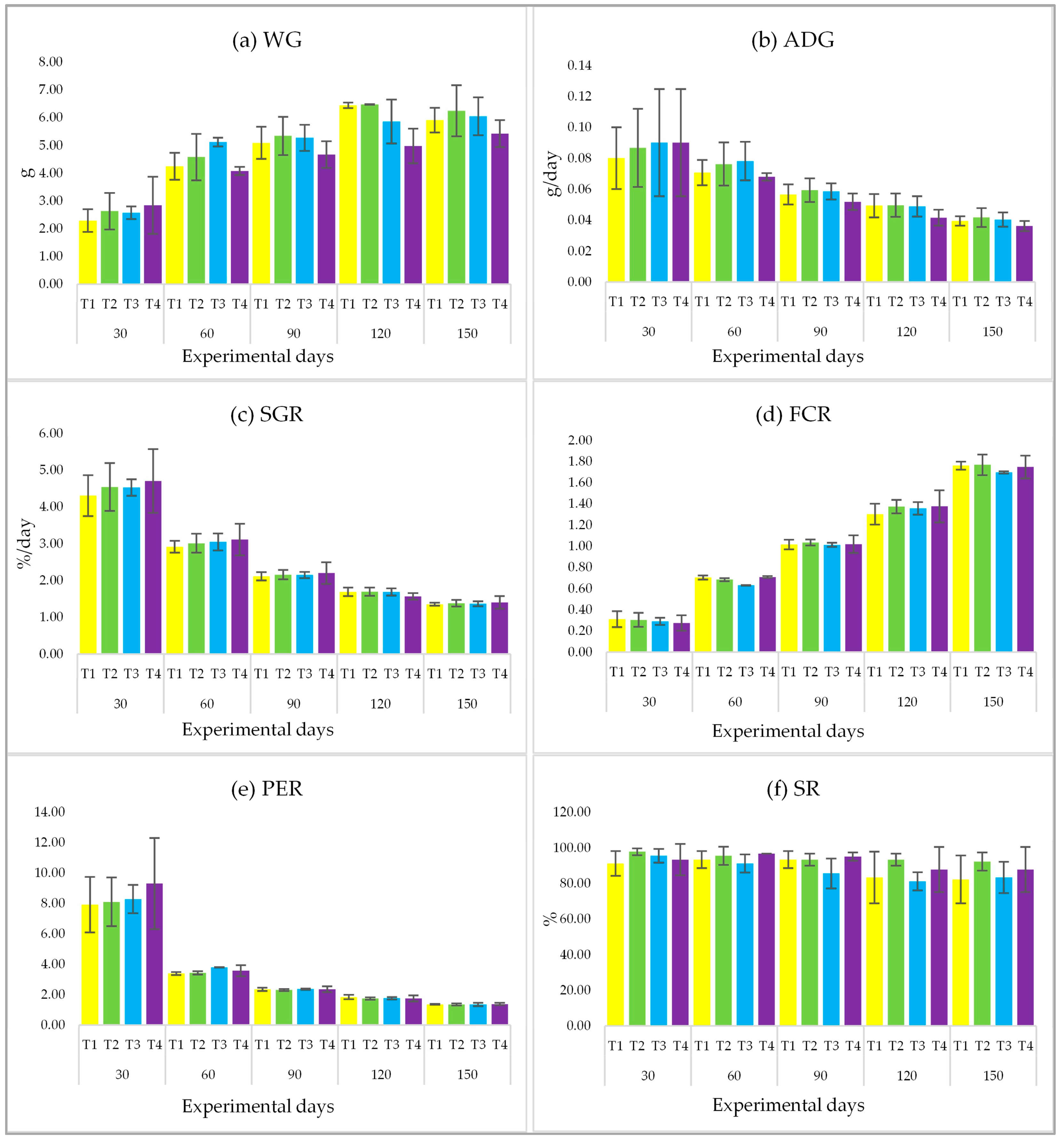
3.3. Growth Performance and Survival Rate

Throughout the 150-day experimental period of feeding with PPE at four different concentration levels (including the control group), it was found that all parameters, i.e., WG, ADG, SGR, FCR, PER, and SR, showed no statistical difference (p > 0.05) (Figure 1a–f).

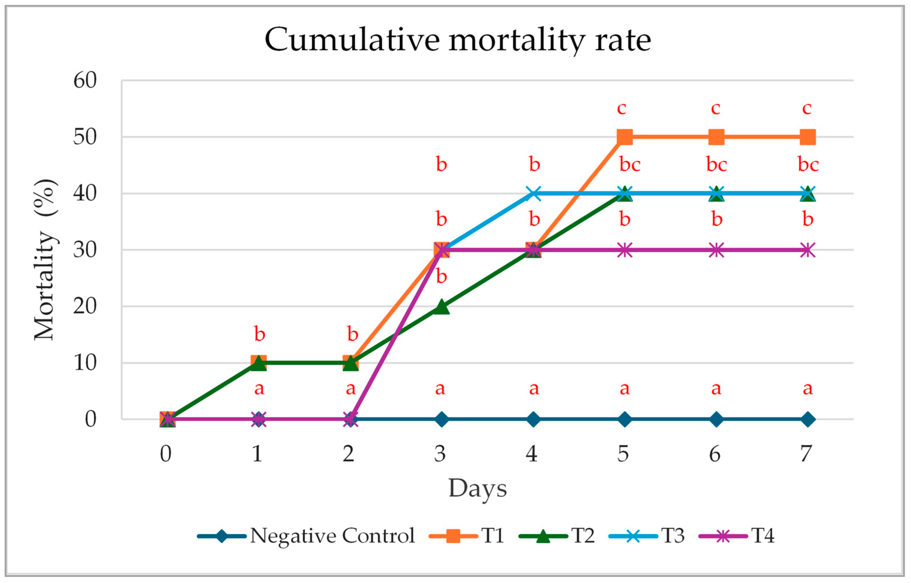
3.4. Cumulative Mortality Rate Following Pathogenic Exposure

After a 150-day experimental period, each group of test fish received an injection of A. hydrophila suspension at a concentration of 108 CFU/mL, while the negative control group was injected solely with 0.85% saline. The injections were administered only on the first day. Subsequently, the fish were monitored for clinical symptoms, and mortality data were collected continuously over a period of 7 days. It was found that the control group experienced the highest cumulative mortality rate, with deaths starting from the first day of A. hydrophila exposure. Groups T2 and T3 showed identical cumulative mortality rates. The highest cumulative mortality rate in group T3 was observed on day 4, whereas that of group T2 occurred on day 5 post-injection. Group T4 had a lower cumulative mortality rate compared to groups T1, T2, and T3. As expected, the negative control group did not experience any mortality during the 7-day period after the bacterial challenge (Figure 2).

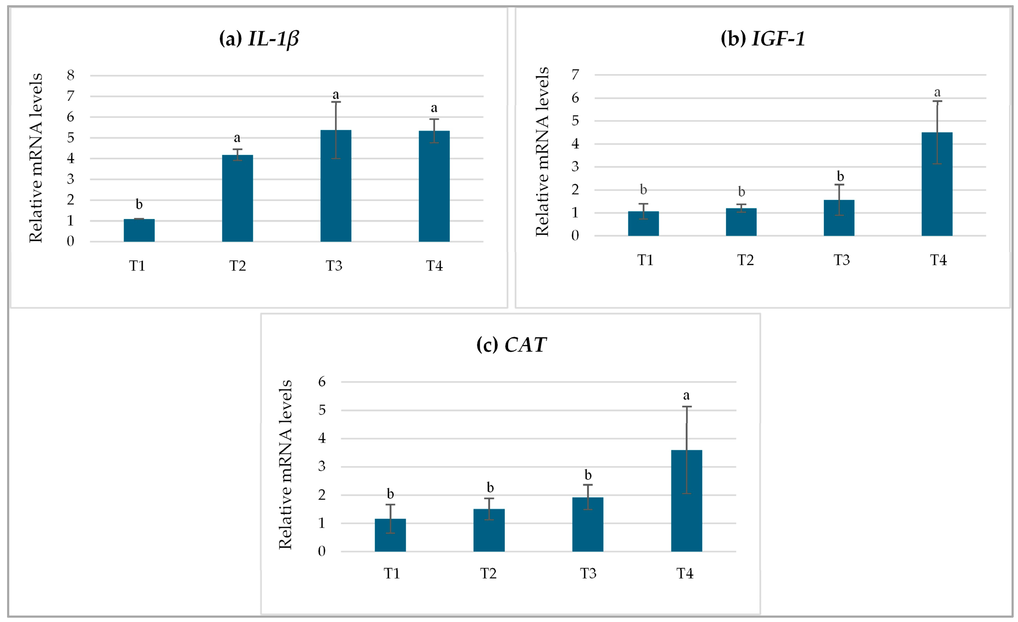
3.5. Relative Gene Expression Levels

Based on a comparison with sequences available in GenBank, the two amplified sequence samples from each gene were confirmed to be the target genes, showing 96–100% sequence identity with L. rohita. The sequences of four genes of L. chrysophekadion were submitted under GenBank accession number PV926119-PV926122. Therefore, the nucleotide sequences of these primers are suitable for use in gene expression studies in L. chrysophekadion.
On day 150, the relative expression of the IL-1β gene in L. chrysophekadion was significantly elevated (p < 0.05) in all groups administered PPE (T2-T4) compared to the control group (T1) (p < 0.05) (Figure 3a). The highest levels of gene expression were detected in groups T3 and T4, exhibiting an approximate fivefold increase relative to group T1. This was followed by group T2, which showed a fourfold increase compared to group T1. The relative expression levels of the IGF-1 gene varied across the treatment groups. Notably, fish in group T4 exhibited the highest expression level (p < 0.05), with nearly a fourfold increase compared to the other groups (Figure 3b). In contrast, no significant differences were observed among the groups T1, T2, and T3. Group T4 exhibited the highest statistically significant (p < 0.05) relative expression of the CAT gene, showing a 3.5-fold increase compared to group T1 (Figure 3c). While no significant differences were detected among the groups T1, T2, and T3, the relative expression levels of both IGF-1 and CAT genes in all treatment groups were elevated compared to the control.

4. Discussion

The phytochemical analysis of the PPE revealed the presence of several key secondary metabolites, including anthraquinones, phenolics, and triterpenoids, suggesting a diverse biochemical composition with potential biological activities [32,33]. The identification of phenolics is particularly noteworthy, as these compounds are recognized for their antioxidant and free radical scavenging properties, which could contribute to the observed protective effects against oxidative stress [34]. The total phenolic content of the ethanolic extract of P. pulcherimus leaves was found to be 96.00 mg GAE/g extract, indicating a significant concentration of phenolic compounds that could contribute to its antioxidant potential [35]. The total flavonoid content, another important class of phenolic compounds, was measured at 17.55 mg QE/g extract, further supporting the antioxidant capacity of the extract [36]. It is noteworthy that total flavonoid content was detected using the modified aluminum chloride colorimetric assay, albeit at a relatively low concentration. However, preliminary phytochemical screening failed to detect flavonoids. This discrepancy could be explained by two possible reasons: First, the presence of interfering substances in the extract may have affected the colorimetric reaction used in the preliminary test, leading to a false-negative result; second, the flavonoid content may have been too low to be detected by the less sensitive preliminary assay. The antioxidant capacity of the PPE, as indicated by the DPPH assay (IC50 = 1314.08 μg/mL), was relatively weak compared to ascorbic acid (IC50 = 7.53 μg/mL). This difference in potency could be attributed to the specific types and concentrations of phenolic compounds present in the extract, as well as potential synergistic or antagonistic interactions among them [37,38]. The presence of compounds like gallic and salicylic acids, as well as quercetin, could contribute to the observed antioxidant properties [39]. Polyphenols are known to modulate enzyme activity and cell receptor function, offering a range of biological actions beyond antioxidant effects in disease prevention and treatment [40,41]. Phenolic compounds have demonstrated remarkable bioactivities, including anticancer, anti-inflammatory, and antibacterial properties, as well as the ability to reduce the risk of diabetes, cardiovascular disease, and neurodegenerative diseases [42]. Furthermore, the antioxidant activity of phenolics is attributed to their ability to scavenge free radicals and modulate the production of reactive oxygen species, thereby protecting cellular components from oxidative damage [43].
While the present study did not observe significant improvements in growth parameters with P. pulcherimus extract supplementation, it is important to note that the effects of dietary supplements can vary depending on the specific compound, dosage, fish species, and rearing conditions. Herbal bioactive compounds, including flavonoids, alkaloids, terpenoids, and phenolics, primarily act as immunomodulators and antioxidants. Their mechanisms focus on activating immune cells such as macrophages and neutrophils, promoting cytokine production like interleukin-1β, and boosting antioxidant enzyme systems such as catalase. These compounds do not directly enhance nutrient digestibility, absorption, or anabolic metabolism [44,45]. Moreover, the dose used in this instance may be too low to observe a notable difference in growth. However, the effective dose for stimulating the immune system or antioxidant defenses is often lower than the dose required to induce significant growth response. The doses employed in many studies are typically optimized based on immune parameters or challenge tests, rather than on maximizing growth [46]. Meanwhile, higher doses intended for growth might sometimes even be immunosuppressive or cause palatability issues, counteracting potential benefits [47]. In systems that are well-managed with nutritionally balanced diets and low levels of pathogen pressure, healthy fish are likely already achieving their genetic growth potential. Under such optimal conditions, the addition of an immunostimulant may not significantly enhance growth. Instead, its main advantage emerges during challenges such as bacterial infections or stress, where the immune system’s readiness offers protection [48]. Further research may be warranted to explore the potential benefits of P. pulcherimus extract on growth performance under different stress conditions or in combination with other dietary additives. The improved survival rate observed in the 0.75 g/kg group suggests that P. pulcherimus extract may possess antibacterial or immunostimulatory properties that enhance the fish’s ability to combat A. hydrophila infection. Similar studies have demonstrated the protective effects of plant extracts against bacterial infections in fish, highlighting the potential of natural compounds as alternatives to antibiotics in aquaculture [49]. Furthermore, herbal–probiotic combinations have shown promise in enhancing hematological parameters, immune function, and antioxidant capacity in fish [50]. The use of herbal extracts can be a valuable component of efforts to improve fish health and reduce reliance on synthetic drugs in aquaculture [51].
The analysis of P. pulcherrimus extract’s influence on gene expression in L. chrysophekadion revealed an involved relationship of immune, growth, and antioxidant responses. The initial verification of primer specificity, demonstrating 96–100% sequence identity with target genes in GenBank, establishes an adequate foundation for subsequent gene expression analyses, promising that the observed alterations in gene expression are related to the intended targets rather than to non-specific amplification or primer mismatches [52]. The administration of P. pulcherrimus extract significantly affected the relative expression of immune-, growth- and antioxidant enzyme-related genes in L. chrysophekadion, demonstrating dose-dependent effects. The upregulation of IL-1β across all treatment groups compared to the control group suggests that the extract possesses immunostimulatory properties. IL-1β is a pro-inflammatory cytokine critical for initiating immune responses [53], and its elevated expression indicates enhanced immune activation [54]. Therefore, the extract from P. pulcherrimus may contribute to the enhancement of the immune response in L. chrysophekadion. This finding is supported by the lower mortality rates observed in fish treated with P. pulcherrimus extract when challenged with A. hydrophila, indicating enhanced disease resistance compared to the untreated control group. The IGF-1 gene, which plays a pivotal role in growth [55], exhibited significantly higher expression only at the highest extract concentration at 0.75 g/kg. These findings indicate that P. pulcherrimus extract may promote growth performance at appropriate dosage levels, aligning with previous studies in other fish species where phytogenic feed additives have been shown to enhance growth through IGF-1 upregulation. For instance, curcumin derived from Curcuma longa has demonstrated growth-promoting effects in tilapia (Oreochromis mossambicus) [56], while the combined use of limonene and thymol, as well as bay leaf (Laurus nobilis) aqueous extract, have similarly been reported to improve growth and IGF-1 expression in Nile tilapia (Oreochromis niloticus) [57,58]. Interestingly, although IGF-1 gene expression was elevated, various physiological and environmental factors such as reproductive maturation, acute stress, or changes in temperature can interfere with the normal relationship between IGF-1 levels and somatic growth [59]. Similarly, CAT gene expression was highest at 0.75 g/kg, indicating enhanced antioxidant capacity. CAT is a key antioxidant enzyme involved in the detoxification of hydrogen peroxide (H2O2), a reactive oxygen species commonly generated during metabolic processes and under stress conditions. The enzyme catalyzes the conversion of H2O2 into water and molecular oxygen [60], which are non-toxic to cells. Magnesium, which is abundantly present in P. pulcherrimus, plays a crucial role as a cofactor for several enzymes involved in antioxidant biosynthesis [61], thereby contributing to its potential antioxidant properties. However, elevated catalase levels may also indicate that the fish are experiencing significant oxidative stress, which is not a normal physiological condition, such as exposure to heavy metals [62], pesticides [63], and thermal stress [64]. Despite this, the present study found that the trends of growth rate, survival rate, and disease resistance of fish supplemented with P. pulcherrimus extract were higher than those of the control group. This suggests that the tested extract concentration was non-toxic and potentially beneficial for the experimental fish.

5. Conclusions

This research elucidates that dietary incorporation of PPE significantly enhances disease resistance and modulates crucial immune and antioxidant gene expressions in L. chrysophekadion, despite having no impact on growth performance in unstressed conditions. The phytochemical composition of PPE, abundant in phenolics, flavonoids, anthraquinones, and triterpenoids, endows it with moderate antioxidant properties and pronounced immunostimulatory effects. Although growth parameters such as WG, ADG, SGR, FCR, and survival rate remained constant across varying PPE dosages, there was a dose-dependent upregulation of pivotal genes: IL-1β (indicative of immune activation), IGF-1 (related to growth regulation), and CAT (involved in oxidative stress mitigation). The highest PPE concentration (0.75 g/kg) resulted in the most pronounced increase in IGF-1 and CAT expressions and significantly decreased mortality by 20% when challenged with A. hydrophila and compared to the control group, highlighting PPE’s role in bolstering pathogen resistance. These findings advocate for PPE as a practical functional additive in sustainable aquaculture, particularly in enhancing immune response in economically valuable species such as L. chrysophekadion. However, the concentration of the extract used in this study was tested solely on L. chrysophekadion, a single species. When applied to other species or under different rearing conditions, the experimental outcomes may vary from those observed here. Additionally, the experiment was conducted in a real field setting, rather than in a controlled laboratory environment where environmental factors can be regulated. As a result, fluctuating weather conditions—such as rain, overcast skies, high temperatures, and cold weather—significantly influenced the fish’s feeding behavior and overall activity throughout the experimental period. Future research should investigate the efficacy of PPE under environmental stress conditions or in conjunction with probiotics to maximize its health benefits.

Author Contributions

S.S.: Investigation, resources, methodology, funding acquisition, project administration, writing—original draft, writing—review and editing. P.A.: methodology, validation, visualization. D.P.: supervision, validation, visualization, methodology, writing—original draft, writing—review and editing. P.P.: conceptualization, resources, methodology, data curation, writing—original draft, writing—review and editing. All authors have read and agreed to the published version of the manuscript.

Funding

This study was financially supported by University of Phayao and Thailand Science Research and Innovation Fund (Fundamental Fund 2024) (grant No. 1833/2567) and partially supported by the Thailand Science Research and Innovation Fund and the University of Phayao (Fundamental Fund 2025, grant No. 5032/2567).

Institutional Review Board Statement

The animal study protocol was approved by the Committee of Institutional Animal Care, University of Phayao, Thailand (approval ID: 1-010-67; date of approval: 1 August 2024).

Informed Consent Statement

Not applicable.

Data Availability Statement

Data are contained within the article.

Acknowledgments

The authors would like to thank the School of Agriculture and Natural Recourses and the School of Pharmaceutical Sciences at the University of Phayao, Thailand, for providing facilities for the present research. Moreover, the authors would like to thank all the staff and students in the fisheries program.

Conflicts of Interest

The authors declare no conflicts of interest.

Abbreviations

The following abbreviations are used in this manuscript:
PPEPhlogacanthus pulcherrimus leaf ethanol extract
GAEGallic acid equivalent
QEQuercetin equivalent
IC50Inhibition concentration 50%
IL-1βInterleukin-1β
IGF-1Insulin-like growth factor 1
CATCatalase
CFUColony-forming unit
DPPH2,2-diphenyl-1-picrylhydrazyl
RT-qPCRReverse transcription-quantitative polymerase chain reaction
DODissolved oxygen
WGWeight gain
ADGAverage daily gain
SGRSpecific growth rate
FCRFeed conversion rate
PERProtein efficiency ratio
SRSurvival rate
LD50Lethal dose 50%
SDStandard deviation
H2O2Hydrogen peroxide

References

  1. Froese, R.; Pauly, D. Fish Base. Available online: https://www.fishbase.org.au/summary/SpeciesSummary.php?ID=12102&genusname=Labeo&speciesname=chrysophekadion&AT=Labeo+chrysophekadion&lang=English (accessed on 19 April 2025).
  2. Freshwater Aquaculture Research and Development Center, Phrae, Freshwater Fisheries Research and Development Division, Department of Fisheries, Ministry of Agriculture and Cooperatives, Thailand. Breeding and Nursery of Black Sharkminnow (Labeo chrysophekadion). Available online: https://www4.fisheries.go.th/local/pic_activities/202009151826351_pic.pdf (accessed on 10 July 2024). (In Thai)
  3. Duangjai, E. The effect of stocking density on the growth and survival of Labeo chrysophekadion fish in the recirculating system using micro bubbles water. J. Innov. Tech. Res. 2020, 3, 33–43. (In Thai) [Google Scholar]
  4. Aquarium Glaser. Ornamental Fish from All over the World to Any Place Around the World, Labeo chrysophekadion. Available online: https://www.aquariumglaser.de/en/08-carp-like-fishes-2-barbs-minnows-carps-goldfish-etc/labeo_chrysophekadion_en/ (accessed on 19 April 2025).
  5. Research and Development Group for Ornamental Aquaculture and Aquatic Plants, Aquaculture Research and Development Division, Department of Fisheries, Ministry of Agriculture and Cooperatives, Thailand. Native Ornamental Fish and Aquatic Plants in Thailand: Knowledge Management on Research for Commercial Production and Sustainable Resource Utilization. Available online: https://www4.fisheries.go.th/local/index.php/main/view_activities/1378/238420 (accessed on 13 July 2025). (In Thai).
  6. Herbal Database, Faculty of Pharmaceutical Sciences, Ubon Ratchathani University. “Dee Pla Kang”. Available online: https://phar.ubu.ac.th/herb-DetailPhargarden/142#:~:text=%E0%B8%8A%E0%B8%B7%E0%B9%88%E0%B8%AD%E0%B8%AA%E0%B8%A1%E0%B8%B8%E0%B8%99%E0%B9%84%E0%B8%9E%E0%B8%A3,.th/herb%2Dthaiherbarium/ (accessed on 10 April 2025). (In Thai).
  7. Panyadee, P.; Balslev, H.; Wangpakapattanawong, P.; Inta, A. Medicinal plants in homegardens of four ethnic groups in Thailand. J. Ethnopharmacol. 2019, 239, 111927. [Google Scholar] [CrossRef]
  8. Wongnaya, N.; Maneerat, T.; Phousamanee, S. Conservation and restoration of food and herb diversity of Karen community in Klonglan National Park, Kamphaeng Phet. Gold. Teak Humanit. Soc. Sci. J. 2020, 26, 56–71. [Google Scholar]
  9. Boontha, S.; Buranrat, B.; Temkhitthawon, P.; Pitaksuteepong, T. Anticancer activities of Phlogacanthus pulcherrimus T. Anderson leaves extract on MCF-7 breast cancer cells. Key Eng. Mater. 2021, 901, 16–21. [Google Scholar] [CrossRef]
  10. Kheawchaum, S.; Mahidol, C.; Thongnest, S.; Boonsombat, J.; Batsomboon, P.; Sitthimonchai, S.; Ruchirawat, S.; Prawat, H. Ent-abietane diterpenoid lactone glycosides and a phenolic glycoside from Phlogacanthus pulcherrimus T. Anderson with cytotoxic and cancer chemopreventive activities. Phytochemistry 2022, 201, 113261. [Google Scholar] [CrossRef]
  11. Athipornchai, A.; Homvisasevongsa, S.; Semsri, S. Discovery and Development of Thai Medicinal Plants with Potential Antidiabetic Activity; Research Report; Faculty of Science, Burapha University: Saen Suk, Thailand, 2019; Available online: https://buuir.buu.ac.th/xmlui/bitstream/handle/1234567890/3970/2564_111.pdf?sequence=1&isAllowed=y (accessed on 1 June 2025). (In Thai)
  12. Kaewsoongnern, T.; Nukulkit, C.; Chan-ae, P.; Pongnaratorn, P.; Pakdee, N.; Wechvitan, P.; Chaweerak, S.; Jitcharoentham, A.; Prapawinee Padannok, P.; Hongwilai, C. Inhibitory effect on alpha glucosidase of herbs and local vegetables in Sakon Nakhon province. J. Thai. Trad. Alt. Med. 2021, 7, 15–28. (In Thai) [Google Scholar]
  13. Noontum, P. Antioxidant and α- Glucosidase Inhibitory Activities of Phlogacanthus pulcherrimus (T. Anderson) Leaf Extract. Master’s Thesis, Mahasarakham University, Mahasarakham, Thailand, August 2019. [Google Scholar]
  14. Chaichana, N. Nutrition information, element and antioxidant activity of native plant in Chiang Rai, Thailand. SWU Sci. J. 2020, 36, 144–153. [Google Scholar]
  15. Hoseinifar, S.H.; Fazelan, Z.; El-Haroun, E.; Yousefi, M.; Yazici, M.; Van Doan, H.; Paolucci, M. The effects of grapevine (Vitis vinifera L.) leaf extract on growth performance, antioxidant status, and immunity of zebrafish (Danio rerio). Fishes 2023, 8, 326. [Google Scholar] [CrossRef]
  16. Yue, R.; Dong, W.; Feng, Z.; Jin, T.; Wang, W.; Chen, Y.; He, Y.; Lin, S. Effects of Three Tested Medicinal Plant Extracts on Growth, Immune Function and Microflora in Juvenile Largemouth Bass (Micropterus salmoides). Aquac. Rep. 2024, 36, 102075. [Google Scholar] [CrossRef]
  17. Esen, R.; Öz, M.; Dikel, S. Effects of Artichoke (Cynara scolymus) Leaf Extract on the Growth, Blood, and Biochemistry Parameters of Nile Tilapia (Oreochromis niloticus). Trop. Anim. Health Prod. 2025, 57, 284. [Google Scholar] [CrossRef]
  18. Sookying, S.; Srisuttha, P.; Rodprasert, V.; Chaodon, C.; Phinrub, W.; Sutthi, N.; Panase, P. Utilizing invasive Pterygoplichthys pardalis as a sustainable fish meal substitute and Euphorbia hirta extract supplement: Effects on growth performance, organosomatic indices, hematological profiles, and serum biochemistry in Chinese Bullfrogs (Hoplobatrachus chinensis). Life 2025, 15, 115. [Google Scholar] [CrossRef]
  19. Pant, D.R.; Pant, N.D.; Saru, D.B.; Yadav, U.N.; Khanal, D.P. Phytochemical screening and study of antioxidant, antimicrobial, antidiabetic, anti-inflammatory and analgesic activities of extracts from stem wood of Pterocarpus marsupium Roxburgh. J. Intercult. Ethnopharmacol. 2017, 6, 170–176. [Google Scholar] [CrossRef]
  20. Hartanti, D.; Cahyani, A.N. Plant cyanogenic glycosides: An overview. Farmasains J. Farm. Dan Ilmu Kesehat. 2020, 5, 1–6. [Google Scholar]
  21. Shaikh, J.R.; Patil, M.K. Qualitative Tests for Preliminary Phytochemical Screening: An Overview. Int. J. Chem. Stud. 2020, 8, 603–608. [Google Scholar] [CrossRef]
  22. Chandra, S.; Khan, S.; Avula, B.; Lata, H.; Yang, M.H.; Elsohly, M.A.; Khan, I.A. Assessment of total phenolic and flavonoid content, antioxidant properties, and yield of aeroponically and conventionally grown leafy vegetables and fruit crops: A comparative study. Evid.-Based Complement. Altern. Med. 2014, 2014, 253875. [Google Scholar] [CrossRef]
  23. Bello, A.A.; Katta, A.; Obaydo, R.H.; Jazmati, A. Phytochemical analysis and antioxidant efficacy of Chrysojasminum fruticans (L.) Banfi in Syrian flora. Heliyon 2024, 10, e37322. [Google Scholar] [CrossRef]
  24. Sutthi, N.; Panase, A.; Chitmanat, C.; Sookying, S.; Ratworawong, K.; Panase, P. Effects of dietary leaf ethanolic extract of Apium graveolens L. on growth performance, serum biochemical indices, bacterial resistance and lysozyme activity in Labeo chrysophekadion (Bleeker, 1849). Aquac. Rep. 2020, 18, 100551. [Google Scholar] [CrossRef]
  25. Bagenal, T. Methods for the Assessment of Fish Production in Fresh Waters, 3rd ed.; Blackwell Scientific Publication: Oxford, UK, 1978; 365p. [Google Scholar]
  26. Sievers, F.; Wilm, A.; Dineen, D.G.; Gibson, T.J.; Karplus, K.; Li, W.; Lopez, R.; McWilliam, H.; Remmert, M.; Söding, J.; et al. Fast, scalable generation of high-quality protein multiple sequence alignments using Clustal Omega. Mol. Syst. Biol. 2011, 7, 539. [Google Scholar] [CrossRef]
  27. Altschul, S.F.; Gish, W.; Miller, W.; Myers, E.W.; Lipman, D.J. Basic local alignment search tool. J. Mol. Biol. 1990, 215, 403–410. [Google Scholar] [CrossRef]
  28. Kole, S.; Anand, D.; Sharma, R.; Tripathi, G.; Makesh, M.; Rajendran, K.V.; Kadam Bedekar, M. Tissue specific expression profile of some immune related genes in Labeo rohita to Edwardsiella tarda infection. Fish Shellfish Immunol. 2017, 66, 575–582. [Google Scholar] [CrossRef]
  29. Kumar, S.; Sahu, N.P.; Ranjan, A. Feeding de-oiled rice bran (DORB) to Rohu, Labeo rohita: Effect of varying dietary protein and lipid level on growth, body composition, and insulin like growth factor (IGF) expression. Aquaculture 2018, 492, 59–66. [Google Scholar] [CrossRef]
  30. Parida, S.; Sahoo, P.K. Antioxidant defence in Labeo rohita to biotic and abiotic stress: Insight from mRNA expression, molecular characterization and recombinant protein-based ELISA of catalase, glutathione peroxidase, CuZn superoxide dismutase, and glutathione s-transferase. Antioxidants 2024, 13, 18. [Google Scholar] [CrossRef]
  31. Livak, K.J.; Schmittgen, T.D. Analysis of relative gene expression data using real-time quantitative PCR and the 2-ΔΔCt method. Methods 2001, 25, 402–408. [Google Scholar] [CrossRef]
  32. Sauceda, A.E.Q.; Sáyago-Ayerdi, S.G.; Ayala-Zavala, J.F.; Wall-Medrano, A.; de la Rosa, L.A.; González-Aguilar, G.A.; Álvarez-Parrilla, E. Biological actions of phenolic compounds. In Fruit and Vegetable Phytochemicals: Chemistry and Human Health, 2nd ed.; Elhadi, M., Yahia, E.M., Eds.; John Wiley & Sons Ltd.: New Jersey, UK, 2018; Volume 1, pp. 125–138. [Google Scholar] [CrossRef]
  33. Zheng, K.; Liang, M.; Yao, H.; Wang, J.; Chang, Q. Effect of size-fractionated fish protein hydrolysate on growth and feed utilization of turbot (Scophthalmus maximus L.). Aquac. Res. 2012, 44, 895–902. [Google Scholar] [CrossRef]
  34. Espín, J.C.; González-Sarrías, A.; Tomás-Barberán, F.A. The gut microbiota: A key factor in the therapeutic effects of (poly)phenols. Biochem. Pharmacol. 2017, 139, 82–93. [Google Scholar] [CrossRef]
  35. Vuolo, M.M.; Lima, V.S.; Maróstica Junior, M.R. Phenolic compounds: Structure, classification, and antioxidant power. In Bioactive Compounds; Campos, M.R.S., Ed.; Woodhead Publishing: Cambridge, UK, 2019; Volume 1, pp. 33–50. [Google Scholar] [CrossRef]
  36. Hassanpour, S.H.; Doroudi, A. Review of the antioxidant potential of flavonoids as a subgroup of polyphenols and partial substitute for synthetic antioxidants. Avicenna J. Phytomed. 2023, 13, 354–376. [Google Scholar]
  37. Chen, X.; Li, H.; Zhang, B.; Deng, Z. The synergistic and antagonistic antioxidant interactions of dietary phytochemical combinations. Crit. Rev. Food. Sci. Nutr. 2022, 62, 5658–5677. [Google Scholar] [CrossRef]
  38. Sonam, K.S.; Guleria, S. Synergistic antioxidant activity of natural products. Ann. Pharmacol. Pharm. 2017, 2, 1086. [Google Scholar]
  39. Sawicki, T.; Jabłońska, M.; Danielewicz, A.; Przybyłowicz, K.E. Phenolic compounds profile and antioxidant capacity of plant-based protein supplements. Molecules 2024, 29, 2101. [Google Scholar] [CrossRef]
  40. Dai, J.; Mumper, R.J. Plant Phenolics: Extraction, analysis and their antioxidant and anticancer properties. Molecules 2010, 15, 7313. [Google Scholar] [CrossRef]
  41. Li, N.; Sun, C.; Zhou, B.; Xing, H.; Ma, D.; Chen, G.; Weng, D. Low concentration of quercetin antagonizes the cytotoxic effects of anti-neoplastic drugs in ovarian cancer. PLoS ONE 2014, 9, e100314. [Google Scholar] [CrossRef]
  42. Shahidi, F.; Yeo, J. Bioactivities of phenolics by focusing on suppression of chronic diseases: A review. Int. J. Mol. Sci. 2018, 19, 1573. [Google Scholar] [CrossRef]
  43. Kiokias, S.; Oreopoulou, V. A Review of the health orotective effects of phenolic acids against a range of severe pathologic conditions (including Coronavirus-based infections). Molecules 2021, 26, 5405. [Google Scholar] [CrossRef]
  44. Hoseinifar, S.H.; Sun, Y.Z.; Wang, A.; Zhou, Z. Probiotics as means of diseases control in aquaculture, a review of current knowledge and future perspectives. Front. Microbiol. 2018, 9, 2429. [Google Scholar] [CrossRef]
  45. Dawood, M.A.; Koshio, S.; Esteban, M.Á. Beneficial roles of feed additives as immunostimulants in aquaculture: A review. Rev. Aquac. 2017, 10, 950–974. [Google Scholar] [CrossRef]
  46. Chakraborty, S.B.; Hancz, C. Application of phytochemicals as immunostimulant, antipathogenic and antistress agents in finfish culture. Rev. Aquac. 2011, 3, 103–119. [Google Scholar] [CrossRef]
  47. Awad, E.; Awaad, A. Role of medicinal plants on growth performance and immune status in fish. Fish Shellfish Immunol. 2017, 67, 40–54. [Google Scholar] [CrossRef]
  48. Van Doan, H.; Hoseinifar, S.H.; Ringø, E.; Ángeles Esteban, M.; Dadar, M.; Dawood, M.A.; Faggio, C. Host-associated probiotics: A key factor in sustainable aquaculture. Rev. Fish. Sci. Aquac. 2020, 28, 16–42. [Google Scholar] [CrossRef]
  49. Nafiqoh, N.; Sukenda, S.; Zairin, M., Jr.; Alimuddin, A.; Lusiastuti, A.; Sarter, S.; Caruso, D.; Avarre, J.C. Antimicrobial properties against Aeromonas hydrophila and immunostimulant effect on Clarias gariepinus of Piper betle, Psidium guajava, and Tithonia diversifolia plants. Aquac. Intern. 2020, 28, 1–13. [Google Scholar] [CrossRef]
  50. Abarike, E.D.; Jian, J.; Tang, J.; Cai, J.; Sakyi, E.M.; Kuebutornye, F.K. A mixture of Chinese herbs and a commercial probiotic Bacillus species improves hemato-immunological, stress, and antioxidant parameters, and expression of HSP70 and HIF-1α mRNA to hypoxia, cold, and heat stress in Nile tilapia, Oreochromis niloticus. Aquac. Rep. 2020, 18, 100438. [Google Scholar] [CrossRef]
  51. Olusola, S.E.; Nwokike, C.C. Effects of dietary leaves extracts of bitter (Vernonia amygdalina) and pawpaw (Carica papaya) on the growth, feed conversion efficiency and disease resistance on juveniles Clarias gariepinus. Aquac. Res. 2018, 49, 1858–1865. [Google Scholar] [CrossRef]
  52. Ye, J.; Coulouris, G.; Zaretskaya, I.; Cutcutache, I.; Rozen, S.; Madden, T.L. Primer-BLAST: A tool to design target-specific primers for polymerase chain reaction. BMC Bioinformatics 2012, 13, 134. [Google Scholar] [CrossRef]
  53. Secombes, C.J.; Wang, T.; Bird, S. The interleukins of fish. Dev. Comp. Immunol. 2011, 35, 1336–1345. [Google Scholar] [CrossRef]
  54. Secombes, C.J.; Wang, T.; Hong, S.; Peddie, S.; Crampe, M.; Laing, K.J.; Cunningham, C.; Zou, J. Cytokines and innate immunity of fish. Dev. Comp. Immunol. 2001, 25, 713–723. [Google Scholar] [CrossRef]
  55. Reinecke, M. Insulin-like growth factors and fish reproduction. Biol. Reprod. 2010, 82, 656–661. [Google Scholar] [CrossRef]
  56. Midhun, S.J.; Arun, D.; Edatt, L.; Sruthi, M.V.; Thushara, V.V.; Oommen, O.V.; Kumar, V.B.S.; Divya, L. Modulation of digestive enzymes, GH, IGF-1 and IGF-2 genes in the teleost, tilapia (Oreochromis mossambicus) by dietary curcumin. Aquac. Int. 2016, 24, 1277–1286. [Google Scholar] [CrossRef]
  57. Aanyu, M.; Betancor, M.B.; Monroig, Ó. The effects of combined phytogenics on growth and nutritional physiology of Nile tilapia Oreochromis niloticus. Aquaculture 2020, 519, 734867. [Google Scholar] [CrossRef]
  58. Shehata, A.I.; Taha, S.A.; Elmaghraby, A.M.; Elhetawy, A.I.G.; Srour, T.M.; El Basuini, M.F.; Shahin, S.A. Effects of dietary bay leaf (Laurus nobilis) aqueous extract on growth performance, feed utilization, antioxidant activity, immunity, and gene expression in Nile tilapia (Oreochromis niloticus). Aquaculture 2025, 599, 742155. [Google Scholar] [CrossRef]
  59. Beckman, B.R. Perspectives on concordant and discordant relations between insulin-like growth factor 1 (IGF1) and growth in fishes. Gen. Comp. Endocrinol. 2011, 170, 233–252. [Google Scholar] [CrossRef]
  60. Von Ossowski, I.; Hausner, G.; Loewen, P.C. Molecular evolutionary analysis based on the amino acid sequence of catalase. J. Mol. Evol. 1993, 37, 71–76. [Google Scholar] [CrossRef]
  61. Monteiro, C.P.; Matias, C.N.; Bicho, M.; Santa-Clara, H.; Laires, M.J. Coordination between antioxidant defences might be partially modulated by magnesium status. Magnes. Res. 2016, 29, 161–168. [Google Scholar] [CrossRef]
  62. Hansen, B.H.; Rømma, S.; Garmo, Ø.A.; Olsvik, P.A.; Andersen, R.A. Antioxidative stress proteins and their gene expression in brown trout (Salmo trutta) from three rivers with different heavy metal levels. Comp. Biochem. Physiol. C 2006, 143, 263–274. [Google Scholar] [CrossRef]
  63. Jin, Y.; Zhang, X.; Shu, L.; Chen, L.; Sun, L.; Qian, H.; Liu, W.; Fu, Z. Oxidative stress response and gene expression with atrazine exposure in adult female zebrafish (Danio rerio). Chemosphere 2010, 78, 846–852. [Google Scholar] [CrossRef]
  64. Vinagre, C.; Madeira, D.; Narciso, L.; Cabral, H.N.; Diniz, M. Effect of temperature on oxidative stress in fish: Lipid peroxidation and catalase activity in the muscle of juvenile seabass, Dicentrarchus labrax. Ecol. Indic. 2012, 23, 274–279. [Google Scholar] [CrossRef]
Figure 1. Growth indices consisting of (a) weight gain: WG, (b) average daily gain: ADG, (c) specific growth rate: SGR, (d) feed conversion rate: FCR, (e) protein efficiency ratio: PER, and (f) survival rate: SR of Labeo chrysophekadion, which were fed different levels of Phlogacanthus pulcherrimus extract. T1 (0.00 g/kg), T2 (0.25 g/kg), T3 (0.50 g/kg), and T4 (0.75 g/kg).
Life 15 01220 g001
Figure 2. Cumulative mortality rate (%) of Labeo chrysophekadion, fed with the four different concentrations of Phlogacanthus pulcherrimus extract against Aeromonas hydrophila for 7 days. T1 (0.00 g/kg), T2 (0.25 g/kg), T3 (0.50 g/kg), T4 (0.75 g/kg), and negative control (0.85% NaCl). Different letters indicate statistically significant differences (p < 0.05) for each day.
Life 15 01220 g002
Figure 3. Relative expression levels of (a) IL-1β, (b) IGF-1, and (c) CAT genes in Labeo chrysophekadion following administration of Phlogacanthus pulcherrimus extract on day 150. T1 (0.00 g/kg), T2 (0.25 g/kg), T3 (0.50 g/kg), and T4 (0.75 g/kg). Results are presented as mean values ± standard deviation (SD). Different letters indicate statistically significant differences (p < 0.05).
Life 15 01220 g003
Table 1. Primers used for reverse transcription–quantitative polymerase chain reaction analysis.
GenePrimer NameSequenceAnnealing Temperature (°C)Amplicon Size (bp)References
Immune-related genes
Interleukin-1β (IL-1β)IL-1β-qFTTGAAGGCCGTGACACTGACT60114[28]
IL-1β-qRGATTCCCAGGCACACAGGTT
Growth-related genes
Insulin-like growth factors 1 (IGF-1)FGCAAACCGACAGGCTATGGGC60166[29]
RGTGTCTGTGTGCCGTTCCGC
Antioxidant enzyme-related genes
Catalase (CAT)FACCTCTACAACGCCATCT5795[30]
RATTCCACTTCCAGTTCTCAG
Housekeeping gene
β-actinFCACTGCTGCTTCCTCCTCCTCC60139[29]
RGATACCGCAAGACTCCATACCCAAG
Table 2. Phytochemical composition of Phlogacanthus pulcherrimus extract.
PhytochemicalsResultsPhytochemicalsResults
AlkaloidsPhenolics+
AnthocyaninsFlavonoids
Anthraquinones+Hydrolysable tannins
SteroidsCondensed tannins
SaponinsCarbohydrates+
Triterpenoids+Cyanogenic glycosides
Volatile coumarinsCardiac glycosides
Nonvolatile coumarins
Note: (+) present, (−) absent.
Table 3. Total phenolic content, total flavonoid content, and antioxidant capacity of Phlogacanthus pulcherrimus extract.
AnalysisTotal Phenolics
(mg GAE/g Extract)
Total Flavonoids
(mg QE/g Extract)
Antioxidant Capacity
(IC50) (μg/mL)
P. pulcherimus extract96.00 ± 14.5817.55 ± 3.181314.08 ± 3.60
Ascorbic acid--7.53 ± 3.19
Values are presented as mean ± SD (n = 3). IC50 = the concentration of the test sample that produces 50% inhibition. The IC50 of Phlogacanthus pulcherrimus extract was extrapolated by GraphPad Prism 10.4.2 due to its limit of solubility. The SDs of antioxidant capacities were calculated using the lack-of-fit model.
Disclaimer/Publisher’s Note: The statements, opinions and data contained in all publications are solely those of the individual author(s) and contributor(s) and not of MDPI and/or the editor(s). MDPI and/or the editor(s) disclaim responsibility for any injury to people or property resulting from any ideas, methods, instructions or products referred to in the content.

