Review

Longitudinal Myocardial Deformation as an Emerging Biomarker for Post-Traumatic Cardiac Dysfunction

1
Heart Center, “University Medical Center” Corporate Fund, Nazarbayev University, Astana 010000, Kazakhstan
2
Institute of Fundamental and Applied Medical Research, S.D. Asfendiyarov Kazakh National Medical University, Tole Bi Street 94, Almaty 050000, Kazakhstan
3
National Scientific Center of Traumatology and Orthopedics Named After Academician N.D. Batpenov, Astana 010000, Kazakhstan
*
Author to whom correspondence should be addressed.
Life 2025, 15(7), 1052; https://doi.org/10.3390/life15071052
Submission received: 26 May 2025 / Revised: 23 June 2025 / Accepted: 27 June 2025 / Published: 30 June 2025
(This article belongs to the Special Issue Management of Ischemia and Heart Failure—3rd Edition)

Abstract

Post-traumatic cardiac dysfunction is a clinically under-recognized complication of polytrauma, often occurring in the absence of overt structural injury. Traditional diagnostic tools frequently fail to detect early or subclinical myocardial impairment, underscoring the need for more sensitive assessment methods. This review explores the utility of global longitudinal strain (GLS), derived from speckle-tracking echocardiography (STE), as a sensitive biomarker for identifying and managing cardiac dysfunction following traumatic injury. It outlines the complex pathophysiology of trauma-induced myocardial impairment, including mechanical injury, systemic inflammation, oxidative stress, and neuro-hormonal activation. The limitations of conventional diagnostic approaches, such as electrocardiography, left ventricular ejection fraction (LVEF), and cardiac biomarkers, are critically assessed and contrasted with the enhanced diagnostic performance of GLS. GLS has demonstrated superior sensitivity in detecting subclinical myocardial dysfunction even when LVEF remains preserved and is associated with increased risk of long-term cardiovascular complications, including arrhythmias and heart failure. The manuscript highlights the clinical utility of GLS in early diagnosis, risk stratification, treatment monitoring, and long-term follow-up. Integration of GLS with inflammatory and oxidative biomarkers (e.g., IL-6, TNF-α, and MPO) and artificial intelligence-based diagnostic models offers potential for improved precision in trauma cardiology.

1. Introduction

Post-traumatic cardiac dysfunction is an under-recognized yet critical consequence of severe trauma, often overlooked in the acute management of polytrauma patients. While the primary focus in trauma care is on immediate life-threatening injuries, subtle myocardial impairment can develop due to direct cardiac contusion, systemic inflammation, neurohormonal dysregulation, and oxidative stress [1]. Traditional diagnostic tools, including electrocardiography (ECG), cardiac biomarkers, and conventional echocardiography, frequently lack the sensitivity to detect early or subclinical myocardial dysfunction, leading to missed opportunities for timely intervention [2,3,4]. Given the increasing recognition of cardiovascular complications following trauma, there is a growing need for more precise diagnostic methods to assess myocardial injury and guide clinical management [5].
Although the exact prevalence of subclinical myocardial dysfunction following trauma is underreported due to diagnostic limitations, data from intensive care settings suggest that approximately 25–30% of critically ill patients exhibit impaired myocardial strain despite preserved ejection fraction [6]. This prospective cohort study evaluated left ventricular global longitudinal strain (LV GLS) using speckle-tracking echocardiography in 152 normotensive septic ICU patients [6]. Non-survivors had worse LV GLS (−15.2 vs. −17.3, p < 0.001) and a cut-off of −17% predicted ICU mortality (AUC 0.728). Patients with LV GLS >−17% had higher mortality (39.2% vs. 13.7%, p < 0.001). Multivariate analysis confirmed LV GLS as an independent mortality predictor (OR 1.326, p = 0.024), alongside mechanical ventilation and severity scores. Impaired LV GLS is linked to higher mortality in normotensive septic patients. Similarly, among septic patients with normal LVEF, GLS ≥ −13% was independently predictive of ICU and hospital mortality (HR ~4.3), even when troponin and LF-EF were unremarkable [7]. These findings underscore the clinical importance of incorporating strain imaging into early trauma assessment as GLS abnormalities frequently precede overt symptoms and traditional diagnostic markers, offering a crucial window for intervention.
Longitudinal myocardial deformation, assessed through speckle-tracking echocardiography (STE), has emerged as a promising tool in the evaluation of post-traumatic cardiac dysfunction. Unlike left ventricular ejection fraction (LVEF), which reflects global ventricular function and may remain preserved in early myocardial impairment, global longitudinal strain (GLS) provides a more detailed and quantitative measure of myocardial contractility. GLS has been shown to detect subclinical myocardial dysfunction in various cardiac conditions, including heart failure, myocardial infarction, and cardiomyopathies, making it an ideal candidate for trauma-related myocardial assessment [8,9]. The application of GLS in trauma care holds significant promise for improving early detection, monitoring myocardial recovery, and guiding treatment strategies. Trauma patients with preserved LVEF but impaired GLS may be at higher risk for long-term cardiovascular complications, necessitating closer cardiac surveillance and potential pharmacologic interventions [10,11]. Additionally, GLS has been proposed as a valuable tool for evaluating treatment response as improvements in myocardial strain over time can provide insights into cardiac recovery and therapeutic efficacy.
GLS was selected for its superior sensitivity in detecting subclinical myocardial dysfunction compared to traditional measures such as left ventricular ejection fraction (LVEF), tissue Doppler imaging (TDI), or cardiac biomarkers. Unlike LVEF, which often remains preserved in early myocardial injury, GLS can quantify subtle changes in myocardial fiber deformation before overt systolic dysfunction develops. Additionally, GLS is angle-independent, and reproducible, and can be measured via standard 2D speckle-tracking echocardiography, making it a feasible and non-invasive tool in the acute trauma setting.
Despite its advantages, the clinical implementation of GLS in trauma settings faces several challenges. Variability in strain measurement techniques across different echocardiographic platforms, the need for specialized imaging expertise, and the lack of established reference values for trauma patients limit the widespread adoption of this technique [10,12]. Further research is needed to validate GLS as a reliable biomarker for post-traumatic cardiac dysfunction and to establish standardized protocols for its use in trauma populations. Moreover, integrating GLS with complementary diagnostic tools, such as cardiac biomarkers and emerging artificial intelligence (AI)-driven imaging analytics, could enhance its predictive value and clinical applicability [13,14].
Traditional diagnostic tools such as left ventricular ejection fraction (LVEF) and standard electrocardiography (ECG) often fail to detect subtle myocardial dysfunction in the early post-trauma phase. For instance, several studies have reported that trauma patients with normal LVEF on echocardiography exhibited significantly reduced global longitudinal strain (GLS), indicating occult systolic impairment [15,16]. In these cases, GLS abnormalities preceded any decline in ejection fraction or visible ECG changes, underscoring its sensitivity for identifying early myocardial injury that would otherwise remain undiagnosed.
Trauma-induced myocardial dysfunction is multifactorial, involving both direct mechanical injury and systemic physiological responses. Blunt chest trauma, often resulting from motor vehicle accidents, falls, or physical impact, can cause myocardial contusion, leading to localized ischemia, oedema, and wall motion abnormalities [17,18]. However, myocardial impairment is not limited to direct injury. Trauma triggers a systemic inflammatory response, characterized by the release of cytokines such as interleukin-6 (IL-6) and tumor necrosis factor-alpha (TNF-α), which contribute to endothelial dysfunction, increased vascular permeability, and myocardial inflammation [19]. Oxidative stress and neurohormonal activation further exacerbate myocardial damage, leading to myocardial stunning, impaired contractility, and potential long-term cardiovascular complications [20,21]. These complex pathophysiological mechanisms underscore the need for advanced diagnostic approaches that can accurately capture myocardial dysfunction beyond traditional methods.
This narrative review aims to explore the role of longitudinal myocardial deformation as an emerging biomarker for post-traumatic cardiac dysfunction. It examines the pathophysiological mechanisms underlying myocardial impairment in trauma patients, the limitations of traditional diagnostic methods, and the potential advantages of GLS in detecting subclinical myocardial dysfunction. Furthermore, the review discusses the clinical applications of GLS in trauma care, including its role in early diagnosis, risk stratification, treatment monitoring, and long-term follow-up. Future research directions, including the integration of AI in strain analysis and the development of standardized strain assessment protocols, are also highlighted.

2. Pathophysiology of Post-Traumatic Cardiac Dysfunction

Post-traumatic cardiac dysfunction is a complex and multifaceted condition that arises from a combination of direct myocardial injury, systemic inflammation, oxidative stress, neurohormonal activation, and immune dysregulation [22]. Unlike ischemic heart disease, where coronary artery occlusion is the primary insult, cardiac dysfunction following trauma results from a cascade of pathophysiological events that impair myocardial function and increase long-term cardiovascular risk. Trauma-induced myocardial dysfunction can occur even in the absence of direct cardiac injury, making it a challenging condition to diagnose and manage [23]. Understanding the intricate interplay between inflammatory, oxidative, and neurohormonal pathways is crucial for identifying high-risk patients and developing targeted therapeutic interventions.
Blunt chest trauma is one of the most direct causes of myocardial dysfunction in trauma patients [24]. High-impact injuries, such as those sustained in motor vehicle accidents or falls, can lead to myocardial contusion, hemorrhage, and necrosis. These localized effects contribute to regional wall motion abnormalities, impaired contractility, arrhythmias, and conduction disturbances. In severe cases, myocardial rupture or cardiac tamponade can occur, leading to hemodynamic instability and life-threatening consequences [25]. Even when structural damage is not apparent, mechanical stress can lead to subcellular injury, calcium dysregulation, and metabolic disturbances that compromise cardiac function. However, beyond mechanical trauma, systemic factors play a crucial role in the development of cardiac dysfunction.
A systemic inflammatory response following severe injury is a major driver of trauma-induced cardiac dysfunction. Polytrauma triggers a massive immune response characterized by the release of pro-inflammatory cytokines, including interleukin-6 (IL-6), tumor necrosis factor-alpha (TNF-α), and interleukin-1 beta (IL-1β) [26]. These cytokines contribute to endothelial activation, increased vascular permeability, and leukocyte infiltration into the myocardium, leading to myocardial oedema and contractile dysfunction. Elevated levels of IL-6 correlate with increased myocardial injury and worse clinical outcomes, while TNF-α and IL-1β have been implicated in endothelial dysfunction, smooth muscle proliferation, and cardiac fibrosis [27]. Persistent immune activation exacerbates myocardial stress and increases the risk of long-term complications such as heart failure and arrhythmias.
Oxidative stress further amplifies myocardial injury in trauma patients. The excessive generation of reactive oxygen species (ROS) due to ischemia–reperfusion injury, hypoxia, and excessive neutrophil activation results in mitochondrial dysfunction, lipid peroxidation, and cardiomyocyte apoptosis [28]. Oxidative damage is exacerbated by myeloperoxidase (MPO), a pro-oxidant enzyme released by neutrophils, which contributes to endothelial damage, microvascular thrombosis, and nitric oxide depletion [29]. The cumulative effects of oxidative stress impair myocardial energy metabolism, reduce contractility, and promote fibrosis. These changes not only worsen acute cardiac dysfunction but also set the stage for chronic myocardial remodeling, increasing the risk of diastolic dysfunction and heart failure.
The neurohormonal response to trauma plays a significant role in cardiac dysfunction. Following severe injury, the sympathetic nervous system is activated, leading to a surge in catecholamines such as epinephrine and norepinephrine [30,31]. While this response is initially adaptive, prolonged sympathetic overactivity places excessive stress on the myocardium. Increased heart rate, contractility, and vascular resistance result in heightened myocardial oxygen demand, potentially leading to ischemia and cardiomyocyte injury. Catecholamine excess also disrupts calcium homeostasis, triggering hypercontractility, arrhythmias, and cellular apoptosis. In extreme cases, this neurohormonal stress response manifests as stress cardiomyopathy (Takotsubo cardiomyopathy), characterized by transient left ventricular dysfunction and wall motion abnormalities [32,33].
Endothelial dysfunction and microvascular thrombosis contribute to impaired myocardial perfusion and exacerbate cardiac injury. The inflammatory response following trauma promotes the upregulation of adhesion molecules such as intercellular adhesion molecule-1 (ICAM-1) and vascular cell adhesion molecule-1 (VCAM-1), facilitating monocyte and neutrophil adhesion to the endothelium [34]. This process leads to increased vascular inflammation, microvascular dysfunction, and reduced nitric oxide bioavailability, impairing vasodilation and myocardial perfusion. In addition, platelet activation and increased thromboxane A2 (TXA2) levels promote clot formation, increasing the risk of microvascular thrombosis and ischemia [35]. These factors create a self-perpetuating cycle of inflammation, vascular dysfunction, and cardiac stress that worsens myocardial injury over time.
As the inflammatory and oxidative cascades progress, ventricular remodeling and fibrosis develop, leading to long-term structural and functional alterations in the heart. The persistent activation of fibroblasts and excessive collagen deposition stiffen the myocardium, impairing relaxation and reducing compliance [36]. Over time, this process contributes to diastolic dysfunction, increased left ventricular filling pressures, and eventual heart failure. Patients who experience post-traumatic cardiac dysfunction are at a heightened risk of developing chronic cardiovascular diseases, including hypertensive heart disease, arrhythmias, and thromboembolic complications [37]. Therefore, early recognition and intervention are critical to improving long-term cardiac outcomes.
Given the complex and multifactorial nature of trauma-induced cardiac dysfunction, early identification of high-risk patients is essential. Conventional diagnostic tools such as electrocardiography (ECG) and standard echocardiography may not adequately detect subtle myocardial dysfunction. Advanced imaging modalities, including speckle-tracking echocardiography (STE) and cardiac MRI, provide more detailed assessments of myocardial mechanics, allowing for earlier detection of contractile abnormalities [38]. In addition, biomarker profiling, including IL-6, TNF-α, MPO, and ICAM-1, may offer valuable insights into the inflammatory and oxidative pathways driving myocardial dysfunction. Integrating these biomarkers into clinical practice may help refine risk stratification and guide targeted treatment approaches.
Anti-inflammatory agents such as IL-1β inhibitors (e.g., canakinumab) have demonstrated potential in reducing cardiovascular events in patients with chronic inflammation and may offer benefits in trauma-related cardiac dysfunction [39]. Similarly, antioxidant therapies targeting ROS and MPO may help reduce oxidative damage and preserve myocardial function [40]. In addition, interventions that modulate the neurohormonal response, such as beta-blockers, may help mitigate catecholamine-induced myocardial stress and prevent long-term dysfunction [41].
The pathophysiology of post-traumatic cardiac dysfunction is a complex interplay of mechanical, inflammatory, oxidative, and neurohormonal factors that contribute to acute and chronic myocardial impairment. Understanding these mechanisms is critical for improving early detection, risk stratification, and treatment strategies in trauma patients. By integrating advanced imaging techniques, biomarker profiling, and targeted therapeutic interventions, clinicians can better manage trauma-induced cardiac dysfunction and improve long-term cardiovascular outcomes.

3. Limitations of Traditional Diagnostic Methods in Trauma-Related Cardiac Dysfunction

The diagnosis of trauma-related cardiac dysfunction remains a significant challenge due to the complex interplay of mechanical injury, systemic inflammation, oxidative stress, and neurohormonal activation. While traditional diagnostic tools such as electrocardiography (ECG), conventional echocardiography, and cardiac biomarkers provide valuable information, they often fail to detect subtle myocardial dysfunction, leading to delays in diagnosis and treatment [42]. Many cases of post-traumatic myocardial impairment occur without overt structural damage, making it difficult to identify at-risk patients using standard diagnostic approaches. The limitations of traditional diagnostic methods underscore the need for more advanced, sensitive techniques that can detect early myocardial changes and guide timely intervention (Table 1).
Electrocardiography (ECG) is widely used in the initial evaluation of trauma patients with suspected cardiac involvement. It is useful for detecting arrhythmias, conduction abnormalities, and ischemic changes that may indicate myocardial injury. However, ECG has several limitations in the context of trauma-related cardiac dysfunction. Many trauma patients present with non-specific ECG findings that do not provide definitive evidence of myocardial impairment [43]. Transient ST-segment and T-wave abnormalities are common but lack specificity for diagnosing myocardial contusion or inflammation. In addition, ECG changes often do not correlate with the severity of myocardial dysfunction, and some patients with significant trauma-induced cardiac injury may have normal ECG findings. The reliance on ECG alone can lead to underdiagnosis of subtle myocardial dysfunction, particularly in cases where structural damage is not apparent (Table 1).
Conventional echocardiography is another key tool for assessing cardiac function in trauma patients. It provides real-time imaging of ventricular contractility, wall motion abnormalities, and pericardial effusion, which are critical for diagnosing myocardial contusion, cardiac tamponade, and other forms of post-traumatic cardiac dysfunction [44]. However, traditional echocardiographic parameters, such as left ventricular ejection fraction (LVEF), have limitations in detecting early myocardial dysfunction. LVEF is a measure of systolic function that reflects the percentage of blood ejected from the left ventricle during each contraction [45]. While a reduced LVEF is indicative of severe cardiac impairment, many trauma patients with myocardial dysfunction have a preserved ejection fraction, masking underlying contractile abnormalities. Standard echocardiographic techniques may not be sensitive enough to detect subtle myocardial strain, which can be an early sign of trauma-induced myocardial impairment (Table 1).
Another limitation of conventional echocardiography is its inability to provide detailed insights into myocardial mechanics. Speckle-tracking echocardiography (STE) and strain imaging have emerged as more advanced tools that can assess myocardial deformation, but these techniques are not yet widely used in routine trauma care [38]. Strain analysis provides a quantitative assessment of myocardial contractility by measuring the percentage of myocardial shortening during systole. GLS, in particular, has been shown to be a more sensitive marker of myocardial dysfunction than LVEF [46]. Studies have demonstrated that trauma patients with preserved LVEF but impaired GLS are at higher risk of developing long-term cardiovascular complications [47]. The limited availability of strain imaging in trauma settings represents a significant gap in current diagnostic capabilities.
Cardiac biomarkers, including high-sensitivity troponin (hs-Tn) and brain natriuretic peptide (BNP), play a crucial role in diagnosing myocardial injury [48] (Table 1). Troponin is a highly specific marker of myocardial cell damage and is widely used in the evaluation of suspected myocardial infarction [49]. In trauma patients, elevated troponin levels may indicate myocardial contusion, ischemia, or stress cardiomyopathy [50]. However, troponin elevation alone is not always indicative of direct cardiac injury as trauma-related factors such as systemic inflammation, hypoxia, and catecholamine surge can also contribute to elevated troponin levels [51]. This makes it challenging to distinguish between primary myocardial injury and secondary myocardial stress responses. Additionally, some trauma patients with significant myocardial dysfunction may have normal troponin levels, particularly if the damage is more functional than structural.
Brain natriuretic peptide (BNP) and N-terminal pro-brain natriuretic peptide (NT-proBNP) are markers of cardiac stress and heart failure that have been investigated in trauma patients [52,53]. Elevated BNP levels may reflect increased ventricular wall stress due to fluid overload, pulmonary hypertension, or myocardial dysfunction. While BNP measurements can provide valuable insights into cardiac status, they are non-specific and may be elevated due to non-cardiac factors such as renal dysfunction, sepsis, or severe trauma-related inflammation [54,55]. The lack of specificity of BNP and troponin limits their ability to serve as standalone diagnostic markers for trauma-related cardiac dysfunction.
Computed tomography (CT) and magnetic resonance imaging (MRI) are additional imaging modalities used in the evaluation of trauma patients [56,57]. CT angiography is highly effective in detecting aortic injuries, pericardial effusion, and vascular abnormalities but has limited utility in assessing myocardial function. Cardiac MRI is the gold standard for evaluating myocardial fibrosis, oedema, and tissue characterization, making it an excellent tool for diagnosing myocardial contusion and stress cardiomyopathy. However, its limited availability, high cost, and prolonged scan times make it impractical for use in the acute trauma setting.
The limitations of traditional diagnostic methods highlight the need for a more comprehensive, multi-modal approach to assessing trauma-related cardiac dysfunction. Integrating advanced imaging techniques such as strain imaging, cardiac MRI, and artificial intelligence-driven diagnostics may improve the early detection and management of myocardial impairment. Additionally, the use of inflammatory and oxidative stress biomarkers, such as interleukin-6 (IL-6), tumor necrosis factor-alpha (TNF-α), myeloperoxidase (MPO), and intercellular adhesion molecule-1 (ICAM-1), could enhance risk stratification and provide a more detailed understanding of trauma-induced cardiac injury.
Moving forward, the development of personalized diagnostic strategies that incorporate both functional and molecular assessments will be essential in optimizing trauma care. By combining traditional diagnostic tools with emerging technologies, clinicians can improve the accuracy of cardiac assessments, facilitate early intervention, and reduce the long-term cardiovascular burden in trauma survivors. The integration of artificial intelligence and machine learning in trauma cardiology could further enhance the predictive value of diagnostic models by analyzing complex data patterns and identifying high-risk patients more effectively.
In conclusion, while traditional diagnostic methods such as ECG, conventional echocardiography, and cardiac biomarkers remain valuable in the evaluation of trauma-related cardiac dysfunction, they have significant limitations in detecting early and subclinical myocardial impairment. More advanced techniques, including speckle-tracking echocardiography, biomarker profiling, and cardiac MRI, offer greater sensitivity and specificity in assessing myocardial injury but are not yet widely implemented in trauma care. A multi-disciplinary approach that incorporates emerging diagnostic technologies, personalized risk assessment, and advanced imaging modalities is essential to improving outcomes for trauma patients with cardiac involvement. Continued research into novel diagnostic strategies and the integration of artificial intelligence in trauma cardiology will be crucial in addressing the current limitations and enhancing the precision of post-trauma cardiac evaluations.
Table 1. Traditional and emerging diagnostic approaches in trauma-related cardiac dysfunction.
Diagnostic
Modality
Clinical RoleLimitations in Trauma
Context
Emerging Enhancements/SolutionsRef.
Electrocardiography (ECG)Rapid bedside assessment; identifies arrhythmias, ischemia, and conduction abnormalities.Low sensitivity for subtle myocardial injury. Non-specific changes such as transient ST-segment or T-wave abnormalities. May be normal despite significant dysfunction.AI-enhanced ECG interpretation. Combined use with biomarkers and imaging for improved diagnostic yield.[58,59]
Conventional
Echocardiography
Assesses systolic function, wall motion, and pericardial effusion. Detects cardiac tamponade and contusion.Left ventricular ejection fraction (LVEF) may be preserved despite dysfunction. Poor sensitivity to early or subclinical myocardial changes. Limited insight into myocardial mechanics.Speckle-tracking echocardiography (STE) and global longitudinal strain (GLS) for early detection of myocardial dysfunction.[60,61]
Cardiac Biomarkers (e.g., Troponin,
hs-Tn)
Detects myocardial cell injury. Commonly used to evaluate myocardial infarction or contusion.Elevated levels may result from non-cardiac trauma-related stress (inflammation, hypoxia). May be normal in cases of functional rather than structural injury. Lack of anatomical or functional context.Multiparametric biomarker panels including IL-6, TNF-α, MPO, and ICAM-1. Use in combination with imaging and clinical scoring systems.[62,63]
Natriuretic Peptides (BNP, NT-proBNP)Reflects ventricular wall stress. Useful in evaluating potential heart failure.Elevated in non-cardiac conditions such as renal dysfunction, sepsis, or volume overload. Low specificity for direct cardiac injury.Adjunctive role in multimodal risk stratification. Should not be used as standalone diagnostics.[64,65]
Computed
Tomography
(CT Angiography)
Visualizes vascular trauma, aortic injury, and pericardial effusion.Limited capability in assessing myocardial function. Exposure to radiation. Not suitable for dynamic or functional cardiac evaluation.Used primarily for vascular and structural assessment. Limited utility in evaluating myocardial performance.[66,67]
Cardiac Magnetic Resonance Imaging (MRI)High-resolution myocardial tissue characterization. Differentiates fibrosis, oedema, and inflammation. Limited availability and high cost. Long scan times. Not practical in acute trauma settings.Selective use in stable or subacute trauma patients. Valuable for follow-up and definitive tissue-level assessment.[68,69]
Overall Strategy and Future DirectionFoundational role in initial cardiac assessment and
Triage.
No single modality adequately captures structural, functional, and molecular abnormalities. Risk of delayed or missed diagnosis of subclinical dysfunction.Multimodal diagnostic strategies integrating ECG, strain imaging, biomarkers, and MRI. Use of AI for risk stratification and predictive analytics. Personalized diagnostics based on trauma severity comorbidities and resource availability.[70,71]

4. Longitudinal Myocardial Deformation: A Sensitive Marker for Post-Traumatic Cardiac Dysfunction

In post-traumatic cardiac dysfunction, myocardial deformation abnormalities may arise due to a combination of direct mechanical injury, systemic inflammation, oxidative stress, and neurohormonal dysregulation. Trauma patients who sustain blunt chest injuries, such as those caused by motor vehicle accidents or falls, may experience myocardial contusion, leading to localized wall motion abnormalities that can be detected using strain imaging [72,73]. However, even in cases where no overt structural damage is evident, myocardial strain analysis can reveal impairments in contractility that are not apparent on standard echocardiography.
Longitudinal myocardial deformation has emerged as a highly sensitive marker for detecting post-traumatic cardiac dysfunction, offering a more precise and quantitative assessment of myocardial mechanics than traditional diagnostic measures [74] (Table 2). Unlike conventional echocardiographic parameters, such as left ventricular ejection fraction (LVEF), which primarily reflect global systolic function, longitudinal strain provides detailed insights into myocardial contractility at the fiber level [75]. This distinction is particularly important in trauma patients, where subtle myocardial dysfunction often remains undetected using standard assessment tools. Speckle-tracking echocardiography (STE) enables clinicians to assess myocardial deformation by tracking the movement of naturally occurring acoustic markers within the myocardium throughout the cardiac cycle [76]. By analyzing the degree of myocardial shortening during systole, STE-derived GLS provides a quantitative measure of myocardial function, making it a valuable tool for early diagnosis, risk stratification, and management of trauma-induced cardiac dysfunction.
One of the key advantages of GLS over LVEF is its ability to detect myocardial dysfunction before significant changes in ejection fraction occur [77]. LVEF, while commonly used in clinical practice, is a relatively crude measure of ventricular function that can remain preserved even in the presence of significant myocardial impairment (Table 2). This phenomenon is particularly relevant in trauma patients, where compensatory mechanisms may temporarily maintain LVEF despite underlying myocardial injury. GLS, on the other hand, is more sensitive to early contractile dysfunction, allowing for the detection of subtle changes in myocardial mechanics that precede overt cardiac dysfunction [78]. Studies have shown that reduced GLS is associated with worse clinical outcomes, including increased risk of heart failure, arrhythmias, and mortality, underscoring its prognostic value in trauma-related cardiac dysfunction.
Some individuals may experience transient myocardial stunning, a reversible condition characterized by temporary contractile dysfunction due to excessive catecholamine release and ischemia–reperfusion injury. In such cases, GLS can help distinguish between reversible myocardial dysfunction and more severe forms of cardiac injury that may require long-term management. Additionally, in patients with systemic inflammatory responses following trauma, GLS can serve as an indicator of inflammation-related myocardial impairment, offering a potential link between immune activation and cardiac dysfunction.
Incorporating GLS into trauma care has the potential to improve early detection and guide targeted interventions aimed at preventing long-term complications [79]. Trauma patients with reduced GLS, even in the absence of significant LVEF reduction, may benefit from closer cardiac monitoring and early initiation of cardioprotective therapies. In particular, beta-blockers and angiotensin-converting enzyme (ACE) inhibitors have been shown to improve myocardial strain and reduce the risk of adverse cardiovascular events in patients with subclinical myocardial dysfunction. Additionally, GLS may be useful in assessing the effectiveness of anti-inflammatory and antioxidant therapies in trauma patients, providing a non-invasive means of tracking myocardial recovery over time [80,81].
The prognostic significance of GLS extends beyond acute trauma care, with implications for long-term cardiovascular health [79,82]. Trauma patients with persistently reduced GLS are at higher risk for developing chronic cardiac conditions, including heart failure and arrhythmias. As such, incorporating longitudinal strain analysis into follow-up assessments can help identify individuals who may require long-term cardiac care. Serial GLS measurements can provide valuable information on myocardial recovery, allowing clinicians to tailor treatment strategies based on individual patient trajectories. By identifying patients at risk for long-term cardiovascular complications, GLS can play a crucial role in improving long-term outcomes and reducing the burden of post-traumatic cardiac disease.
While GLS has shown significant promise in the evaluation of post-traumatic cardiac dysfunction, its widespread adoption in trauma settings faces several challenges. One limitation is the need for specialized imaging equipment and expertise in strain analysis, which may not be readily available in all healthcare settings. Additionally, interobserver variability and differences in vendor-specific strain measurement algorithms can impact the reproducibility of GLS results. Standardization of strain imaging protocols and the development of automated strain analysis tools could help address these challenges and facilitate the broader implementation of GLS in trauma care.
Future research should focus on refining the clinical applications of GLS in trauma-related cardiac dysfunction, including the establishment of standardized GLS cut-off values for identifying high-risk patients. Large-scale prospective studies are needed to validate the prognostic utility of GLS in trauma populations and determine its role in guiding therapeutic interventions. Additionally, the integration of GLS with other emerging biomarkers, such as inflammatory cytokines and oxidative stress markers, may enhance risk stratification and provide a more comprehensive understanding of the mechanisms underlying post-traumatic myocardial dysfunction.
Table 2. Longitudinal myocardial deformation in trauma-related cardiac dysfunction: a multidimensional overview.
ComponentDescriptionClinical
Relevance
Advantages Over
Traditional
Methods
Limitations/ChallengesFuture
Perspectives
Ref.
Global Longitudinal Strain (GLS)Quantitative measure of myocardial fiber shortening during systole, assessed via speckle-tracking echocardiography (STE)Detects subclinical dysfunction in trauma patients, even with normal LVEFHigher sensitivity to early dysfunction; fiber-level analysisRequires advanced echocardiographic equipment and trained operatorsStandardization and integration into trauma protocols[83,84]
Mechanisms of
Dysfunction
Includes direct injury, inflammation, oxidative stress, neuro-
hormonal imbalance
Identifies myocardial injury even without visible structural damageCan localize injury patterns undetectable by ECG or LVEFCannot differentiate exact ethology (e.g., contusion vs. inflammation) without adjunct biomarkersCombined use with cardiac biomarkers and imaging (e.g., MRI)[85,86]
Early
Detection
Identifies early myocardial impairment prior to LVEF dropEnables prompt intervention and monitoringBetter than conventional echocardiography for initial trauma screeningLimited access in emergency trauma settingsImplementation of point-of-care STE platforms[87,88]
Risk
Stratification
Differentiates high- from low-risk patients based on GLS thresholdsGuides intensity of monitoring and therapyProvides prognostic information beyond ECG or biomarkersNo universally accepted GLS cut-offs in traumaDevelopment of trauma-specific GLS algorithms[89,90]
Prognostic ValueReduced GLS predicts worse outcomes (HF, arrhythmias, mortality)Long-term risk assessment and patient counselingTracks recovery or deterioration longitudinallyLong-term data still limited in trauma populationsProspective multicenter cohort validation[91,92]
Therapeutic MonitoringMonitors response to cardioprotective (e.g., beta-blockers) or anti-inflammatory therapiesTailors treatment to myocardial recovery trajectoryNon-invasive and repeatable compared to cardiac MRIRequires repeat echocardiographic accessIntegration with biomarker panels and AI for monitoring[93,94]
Versus LVEFGLS detects dysfunction with preserved LVEFIdentifies hidden myocardial damage earlyAdds value where LVEF is insensitiveRequires familiarity with strain analysisPromote educational training for clinicians[95,96]
Versus Biomarkers (e.g., Troponin, BNP)GLS reflects mechanical function, while biomarkers reflect biochemical stressUseful where biomarker levels are normal despite dysfunctionMore specific to mechanical impairmentBiomarkers remain useful for systemic contextCombine GLS with inflammatory and oxidative stress
markers
[97,98]
As the field of trauma cardiology continues to evolve, longitudinal myocardial deformation analysis represents a promising tool for improving the diagnosis and management of trauma-induced cardiac dysfunction. By providing a more sensitive and quantitative assessment of myocardial function, GLS has the potential to bridge the gap between early myocardial injury detection and long-term cardiovascular risk reduction. The incorporation of strain imaging into routine trauma care could lead to earlier interventions, improved patient outcomes, and a more personalized approach to post-traumatic cardiac management. With continued advancements in imaging technology and a growing body of clinical evidence supporting its use, GLS is poised to become an essential component of trauma cardiology, helping to redefine how myocardial dysfunction is detected and treated in trauma patients.

5. Clinical Applications of Longitudinal Myocardial Deformation in Trauma Care

Longitudinal myocardial deformation has become a valuable tool for assessing heart function in trauma patients, offering greater sensitivity than traditional methods. Unlike conventional measures such as left ventricular ejection fraction (LVEF), which may miss early signs of cardiac impairment, this technique provides detailed insights into trauma-related myocardial dysfunction [99,100]. Its clinical uses range from early detection and risk assessment to guiding treatment and monitoring long-term heart health. A key advantage is its ability to identify myocardial dysfunction early in patients with blunt chest trauma, such as those from car accidents or falls, where injuries may not be apparent on standard imaging or ECG.
Speckle-tracking echocardiography (STE), which measures GLS, can detect subtle changes in heart muscle contractility even when LVEF appears normal [88]. This makes GLS a highly effective tool for diagnosing and managing cardiac injury in trauma care (Table 3).
In addition to its role in detecting myocardial contusion, longitudinal myocardial deformation is particularly useful in assessing myocardial dysfunction in patients with systemic inflammatory responses following severe trauma [101]. Trauma induces a cascade of inflammatory and oxidative stress responses that can lead to myocardial stunning and impaired contractility. Elevated levels of inflammatory cytokines, such as interleukin-6 (IL-6) and tumor necrosis factor-alpha (TNF-α), have been associated with myocardial dysfunction in trauma patients, contributing to contractile impairment and ventricular remodeling [102]. GLS provides a quantitative assessment of myocardial function that correlates with the extent of inflammatory-mediated myocardial injury, allowing clinicians to better understand the interplay between systemic inflammation and cardiac dysfunction [103]. By integrating GLS with biomarker analysis, clinicians can more accurately assess the severity of trauma-induced myocardial dysfunction and tailor treatment strategies accordingly (Table 3).
Another critical application of longitudinal myocardial deformation in trauma care is its use in risk stratification and prognostication (Table 3). Trauma patients who exhibit reduced GLS, even in the absence of overt cardiac symptoms, may be at increased risk for developing long-term cardiovascular complications, such as heart failure and arrhythmias. The ability to identify high-risk patients early in their clinical course enables the implementation of more aggressive monitoring and therapeutic interventions aimed at preventing disease progression. Studies have demonstrated that GLS abnormalities are predictive of adverse cardiovascular events in various clinical settings, including heart failure and myocardial infarction, suggesting that similar principles may apply in trauma populations [38,103,104]. In this context, GLS could serve as a valuable risk assessment tool to guide clinical decision-making and improve patient outcomes.
Beyond risk stratification, longitudinal myocardial deformation plays a crucial role in guiding therapeutic interventions in trauma patients with cardiac dysfunction. The ability to detect early myocardial impairment allows for timely initiation of cardioprotective therapies, such as beta-blockers and angiotensin-converting enzyme (ACE) inhibitors, which have been shown to improve myocardial function and reduce the risk of long-term complications [105]. Patients with evidence of strain abnormalities may benefit from closer hemodynamic monitoring, fluid optimization, and early intervention to prevent the development of heart failure. Furthermore, GLS can be used to evaluate the efficacy of anti-inflammatory and antioxidant therapies in trauma patients, providing a non-invasive means of assessing treatment response and myocardial recovery.
The role of longitudinal myocardial deformation extends beyond the acute phase of trauma care, with significant implications for long-term cardiovascular monitoring [106]. Trauma patients who survive their initial injuries may continue to experience subclinical myocardial dysfunction that places them at risk for developing chronic cardiac conditions [107]. GLS can be utilized as a longitudinal monitoring tool to track myocardial function over time and assess the trajectory of cardiac recovery. Serial GLS measurements can provide valuable information on myocardial remodeling and help determine whether patients require ongoing cardiac management. This is particularly important for patients with persistent strain abnormalities as they may benefit from continued medical therapy and lifestyle modifications to mitigate the risk of future cardiovascular events.
Another promising application of longitudinal myocardial deformation in trauma care is its integration with other advanced imaging modalities, such as cardiac magnetic resonance imaging (MRI) and computed tomography (CT) [108]. While GLS provides detailed insights into myocardial mechanics, cardiac MRI offers superior tissue characterization, allowing for the detection of myocardial fibrosis, oedema, and scarring [109]. Combining these modalities could enhance diagnostic accuracy and provide a more comprehensive assessment of trauma-induced myocardial injury. Additionally, the integration of artificial intelligence and machine learning algorithms into strain imaging analysis holds the potential to further refine risk prediction models and improve the clinical utility of GLS in trauma populations.
Table 3. Clinical applications of longitudinal myocardial deformation in trauma care.
Clinical
Application
DescriptionKey BenefitsChallengesRef.
Early Detection of Cardiac
Injury
Detects subclinical myocardial dysfunction in blunt chest trauma, even when ECG and LVEF are normal.Enables early diagnosis and intervention for myocardial contusion or trauma-induced contractility impairment.May be overlooked in patients without visible trauma; requires high-resolution imaging and trained operators.[110,111]
Assessment
During Systemic
Inflammatory
Response
Evaluates myocardial dysfunction linked to inflammation and oxidative stress post-trauma (e.g., IL-6, TNF-α).Provides insight into inflammation-induced cardiac injury and helps guide anti-inflammatory treatment.Requires biomarker correlation; inflammation-related changes may be transient or confounded by other injuries.[112,113]
Risk Stratification and
Prognostication
Identifies high-risk patients with reduced GLS for future cardiac complications, even if asymptomatic.Supports targeted monitoring and early therapeutic strategies to prevent heart failure and arrhythmias.No established trauma-specific GLS thresholds; prognostic implications need further validation.[114,115]
Guiding
Therapeutic
Interventions
Informs timely initiation of cardioprotective therapies (e.g., beta-blockers, ACE inhibitors) and fluid optimization.Improves outcomes by tailoring treatments to individual myocardial function.Requires dynamic monitoring and clinical interpretation; risk of over-treatment in ambiguous cases.[116]
Long-Term
Cardiac
Monitoring
Tracks myocardial recovery and remodeling in trauma survivors through serial GLS measurements.Enables personalized follow-up and long-term care planning.Long-term access to imaging may be limited; adherence to follow-up may be poor.[117]
Integration with Advanced
Imaging
Modalities
Combines with cardiac MRI/CT to assess structural damage (e.g., fibrosis, oedema, scarring).Enhances diagnostic accuracy and understanding of myocardial pathology.MRI/CT access may be limited; high cost and patient instability may preclude use in acute trauma.[118,119]
AI and Machine Learning
Integration
Enhances GLS analysis and risk prediction through automated algorithms and data-driven models.Improves consistency, speed, and predictive power of strain interpretation.Requires robust datasets; AI systems must be validated for trauma populations.[120]
Implementation in Clinical PracticeOffers a non-invasive, sensitive tool for routine trauma cardiac assessment.Facilitates evidence-based, personalized trauma cardiology care.Requires specialized echocardiography equipment and training; vendor variability in GLS measurements.[38]
Future Research and ValidationNeeded for establishing trauma-specific GLS reference values and studying utility in subgroups (e.g., TBI, hemorrhagic shock).Expands clinical relevance and optimizes use in diverse trauma settings.Limited large-scale prospective studies; subgroup-specific data currently sparse.[121]
Despite its numerous advantages, the widespread adoption of longitudinal myocardial deformation in trauma care faces several challenges. One of the primary limitations is the need for specialized imaging equipment and expertise in strain analysis, which may not be readily available in all healthcare settings. Additionally, variability in strain measurement techniques between different echocardiographic vendors can impact the reproducibility of GLS results, highlighting the need for standardized protocols and calibration methods. Efforts to address these challenges through training programs, standardized guidelines, and automated strain analysis tools will be critical in facilitating the broader implementation of GLS in trauma care.
While speckle tracking echocardiography (STE) offers promising sensitivity for detecting subclinical myocardial dysfunction, its broader clinical application is limited by several important factors. First, the accuracy of strain measurements is highly dependent on the operator’s experience, both in image acquisition and post-processing interpretation. Second, the quality of the echocardiographic images themselves, including endocardial border definition, plays a critical role, particularly in trauma settings where imaging conditions may be suboptimal. Third, frame rate settings must be carefully optimized as strain analysis can be significantly affected by excessively low or high temporal resolution. Fourth, STE is sensitive to changes in loading conditions (preload and afterload), which can alter myocardial deformation patterns independent of intrinsic cardiac function. Fifth, extrinsic mechanical factors, especially chest wall conformation and soft tissue variability, can degrade image quality and strain reliability, particularly in patients with obesity, chest trauma, or surgical dressings [122,123]. Finally, it is important to note that STE remains underutilized in daily clinical practice due to barriers such as limited training, a lack of familiarity among general practitioners, and time constraints in emergency and ICU environments [124]. These limitations underscore the need for standardized protocols, automated software tools, and broader clinician education to enable more consistent use of STE in trauma care settings.
Future research should focus on further validating the clinical applications of longitudinal myocardial deformation in trauma populations through large-scale prospective studies. Establishing GLS reference values for trauma patients and determining optimal cut-off thresholds for risk stratification will enhance its utility as a clinical tool. Additionally, exploring the role of GLS in specific trauma subgroups, such as patients with traumatic brain injury or hemorrhagic shock, could provide valuable insights into its broader applicability in critical care settings. As the field of trauma cardiology continues to evolve, longitudinal myocardial deformation is poised to play an increasingly important role in improving the diagnosis, management, and long-term outcomes of patients with trauma-induced cardiac dysfunction.
The ability of GLS to detect early myocardial dysfunction, guide treatment decisions, and predict long-term cardiovascular risk makes it a highly valuable tool in trauma care. By incorporating strain imaging into routine clinical practice, clinicians can achieve a more comprehensive understanding of myocardial function and provide more personalized, evidence-based care for trauma patients. With continued advancements in imaging technology and increasing recognition of the importance of myocardial strain analysis, longitudinal myocardial deformation is set to become a cornerstone of trauma cardiology, paving the way for improved patient outcomes and more effective management of post-traumatic cardiac dysfunction.

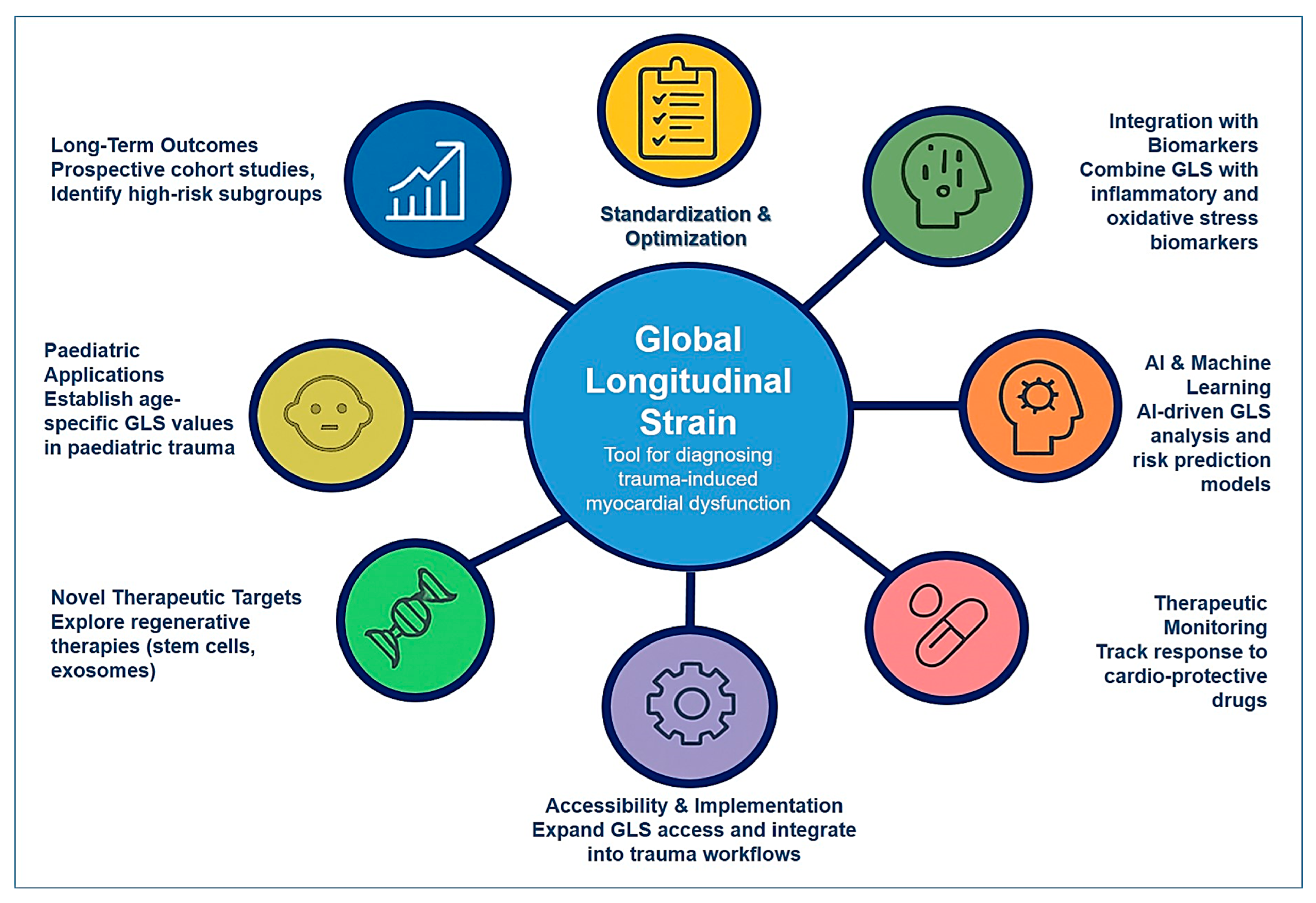
6. Future Directions and Research Opportunities

Future directions and research opportunities in the field of longitudinal myocardial deformation and post-traumatic cardiac dysfunction are vast, offering significant potential to improve early diagnosis, risk stratification, and patient outcomes. As the understanding of trauma-induced myocardial dysfunction continues to evolve, there is a growing need for interdisciplinary approaches that integrate advanced imaging techniques, biomarker analysis, and artificial intelligence-driven diagnostics. Expanding research in these areas will enhance clinicians’ ability to detect myocardial dysfunction at its earliest stages, refine therapeutic strategies, and develop personalized treatment plans that reduce long-term cardiovascular risk in trauma patients.
One of the most promising areas for future research is the optimization and standardization of GLS measurement in trauma populations. While GLS has demonstrated superior sensitivity in detecting subclinical myocardial dysfunction compared to traditional metrics such as left ventricular ejection fraction (LVEF), significant variability remains in strain measurement techniques across different imaging platforms and vendors [125,126]. Standardizing GLS protocols and establishing trauma-specific reference values will be essential to improving its clinical utility. Large-scale, multi-center studies should be conducted to define normative GLS ranges in trauma patients and determine the optimal cut-off values for identifying individuals at risk for adverse cardiac outcomes. Additionally, further investigation into the correlation between GLS abnormalities and long-term cardiovascular events in trauma survivors will help refine risk prediction models and inform clinical decision-making.
The integration of GLS with emerging cardiac biomarkers represents another exciting avenue for future research. While strain imaging provides valuable mechanical insights into myocardial function, the addition of biomarker profiling could offer a more comprehensive assessment of trauma-induced cardiac dysfunction. Inflammatory mediators such as interleukin-6 (IL-6), tumor necrosis factor-alpha (TNF-α), and interleukin-1 beta (IL-1β) have been shown to play critical roles in post-traumatic myocardial impairment, and their relationship with myocardial strain abnormalities warrants further investigation [127]. Additionally, oxidative stress markers, including myeloperoxidase (MPO) and reactive oxygen species (ROS), may provide complementary information on the pathophysiological mechanisms underlying myocardial dysfunction in trauma patients. Future research should explore the potential of combining GLS with biomarker panels to create multi-modal risk assessment tools that enhance early detection and treatment strategies.
Artificial intelligence (AI) and machine learning have the potential to revolutionize the application of longitudinal myocardial deformation analysis in trauma care [128,129]. AI-driven algorithms can be trained to detect subtle myocardial strain abnormalities with greater accuracy and reproducibility, reducing operator-dependent variability in GLS measurements. Machine learning models could also integrate data from echocardiographic imaging, biomarker profiling, and electronic health records to predict which trauma patients are at the highest risk for developing long-term cardiovascular complications [129]. Future research should focus on developing AI-assisted diagnostic platforms that leverage big data to enhance the clinical interpretation of myocardial strain analysis and improve patient outcomes.
The role of longitudinal myocardial deformation in guiding therapeutic interventions also warrants further exploration [130]. While GLS has primarily been used as a diagnostic and prognostic tool, its potential as a treatment-monitoring parameter remains largely unexplored. Future studies should investigate whether serial GLS measurements can be used to track the efficacy of cardioprotective therapies in trauma patients. For example, beta-blockers, angiotensin-converting enzyme (ACE) inhibitors, and anti-inflammatory agents have shown promise in improving myocardial function in other cardiac conditions, but their impact on myocardial strain recovery in trauma patients remains unknown. Conducting clinical trials that evaluate how GLS responds to specific pharmacologic interventions could help establish evidence-based treatment guidelines for post-traumatic cardiac dysfunction.
Longitudinal studies assessing the long-term cardiovascular impact of trauma-related myocardial dysfunction are critically needed. Many trauma patients, even those who appear to recover fully, may experience lingering cardiac effects that predispose them to heart failure, arrhythmias, and ischemic heart disease later in life. Prospective cohort studies following trauma patients over extended periods will help clarify the relationship between early GLS abnormalities and long-term cardiovascular risk. Additionally, identifying subgroups of trauma patients who are particularly susceptible to chronic cardiac complications—such as those with pre-existing cardiovascular conditions, systemic inflammatory responses, or prolonged ICU stays—will enable the development of targeted monitoring and intervention strategies.
Advancing the role of GLS in pediatric trauma populations is another important area for future research. While myocardial strain imaging has been extensively studied in adults, there are limited data on its application in pediatric trauma patients. Children and adolescents may exhibit different myocardial adaptation responses following trauma, necessitating age-specific GLS reference values and diagnostic criteria. Research efforts should focus on evaluating how longitudinal myocardial deformation evolves across different pediatric age groups and determining whether early GLS abnormalities in children correlate with long-term cardiovascular consequences.
The exploration of novel therapeutic targets for mitigating trauma-induced myocardial dysfunction is another crucial direction for future research. Beyond conventional pharmacologic interventions, regenerative medicine approaches, such as stem cell therapy and tissue engineering, hold promise for repairing myocardial damage following trauma. Preclinical studies suggest that mesenchymal stem cells (MSCs) and exosome-based therapies may enhance myocardial regeneration and improve cardiac function in animal models of injury [131]. Translating these findings into clinical practice will require rigorous clinical trials to assess the safety and efficacy of regenerative therapies in trauma patients.
Finally, research should focus on improving the accessibility and implementation of strain imaging in diverse healthcare settings. While GLS is a powerful diagnostic tool, its widespread adoption is hindered by equipment limitations, training requirements, and cost barriers. Efforts to develop more user-friendly, automated strain imaging technologies could help expand the availability of GLS in both high-resource and low-resource settings. Additionally, integrating GLS into trauma care protocols and emergency medicine workflows will be essential for ensuring its timely use in high-acuity settings.
Future directions and research opportunities in the field of longitudinal myocardial deformation and trauma-related cardiac dysfunction hold immense potential to transform patient care (Figure 1). By refining diagnostic methodologies, integrating biomarker-based risk assessment, leveraging artificial intelligence, exploring novel therapies, and conducting long-term follow-up studies, researchers can advance the understanding and management of trauma-induced myocardial dysfunction. As these advancements continue to unfold, the clinical application of GLS is likely to become an integral component of trauma cardiology, leading to earlier detection, improved treatment strategies, and better long-term cardiovascular outcomes for trauma patients.

7. Conclusions

Longitudinal myocardial deformation, assessed through global longitudinal strain (GLS), represents a significant advancement in the evaluation of cardiac function following trauma. Unlike traditional diagnostic tools such as electrocardiography, left ventricular ejection fraction (LVEF), or isolated biomarker measurements, GLS offers a sensitive and quantitative measure of subclinical myocardial impairment that often remains undetected in the acute trauma setting. Its ability to identify early contractile dysfunction before structural or hemodynamic deterioration occurs makes it an invaluable tool for timely risk stratification and clinical decision-making.
This review underscores the complex, multifactorial nature of post-traumatic cardiac dysfunction, driven by direct mechanical injury, systemic inflammation, oxidative stress, and neuro-hormonal activation. GLS has demonstrated clinical relevance across the entire care continuum, from early detection and prognostication to monitoring treatment response and guiding long-term cardiovascular follow-up. Furthermore, the integration of GLS with inflammatory and oxidative stress biomarkers, along with artificial intelligence-driven analytics, holds promise for developing a precision medicine framework in trauma cardiology.
Despite the growing interest in global longitudinal strain (GLS) as a sensitive and non-invasive marker for post-traumatic myocardial dysfunction, several limitations currently constrain its widespread clinical adoption in trauma settings. First, most of the available data regarding GLS in trauma patients are derived from small-scale, single-center observational studies with limited generalizability. There remains a lack of large, prospective, multicenter trials specifically focused on trauma populations, which hampers the development of standardized GLS-based diagnostic or prognostic algorithms in this context. Second, technical variability in image acquisition protocols and post-processing algorithms used for strain analysis, such as differences between vendor-specific software and inter-observer variability, introduces challenges in reproducibility and cross-institutional consistency. These issues are particularly relevant in acute trauma care, where echocardiographic imaging conditions may be suboptimal. Addressing these limitations through standardized protocols and multicenter validation efforts will be essential to fully integrate GLS into trauma care pathways.
In conclusion, GLS has the potential to redefine the diagnostic and prognostic landscape of trauma-induced cardiac dysfunction. By enabling earlier detection, more accurate risk assessment, and individualized patient management, longitudinal myocardial deformation may significantly improve outcomes for trauma patients and reduce the long-term burden of cardiovascular complications.

Author Contributions

M.B.: writing, review, and editing, supervision, and project administration. T.S.: writing––original draft, conceptualization, and editing. M.M.: collecting data and literature analysis. M.S.: writing, analysis, and table preparations. A.B.: review and editing. A.K.: literature analysis. Z.S.: collecting data and literature analysis. All authors have read and agreed to the published version of the manuscript.

Funding

This research was funded by the Committee of Science of the Ministry of Science and Higher Education of the Republic of Kazakhstan through the grant “Clinical, functional, and immunological predictors on the severity of asymptomatic post-traumatic myocardial dysfunction in polytrauma study” (grant number AP19678310).

Institutional Review Board Statement

Not applicable.

Data Availability Statement

Not applicable.

Acknowledgments

The authors are thankful to the Nazarbayev University for the administrative and technical support provided.

Conflicts of Interest

The authors declare no conflicts of interest.

References

  1. Weber, B.; Lackner, I.; Gebhard, F.; Miclau, T.; Kalbitz, M. Trauma, a Matter of the Heart-Molecular Mechanism of Post-Traumatic Cardiac Dysfunction. Int. J. Mol. Sci. 2021, 22, 737. [Google Scholar] [CrossRef] [PubMed]
  2. Cardoso, C.D.; Ohe, N.T.; Bader, Y.; Afify, N.; Al-Homedi, Z.; Alwedami, S.M.; O’Sullivan, S.; Campos, L.A.; Baltatu, O.C. Heart Rate Variability Indices as Possible Biomarkers for the Severity of Post-traumatic Stress Disorder Following Pregnancy Loss. Front. Psychiatry 2022, 12, 700920. [Google Scholar]
  3. Weber, B.; Lackner, I.; Braun, C.K.; Kalbitz, M.; Huber-Lang, M.; Pressmar, J. Laboratory Markers in the Management of Pediatric Polytrauma: Current Role and Areas of Future Research. Front. Pediatr. 2021, 9, 622753. [Google Scholar] [CrossRef] [PubMed]
  4. Wang, N.; Huang, J.L.; Fang, Y.; Du, H.L.; Chen, Y.L.; Zhao, S.Q. Molecular biomarkers of blunt cardiac injury: Recent advances and future perspectives. Expert Rev. Mol. Diagn. 2024, 24, 1023–1031. [Google Scholar] [CrossRef] [PubMed]
  5. Kalil, R.; Saretta, R.; Franci, A.; Baracioli, L.M.; Galas, F.R.B.G.; Gil, J.S.; Ferino, A.; Giacovone, C.; Oliveira, I.; Souza, J.; et al. Post-COVID-19 Cardiopulmonary Symptoms: Predictors and Imaging Features in Patients after Hospital Discharge. Arq. Bras. Cardiol. 2023, 120, e20220642. [Google Scholar]
  6. Omar, T.; İnci, K.; Oflu, Y.; Dilek, M.; Çelik, Z.B.; Kına, S.; İliş, D.; Bucak, H.M. The predictive value of left ventricular global longitudinal strain in normotensive critically ill septic patients. Crit. Care Sci. 2023, 35, 187–195. [Google Scholar] [CrossRef]
  7. Lima, M.R.; Silva, D. Septic cardiomyopathy: A narrative review. Rev. Port. Cardiol. 2023, 42, 471–481. [Google Scholar] [CrossRef]
  8. Cho, S.K.S.; Darby, J.R.T.; Williams, G.K.; Holman, S.L.; Rai, A.; Van Amerom, J.F.P.; Fan, C.P.; Macgowan, C.K.; Selvanayagam, J.B.; Morrison, J.L.; et al. Post-Myocardial Infarction Remodeling and Hyperkinetic Remote Myocardium in Sheep Measured by Cardiac MRI Feature Tracking. J. Magn. Reson. Imaging 2025, 61, 1323–1335. [Google Scholar] [CrossRef]
  9. Villar-Calle, P.; Kochav, J.D.; Vadaketh, K.; Chiu, C.; Tak, K.; Agoglia, H.; Liberman, N.; Nguyen, K.L.; Vizcarra-Tellez, A.; Wu, A.L.; et al. Tissue-Based Predictors of Impaired Right Ventricular Strain in Coronary Artery Disease: A Multicenter Stress Perfusion Study. Circ.-Cardiovasc. Imaging 2024, 17, e016852. [Google Scholar] [CrossRef]
  10. Shvets, D.A.; Povetkin, S. Limitations of Diagnosis of Ischemic Left Ventricular Dysfunction Using the Values of Strain, Twist and Untwist in Patients with Myocardial Infarction of Various Localization. Kardiologiya 2024, 64, 55–62. [Google Scholar] [CrossRef]
  11. Roger, G.; Denormandie, P.; Gobe, T.; Azzolina, D.; Pham, T.; Chantalat, C.; Cuveillier, D.; Bouchachi, A.; Jourdain, P.; Lai, C.; et al. Left ventricular global longitudinal strain and acute myocardial injury in patients with sickle cell disease admitted to the intensive care unit for vaso-occlusive crisis. Br. J. Haematol. 2024, 204, 2007–2015. [Google Scholar] [CrossRef] [PubMed]
  12. Liu, T.T.; Liu, H.L.; Song, Y.; Huang, Y.L.; Zhang, C.Q. Quantitative assessment of left ventricular myocardial work in patients with different types of atrial fibrillation by non-invasive pressure-strain loop technique. Echocardiography 2024, 41, e15801. [Google Scholar] [CrossRef]
  13. Tolu-Akinnawo, O.Z.; Ezekwueme, F.; Omolayo, O.; Batheja, S.; Awoyemi, T. Advancements in Artificial Intelligence in Noninvasive Cardiac Imaging: A Comprehensive Review. Clin. Cardiol. 2025, 48, e70087. [Google Scholar] [CrossRef] [PubMed]
  14. Udoy, I.A.; Hassan, O. AI-Driven Technology in Heart Failure Detection and Diagnosis: A Review of the Advancement in Personalized Healthcare. Symmetry 2025, 17, 469. [Google Scholar] [CrossRef]
  15. Geneş, M.; Çelik, M. Evaluation of Left Ventricular Systolic Functions of Patients with Exaggerated High Blood Pressure Response to Treadmill Exercise Test with Two-Dimensional Longitudinal Strain Imaging. Anatol. J. Cardiol. 2024, 29, 36–41. [Google Scholar] [CrossRef] [PubMed]
  16. Baykiz, D.; Govdeli, E.A.; Ozer, P.K.; Karaayvaz, E.B.; Catma, Y.; Medetalibeyoglu, A.; Cagatay, A.; Umman, B.; Tukek, T.; Bugra, Z. Evaluation the relationship of left ventricular global longitudinal strain and laboratory parameters in discharged patients with COVID-19: A follow-up study. Int. J. Cardiovasc. Imaging 2021, 37, 2451–2464. [Google Scholar] [CrossRef]
  17. Biffl, W.L.; Fawley, J.A.; Mohan, R.C. Diagnosis and management of blunt cardiac injury: What you need to know. J. Trauma Acute Care Surg. 2024, 96, 685–693. [Google Scholar] [CrossRef]
  18. Akbar, M.; Raju, V.S.; Setia, P.; Chaturvedi, K.; Rao, M. Blunt chest trauma-induced myocardial infarction: A case of sudden death by homicide. Egypt. J. Forensic Sci. 2024, 14, 4. [Google Scholar] [CrossRef]
  19. Bekbossynova, M.; Mukarov, M.; Kanabekova, P.; Shaktybek, Z.; Sugralimova, M.; Batpen, A.; Kozhakhmetova, A.; Sholdanova, Z.; Zhanbolat, A. Biochemical markers of myocardial contusion after blunt chest trauma. Eur. J. Trauma Emerg. Surg. 2025, 51, 189. [Google Scholar] [CrossRef]
  20. Zhou, W.M.; Wang, N.; Dong, S.; Huan, Z.R.; Sui, L.J.; Ge, X. PRG4 mitigates hemorrhagic shock-induced cardiac injury by inhibiting mitochondrial dysregulation, oxidative stress and NLRP3-mediated pyroptosis. Int. Immunopharmacol. 2024, 137, 112507. [Google Scholar] [CrossRef]
  21. Li, A.L.; Lian, L.; Chen, X.N.; Cai, W.H.; Fan, X.B.; Fan, Y.J.; Li, T.T.; Xie, Y.Y.; Zhang, J.P. The role of mitochondria in myocardial damage caused by energy metabolism disorders: From mechanisms to therapeutics. Free. Radic. Biol. Med. 2023, 208, 236–251. [Google Scholar] [CrossRef] [PubMed]
  22. Singh, J.; Carleton, R.N.; Neary, J.P. Cardiac function and posttraumatic stress disorder: A review of the literature and case report. Health Promot. Chronic Dis. Prev. Can. 2023, 43, 472–480. [Google Scholar] [CrossRef]
  23. Ma, D.S. Effects of Trauma-Related Shock on Myocardial Function in the Early Period Using Transthoracic Echocardiography. J. Trauma Inj. 2021, 34, 119–125. [Google Scholar] [CrossRef]
  24. Nair, L.; Winkle, B.; Senanayake, E. Managing blunt cardiac injury. J. Cardiothorac. Surg. 2023, 18, 71. [Google Scholar] [CrossRef] [PubMed]
  25. Kyriazidis, I.P.; Jakob, D.A.; Vargas, J.A.H.; Franco, O.H.; Degiannis, E.; Dorn, P.; Pouwels, S.; Patel, B.; Johnson, I.; Houdlen, C.J.; et al. Accuracy of diagnostic tests in cardiac injury after blunt chest trauma: A systematic review and meta-analysis. World J. Emerg. Surg. 2023, 18, 36. [Google Scholar] [CrossRef]
  26. Li, R.; Ye, J.J.; Gan, L.; Zhang, M.; Sun, D.; Li, Y.; Wang, T.; Chang, P. Traumatic inflammatory response: Pathophysiological role and clinical value of cytokines. Eur. J. Trauma Emerg. Surg. 2023, 50, 1313–1330. [Google Scholar] [CrossRef]
  27. Katkenov, N.; Mukhatayev, Z.; Kozhakhmetov, S.; Sailybayeva, A.; Bekbossynova, M.; Kushugulova, A. Systematic Review on the Role of IL-6 and IL-1β in Cardiovascular Diseases. J. Cardiovasc. Dev. Dis. 2024, 11, 206. [Google Scholar] [CrossRef]
  28. Dhalla, N.S.; Ostadal, P.; Tappia, P.S. Involvement of Oxidative Stress and Antioxidants in Modification of Cardiac Dysfunction Due to Ischemia–Reperfusion Injury. Antioxidants 2025, 14, 340. [Google Scholar] [CrossRef]
  29. Lin, W.; Chen, H.; Chen, X.; Guo, C. The Roles of Neutrophil-Derived Myeloperoxidase (MPO) in Diseases: The New Progress. Antioxidants 2024, 13, 132. [Google Scholar] [CrossRef]
  30. Suh, J.I.; da Roza, D.L.; Cadamuro, F.M.; Malbouisson, L.M.S.; Sanches, T.R.; Andrade, L. Catecholamine concentration as a predictor of mortality in emergency surgical patients. Int. J. Emerg. Med. 2024, 17, 95. [Google Scholar] [CrossRef]
  31. Triposkiadis, F.; Briasoulis, A.; Sarafidis, P.; Magouliotis, D.; Athanasiou, T.; Paraskevaidis, I.; Skoularigis, J.; Xanthopoulos, A. The Sympathetic Nervous System in Hypertensive Heart Failure with Preserved LVEF. J. Clin. Med. 2023, 12, 6486. [Google Scholar] [CrossRef]
  32. Omerovic, E.; Citro, R.; Bossone, E.; Redfors, B.; Backs, J.; Bruns, B.; Ciccarelli, M.; Couch, L.S.; Dawson, D.; Grassi, G.; et al. Pathophysiology of Takotsubo syndrome—A joint scientific statement from the Heart Failure Association Takotsubo Syndrome Study Group and Myocardial Function Working Group of the European Society of Cardiology—Part 1: Overview and the central role for catecholamines and sympathetic nervous system. Eur. J. Heart Fail. 2022, 24, 257–273. [Google Scholar] [CrossRef] [PubMed]
  33. Wang, T.; Xiong, T.; Yang, Y.; Zuo, B.; Chen, X.; Wang, D. Metabolic remodeling in takotsubo syndrome. Front. Cardiovasc. Med. 2022, 9, 1060070. [Google Scholar] [CrossRef] [PubMed]
  34. Singh, V.; Kaur, R.; Kumari, P.; Pasricha, C.; Singh, R. ICAM-1 and VCAM-1: Gatekeepers in various inflammatory and cardiovascular disorders. Clin. Chim. Acta 2023, 548, 117487. [Google Scholar] [CrossRef] [PubMed]
  35. Kanuparthy, M.; Manthana, R.; Kaushik, H.; Xiang, K.; Hamze, J.; Marimekala, D.; Feng, J.; Sellke, F.W. Microvascular Dysfunction Following Cardioplegic Arrest and Cardiopulmonary Bypass: Impacts of Diabetes and Hypertension. Biomedicines 2025, 13, 409. [Google Scholar] [CrossRef]
  36. BaniHani, H.A.; Khaled, L.H.; Al Sharaa, N.M.; Al Saleh, R.A.; Bin Ghalaita, A.K.; Bin Sulaiman, A.S.; Holeihel, A. Causes, Diagnosis, Treatment, and Prognosis of Cardiac Fibrosis: A Systematic Review. Cureus 2025, 17, e81264. [Google Scholar] [CrossRef]
  37. Wang, B.X. Diagnosis and Management of Hypertensive Heart Disease: Incorporating 2023 European Society of Hypertension and 2024 European Society of Cardiology Guideline Updates. J. Cardiovasc. Dev. Dis. 2025, 12, 46. [Google Scholar] [CrossRef]
  38. Smiseth, O.A.; Rider, O.; Cvijic, M.; Valkovič, L.; Remme, E.W.; Voigt, J.-U. Myocardial Strain Imaging. JACC Cardiovasc. Imaging 2025, 18, 340–381. [Google Scholar] [CrossRef]
  39. Alexeeva, E.; Shingarova, M.; Dvoryakovskaya, T.; Lomakina, O.; Fetisova, A.; Isaeva, K.; Chomakhidze, A.; Chibisova, K.; Krekhova, E.; Kozodaeva, A.; et al. Safety and efficacy of canakinumab treatment for undifferentiated autoinflammatory diseases: The data of a retrospective cohort two-centered study. Front. Med. 2023, 10. [Google Scholar] [CrossRef]
  40. Kozlov, A.V.; Javadov, S.; Sommer, N. Cellular ROS and Antioxidants: Physiological and Pathological Role. Antioxidants 2024, 13, 602. [Google Scholar] [CrossRef]
  41. Johri, N.; Matreja, P.S.; Maurya, A.; Varshney, S.; Smritigandha. Role of β-blockers in Preventing Heart Failure and Major AdverseCardiac Events Post Myocardial Infarction. Curr. Cardiol. Rev. 2023, 19, 24–31. [Google Scholar] [CrossRef]
  42. Tanisha; Amudha, C.; Raake, M.; Samuel, D.; Aggarwal, S.; Bashir, Z.M.D.; Marole, K.K.; Maryam, I.; Nazir, Z. Diagnostic Modalities in Heart Failure: A Narrative Review. Cureus 2024, 16, e67432. [Google Scholar] [CrossRef]
  43. El-Andari, R.; O’Brien, D.; Bozso, S.J.; Nagendran, J. Blunt cardiac trauma: A narrative review. Mediastinum 2021, 5, 28. [Google Scholar] [CrossRef] [PubMed]
  44. Latif, R.K.; Clifford, S.P.; Ghafghazi, S.; Phipps, Z.; Chen, J.J.; Sangroula, D.; Khan, A.Z.; Saleem, J.; Farah, I.; Huang, J.; et al. Echocardiography and Management for Cardiac Trauma. J. Cardiothorac. Vasc. Anesth. 2022, 36, 3265–3277. [Google Scholar] [CrossRef] [PubMed]
  45. Robinson, S.; Ring, L.; Oxborough, D.; Harkness, A.; Bennett, S.; Rana, B.; Sutaria, N.; Lo Giudice, F.; Shun-Shin, M.; Paton, M.; et al. The assessment of left ventricular diastolic function: Guidance and recommendations from the British Society of Echocardiography. Echo Res. Pract. 2024, 11, 16. [Google Scholar] [CrossRef]
  46. Yeong, C.C.; Harrop, D.L.; Ng, A.C.T.; Wang, W.Y.S. Global longitudinal strain manually measured from mid-myocardial lengths is a reliable alternative to speckle tracking global longitudinal strain. J. Cardiovasc. Imaging 2024, 32, 35. [Google Scholar] [CrossRef] [PubMed]
  47. Le, T.-T.; Huang, W.; Singh, G.K.; Toh, D.-F.; Ewe, S.H.; Tang, H.C.; Loo, G.; Bryant, J.A.; Ang, B.; Tay, E.L.-W.; et al. Echocardiographic Global Longitudinal Strain Is Associated with Myocardial Fibrosis and Predicts Outcomes in Aortic Stenosis. Front. Cardiovasc. Med. 2021, 8, 750016. [Google Scholar] [CrossRef]
  48. Batchelor, R.J.; Dipnall, J.F.; Read, D.; Cameron, P.; Fitzgerald, M.; Stub, D.; Lefkovits, J. Prevalence and clinical outcomes of acute myocardial infarction in patients presenting with major trauma. Injury 2025, 56, 111996. [Google Scholar] [CrossRef]
  49. Loutati, R.; Bruoha, S.; Taha, L.; Karmi, M.; Perel, N.; Maller, T.; Sabouret, P.; Galli, M.; Zoccai, G.B.; De Rosa, S.; et al. Association between peak troponin level and prognosis among patients admitted to intensive cardiovascular care unit. Int. J. Cardiol. 2024, 417, 132556. [Google Scholar] [CrossRef]
  50. Desai, R.; Damarlapally, N.; Bareja, S.; Arote, V.; SuryaVasudevan, S.; Mehta, K.; Ashfaque, M.; Jayachandran, Y.; Sampath, S.; Behera, A.; et al. A systematic review and meta-analysis evaluating the association of high sensitivity troponin levels with outcomes in patients with stable coronary artery disease. Curr. Med. Res. Opin. 2024, 40, 1685–1695. [Google Scholar] [CrossRef]
  51. Desaegher, A.; Marin, V.; Beauvieux, M.C.; Colombiès, B.; Lauga, M.; Alloug, S.; Kalkan, S.; Castaing-Mouhica, G.; Lacape, G.; Rucheton, B.; et al. Exploring strategies to rapidly identify false positives in high-sensitivity cardiac troponin I assay: A prospective study. Clin. Chim. Acta 2025, 565, 119996. [Google Scholar] [CrossRef]
  52. Ludwikowska, K.M.; Tokarczyk, M.; Paleczny, B.; Tracewski, P.; Szenborn, L.; Kusa, J. Clinical Significance of B-Type Natriuretic Peptide and N-Terminal Pro-B-Type Natriuretic Peptide in Pediatric Patients: Insights into Their Utility in the Presence or Absence of Pre-Existing Heart Conditions. Int. J. Mol. Sci. 2024, 25, 8781. [Google Scholar] [CrossRef] [PubMed]
  53. Kryczka, K.E.; Demkow, M.; Dzielinska, Z. Biomarkers in Peripartum Cardiomyopathy-What We Know and What Is Still to Be Found. Biomolecules 2024, 14, 103. [Google Scholar] [CrossRef]
  54. Abraham, M.K.; Madanan, A.S.; Varghese, S.; Shkhair, A.I.; Indongo, G.; Rajeevan, G.; Kala, A.B.; George, S. Luminescence “Turn-On” Sensing of Brain Natriuretic Peptide (BNP)—Dilated Cardiomyopathy Biomarker Based on the MoS Nanosheet Quenched Terbium Citrate Complex. ACS Appl. Bio Mater. 2024, 7, 6044–6054. [Google Scholar] [CrossRef]
  55. Song, J.L.; Fan, B.; Qiu, L.Q.; Li, Q.; Chen, G.Y. Brain natriuretic peptide as a predictive marker of mortality in sepsis: An updated systematic review and meta-analysis. BMC Anesthesiol. 2024, 24, 276. [Google Scholar] [CrossRef] [PubMed]
  56. Tamaki, T.; Isayama, K.; Yamamoto, Y.; Teramoto, A. Cardiopulmonary haemodynamic changes after severe head injury. Br. J. Neurosurg. 2004, 18, 158–163. [Google Scholar] [CrossRef] [PubMed]
  57. Dujardin, K.S.; McCully, R.B.; Wijdicks, E.F.M.; Tazelaar, H.D.; Seward, J.B.; McGregor, C.G.A.; Olson, L.J. Myocardial dysfunction associated with brain death: Clinical, echocardiographic, and pathologic features. J. Heart Lung Transpl. 2001, 20, 350–357. [Google Scholar] [CrossRef]
  58. Zafeiri, M.; Knott, K.; Lampejo, T. Acute myocarditis: An overview of pathogenesis, diagnosis and management. Panminerva Med. 2024, 66, 174–187. [Google Scholar] [CrossRef]
  59. Abohelwa, M.; Mohamed, A.A.; Del-Rio-Pertuz, G.; Elgwairi, E.; Nguyen, T.H.; Elmassry, M.; Parmar, K.; Rao, S.J.A.; Patel, B.; Hamous, K.; et al. Cardiac Muscle Injury and Echocardiographic Plus Electrocardiographic Findings in Patients with 2019 Novel Coronavirus (COVID-19): A Retrospective Cohort Study. Cjc Open 2024, 6, 108–117. [Google Scholar] [CrossRef]
  60. Xourgia, E.; Brignoli, K.; Linder, O.; Neagoe, A.M.; Capek, L.; Bruno, J.; Strickler, E.; Bakula, A.; Pavlicek-Bahlo, M.; Fürholz, M.; et al. Speckle-tracking echocardiography of left and right ventricle and acute cellular rejection in orthotropic heart transplantation: A systematic review and meta-analysis. Int. J. Cardiovasc. Imaging 2025, 41, 669–679. [Google Scholar] [CrossRef]
  61. Prajapati, R.; Qin, T.T.; Connelly, K.A.; Merdad, A.; Chow, C.M.; Leong-Poi, H.; Ong, G. Echocardiographic Assessment of Cardiac Remodeling According to Obesity Class. Am. J. Cardiol. 2025, 236, 34–41. [Google Scholar] [CrossRef]
  62. Christenson, R.H. Cardiac Markers: A Chest Pain Center Focus. In Short Stay Management of Chest Pain; Humana Press: Totowa, NJ, USA, 2009; pp. 91–113. [Google Scholar] [CrossRef]
  63. Ahmed, N.A.; Redwan, F.N.; Jahjah, A.S.; Al-Shehabi, Z.A. Evaluating high-sensitivity cardiac troponin I for early detection of treatment-related cardiotoxicity in HER2-positive breast cancer patients. Ann. Med. Surg. 2025, 87, 93–102. [Google Scholar] [CrossRef] [PubMed]
  64. Nishi, H.; Nangaku, M.; Sofue, T.; Kagimura, T.; Narita, I. N-terminal pro-brain natriuretic peptide and cardiorenal outcome in patients with anaemia in chronic kidney disease. Esc Heart Fail. 2025, 12, 848–858. [Google Scholar] [CrossRef] [PubMed]
  65. Ari, M.; Cevval, Z.K.; Usul, E.; Ari, E.; Kahveci, U. Role of Cardiac Biomarkers and Tomographic Right Ventricular Dysfunction Findings in the Treatment of Pulmonary Thromboembolism. Eurasian J. Emerg. Med. 2024, 23, 224–230. [Google Scholar] [CrossRef]
  66. Martinez-Lucio, T.S.; Mendoza-Ibañez, O.I.; Liu, W.L.; Mostafapour, S.; Li, Z.K.; Providência, L.; de Souza, G.S.; Mohr, P.; Dobrolinska, M.M.; van Leer, B.; et al. Long Axial Field of View PET/CT: Technical Aspects in Cardiovascular Diseases. Semin. Nucl. Med. 2025, 55, 52–66. [Google Scholar] [CrossRef]
  67. Tore, D.; Faletti, R.; Palmisano, A.; Salto, S.; Rocco, K.; Santonocito, A.; Gaetani, C.; Biondo, A.; Bozzo, E.; Giorgino, F.; et al. Cardiac computed tomography with late contrast enhancement: A review. Heliyon 2024, 10, e32436. [Google Scholar] [CrossRef]
  68. Fu, Q.H.; Tong, L.; Zhang, H.Z.; Xu, H. Multimodal Imaging Diagnosis of Apical Ventricular Aneurysm with Thrombosis Resulting From Blunt Myocardial Injury: A Case Report. J. Clin. Ultrasound 2025, early view. [Google Scholar] [CrossRef]
  69. Mortezaeian, H.; Tabib, A.; Pouraliakbar, H.; Anafje, M.; Ebrahimi, P.; Soltani, P. Ventricular Septal Defect and Mitral Regurgitation Due to Penetrating Cardiac Trauma; a Case Report and Review of Literature. Arch. Acad. Emerg. Med. 2024, 12, e25. [Google Scholar]
  70. Rivera Boadla, M.E.; Sharma, N.R.; Varghese, J.; Lamichhane, S.; Khan, M.H.; Gulati, A.; Khurana, S.; Tan, S.; Sharma, A. Multimodal Cardiac Imaging Revisited by Artificial Intelligence: An Innovative Way of Assessment or Just an Aid? Cureus 2024, 16, e64272. [Google Scholar] [CrossRef]
  71. Shaikh, M.F.W.; Mama, M.S.; Proddaturi, S.H.; Vidal, J.; Gnanasekaran, P.; Kumar, M.S.; Clarke, C.J.; Reddy, K.S.; Bello, H.M.; Raquib, N.; et al. The Role of Artificial Intelligence in the Prediction, Diagnosis, and Management of Cardiovascular Diseases: A Narrative Review. Cureus 2025, 17, e81332. [Google Scholar] [CrossRef]
  72. Qamar, S.R.; Wu, Y.H.; Nicolaou, S.; Murray, N. State of the Art Imaging Review of Blunt and Penetrating Cardiac Trauma. Can. Assoc. Radiol. J. 2020, 71, 301–312. [Google Scholar] [CrossRef] [PubMed]
  73. Wu, Y.H.; Qamar, S.R.; Murray, N.; Nicolaou, S. Imaging of Cardiac Trauma. Radiol. Clin. N. Am. 2019, 57, 795–808. [Google Scholar] [CrossRef] [PubMed]
  74. Lange, T.; Schuster, A. Quantification of Myocardial Deformation Applying CMR-Feature-Tracking—All About the Left Ventricle? Curr. Heart Fail. Rep. 2021, 18, 225–239. [Google Scholar] [CrossRef] [PubMed]
  75. Leitman, M.; Tyomkin, V. Evaluating Right Ventricular Function Using Longitudinal Displacement. Medicina 2025, 61, 446. [Google Scholar] [CrossRef] [PubMed]
  76. Ji, X.; Zhang, J.; Xie, Y.; Wang, W.; Zhang, Y.; Xie, M.; Zhang, L. Speckle-Tracking Echocardiography in Right Ventricular Function of Clinically Well Patients with Heart Transplantation. Diagnostics 2024, 14, 1305. [Google Scholar] [CrossRef]
  77. Pambianchi, G.; Marchitelli, L.; Cundari, G.; Ruoli, L.; Conia, L.; Catalano, C.; Galea, N. Takotsubo syndrome: Left atrial and ventricular myocardial strain impairment in the subacute and convalescent phases assessed by CMR. Eur. Radiol. Exp. 2024, 8, 34. [Google Scholar] [CrossRef]
  78. Makeev, M.I.; Saidova, M.A.; Safiullina, A.A.; Komlev, A.E.; Kuchin, I.V.; Kantemirova, M.M.; Imaev, T.E. Prediction of Cardiovascular Events and Structural and Functional Remodeling of the Heart in Patients with Severe Mitral Regurgitation of Various Genesis Underwent Transcatheter Mitral Valve Repair “Edge-to-Edge”. Kardiologiya 2024, 64, 3–15. [Google Scholar] [CrossRef]
  79. Liu, S.Q.; Laghzali, O.; Shalikar, S.; Rusu, M.C.; Carrier, L.; Niendorf, T.; Ku, M.C. Cardiac MRI Strain as an Early Indicator of Myocardial Dysfunction in Hypertrophic Cardiomyopathy. Int. J. Mol. Sci. 2025, 26, 1407. [Google Scholar] [CrossRef]
  80. Neveu, A.; Aghezzaf, S.; Oger, E.; L’official, G.; Curtis, E.; Galli, E.; Montaigne, D.; Coisne, A.; Donal, E. Primary mitral regurgitation: Toward a better quantification on left ventricular consequences. Clin. Cardiol. 2024, 47, e24190. [Google Scholar] [CrossRef]
  81. Slawinski, G.; Hawryszko, M.; Lizewska-Springer, A.; Nabialek-Trojanowska, I.; Lewicka, E. Global Longitudinal Strain in Cardio-Oncology: A Review. Cancers 2023, 15, 986. [Google Scholar] [CrossRef]
  82. Myers, S.; Gupta, D.K.; Izzy, M. The clinical relevance of the new criteria for cirrhotic cardiomyopathy and future directions. Liver Transplant. 2025, 31, 521–530. [Google Scholar] [CrossRef] [PubMed]
  83. Trimarchi, G.; Carerj, S.; Di Bella, G.; Manganaro, R.; Pizzino, F.; Restelli, D.; Pelaggi, G.; Lofrumento, F.; Licordari, R.; Taverna, G.; et al. Clinical Applications of Myocardial Work in Echocardiography: A Comprehensive Review. J. Cardiovasc. Echogr. 2024, 34, 99–113. [Google Scholar] [CrossRef]
  84. Paelinck, B.P.; Bondue, A.; Robyns, T.; Eyskens, F. Left ventricular hypertrophy: Do not forget Fabry disease. Diagnostic work-up and differential diagnosis. Acta Cardiol. 2024, 79, 642–649. [Google Scholar] [CrossRef]
  85. Nemes, A. Cardiac Mechanics and Valvular and Vascular Abnormalities in Hypereosinophilic Syndrome. J. Clin. Med. 2024, 13, 1403. [Google Scholar] [CrossRef] [PubMed]
  86. Tsampasian, V.; Victor, K.; Bhattacharyya, S.; Oxborough, D.; Ring, L.M. Echocardiographic assessment of aortic regurgitation: A narrative review. Echo Res. Pract. 2024, 11, 1. [Google Scholar] [CrossRef]
  87. Sonaglioni, A.; Fagiani, V.; Nicolosi, G.L.; Lombardo, M. Echocardiographic assessment of left ventricular mechanics in individuals with mitral valve prolapse: A systematic review and meta-analysis. Int. J. Cardiovasc. Imaging 2024, 40, 1617–1629. [Google Scholar] [CrossRef]
  88. Martini, L.; Lisi, M.; Pastore, M.C.; Righini, F.M.; Rubboli, A.; Henein, M.Y.; Cameli, M. The Role of Speckle Tracking Echocardiography in the Evaluation of Advanced-Heart-Failure Patients. J. Clin. Med. 2024, 13, 4037. [Google Scholar] [CrossRef] [PubMed]
  89. Kashlan, B.; Kinno, M.; Syed, M. Perioperative myocardial injury and infarction after noncardiac surgery: A review of pathophysiology, diagnosis, and management. Front. Cardiovasc. Med. 2024, 11, 1323425. [Google Scholar] [CrossRef]
  90. Eichhorn, C.; Koeckerling, D.; Reddy, R.K.; Ardissino, M.; Rogowski, M.; Coles, B.; Hunziker, L.; Greulich, S.; Shiri, I.; Frey, N.; et al. Risk Stratification in Nonischemic Dilated Cardiomyopathy Using CMR Imaging A Systematic Review and Meta-Analysis. JAMA-J. Am. Med. Assoc. 2024, 332, 1535–1550. [Google Scholar] [CrossRef]
  91. Saito, N.; Kato, S.; Azuma, M.; Horita, N.; Utsunomiya, D. Prognostic impact of MRI-derived feature tracking myocardial strain in patients with non-ischaemic dilated cardiomyopathy: A systematic review and meta-analysis. Clin. Radiol. 2024, 79, e702–e714. [Google Scholar] [CrossRef]
  92. He, X.G.; Li, Y.X.; Wang, Y.; Tian, W.; Li, Z.F.; Ge, L.; Wang, G.; Chen, Z.X. Prognostic Value of CT-Derived Myocardial Biomarkers: Extracellular Volume Fraction and Strain in Patients with Severe Aortic Stenosis Undergoing Transcatheter Aortic Valve Replacement: A Systematic Review and Meta-analysis. Acad. Radiol. 2024, 31, 4352–4364. [Google Scholar] [CrossRef] [PubMed]
  93. Bright, C.; Rizvi, A.; Ezekwueme, F.; Schiff, M.; Kliner, J.; Hindes, M.; Thorn, K.; Kowalski, V.; Hovanec, P.; Draxinger, A.; et al. Impact of guideline directed medical therapy on myocardial function in adults with congenital heart disease. Int. J. Cardiol. 2024, 414, 132413. [Google Scholar] [CrossRef] [PubMed]
  94. Bohbot, Y.; Pezel, T.; Demirkiran, A.; Androulakis, E.; Houshmand, G.; Szabo, L.; Manka, R.; Botezatu, S.B.; Rodríguez-Palomares, J.F.; Biering-Sorensen, T.; et al. European Association of Cardiovascular Imaging survey on cardiovascular multimodality imaging in acute myocarditis. Eur. Heart J.-Cardiovasc. Imaging 2024, 25, 892–900. [Google Scholar] [CrossRef]
  95. Ilkhomova, L.T.; Bekmetova, F.M.; Fozilov, K.G.; Tursunova, N.B.; Abdullaeva, S.Y.; Khotamova, M.; Doniyorov, S.N.; Usmonova, N.A.; Bekmetova, S.I. Left Ventricular Mechanical Dispersion in the Development of Ventricular Arrhythmia in Patients after Q-Wave Myocardial Infarction. Int. J. Biomed. 2024, 14, 558–562. [Google Scholar] [CrossRef]
  96. Gao, X.; Xiao, W.W.; Ji, L.Q.; Li, H.Y.; Zou, A.L.Z.; Zhang, X.R.; Miao, Z.M.; Yang, S.Y.; Yu, S.M. Assessment of Left Ventricular Functional Impairment in Patients with Chronic Kidney Disease Using Three-Dimensional Speckle Tracking Imaging. Echocardiography 2024, 41, e15928. [Google Scholar] [CrossRef]
  97. Carrizales-Sepúlveda, E.; Ordaz-Farías, A.; Vera-Pineda, R.; Rodríguez-Gutierrez, R.; Flores-Ramírez, R. Comprehensive echocardiographic and biomarker assessment of patients with diabetic ketoacidosis. Cardiovasc. Diabetol. 2024, 23, 385. [Google Scholar] [CrossRef]
  98. Netea, S.A.; Biesbroek, G.; Groenink, M.; Planken, R.N.; de Winter, R.J.; Blom, N.A.; Kuijpers, T.W.; Kuipers, I.M. Long-term global longitudinal strain abnormalities in paediatric patients after multisystem inflammatory syndrome in children correlate with cardiac troponin T: A single-centre cohort study. Cardiol. Young 2024, 34, 1683–1692. [Google Scholar] [CrossRef] [PubMed]
  99. Sartorio, A.; Dal Pont, C.; Romano, S. Standard and New Echocardio Techniques, Such as Global Longitudinal Strain, to Monitor the Impact of Diets on Cardiovascular Diseases and Heart Function. Nutrients 2024, 16, 1471. [Google Scholar] [CrossRef]
  100. Anwar, A.M. Potential Diagnostic and Prognostic Values of Left Atrial Strain in Valvular Heart Disease. J. Cardiovasc. Echogr. 2024, 34, 41–49. [Google Scholar] [CrossRef]
  101. Matsubara, D.; Chang, J.; Kauffman, H.L.; Wang, Y.; Nadaraj, S.; Patel, C.; Paridon, S.M.; Fogel, M.A.; Quartermain, M.D.; Banerjee, A. Longitudinal Assessment of Cardiac Outcomes of Multisystem Inflammatory Syndrome in Children Associated with COVID-19 Infections. J. Am. Heart Assoc. 2022, 11, e023251. [Google Scholar] [CrossRef]
  102. Lazzerini, P.E.; Abbate, A.; Boutjdir, M.; Capecchi, P.L. Fir(e)ing the Rhythm. JACC Basic Transl. Sci. 2023, 8, 728–750. [Google Scholar] [CrossRef]
  103. Kadoglou, N.P.E.; Dimopoulou, A.; Korakianitis, I.; Parperis, K. Advanced Parameters of Myocardial Strain and Cardiac Biomarkers Indicate Subclinical Systolic Myocardial Dysfunction in Patients with Systemic Lupus Erythematous. Biomedicines 2024, 12, 2638. [Google Scholar] [CrossRef] [PubMed]
  104. Jain, H.; Shahzad, M.; Usman, M.; Kc, A.; Singh, J.; Jain, J.; Odat, R.M.; Goyal, A.; Ahmed, F.; Ahmed, R. Detection of Myocardial Deformation Patterns and Prognostic Value of Routine Echocardiographic Parameters in Patients with Cardiac Sarcoidosis Versus Extracardiac Sarcoidosis: Systematic Review and Meta-Analysis. Diagnostics 2025, 15, 518. [Google Scholar] [CrossRef]
  105. Strauss, M.H.; Hall, A.S.; Narkiewicz, K. The Combination of Beta-Blockers and ACE Inhibitors Across the Spectrum of Cardiovascular Diseases. Cardiovasc. Drugs Ther. 2021, 37, 757–770. [Google Scholar] [CrossRef]
  106. Lange, T.; Gertz, R.J.; Schulz, A.; Backhaus, S.J.; Evertz, R.; Kowallick, J.T.; Hasenfuß, G.; Desch, S.; Thiele, H.; Stiermaier, T.; et al. Impact of myocardial deformation on risk prediction in patients following acute myocardial infarction. Front. Cardiovasc. Med. 2023, 10, 1199936. [Google Scholar] [CrossRef] [PubMed]
  107. Dietrich, M.; Weilbacher, F.; Katzenschlager, S.; Weigand, M.A.; Popp, E. Severe trauma associated cardiac failure. Scand. J. Trauma Resusc. Emerg. Med. 2024, 32, 4. [Google Scholar] [CrossRef]
  108. Lisi, C.; Moser, L.J.; Mergen, V.; Klambauer, K.; Uçar, E.; Eberhard, M.; Alkadhi, H. Advanced myocardial characterization and function with cardiac CT. Int. J. Cardiovasc. Imaging 2024. [Google Scholar] [CrossRef] [PubMed]
  109. Pezzini, S.; Daus, F.; Galli, G.; Farina, A.; Fragasso, G.; Spoladore, R. Cardiac Magnetic Resonance in Heart Failure: Diagnostic and Prognostic Assessments. J. Cardiovasc. Dev. Dis. 2025, 12, 200. [Google Scholar] [CrossRef]
  110. Ghita-Pettigrew, M.; Edgar, K.S.; Kuburas, R.; Brown, K.H.; Walls, G.M.; Facchi, C.; Grieve, D.J.; Watson, C.J.; McWilliam, A.; van Herk, M.; et al. Dose-dependent changes in cardiac function, strain and remodelling in a preclinical model of heart base irradiation. Radiother. Oncol. 2024, 193, 110113. [Google Scholar] [CrossRef]
  111. Vago, H.; Szabo, L.; Dohy, Z.; Czimbalmos, C.; Toth, A.; Suhai, F.I.; Barczi, G.; Gyarmathy, V.A.; Becker, D.; Merkely, B. Early cardiac magnetic resonance imaging in troponin-positive acute chest pain and non-obstructed coronary arteries. Heart 2020, 106, 992–1000. [Google Scholar] [CrossRef]
  112. Triantafyllou, C.; Nikolaou, M.; Ikonomidis, I.; Bamias, G.; Kouretas, D.; Andreadou, I.; Tsoumani, M.; Thymis, J.; Papaconstantinou, I. Effects of Anti-Inflammatory Treatment and Surgical Intervention on Endothelial Glycocalyx, Peripheral and Coronary Microcirculatory Function and Myocardial Deformation in Inflammatory Bowel Disease Patients: A Two-Arms Two-Stage Clinical Trial. Diagnostics 2021, 11, 993. [Google Scholar] [CrossRef]
  113. Sun, L.J.; Qiao, W.; Xiao, Y.J.; Ren, W.D. Layer-specific strain for assessing the effect of naringin on systolic myocardial dysfunction induced by sepsis and its underlying mechanisms. J. Int. Med. Res. 2021, 49, 300060520986369. [Google Scholar] [CrossRef]
  114. Wazzan, A.A.; Taconne, M.; Rolle, V.L.; Forsaa, M.I.; Haugaa, K.H.; Galli, E.; Hernandez, A.; Edvardsen, T.; Donal, E. Risk profiles for ventricular arrhythmias in hypertrophic cardiomyopathy through clustering analysis including left ventricular strain. Int. J. Cardiol. 2024, 409, 132167. [Google Scholar] [CrossRef]
  115. Ghanbari, F.; Cirillo, J.; Rodriguez, J.; Yue, J.; Morales, M.A.; Kramer, D.B.; Manning, W.J.; Nezafat, R.; Ngo, L.H. MRI Assessment of Myocardial Deformation for Risk Stratification of Major Arrhythmic Events in Patients with Non-Ischemic Cardiomyopathy Eligible for Primary Prevention Implantable Cardioverter Defibrillators. J. Magn. Reson. Imaging 2024, 60, 1976–1986. [Google Scholar] [CrossRef] [PubMed]
  116. Yurdam, F.S.; Gurses, E. Myocardial bridge and beta blockers: Effect on left ventricular strain parameters. Acta Cardiol. 2023, 78, 623–629. [Google Scholar] [CrossRef] [PubMed]
  117. Cuomo, G.; Iannone, F.P.; Di Lorenzo, A.; Testa, C.; Ciccarelli, M.; Venturini, E.; Cesaro, A.; Pacileo, M.; Tagliamonte, E.; D’Andrea, A.; et al. Potential Role of Global Longitudinal Strain in Cardiac and Oncological Patients Undergoing Cardio-Oncology Rehabilitation (CORE). Clin. Pract. 2023, 13, 384–397. [Google Scholar] [CrossRef]
  118. Gao, L.; Zhang, L.; Zhang, Z.; Lin, Y.; Ji, M.; He, Q.; Xie, M.; Li, Y. Clinical Utility of Strain Imaging in Assessment of Myocardial Fibrosis. J. Clin. Med. 2023, 12, 743. [Google Scholar] [CrossRef]
  119. Li, R.; Xu, X.-X.; Li, C.-P.; Yang, H.-F.; Wang, L.-L.; Yang, F.; He, W.-F.; Yang, Y.-X.; Zheng, Y.-C.; Feng, X.-Y. Utilization of Cardiac Magnetic Resonance Imaging for Assessing Myocardial Fibrosis in Prognosis Evaluation and Risk Stratification of Patients with Dilated Cardiomyopathy. Rev. Cardiovasc. Med. 2025, 26, 25654. [Google Scholar] [CrossRef]
  120. Kwan, A.C.; Chang, E.W.; Jain, I.; Theurer, J.; Tang, X.; Francisco, N.; Haddad, F.; Liang, D.; Fábián, A.; Ferencz, A.; et al. Deep Learning-Derived Myocardial Strain. JACC Cardiovasc. Imaging 2024, 17, 715–725. [Google Scholar] [CrossRef]
  121. Kitov, S.; Vladimirova-Kitova, L. A review of alternative measurements in strain imaging for ventricular arrhythmia prediction. Folia Medica 2024, 66, 599–607. [Google Scholar] [CrossRef]
  122. Negishi, T.; Negishi, K.; Thavendiranathan, P.; Cho, G.Y.; Popescu, B.A.; Vinereanu, D.; Kurosawa, K.; Penicka, M.; Marwick, T.H.; Investigators, S. Effect of Experience and Training on the Concordance and Precision of Strain Measurements. JACC Cardiovasc. Imaging 2017, 10, 518–522. [Google Scholar] [CrossRef] [PubMed]
  123. Sonaglioni, A.; Nicolosi, G.L.; Granato, A.; Bonanomi, A.; Rigamonti, E.; Lombardo, M. Influence of chest wall conformation on reproducibility of main echocardiographic indices of left ventricular systolic function. Minerva Cardiol. Angiol. 2024, 72, 111–124. [Google Scholar] [CrossRef]
  124. Sade, L.E.; Joshi, S.S.; Cameli, M.; Cosyns, B.; Delgado, V.; Donal, E.; Edvardsen, T.; Carvalho, R.F.; Manka, R.; Podlesnikar, T.; et al. Current clinical use of speckle-tracking strain imaging: Insights from a worldwide survey from the European Association of Cardiovascular Imaging (EACVI). Eur. Heart J. Cardiovasc. Imaging 2023, 24, 1583–1592. [Google Scholar] [CrossRef] [PubMed]
  125. Carrasco-Ruiz, M.F.; Ruiz-Rivera, A.; Soriano-Ursúa, M.A.; Martinez-Hernandez, C.; Manuel-Apolinar, L.; Castillo-Hernandez, C.; Guevara-Balcazar, G.; Farfán-García, E.D.; Mejia-Ruiz, A.; Rubio-Gayosso, I.; et al. Global longitudinal strain is superior to ejection fraction for detecting myocardial dysfunction in end-stage renal disease with hyperparathyroidism. World J. Cardiol. 2022, 14, 239–249. [Google Scholar] [CrossRef] [PubMed]
  126. Lin, Y.; Xie, M.; Zhang, L.; Zhang, Y.; Zhang, P.; Chen, X.; Ji, M.; Gao, L.; He, Q.; Wu, Z.; et al. Prognostic Value of LV Global Longitudinal Strain by 2D and 3D Speckle-Tracking Echocardiography in Patients with HFpEF. Circ. Cardiovasc. Imaging 2025, 18, e016975. [Google Scholar] [CrossRef]
  127. Tylutka, A.; Walas, Ł.; Zembron-Lacny, A. Level of IL-6, TNF, and IL-1β and age-related diseases: A systematic review and meta-analysis. Front. Immunol. 2024, 15, 1330386. [Google Scholar] [CrossRef]
  128. Doolub, G.; Khurshid, S.; Theriault-Lauzier, P.; Nolin Lapalme, A.; Tastet, O.; So, D.; Labrecque Langlais, E.; Cobin, D.; Avram, R. Revolutionising Acute Cardiac Care with Artificial Intelligence: Opportunities and Challenges. Can. J. Cardiol. 2024, 40, 1813–1827. [Google Scholar] [CrossRef]
  129. Maturi, B.; Dulal, S.; Sayana, S.B.; Ibrahim, A.; Ramakrishna, M.; Chinta, V.; Sharma, A.; Ravipati, H. Revolutionizing Cardiology: The Role of Artificial Intelligence in Echocardiography. J. Clin. Med. 2025, 14, 625. [Google Scholar] [CrossRef]
  130. Chowdhury, M.A.; Rizk, R.; Chiu, C.; Zhang, J.J.; Scholl, J.L.; Bosch, T.J.; Singh, A.; Baugh, L.A.; McGough, J.S.; Santosh, K.C.; et al. The Heart of Transformation: Exploring Artificial Intelligence in Cardiovascular Disease. Biomedicines 2025, 13, 427. [Google Scholar] [CrossRef]
  131. Liu, Y.; Wang, M.; Yu, Y.; Li, C.; Zhang, C. Advances in the study of exosomes derived from mesenchymal stem cells and cardiac cells for the treatment of myocardial infarction. Cell Commun. Signal. 2023, 21, 202. [Google Scholar] [CrossRef]
Figure 1. Future directions and research opportunities in longitudinal myocardial deformation (GLS) in trauma care.
Life 15 01052 g001
Disclaimer/Publisher’s Note: The statements, opinions and data contained in all publications are solely those of the individual author(s) and contributor(s) and not of MDPI and/or the editor(s). MDPI and/or the editor(s) disclaim responsibility for any injury to people or property resulting from any ideas, methods, instructions or products referred to in the content.

