Systematic Review

Proteome-Based Maternal Plasma and Serum Biomarkers for Preeclampsia: A Systematic Review and Meta-Analysis

1
V.I. Kulakov National Medical Research Center for Obstetrics, Gynecology and Perinatology, Ministry of Healthcare of Russian Federation, 117997 Moscow, Russia
2
V.L. Talrose Institute for Energy Problems of Chemical Physics, N.N. Semenov Federal Research Center for Chemical Physics, Russian Academy of Sciences, 119334 Moscow, Russia
3
Moscow Center for Advanced Studies, 123592 Moscow, Russia
4
Lebedev Physical Institute, 119991 Moscow, Russia
*
Author to whom correspondence should be addressed.
Life 2025, 15(5), 776; https://doi.org/10.3390/life15050776
Submission received: 4 April 2025 / Revised: 29 April 2025 / Accepted: 9 May 2025 / Published: 13 May 2025
(This article belongs to the Special Issue Molecular Mechanisms of Preeclampsia)

Abstract

:
Proteomics has emerged as a transformative tool in biomedical research, enabling comprehensive characterization of protein profiles in complex biological systems. In preeclampsia (PE) research, quantitative proteomic analyses of plasma and serum have revealed critical insights into disease mechanisms and potential biomarkers. Through a systematic review of 17 studies (2009–2024), we identified 561 differentially expressed plasma/serum proteins (p < 0.05) in PE patients versus healthy controls, with 122 proteins consistently replicated across ≥2 independent studies. Stratified analysis by clinical subtype (early-vs. late-onset PE) demonstrated both concordant and divergent protein expression patterns, reflecting heterogeneity in PE pathophysiology, methodological variations (e.g., sample processing, proteomic platforms), and differences between discovery-phase and targeted validation studies. The trimester-specific biomarker panels proposed here offer a framework for future large-scale, multicenter validation. By integrating advanced proteomic technologies with standardized preanalytical and analytical protocols, these findings advance opportunities for early prediction (first-trimester biomarker signatures); mechanistic insight (complement system involvement); and personalized management (subtype-specific therapeutic targets). This work underscores the potential of proteomics to reshape PE research, from molecular discovery to clinical translation, ultimately improving outcomes for this leading cause of maternal and perinatal morbidity.

1. Introduction

Preeclampsia (PE) represents a complex multisystem disorder affecting approximately 8% of pregnancies worldwide, remaining a leading cause of maternal and perinatal mortality [1,2]. The rising global incidence correlates with increasing prevalence of established risk factors including genetic predisposition, advanced maternal age, and preexisting medical conditions such as chronic hypertension, diabetes mellitus, renal disease, and autoimmune disorders [3]. Current diagnostic criteria, as outlined by the UK National Institute for Health and Care Excellence (NICE), require the presence of new-onset hypertension (defined as systolic/diastolic blood pressure ≥ 140/90 mmHg) accompanied by either significant proteinuria (≥300 mg/24 h) or systemic manifestations including oliguria, persistent headache, or right upper quadrant pain [4]. Clinicians typically classify PE based on gestational timing, distinguishing between early-onset (diagnosis before 34 weeks) and late-onset (after 34 weeks) forms, with the former associated with more severe complications and poorer outcomes [5,6]. Additional classification considers delivery timing, with preterm PE requiring delivery before 37 weeks and term PE managed at or beyond 37 weeks gestation.
When undiagnosed or inadequately managed, PE can progress to life-threatening maternal complications including eclampsia, HELLP syndrome (characterized by hemolysis, elevated liver enzymes, and low platelets), and acute kidney injury. Notably, cerebrovascular events such as strokes and cerebral edema account for nearly 40% of PE-related maternal deaths [7]. Cardiovascular research conducted in collaboration with the American Heart Association has demonstrated significant cardiac involvement in early-onset PE, manifesting as impaired myocardial contractility, diastolic dysfunction, ventricular hypertrophy, and pathological remodeling [8,9]. From a fetal perspective, PE presents significant risks, including chronic intrauterine hypoxia and growth restriction, which may predispose offspring to long-term cardiovascular, respiratory, and metabolic disorders. These include hypertension, respiratory distress syndrome, bronchopulmonary dysplasia, obesity, and dysregulation of the renin–angiotensin–aldosterone system. Additionally, children exposed to PE face an elevated risk of neurodevelopmental impairments, ranging from cerebral palsy to intellectual disability and autism spectrum disorder [7]. These widespread consequences underscore the urgent need for improved early detection and diagnostic strategies in modern obstetric practice.
PE has long been termed a “disease of theories” due to persistent uncertainties surrounding its pathogenesis. The prevailing hypothesis centers on inadequate cytotrophoblast invasion during early placentation, resulting in defective spiral artery remodeling. This placental dysfunction initiates a cascade of systemic inflammation and widespread endothelial damage [10]. The consequent vascular abnormalities not only impair uteroplacental perfusion but may also promote atherosclerotic-like changes in maternal vasculature, further aggravating placental ischemia and endothelial injury [10,11,12]. The American College of Obstetricians and Gynecologists (ACOG) recommends low-dose aspirin prophylaxis to mitigate PE risk [13]. Although aspirin reduces preterm PE incidence by 18–48%—primarily by preventing spiral artery thrombosis—its efficacy remains partial [14]. Emerging evidence also underscores the role of systemic angiogenic imbalance, bridging placental pathology to clinical disease manifestations.
Modern investigations into PE pathogenesis increasingly employ multi-omics approaches. These high-throughput technologies enable comprehensive molecular profiling by simultaneously analyzing diverse biomolecules-including genomic, transcriptomic, proteomic, and metabolomic markers-through advanced analytical platforms such as high-performance liquid chromatography–tandem mass spectrometry (HPLC-MS/MS); next-generation sequencing (NGS); nuclear magnetic resonance (NMR) spectroscopy; multiplex immunoassays. Among these approaches, proteomics has emerged as particularly valuable for PE research, providing detailed protein signatures that reveal disease mechanisms and identify clinically relevant biomarkers [15]. The most well-validated biomarker combination is the ratio of soluble fms-like tyrosine kinase-1 (sFlt-1), an anti-angiogenic factor, to Placental growth factor (PlGF), a pro-angiogenic protein. This ratio demonstrates exceptional predictive value, with area-under-the-curve (AUC) values of 0.92 for early-onset PE and 0.87 for late-onset PE.
Clinically, the sFlt-1/PlGF ratio enables PE prediction 4–5 weeks before symptom onset in early cases and 1–2 weeks in late presentations. When combined with standard clinical evaluation, PlGF testing enhances diagnostic accuracy and facilitates earlier intervention [16,17,18]. Compared to ultrasound-based methods, quantitative proteomic analysis of plasma and serum offers distinct advantages, including greater objectivity, superior reproducibility, and reduced operator dependence-characteristics particularly valuable for standardized screening programs.
Recently, Than et al. (2024) proposed a pragmatic molecular theory of PE based on placental and plasma proteomic analyses, identifying four distinct PE subclasses [19]. The placental subclass is characterized by impaired vascular perfusion and a maternal anti-angiogenic state, while the metabolic subclass exhibits hypercoagulability, leading to placental vascular thrombosis. The maternal anti-fetal rejection subclass features CXCL10 overexpression and cytotoxic T-cell infiltration, resulting in inflammatory placental lesions. Finally, the extracellular matrix (ECM)-related subclass, typically associated with late-onset PE, presents with lower mean arterial pressure and higher neonatal birth weight percentiles. This subclass involves dysregulated ECM biochemical interactions, such as altered signaling between collagen receptors and interstitial collagenases (e.g., matrix metalloproteinase-1) [19].
The Fetal Medicine Foundation (FMF) has developed an advanced first-trimester screening algorithm (10–14 weeks’ gestation) that integrates maternal risk factors with biophysical and biochemical markers—including pregnancy-associated plasma protein-A (PAPP-A) and PlGF—to identify high-risk PE pregnancies (>1:100). This model surpasses conventional 11–13 week screening, which detects fewer than 40% of preterm PE cases due to its reliance on biophysical markers alone. Notably, the FMF algorithm demonstrates high sensitivity for the “placental” PE subtype described by Prof. Romero’s group and has been endorsed by the International Federation of Gynecology and Obstetrics (FIGO) as a superior predictive tool [20,21]. Currently, the PlGF-based algorithm is implemented in the Astraia company’s software (Nexus/Astraia, Ismaning, Germany) and has been incorporated into multiple clinical guidelines for routine first-trimester screening.
This systematic review critically evaluates emerging evidence on circulating blood proteins as potential biomarkers for PE, with particular emphasis on the necessity for standardized proteomic analysis protocols in plasma and serum studies. Establishing such methodological consistency could facilitate the development of targeted predictive models and accelerate the translation of proteomic discoveries into clinical applications for PE risk stratification and management.

2. Materials and Methods

2.1. Literature Search Strategy

This systematic review was performed in accordance with the PRISMA 2020 guidelines and checklist [22]. A systematic literature search was conducted to identify original articles containing data on proteins identified through quantitative proteomic analysis of blood serum or plasma. The search was performed in the PubMed and MEDLINE databases, covering publications from January 2019 to October 2024. The following search string was used: ‘(“proteomics”[Title/Abstract]) AND (“preeclampsia”[Title/Abstract] OR “intrauterine growth restriction”[Title/Abstract] OR “fetal growth restriction”[Title/Abstract])’. Filters were applied to limit results to human studies, original research articles, and publications in English.

2.2. Eligibility Criteria

Studies were included if they met all of the following criteria: (1) information was provided on gestational age at the time of blood collection, (2) timing of delivery was reported, and (3) quantitative data were available on protein marker levels comparing PE and/or fetal growth restriction (FGR) and control groups. Studies were also required to utilize quantitative proteomic analysis of maternal blood serum or plasma. If a biomarker was evaluated in multiple cohorts within the same study, each cohort’s results were analyzed separately.

2.3. Study Selection Process

Duplicate records were removed using EndNote (version 20.6 Bld 17174, Clarivate, Philadelphia, PA, USA) before screening. In the first screening phase, studies were excluded if they met any of the following criteria: (1) non-original research types, (2) non-pregnant or non-human study populations, (3) fewer than five pregnant women in any study group, or (4) analysis of biological samples other than maternal blood serum or plasma.
In the second selection phase, full-text articles and related supplementary materials flagged as potentially relevant during title/abstract screening were evaluated. Studies were excluded if they met any of the following criteria: (1) did not provide a list of proteins identified through quantitative proteomic analysis of maternal serum in PE and/or FGR compared to healthy controls, (2) lacked data on gestational age at sampling, or (3) had inaccessible full texts (Figure 1).
Two independent reviewers screened all titles and abstracts using predefined criteria. Disagreements were resolved through discussion or, if needed, by a third reviewer. Full texts of potentially eligible studies were independently assessed by the same reviewers.
Ultimately, 17 articles meeting the inclusion criteria were selected for meta-analysis (Table S1). Three comparison groups were established based on PE subtypes: late-onset (healthy women vs. those developing PE after 34 weeks), early-onset (healthy women vs. cases diagnosed before 34 weeks), and mixed-type (healthy women vs. PE cases without onset timing restrictions). Additional comparison groups reflected blood sampling timelines: first trimester (weeks 1–13), second trimester (weeks 14–27), and third trimester (weeks 28–40). The studies included in the meta-analysis may contain multiple independent PE cohorts, representing different PE subtypes. Additionally, some studies collected maternal blood at multiple timepoints during pregnancy. Thus, a single study could contribute multiple comparisons—either due to the inclusion of distinct PE subtypes or repeated blood sampling over time. The extracted variables comprised the following: study identification (lead author names, publication years, and reference details); protein quantification: (fold-change values between PE and control groups with corresponding sample sizes, measurement units, and statistical significance (p-values with specified tests)); methodological details: blood matrix (serum/plasma), sampling timing relative to gestation, and concise proteomic workflow description. Clinical parameters (cohort size, maternal age, gestation age at delivery, birth weight) were selected as additional information.

2.4. Quality Assessment

The methodological quality of each included study was assessed using the Newcastle–Ottawa Scale (NOS) [23], which evaluates three domains: selection of study groups, comparability between groups, and ascertainment of outcomes. Scores were assigned out of a maximum of 10 points, with higher scores indicating lower risk of bias.

2.5. Data Synthesis and Bioinformatics

Proteins identified in at least two independent studies were considered reproducible markers. We assessed two levels of reproducibility: (1) across all studies and (2) within specific trimesters. Reproducible markers were further categorized based on the consistency of their directional changes (matched vs. unmatched). For proteins detected in three or more comparisons, we calculated the range of fold-change alterations.
Reproducible protein markers were functionally characterized using STRING (version 12.0, STRING Consortium) [24] and PANTHER (version 19.0, University of Southern California, Los Angeles, CA, USA) [25] databases. Pathways according STRING database with false discovery rate <0.05 were selected as importance. Data visualization was performed using ggplot2 (3.5.1 version) by R language (version 4.3.1, The R Foundation for Statistical Computing, Viene, Austria) and STRING’s native tools.
For data visualization and synthesis, upper-bound p-values were approximated to significance thresholds while preserving precision.

3. Results

3.1. Quality Assessment, Methodological Trends, and Cohort Characteristics

This meta-analysis, based on a systematic search algorithm, included 17 articles published between January 2009 and October 2024. The included studies were evaluated using a Newcastle–Ottawa Quality Assessment Scale, with a maximum possible score of 10 points. A score of ≥7 was considered indicative of good methodological quality. Based on this criterion, 14 out of 17 studies (82%) were classified as high quality (Table S1). The scores ranged from 6 to 10, with a median value of 8, reflecting an overall acceptable level of methodological rigor across the included literature.
The studies collectively reported 41 comparative analyses of blood proteomes in PE and control groups, with each article containing between one and five comparisons differing by cohort and/or sampling time. Among the included studies, 7 cohorts focused on early-onset PE, 16 on late-onset PE, and 15 did not distinguish PE by time of manifestation, classifying them as mixed cases.
Serum and plasma samples were equally represented across the studies. To enhance the depth of proteome analysis, seven studies employed major protein depletion during sample preparation. Protein fractionation was a critical step, with five studies (29%) utilizing gel-based separation methods such as two-dimensional electrophoresis (2-DE, DIGE) and denaturing electrophoresis (SDS-PAGE). Liquid chromatography coupled with mass spectrometry (LC-MS/MS) emerged as the most widely used technique, appearing in 11 studies (64%) (Table 1).
A notable methodological trend involved structuring studies into multiple phases. The exploratory phase typically relied on semi-quantitative LC-MS/MS to identify potential biomarkers, while the validation phase employed highly sensitive techniques such as LC-MRM-MS with isotope-labeled internal standards and enzyme-linked immunosorbent assay (ELISA) to confirm findings in independent cohorts. Recent advancements have introduced high-throughput proteomic platforms, including those based on chemically modified DNA aptamers (SOMAmer) and highly specific antibodies (Olink), which enable large-scale protein analysis with enhanced specificity and dynamic range. These technologies, which rely on microarray analysis, sequencing, or PCR-based detection, hold significant promise for personalized medicine and pharmacological research.
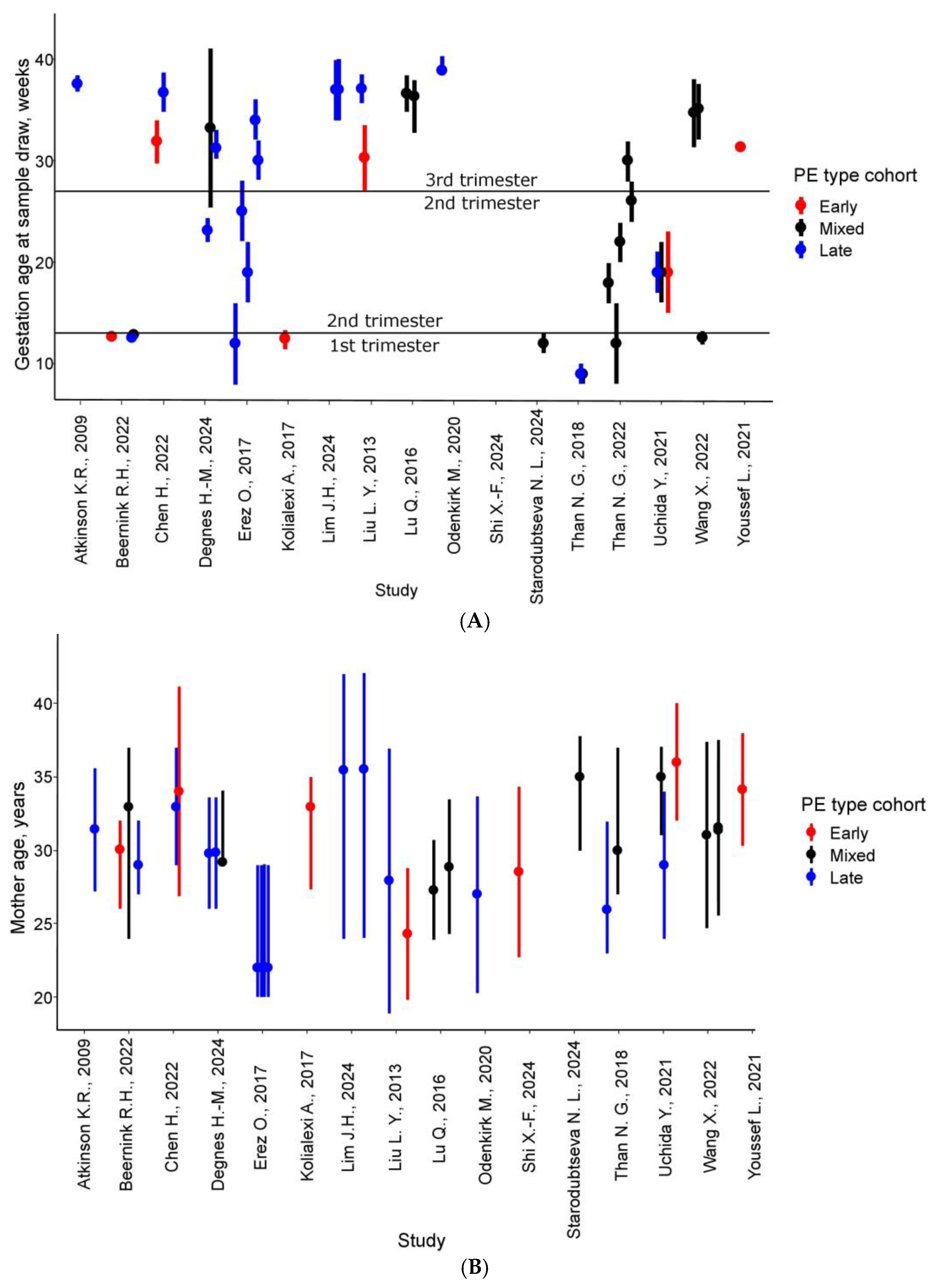
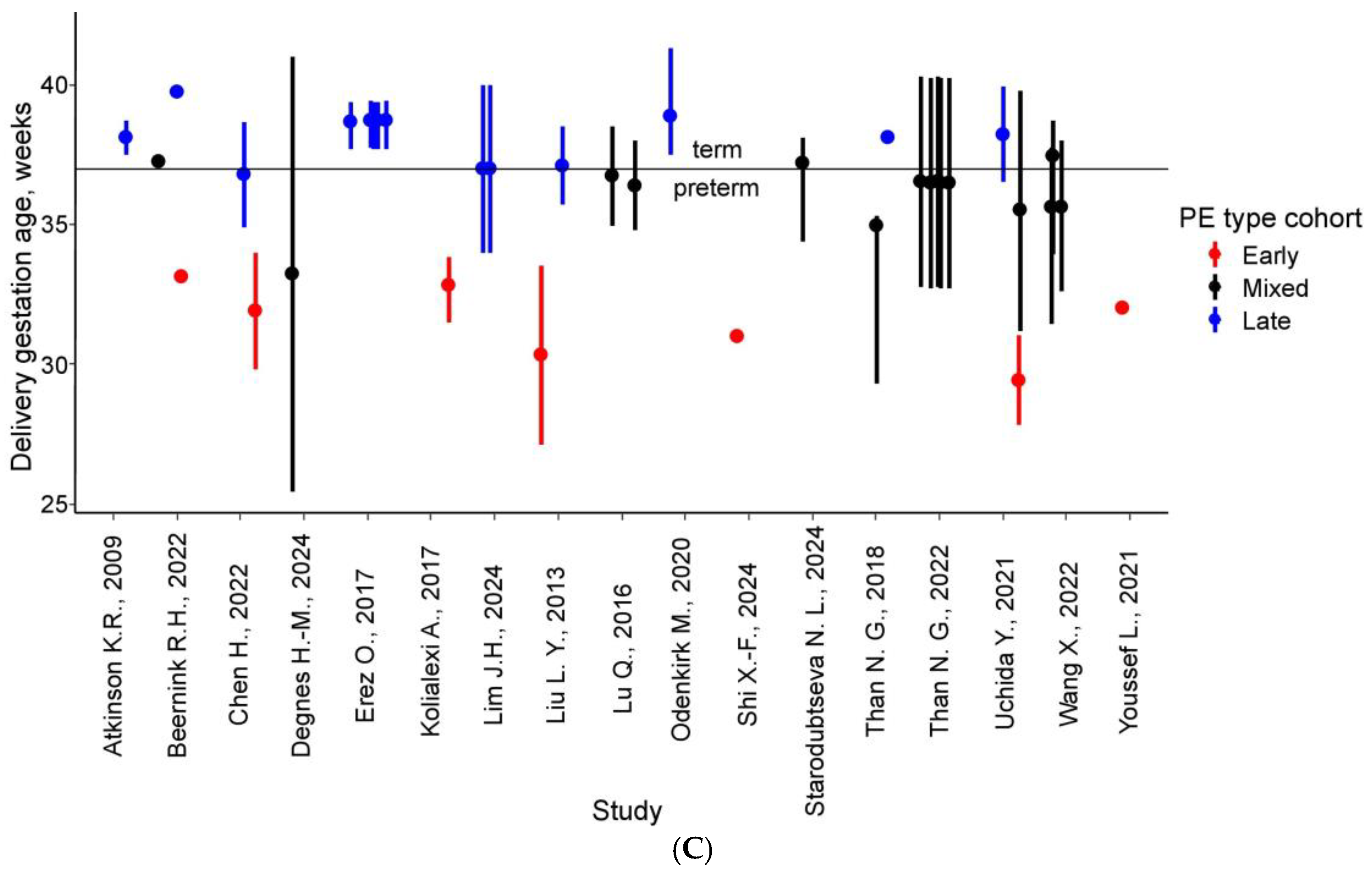
The median cohort size for PE patients was 35 individuals, with the largest cohorts reported by Than et al., 2022 [39] (n = 109) and Erez et al., 2017 [30] (n = 76) (Table S1, Figure S1). Control groups had a median size of 42 patients, reaching up to 120 in one study. Severe PE was analyzed in 15 comparisons, while FGR, frequently associated with PE, was examined in eight. Sample collection predominantly occurred in the third trimester (>27 weeks) [26,28,29,30,32,33,34,35,41,42], though some studies investigated early PE, with blood draws in the first or second trimester (Figure 2) [27,31,40]. Patient ages ranged from 18.9 to 42 years, with weighted averages falling between 22 and 36 years. In ten comparisons, the average gestational age at delivery exceeded 37 weeks, and eight cohorts reported no preterm births.

3.2. PE-Specific Maternal Plasma/Serum Proteomic Profile

The 17 analyzed studies collectively identified 561 proteins with statistically significant alterations in PE, of which 122 were replicated in at least two independent studies (Table S3). Notably, Odenkirk et al. (2020) [35] reported the highest overlap, with 37 consistent protein changes in late third-trimester plasma samples (mean gestational age 38.9 ± 1.4 weeks) (Table S4). A striking confirmation of early findings emerged from Atkinson et al. (2009) [26], where six of seven initially identified proteins were later validated in independent studies (Figure 3, Table S4). The study by Odenkirk et al. (2020) [35] also demonstrated exceptional consistency, with 27 proteins showing uniform directional changes in PE. In contrast, first- and second-trimester studies revealed minimal concordance; for instance, no proteins exhibited consistent changes in the works of Beernink et al. (2022) [27], Degnes et al. (2024) [29], or Uchida et al. (2021) [40]. This striking trimester-specific disparity underscores more pronounced serum/plasma proteome disruptions in late pregnancy, highlighting the need for longitudinal studies to elucidate PE pathogenesis and biomarker potential.
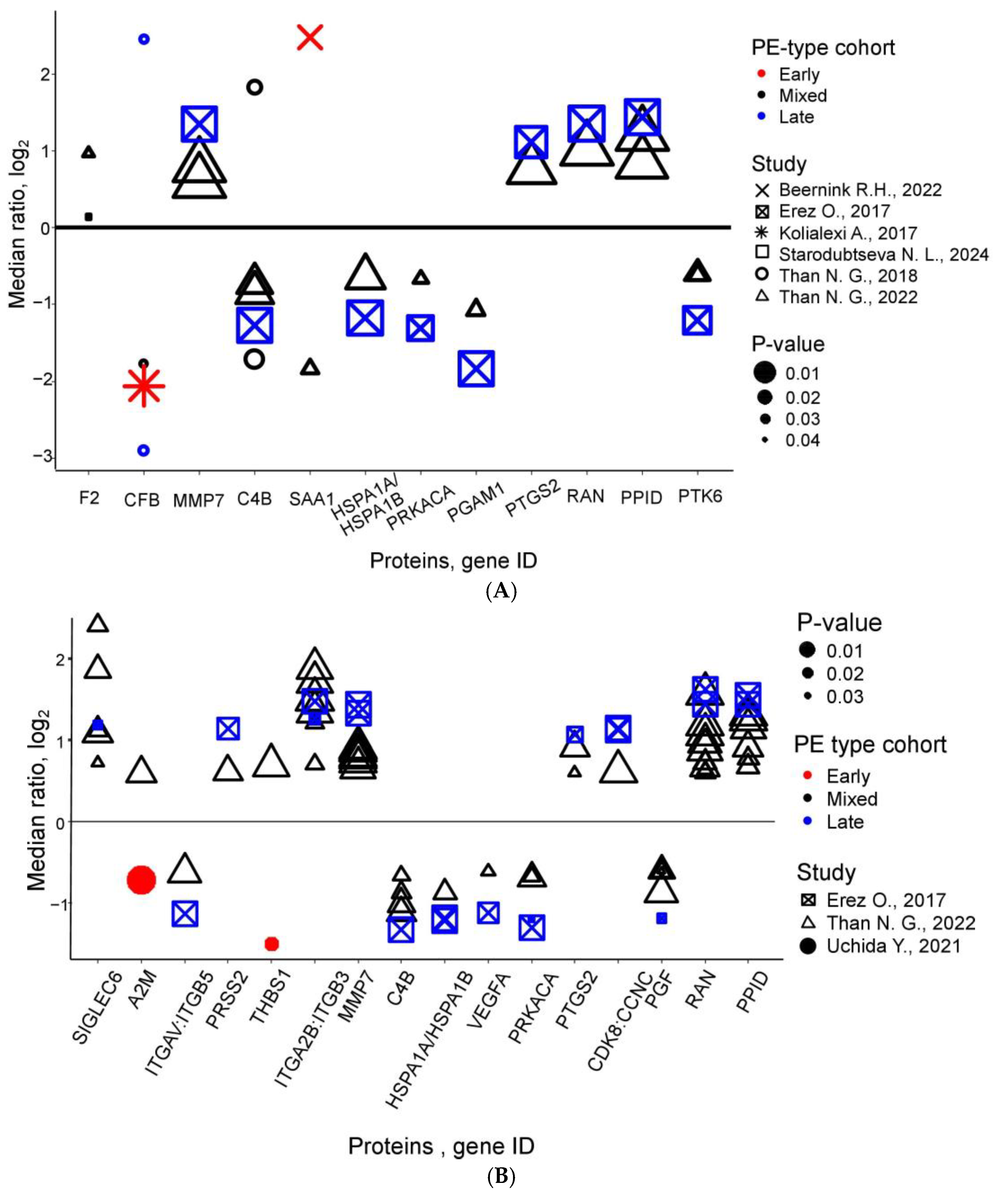
Early-pregnancy proteomics yielded 12 recurrently altered proteins (Figure 4A). Several proteins—including Prothrombin, Matrilysin, Prostaglandin G/H synthase 2, and Peptidyl-prolyl cis-trans isomerase D—demonstrated consistent increases, while others like Heat shock 70 kDa protein 1A/1B, cAMP-dependent protein kinase catalytic subunit alpha, Phosphoglycerate mutase 1 and Protein-tyrosine kinase 6 decreased uniformly. Intriguingly, proteins such as Complement factor B and Serum amyloid A-1 exhibited bidirectional changes, reflecting PE’s pathological complexity (Table 2, Tables S5 and S6).
Second-trimester meta-analysis identified 16 candidate biomarkers according to at least two studies (Figure 4B), with proteins like Sialic acid-binding Ig-like lectin 6, Trypsin-2, Integrin alpha-IIb: beta-3 complex, Matrilysin, Prostaglandin G/H synthase 2, Cyclin-dependent kinase 8:Cyclin-C complex, GTP-binding nuclear protein Ran, and Peptidyl-prolyl cis-trans isomerase D consistently elevated. Integrin alpha-V: beta-5 complex, Complement C4b, Heat shock 70 kDa protein 1A/1B, Vascular endothelial growth factor A, isoform 121, cAMP-dependent protein kinase catalytic subunit alpha, and Peptidyl-prolyl cis-trans isomerase D showed decreased levels (Table S7). However, conflicting data for proteins like Alpha-2-macroglobulin and Thrombospondin-1—observed by Uchida et al. (2021) [40] and Than et al. (2022) [39]—highlight methodological and cohort variability challenges (Table S8).
Third-trimester studies revealed 59 recurrently altered proteins (Figure 4C), with early PE showing consistent increases in maternal blood Fibronectin, Protein AMBP, Inter-alpha-trypsin inhibitor heavy chain H3, and Carboxypeptidase N subunit 2. Late PE exhibited more complex patterns: 15 proteins (e.g., Fibronectin, Vascular endothelial growth factor receptor 1, Inter-alpha-trypsin inhibitor heavy chain H3 and H2) were elevated, 2 (Apolipoprotein A-I, PlGF) decreased, and 11 (including Apolipoprotein E and Inter-alpha-trypsin inhibitor heavy chain H4) showed discordant results across studies (Tables S9 and S10). These trimester-specific signatures and inconsistencies underscore the need for standardized methodologies and refined clinical stratification in PE proteomics research.

3.3. Bioinformatics Analysis

Several proteins identified as PE markers in at least two first-trimester studies—prothrombin, serum amyloid A-1 protein (SAA1), complement C4b, and complement factor B—demonstrate statistically significant associations with kidney disease, while prothrombin and C4b are additionally linked to protein S deficiency (Table S11). A key pathological feature of early PE development is complement system activation (Figure S2). Furthermore, dysregulation of metabolic, hormonal, and neuronal signaling pathways—including those mediated by cAMP-dependent protein kinase A (PRKACA)—plays a critical role in PE pathogenesis (Figure S3).
In the second trimester of pregnancy, PE progression involves pronounced immune dysregulation, particularly complement activation, alongside disruptions in angiogenesis and ECM remodeling. These processes are mediated by integrin-dependent signaling and PI3K/AKT/mTOR cascade activation (Figure S4). Molecular dysregulation patterns resemble those of the first trimester but intensify (Figure S3). Notably, several implicated proteins contribute to fetal developmental processes, including morphogenesis, tube formation, and primary germ layer establishment, with nine proteins expressed in embryonic structures (Table S12). Pathway analyses reveal significant enrichment in cardiomyopathy-related pathways (Figure S3), suggesting a potential link between PE and cardiovascular disorders.
The maternal protein profile exhibits the most pronounced alterations as PE approaches clinical onset. PE-associated markers in this stage correlate not only with eclampsia but also with kidney disease and coagulation disorders (Table S13). Key pathways include complement/coagulation cascades and insulin-like growth factor (IGF) transport regulation by insulin-like growth factor-binding proteins (IGFBPs) (Figure S5, Table S13). Strikingly, 17 PE serum markers are expressed in placental tissues (Table S13). PANTHER analysis highlights coagulation imbalances, metabolic disruptions, and inflammatory signaling dysregulation as dominant features of PE in the third trimester of pregnancy (Figure S3).

4. Discussion

The meta-analysis incorporated studies published between 2009 and 2024 that reported quantitative proteomic analyses of serum and plasma from pregnant women with PE (n = 842) compared to controls with uncomplicated pregnancies (n = 1148). This review systematically evaluated key parameters including patients’ clinical and demographic characteristics, sample collection timing, and PE subtypes (early onset, late onset, and mixed forms). Notably, serum Complement C4b levels demonstrated a consistent reduction across mixed and late-onset PE cohorts during early gestation, suggesting it as a promising candidate for the early detection of preeclampsia. Across the 17 selected studies, researchers identified 122 proteins showing statistically significant expression level alterations in PE cases versus controls, with each protein confirmed by at least 2 independent studies.

4.1. First-Trimester Predictive Biomarkers of PE

Complement system hyperactivation emerges as one of the most significantly enriched biological processes in first-trimester PE, playing a crucial role in fetoplacental immunological tolerance maintenance [43]. Disruption of complement homeostasis—whether through excessive activation or pathway dysregulation—represents a key mechanism in pregnancy complications [44]. This aberrant activation triggers a cascade of pathological events: systemic inflammation, vascular endothelial damage, and subsequent endothelial dysfunction manifesting as hypertension and proteinuria. Furthermore, it elevates oxidative stress and disrupts placental immune regulation, ultimately impairing trophoblast invasion and placental development [43,45,46,47].
The complement system serves as a critical interface between innate immunity and physiological homeostasis, participating in tissue development/repair and interacting with multiple endogenous systems (renin-angiotensin, coagulation, and kallikrein-kinin pathways). Comprising over 50 membrane-bound and circulating proteins, its dysregulation shows distinct biomarker patterns in PE. Notably, serum Complement C4b levels demonstrate consistent reduction across mixed and late-onset PE cohorts during early gestation [30,38,48]. Balduit et al. (2024) attribute this finding to enhanced classical/lectin pathway activity, where increased C4 proteolysis elevates C4b cleavage products [49]. Similarly, Complement Factor B (alternative pathway) shows marked depletion in early pregnancy, independent of PE onset timing [31,38]. These observations support growing interest in complement-targeted interventions for PE prevention and treatment [50,51], with low-dose aspirin-the only internationally recommended PE prophylactic-demonstrating specific inhibitory effects on alternative pathway activity [52,53].
Complement activation is closely linked to coagulation cascades. This complement-coagulation axis manifests in proteomic signatures: meta-analyses reveal elevated first-trimester Prothrombin levels in PE cases versus controls [37,48], while Nguyen et al. (2019) report reduced Fibrinogen activity, suggesting a thrombogenic shift toward fibrin formation [54]. Such findings underscore the bidirectional relationship between complement activation and hemostasis in endothelial dysfunction [55,56].
The first-trimester proinflammatory and hypercoagulable state may induce multiorgan dysfunction, particularly affecting hepatic and renal systems. Associated protein markers correlate with renal impairment and Protein S deficiency (Table S11). Renal dysfunction exacerbates PE risk through multiple mechanisms: impaired pressure natriuresis, fluid retention, and uremic toxin accumulation collectively increase vascular stress. Concurrent Protein S deficiency-by reducing natural anticoagulant activity-potentiates hypercoagulability, placental microthrombosis, and ischemia-driven antiangiogenic factor release, creating a vicious cycle of placental dysfunction.
In conclusion, Prothrombin, Matrilysin (MMP7), Prostaglandin G/H synthase 2 (COX-2), and Peptidyl-prolyl cis-trans isomerase D (PPID) showed consistent increases across studies, while Heat shock 70 kDa protein 1A/1B (HSPA1A/B), cAMP-dependent protein kinase catalytic subunit alpha (PRKACA), Phosphoglycerate mutase 1 (PGAM1), and Protein-tyrosine kinase 6 (PTK6) exhibited uniform decreases. Considering that early-onset PE is generally associated with a more severe clinical course compared to late-onset PE, and that the majority of the identified biomarkers were detected in studies focusing on early or mixed PE cohorts, it can be inferred that alterations in concentrations of these proteins are significantly associated with more severe forms of the disease. Together, these findings highlight a panel of promising biomarkers rather than a single candidate, reflecting the multifactorial nature of PE.

4.2. Blood Proteome Alterations in Second-Trimester PE

Emerging proteomic evidence reveals distinct patterns of integrin dysregulation in PE, with the Integrin αIIbβ3 complex (GPIIb/IIIa) showing consistent elevation during late gestation across multiple studies, while Integrin αVβ5 demonstrates reciprocal downregulation. These transmembrane receptors, critical for cellular adhesion and signaling, play pivotal roles in placental physiology. The platelet-specific GPIIb/IIIa complex becomes activated by thrombin and collagen, enhancing its binding affinity for fibrinogen and von Willebrand factor to promote platelet aggregation and microthrombosis-a prothrombotic state that exacerbates placental hypoperfusion and ischemic injury, central features of PE pathophysiology [57]. While low-dose aspirin prophylaxis effectively reduces platelet aggregation in high-risk pregnancies, the potential therapeutic use of targeted GPIIb/IIIa inhibitors like abciximab requires further investigation for pregnancy-specific safety and efficacy [13].
Concurrent with integrin dysregulation, matrix metalloproteinase-7 (MMP-7) is significantly elevated in both first- and second-trimester mixed and late-onset PE cases [58,59]. Its critical involvement in ECM remodeling during placentation positions MMP-7 as a promising biomarker for PE [30,58,59]. Similarly, the sialic acid-binding lectin Siglec-6 is markedly overexpressed in PE syncytiotrophoblasts [60,61]. However, its detection in maternal circulation relies on advanced SOMAmer-based proteomic techniques rather than conventional assays [30,39]. Siglec-6 interactions with leptin and PlGF implicates it in fetomaternal immune dysregulation [60,61], though clinical utility hinges on the development of ultrasensitive detection methods and large-scale validation studies.
Vascular endothelial growth factor A (VEGF-A), PlGF, and sFlt-1 are among the most extensively studied biomarkers in PE. Our meta-analysis reveals that PlGF levels are significantly reduced in the second and third trimesters of pregnancies complicated by PE. Similarly, VEGF-A levels decline—during the second trimester in late-onset PE and the third trimester in both early- and late-onset PE. These alterations reflect an imbalance in vascular growth regulation, contributing to placental dysfunction and the hallmark symptoms of PE, including hypertension and proteinuria. Moreover, the elevated sFlt-1 levels observed in PE exacerbate this imbalance by neutralizing the biological activity of VEGF-A and PlGF, supporting the hypothesis that PE arises from endothelial dysfunction driven by impaired angiogenic signaling [62,63].
This vascular pathology aligns with broader cardiovascular manifestations revealed through KEGG pathway analysis, which shows significant enrichment of cardiomyopathy-related pathways in PE [64,65]. The condition’s cardiovascular impact becomes particularly evident by mid-gestation, when failed hemodynamic adaptation manifests as pathological cardiac remodeling including left ventricular concentric hypertrophy and diastolic dysfunction, accompanied by elevated biomarkers of myocardial strain such as NT-proBNP and troponin, even in the absence of overt heart failure [66,67]. Most concerning are the long-term implications: women with PE face a four-fold increased risk of subsequent heart failure and double the mortality from ischemic heart disease and stroke [65], underscoring the persistent cardiovascular consequences of this pregnancy complication and the critical need for ongoing postpartum monitoring and targeted prevention strategies.

4.3. Third-Trimester Protein Biomarkers of PE

The data obtained demonstrate substantially more dysregulated serum/plasma proteins in third-trimester (59 proteins) compared to the second (16 proteins) and first trimesters (12 proteins), with each protein alteration confirmed by at least two independent studies. This temporal pattern reflects both biological and methodological factors. From a practical standpoint, third-trimester biomarker research benefits from easier cross-sectional sampling compared to the logistical challenges of longitudinal studies requiring early biospecimen collection for a condition with only ~5% incidence. Biologically, the third trimester represents the culmination of progressive pathological processes-including placental dysfunction, endothelial impairment, and systemic inflammation-which reach maximal intensity as clinical symptoms emerge.
The proteomic heterogeneity observed in late gestation enables identification of distinct biomarker patterns corresponding to different PE subtypes, providing critical insights into disease mechanisms and potential therapeutic targets. Notably, many third-trimester protein alterations are linked not only to PE but also to renal and coagulation disorders, with evidence suggesting these pathophysiological changes originate as early as the first trimester. Complement and coagulation cascade dysregulation, central to PE pathogenesis, demonstrates measurable changes from pregnancy onset. Additionally, third-trimester biomarkers reveal significant enrichment in IGF transport and signaling pathways, underscoring the importance of metabolic dysregulation in PE development.
Metabolic disturbances in PE encompass impaired lipid and carbohydrate metabolism, insulin resistance, and IGF dysregulation, all contributing to endothelial dysfunction, systemic inflammation, and placental insufficiency. Even without overt diabetes, PE patients frequently exhibit insulin resistance leading to abnormal glucose/lipid metabolism that exacerbates oxidative stress and vascular impairment [68,69,70,71,72,73,74]. PE specific metabolic changes include elevated free fatty acids and triglycerides correlating with disease severity [75], along with lipid deposition in spiral arteries resembling early atherosclerotic changes [76].
Our systematic review and meta-analysis revealed multiple maternal blood proteins with consistent and significant alterations during early pregnancy in women who later developed PE. Strikingly, several of these proteins—including GTP-binding nuclear protein Ran, peptidyl-prolyl cis-trans isomerase D (PPID), protein-tyrosine kinase 6 (PTK6), and phosphoglycerate mutase 1 (PGAM1)—have rarely or never been previously associated with PE. Notably, these dysregulations were detectable as early as the first trimester, even in cohorts that subsequently developed late-onset or mixed-type PE. Ran and PPID levels were consistently elevated, whereas PTK6 and PGAM1 were reduced, suggesting their potential as novel early biomarkers or mechanistic contributors to PE pathogenesis. Further studies are needed to validate their clinical utility and elucidate their pathophysiological roles.
Nine proteins involved in IGF transport and signaling show consistent directional changes in third-trimester PE across multiple studies: Apolipoprotein A-I, Ceruloplasmin, Insulin-like growth factor-binding protein 4, Fibronectin, Inter-alpha-trypsin inhibitor heavy chain H2, Pappalysin-2, and Cystatin-C are elevated, while Complement C3 and Kininogen-1 are decreased. Apolipoprotein A-I, the major HDL component crucial for cholesterol transport and vascular protection, becomes dysfunctional in PE despite increased antioxidant capacity-a likely compensatory response to oxidative stress [77,78,79,80]. Ceruloplasmin elevation reflects both oxidative stress and inflammation, with levels correlating with PE severity as this ferroxidase attempts to mitigate free iron-mediated ROS generation [81,82,83,84,85].
Fibronectin, a multifunctional ECM protein critical for placentation, exhibits profound alterations in PE [86,87,88,89]. Although plasma fibronectin levels typically rise modestly during normal pregnancy (31.8% increase), PE patients display markedly elevated concentrations (94.5% increase) [90,91]. Glycosylated fibronectin stands out as a particularly promising early biomarker, detectable in first-trimester samples from women who subsequently develop PE and strongly associated with adverse outcomes, including hypertension, preterm delivery, and FGR [92]. Notably, when integrated into first-trimester screening algorithms, glycosylated fibronectin significantly enhances preterm PE detection rates, improving sensitivity from 64.9% to 82.9% in an Asian cohort [93].

4.4. Standardization of Blood Protein Analysis: Current Challenges and Future Directions for PE Diagnosis and Prediction

Accurate analysis of blood proteins is critical for PE prediction and diagnosis, yet significant concerns persist regarding measurement reliability. A major challenge in plasma/serum proteomics is the substantial inter-laboratory variability in measured protein concentrations (or concentration ratios). These inconsistencies arise from multiple sources: lack of standardized pre-processing protocols (sample collection, handling, and storage); heterogeneity in sample preparation methods (e.g., protein depletion strategies, labeling techniques); diverse fractionation approaches (SDS-PAGE, 2D electrophoresis, preparative LC); variability in detection platforms (HPLC-MS/MS, HPLC-MRM-MS, ELISA, SOMAmer, Olink) [94]. Furthermore, the high correlation between results from Erez O et al. [30] and Than N et al. [48]—both conducted at the same research center—along with their limited overlap of common proteins with other studies, highlights the significant impact of inter-laboratory variation.
The choice of sample type represents a critical methodological consideration, as plasma and serum yield divergent protein profiles due to inherent processing differences. Plasma is the preferred matrix for analyzing complement system proteins, as it circumvents artifactual in vitro activation during serum coagulation and fibrinolysis [95]. Sample preparation must also account for protein–protein interactions, particularly those involving high-abundance species like albumin and apolipoproteins, which can mask or alter biomarker detection [96,97,98]. Furthermore, pre-analytical variables—including storage conditions, thawing protocols, and freeze–thaw cycles—introduce additional variability that must be rigorously controlled to ensure data reproducibility [99,100].
Inconsistent sampling timelines across studies further complicate standardization and hinder meta-analyses. Most studies in our systematic review and meta-analysis measured protein levels at PE diagnosis (third trimester), explaining the scarcity of validated first/second-trimester predictive markers. Notably, five first/second-trimester studies [27,31,37,38,40] were limited by extremely small PE cohort sizes, significantly reducing the statistical power and reliability of their comparative analyses. These findings highlight the urgent need for multicenter longitudinal studies with early-pregnancy sampling (8–12 weeks) and integration of molecular subtyping to account for PE heterogeneity. Given PE’s syndromic nature, no single sampling strategy can predict all pathological variants [101]. Additional confounding factors include cohort heterogeneity (ethnic composition, sample size), disease severity (mild/severe), onset timing (early/late PE), and comorbid conditions.
The identified biomarkers hold significant potential to enhance clinical screening for PE. They could be integrated into first-trimester multi-marker panels alongside established predictors—such as maternal risk factors, mean arterial pressure, uterine artery Doppler indices, and biochemical markers (PAPP-A, PlGF, and β-hCG)—to improve early risk assessment. Beyond screening, these biomarkers may enable more precise individualized risk stratification, particularly for women classified as intermediate risk after standard evaluation. They could also improve early detection of late-onset PE, where current methods lack sensitivity. Additionally, serial monitoring of biomarker dynamics during early pregnancy could help assess biological responses to preventive therapies (e.g., low-dose aspirin), allowing timely adjustments in clinical management. From a research perspective, these biomarkers—linked to cellular stress, protein folding, intracellular transport, and complement regulation—provide novel insights into PE pathophysiology and highlight potential therapeutic targets. Simplified biomarker panels incorporating these proteins could further facilitate the development of point-of-care diagnostic tools, improving accessibility across diverse healthcare settings. However, prospective validation studies remain essential to confirm their clinical utility.
The identified biomarkers could significantly enhance personalized PE management through multiple avenues. By detecting molecular alterations early, these biomarkers may enable precise risk stratification and facilitate tailored preventive strategies—such as low-dose aspirin therapy or intensified monitoring—based on individual patient profiles. Serial assessment of biomarker levels could further provide real-time insights into treatment response, allowing clinicians to adapt management approaches dynamically throughout pregnancy. Beyond clinical applications, these biomarkers’ roles in critical biological pathways—including complement activation, cellular stress response, and protein folding—offer promising opportunities to identify novel therapeutic targets and develop targeted, mechanism-driven interventions.
Recent genomic and proteomic research has identified four molecular PE subtypes: canonical/placental (primarily angiogenic dysfunction); metabolic (associated with obesity/insulin resistance); immunological (maternal/fetal immune dysregulation features); maternal (preexisting cardiovascular/metabolic risk) [38,39,48,102]. These subtypes align with distinct maternal hemodynamic patterns observed across hypertensive pregnancy disorders [103]. Development of subtype-specific PE diagnostic models improved quality of PE prevention. Advancing PE diagnostics requires standardized analytical protocols for sample collection/processing, large multicenter studies incorporating molecular subtyping, and longitudinal designs tracking clinical progression.
To advance methodological standardization in preeclampsia proteomics research, several strategic approaches could be implemented. An international collaborative consortium could be established to unify protocols for sample collection, processing, and data analysis across research institutions. Drawing inspiration from established standards such as MIAME and PRIDE, the field would benefit from developing specialized reporting guidelines to enhance transparency and reproducibility. The creation of shared reference datasets from rigorously characterized PE cohorts would provide valuable validation benchmarks, complemented by interlaboratory proficiency testing to systematically assess and reduce technical variability. Furthermore, aligning these standardization efforts with existing clinical networks like ISSHP or PREPARE would facilitate smoother translation of research findings into clinical practice.
Importantly, this study demonstrates several methodological strengths that reinforce the reliability of its findings. The systematic review was conducted in accordance with the PRISMA 2020 guidelines, and the quality of included studies was systematically evaluated using a modified Newcastle–Ottawa Scale. The majority of studies (82%) achieved a score of 7 or higher out of 10, indicating generally high methodological rigor. Clinical and proteomic data were sufficiently detailed to permit stratified analysis by gestational age and PE subtype. Moreover, the incorporation of studies utilizing advanced high-throughput proteomic platforms, coupled with orthogonal validation approaches, enhances the translational value of the identified protein markers. Nonetheless, limitations remain, including substantial heterogeneity in sample processing protocols and proteomic methodologies, which prevented effect-size pooling in a formal meta-analysis. Early-pregnancy datasets were limited and displayed poor overlap, constraining conclusions regarding first- and second-trimester biomarkers. These findings emphasize the ongoing need for standardized, multi-phase study designs with longitudinal sampling and unified analytical pipelines across laboratories.

5. Conclusions

As a cornerstone discipline of modern biomedical research, proteomics has achieved unprecedented analytical power through the integration of cutting-edge technologies. Contemporary platforms—including high-resolution LC-MS, modified DNA aptamers (SOMAmer), and multiplexed antibody arrays (Olink)—now enable both comprehensive identification and precise quantification of thousands of proteins in complex biological matrices like blood serum and plasma. These technological breakthroughs, coupled with automated sample handling and standardized processing protocols, have transformed proteomics into a robust tool for generating clinically actionable data.
Our comprehensive meta-analysis demonstrates that blood proteome alterations represent a powerful resource for PE prediction, early detection, and mechanistic understanding. These molecular fingerprints not only enhance our understanding of disease pathogenesis but also create new opportunities for developing personalized therapeutic and preventive strategies. These findings emphasize the importance of precision medicine in pregnancy management, grounded in robust molecular data and deep understanding of disease mechanisms.
The refined list of potential PE biomarkers presented in this study provides a valuable foundation for translational research. Key validation strategies include orthogonal verification using Western blotting or ELISA, high-throughput validation via targeted proteomics (SRM/MRM), and multicenter replication studies to assess generalizability. Targeted proteomic approaches offer particular advantages, enabling simultaneous quantification of multiple candidate markers with high specificity and throughput—a crucial step toward clinical implementation.
The clinical utility of proteomic findings requires rigorous standardization across all analytical phases–from pre-analytical processing to data integrity. Only through strict adherence to these principles can we ensure the reproducibility and clinical relevance of proteomic data. The integration of automated systems and artificial intelligence-assisted analysis will further enhance reliability as the field progresses toward routine clinical application.

Supplementary Materials

The following supporting information can be downloaded at https://www.mdpi.com/article/10.3390/life15050776/s1, Figure S1: Cohort size distributions for (A) PE, (B) Control, (C) Severe PE, and (D) FGR study groups, with median values (bold lines); Figure S2: Significantly enriched (FDR < 0.05) first-trimester PE protein associations: (A) GO biological processes; (B) Reactome pathways; Figure S3: Significantly enriched (FDR < 0.05) second-trimester PE protein associations: (A) GO biological processes; (B) KEGG pathways; (C) Reactome pathways; (D) Wikipathways; Figure S4: Significantly enriched (FDR < 0.05) third-trimester PE protein associations: (A) KEGG pathways; (B) GO biological processes; (C) Reactome pathways; (D) Wikipathways; Figure S5: Significantly enriched (FDR<0.05) third-trimester PE protein associations: (A) KEGG pathways; (B) GO biological processes; (C) Reactome pathways; (D) Wikipathways. Table S1: Studied cohort characteristics. Notation: X-Y = range; X(Y-Z) = mean(range); X ± Y=mean ± SD; X [Y;Z] = median [Q1;Q3]. Table S2: Study-specific protein alterations. Table S3: Consistently altered proteins (≥2 studies). Table S4: Counts of significantly altered proteins per study. Table S5: First trimester-specific protein alterations (≥2 studies) with matched directional changes. Table S6: First trimester-specific protein alterations (≥2 studies) with unmatched directional changes. Table S7: Second trimester-specific protein alterations (≥2 studies) with matched directional changes. Table S8: Second trimester-specific protein alterations (≥2 studies) with unmatched directional changes. Table S9: Third trimester-specific protein alterations (≥2 studies) with matched directional changes. Table S10: Third trimester-specific protein alterations (≥2 studies) with unmatched directional changes; Table S11: Pathway enrichment analysis of 1st trimester consistently altered proteins (≥2 studies); Table S12: Pathway enrichment analysis of 2nd trimester consistently altered proteins (≥2 studies); Table S13: Pathway enrichment analysis of 3rd trimester consistently altered proteins (≥2 studies).

Author Contributions

Conceptualization, N.S., E.R. and Z.K.; data curation, N.S., A.P., A.T., E.K. and A.A.; formal analysis, N.S., A.P., A.T., E.K. and E.R.; funding acquisition, N.S., E.R. and Z.K.; investigation, N.S., A.P., A.T., E.K. and E.R.; methodology, N.S., E.R. and A.T.; project administration, N.S. and Z.K.; resources, N.S., A.T. and A.A.; software, A.T. and E.R.; supervision, N.S. and Z.K.; visualization, N.S., A.P., A.T., E.R. and E.K.; writing—original draft, N.S., A.P., A.T., E.K. and A.A.; writing—review and editing, E.R. and Z.K. All authors have read and agreed to the published version of the manuscript.

Funding

This research was funded by the Russian Science Foundation (No. 24-14-00140): https://rscf.ru/en/project/24-14-00140/ (accessed on 11 May 2025).

Institutional Review Board Statement

This systematic review was conducted without prior registration in a public registry, and no formal protocol was prepared before commencing the research.

Informed Consent Statement

Not applicable.

Data Availability Statement

All relevant data are within the manuscript and its Supporting Information Files.

Conflicts of Interest

The authors declare no conflicts of interest.

Abbreviations

The following abbreviations are used in this manuscript:
2-DETwo-dimensional gel electrophoresis
2D-DIGETwo-dimensional difference gel electrophoresis with fluorescent labeling
AUCArea-under-the-curve
CXCL10C-X-C motif chemokine ligand 10
ECMExtracellular matrix
ELISAEnzyme-linked immunosorbent assay
FGRFetal growth restriction
FIGOInternational Federation of Gynecology and Obstetrics
FMFFetal Medicine Foundation
HELLPHemolysis, Elevated Liver Enzymes, Low Platelet Count
HPLC-MS/MSHigh-performance liquid chromatography–tandem mass spectrometry
IQRInter-quartile range
ISIsotope-labeled internal standards (tryptic peptides)
iTRAQIsobaric tags for relative and absolute quantitation
LC-MS/MSLiquid chromatography -tandem mass spectrometry
MALDI-MS/MSMatrix-assisted laser desorption/ionization–tandem mass spectrometry
MRMMultiple reaction monitoring
NGSNext-generation sequencing
NMRNuclear magnetic resonance
PAPP-APregnancy-associated plasma protein-A
PCRPolymerase chain reaction
PEPreeclampsia
PlGFPlacental growth factor
SDStandard deviation
SDS-PAGEPolyacrylamide gel electrophoresis with sodium dodecyl sulfate
sFlt-1Soluble fms-like tyrosine kinase-1
SOMAmerSlow off-rate modified DNA aptamers
SRMSelected reaction monitoring
SWATHSequential window acquisition of all theoretical mass spectra
TMTTandem mass tag

References

  1. Lo, J.O.; Mission, J.F.; Caughey, A.B. Hypertensive disease of pregnancy and maternal mortality. Curr. Opin. Obstet. Gynecol. 2013, 25, 124–132. [Google Scholar] [CrossRef] [PubMed]
  2. Knight, M.; Bunch, K.; Felker, A.; Patel, R.; Kotnis, R.; Kenyon, S.; Kurinczuk, J.J. MBRRACE—UK: Care Saving Lives, Improving Mothers’ Care Lessons learned to Inform Maternity Care from the UK and Ireland Confidential Enquiries into Maternal Deaths and Morbidity State of the Nation Surveillance Report 20; National Perinatal Epidemiology Unit, University of Oxford: Oxford, UK, 2023; ISBN 978-1-7392619-4-8. [Google Scholar]
  3. Boghossian, N.S.; Yeung, E.; Mendola, P.; Hinkle, S.N.; Laughon, S.K.; Zhang, C.; Albert, P.S. Risk factors differ between recurrent and incident preeclampsia: A hospital-based cohort study. Ann. Epidemiol. 2014, 24, 871–877.e3. [Google Scholar] [CrossRef] [PubMed]
  4. NICE Guidelines Institute for Health and Care. Hypertension in Pregnancy: Diagnosis and Management; National Institute for Health and Care Excellence (NICE): London, UK, 2019; Volume 133, ISBN 1179-1411. [Google Scholar]
  5. Lisonkova, S.; Joseph, K.S. Incidence of preeclampsia: Risk factors and outcomes associated with early- versus late-onset disease. Am. J. Obstet. Gynecol. 2013, 209, 544.e1–544.e12. [Google Scholar] [CrossRef] [PubMed]
  6. Lisonkova, S.; Sabr, Y.; Mayer, C.; Young, C.; Skoll, A.; Joseph, K.S. Maternal morbidity associated with early-onset and late-onset Preeclampsia. Obstet. Gynecol. 2014, 124, 771–781. [Google Scholar] [CrossRef]
  7. Khan, S.; Siddique, A.B.; Jabeen, S.; Hossain, A.T.; Haider, M.M.; Zohora, F.T.; Rahman, M.M.; El Arifeen, S.; Rahman, A.E.; Jamil, K. Preeclampsia and eclampsia-specific maternal mortality in Bangladesh: Levels, trends, timing, and care-seeking practices. J. Glob. Health 2023, 13, 07003. [Google Scholar] [CrossRef]
  8. Melchiorre, K.; Sutherland, G.R.; Liberati, M.; Thilaganathan, B. Preeclampsia is associated with persistent postpartum cardiovascular impairment. Hypertension 2011, 58, 709–715. [Google Scholar] [CrossRef]
  9. Melchiorre, K.; Sutherland, G.R.; Liberati, M.; Thilaganathan, B. Maternal cardiovascular impairment in pregnancies complicated by severe fetal growth restriction. Hypertension 2012, 60, 437–443. [Google Scholar] [CrossRef]
  10. Jung, E.; Romero, R.; Yeo, L.; Gomez-Lopez, N.; Chaemsaithong, P.; Jaovisidha, A.; Gotsch, F.; Erez, O. The etiology of preeclampsia. Am. J. Obstet. Gynecol. 2022, 226, S844–S866. [Google Scholar] [CrossRef]
  11. Staff, A.C.; Fjeldstad, H.E.; Fosheim, I.K.; Moe, K.; Turowski, G.; Johnsen, G.M.; Alnaes-Katjavivi, P.; Sugulle, M. Failure of physiological transformation and spiral artery atherosis: Their roles in preeclampsia. Am. J. Obstet. Gynecol. 2022, 226, S895–S906. [Google Scholar] [CrossRef]
  12. Melchiorre, K.; Giorgione, V.; Thilaganathan, B. The placenta and preeclampsia: Villain or victim? Am. J. Obstet. Gynecol. 2022, 226, S954–S962. [Google Scholar] [CrossRef]
  13. American College of Obstetricians and Gynecologists. Low-dose aspirin use during pregnancy. ACOG Committee Opinion No. 743. Obstet. Gynecol. 2018, 132, e44–e52. [Google Scholar] [CrossRef]
  14. Henderson, J.T.; Whitlock, E.P.; O’Connor, E.; Senger, C.A.; Thompson, J.H.; Rowland, M.G. Low-dose aspirin for prevention of morbidity and mortality from preeclampsia: A systematic evidence review for the u.s. preventive services task force. Ann. Intern. Med. 2014, 160, 695–703. [Google Scholar] [CrossRef] [PubMed]
  15. Youssef, L.; Testa, L.; Crovetto, F.; Crispi, F. 10. Role of high dimensional technology in preeclampsia (omics in preeclampsia). Best Pract. Res. Clin. Obstet. Gynaecol. 2024, 92, 102427. [Google Scholar] [CrossRef] [PubMed]
  16. Stepan, H.; Herraiz, I.; Schlembach, D.; Verlohren, S.; Brennecke, S.; Chantraine, F.; Klein, E.; Lapaire, O.; Llurba, E.; Ramoni, A.; et al. Implementation of the sFlt-1/PlGF ratio for prediction and diagnosis of pre-eclampsia in singleton pregnancy: Implications for clinical practice. Ultrasound Obstet. Gynecol. 2015, 45, 241–246. [Google Scholar] [CrossRef]
  17. Zeisler, H.; Llurba, E.; Chantraine, F.; Vatish, M.; Staff, A.C.; Sennström, M.; Olovsson, M.; Brennecke, S.P.; Stepan, H.; Allegranza, D.; et al. Predictive value of the sFlt-1:PlGF ratio in women with suspected preeclampsia. N. Engl. J. Med. 2016, 374, 13–22. [Google Scholar] [CrossRef] [PubMed]
  18. Chaemsaithong, P.; Sahota, D.S.; Poon, L.C. First trimester preeclampsia screening and prediction. Am. J. Obstet. Gynecol. 2022, 226, S1071–S1097.e2. [Google Scholar] [CrossRef]
  19. Than, N.G.; Romero, R.; Posta, M.; Györffy, D.; Szalai, G.; Rossi, S.W.; Szilágyi, A.; Hupuczi, P.; Nagy, S.; Török, O.; et al. Classification of preeclampsia according to molecular clusters with the goal of achieving personalized prevention. J. Reprod. Immunol. 2024, 161, 104172. [Google Scholar] [CrossRef]
  20. Ontario Health Technology Assessment Series. First-Trimester Screening Program for the Risk of Pre-eclampsia Using a Multiple-Marker Algorithm: A Health Technology Assessment. Ont. Health Technol. Assess. Ser. 2022, 22, 1–118. [Google Scholar]
  21. Poon, L.C.; Shennan, A.; Hyett, J.A.; Kapur, A.; Hadar, E.; Divakar, H.; McAuliffe, F.; da Silva Costa, F.; von Dadelszen, P.; McIntyre, H.D.; et al. The International Federation of Gynecology and Obstetrics (FIGO) initiative on pre-eclampsia: A pragmatic guide for first-trimester screening and prevention. Int. J. Gynaecol. Obstet. Off. Organ. Int. Fed. Gynaecol. Obstet. 2019, 145, 1–33. [Google Scholar] [CrossRef]
  22. Page, M.J.; McKenzie, J.E.; Bossuyt, P.M.; Boutron, I.; Hoffmann, T.C.; Mulrow, C.D.; Shamseer, L.; Tetzlaff, J.M.; Akl, E.A.; Brennan, S.E.; et al. The PRISMA 2020 statement: An updated guideline for reporting systematic reviews. BMJ 2021, 372, n71. [Google Scholar] [CrossRef]
  23. Wells, G.; Shea, B.; O’Connell, D.; Robertson, J.; Peterson, J.; Welch, V.; Losos, M.; Tugwell, P. The Newcastle-Ottawa Scale (NOS) for Assessing the Quality of Nonrandomised Studies in Meta-Analyses. 2013, 21. Available online: https://www.ohri.ca/programs/clinical_epidemiology/nosgen.pdf (accessed on 11 May 2025).
  24. Szklarczyk, D.; Kirsch, R.; Koutrouli, M.; Nastou, K.; Mehryary, F.; Hachilif, R.; Gable, A.L.; Fang, T.; Doncheva, N.T.; Pyysalo, S.; et al. The STRING database in 2023: Protein-protein association networks and functional enrichment analyses for any sequenced genome of interest. Nucleic Acids Res. 2023, 51, D638–D646. [Google Scholar] [CrossRef] [PubMed]
  25. Thomas, P.D.; Ebert, D.; Muruganujan, A.; Mushayahama, T.; Albou, L.P.; Mi, H. PANTHER: Making genome-scale phylogenetics accessible to all. Protein Sci. 2022, 31, 8–22. [Google Scholar] [CrossRef] [PubMed]
  26. Atkinson, K.R.; Blumenstein, M.; Black, M.A.; Wu, S.H.; Kasabov, N.; Taylor, R.S.; Cooper, G.J.S.; North, R.A. An altered pattern of circulating apolipoprotein E3 isoforms is implicated in preeclampsia. J. Lipid Res. 2009, 50, 71–80. [Google Scholar] [CrossRef] [PubMed]
  27. Beernink, R.H.J.; Zwertbroek, E.F.; Schuitemaker, J.H.N.; Cremers, T.I.F.H.; Scherjon, S.A. First trimester serum biomarker discovery study for early onset, preterm onset and preeclampsia at term. Placenta 2022, 128, 39–48. [Google Scholar] [CrossRef]
  28. Chen, H.; Aneman, I.; Nikolic, V.; Karadzov Orlic, N.; Mikovic, Z.; Stefanovic, M.; Cakic, Z.; Jovanovic, H.; Town, S.E.L.; Padula, M.P.; et al. Maternal plasma proteome profiling of biomarkers and pathogenic mechanisms of early-onset and late-onset preeclampsia. Sci. Rep. 2022, 12, 19099. [Google Scholar] [CrossRef]
  29. Degnes, M.H.L.; Westerberg, A.C.; Andresen, I.J.; Henriksen, T.; Roland, M.C.P.; Zucknick, M.; Michelsen, T.M. Protein biomarker signatures of preeclampsia—A longitudinal 5000-multiplex proteomics study. Sci. Rep. 2024, 14, 23654. [Google Scholar] [CrossRef]
  30. Erez, O.; Romero, R.; Maymon, E.; Chaemsaithong, P.; Done, B.; Pacora, P.; Panaitescu, B.; Chaiworapongsa, T.; Hassan, S.S.; Tarca, A.L. The prediction of late-onset preeclampsia: Results from a longitudinal proteomics study. PLoS ONE 2017, 12, e0181468. [Google Scholar] [CrossRef]
  31. Kolialexi, A.; Tsangaris, G.T.; Sifakis, S.; Gourgiotis, D.; Katsafadou, A.; Lykoudi, A.; Marmarinos, A.; Mavreli, D.; Pergialiotis, V.; Fexi, D.; et al. Plasma biomarkers for the identification of women at risk for early-onset preeclampsia. Expert Rev. Proteom. 2017, 14, 269–276. [Google Scholar] [CrossRef]
  32. Lim, J.H.; Lim, J.M.; Lee, H.M.; Lee, H.J.; Kwak, D.W.; Han, Y.J.; Kim, M.Y.; Jung, S.H.; Kim, Y.R.; Ryu, H.M.; et al. Systematic Proteome Profiling of Maternal Plasma for Development of Preeclampsia Biomarkers. Mol. Cell Proteom. 2024, 23, 100826. [Google Scholar] [CrossRef]
  33. Liu, L.Y.; Yang, T.; Ji, J.; Wen, Q.; Morgan, A.A.; Jin, B.; Chen, G.; Lyell, D.J.; Stevenson, D.K.; Ling, X.B.; et al. Integrating multiple “omics” analyses identifies serological protein biomarkers for preeclampsia. BMC Med. 2013, 11, 236. [Google Scholar] [CrossRef]
  34. Lu, Q.; Liu, C.; Liu, Y.; Zhang, N.; Deng, H.; Zhang, Z. Serum markers of pre-eclampsia identified on proteomics. J. Obstet. Gynaecol. Res. 2016, 42, 1111–1118. [Google Scholar] [CrossRef] [PubMed]
  35. Odenkirk, M.T.; Stratton, K.G.; Gritsenko, M.A.; Bramer, L.M.; Webb-Robertson, B.J.M.; Bloodsworth, K.J.; Weitz, K.K.; Lipton, A.K.; Monroe, M.E.; Ash, J.R.; et al. Unveiling molecular signatures of preeclampsia and gestational diabetes mellitus with multi-omics and innovative cheminformatics visualization tools. Mol. Omi. 2020, 16, 521–532. [Google Scholar] [CrossRef] [PubMed]
  36. Shi, X.-F.; Zhang, J.-L.; Liu, K.; Wang, L.; Wang, H.-P.; Wu, H.-Y. Detection of serum major histocompatibility complex I (HLA-1) and β2-microglobulin (β2M) in pre-eclampsia using isobaric tags for relative and absolute quantitation (iTRAQ). Int. J. Gynaecol. Obstet. Off. Organ. Int. Fed. Gynaecol. Obstet. 2024, 165, 1072–1084. [Google Scholar] [CrossRef]
  37. Starodubtseva, N.; Tokareva, A.; Kononikhin, A.; Brzhozovskiy, A.; Bugrova, A.; Kukaev, E.; Muminova, K.; Nakhabina, A.; Frankevich, V.E.; Nikolaev, E.; et al. First-Trimester Preeclampsia-Induced Disturbance in Maternal Blood Serum Proteome: A Pilot Study. Int. J. Mol. Sci. 2024, 25, 10653. [Google Scholar] [CrossRef]
  38. Than, N.G.; Romero, R.; Tarca, A.L.; Kekesi, K.A.; Xu, Y.; Xu, Z.; Juhasz, K.; Bhatti, G.; Leavitt, R.J.; Gelencser, Z.; et al. Integrated systems biology approach identifies novel maternal and placental pathways of preeclampsia. Front. Immunol. 2018, 9, 1661. [Google Scholar] [CrossRef]
  39. Than, N.G.; Posta, M.; Györffy, D.; Orosz, L.; Orosz, G.; Rossi, S.W.; Ambrus-Aikelin, G.; Szilágyi, A.; Nagy, S.; Hupuczi, P.; et al. Early pathways, biomarkers, and four distinct molecular subclasses of preeclampsia: The intersection of clinical, pathological, and high-dimensional biology studies. Placenta 2022, 125, 10–19. [Google Scholar] [CrossRef]
  40. Uchida, Y.; Higuchi, T.; Shirota, M.; Kagami, S.; Saigusa, D.; Koshiba, S.; Yasuda, J.; Tamiya, G.; Kuriyama, S.; Kinoshita, K.; et al. Identification and validation of combination plasma biomarker of afamin, fibronectin and sex hormone-binding globulin to predict pre-eclampsia. Biol. Pharm. Bull. 2021, 44, 804–815. [Google Scholar] [CrossRef]
  41. Wang, X.; Yip, K.C.; He, A.; Tang, J.; Liu, S.; Yan, R.; Zhang, Q.; Li, R. Plasma Olink Proteomics Identifies CCL20 as a Novel Predictive and Diagnostic Inflammatory Marker for Preeclampsia. J. Proteome Res. 2022, 21, 2998–3006. [Google Scholar] [CrossRef]
  42. Youssef, L.; Miranda, J.; Blasco, M.; Paules, C.; Crovetto, F.; Palomo, M.; Torramade-Moix, S.; García-Calderó, H.; Tura-Ceide, O.; Dantas, A.P.; et al. Complement and coagulation cascades activation is the main pathophysiological pathway in early-onset severe preeclampsia revealed by maternal proteomics. Sci. Rep. 2021, 11, 3048. [Google Scholar] [CrossRef]
  43. Dimitriadis, E.; Rolnik, D.L.; Zhou, W.; Estrada-Gutierrez, G.; Koga, K.; Francisco, R.P.V.; Whitehead, C.; Hyett, J.; da Silva Costa, F.; Nicolaides, K.; et al. Pre-eclampsia. Nat. Rev. Dis. Prim. 2023, 9, 8. [Google Scholar] [CrossRef]
  44. Burwick, R.M.; Feinberg, B.B. Complement activation and regulation in preeclampsia and hemolysis, elevated liver enzymes, and low platelet count syndrome. Am. J. Obstet. Gynecol. 2022, 226, S1059–S1070. [Google Scholar] [CrossRef] [PubMed]
  45. Girardi, G.; Lingo, J.J.; Fleming, S.D.; Regal, J.F. Essential Role of Complement in Pregnancy: From Implantation to Parturition and Beyond. Front. Immunol. 2020, 11, 1681. [Google Scholar] [CrossRef] [PubMed]
  46. Matsuyama, T.; Tomimatsu, T.; Mimura, K.; Yagi, K.; Kawanishi, Y.; Kakigano, A.; Nakamura, H.; Endo, M.; Kimura, T. Complement activation by an angiogenic imbalance leads to systemic vascular endothelial dysfunction: A new proposal for the pathophysiology of preeclampsia. J. Reprod. Immunol. 2021, 145, 103322. [Google Scholar] [CrossRef] [PubMed]
  47. Fischetti, F.; Tedesco, F. Cross-talk between the complement system and endothelial cells in physiologic conditions and in vascular diseases. Autoimmunity 2006, 39, 417–428. [Google Scholar] [CrossRef]
  48. Than, N.G.; Romero, R.; Györffy, D.; Posta, M.; Bhatti, G.; Done, B.; Chaemsaithong, P.; Jung, E.; Suksai, M.; Gotsch, F.; et al. Molecular subclasses of preeclampsia characterized by a longitudinal maternal proteomics study: Distinct biomarkers, disease pathways and options for prevention. J. Perinat. Med. 2023, 51, 51–68. [Google Scholar] [CrossRef]
  49. Balduit, A.; Agostinis, C.; Mangogna, A.; Zito, G.; Stampalija, T.; Ricci, G.; Bulla, R. Systematic review of the complement components as potential biomarkers of pre-eclampsia: Pitfalls and opportunities. Front. Immunol. 2024, 15, 1419540. [Google Scholar] [CrossRef]
  50. Regal, J.F.; Burwick, R.M.; Fleming, S.D. The Complement System and Preeclampsia. Curr. Hypertens. Rep. 2017, 19, 87. [Google Scholar] [CrossRef]
  51. Pierik, E.; Prins, J.R.; van Goor, H.; Dekker, G.A.; Daha, M.R.; Seelen, M.A.J.; Scherjon, S.A. Dysregulation of Complement Activation and Placental Dysfunction: A Potential Target to Treat Preeclampsia? Front. Immunol. 2020, 10, 3098. [Google Scholar] [CrossRef]
  52. National Collaborating Centre for Women’s and Children’s Health. Hypertension in Pregnancy: The Management of Hypertensive Disorders During Pregnancy Hypertension in Pregnancy the Management of Hypertensive Disorders; Welsh, A., Ed.; RCOG Press: London, UK, 2010. [Google Scholar]
  53. Ducat, A.; Vargas, A.; Doridot, L.; Bagattin, A.; Lerner, J.; Vilotte, J.L.; Buffat, C.; Pontoglio, M.; Miralles, F.; Vaiman, D. Low-dose aspirin protective effects are correlated with deregulation of HNF factor expression in the preeclamptic placentas from mice and humans. Cell Death Discov. 2019, 5, 94. [Google Scholar] [CrossRef]
  54. Nguyen, T.P.H.; Patrick, C.J.; Parry, L.J.; Familari, M. Using proteomics to advance the search for potential biomarkers for preeclampsia: A systematic review and meta-analysis. PLoS ONE 2019, 14, e0214671. [Google Scholar] [CrossRef]
  55. Ricklin, D.; Lambris, J.D. Complement in Immune and Inflammatory Disorders: Pathophysiological Mechanisms. J. Immunol. 2013, 190, 3831–3838. [Google Scholar] [CrossRef] [PubMed]
  56. Levi, M.; Van Der Poll, T. Inflammation and coagulation. Crit. Care Med. 2010, 38, S26–S34. [Google Scholar] [CrossRef] [PubMed]
  57. Huang, J.; Li, X.; Shi, X.; Zhu, M.; Wang, J.; Huang, S.; Huang, X.; Wang, H.; Li, L.; Deng, H.; et al. Platelet integrin αiIbβ3: Signal transduction, regulation, and its therapeutic targeting. J. Hematol. Oncol. 2019, 12, 26. [Google Scholar] [CrossRef] [PubMed]
  58. Tarca, A.L.; Romero, R.; Benshalom-Tirosh, N.; Than, N.G.; Gudicha, D.W.; Done, B.; Pacora, P.; Chaiworapongsa, T.; Panaitescu, B.; Tirosh, D.; et al. The prediction of early preeclampsia: Results from a longitudinal proteomics study. PLoS ONE 2019, 14, e0217273. [Google Scholar] [CrossRef]
  59. Bahabayi, A.; Yang, N.; Xu, T.; Xue, Y.; Ma, L.; Gu, X.; Wang, Y.; Jia, K. Expression of Matrix Metalloproteinase-2,-7,-9 in Serum during Pregnancy in Patients with Pre-Eclampsia: A Prospective Study. Int. J. Environ. Res. Public Health 2022, 19, 14500. [Google Scholar] [CrossRef]
  60. Jia, Y.; Lu, W.; Xie, H.; Sheng, Y.; Wang, L.; Lv, W.; Ling, L.; Dong, J.; Jia, X.; Wu, S.; et al. Upregulation of Siglec-6 induces mitochondrial dysfunction by promoting GPR20 expression in early-onset preeclampsia. J. Transl. Med. 2024, 22, 674. [Google Scholar] [CrossRef]
  61. Rumer, K.K.; Uyenishi, J.; Hoffman, M.C.; Fisher, B.M.; Winn, V.D. Siglec-6 expression is increased in placentas from pregnancies complicated by preterm preeclampsia. Reprod. Sci. 2013, 20, 646–653. [Google Scholar] [CrossRef]
  62. Maynard, S.E.; Min, J.; Merchan, J.; Lim, K.; Li, J.; Mondal, S.; Libermann, T.A.; Morgan, J.P.; Sellke, F.W.; Stillman, I.E.; et al. Excess placental soluble fms-like tyrosine kinase 1 (sFlt1) may contribute to endothelial dysfunction, hypertension, and proteinuria in preeclampsia. J. Clin. Investig. 2003, 111, 649–658. [Google Scholar] [CrossRef]
  63. Maragoudakis, M.E. Angiogenesis in health and disease. Nat. Med. 2003, 9, 653–660. [Google Scholar] [CrossRef]
  64. Bellamy, L.; Casas, J.P.; Hingorani, A.D.; Williams, D.J. Pre-eclampsia and risk of cardiovascular disease and cancer in later life: Systematic review and meta-analysis. Br. Med. J. 2007, 335, 974–977. [Google Scholar] [CrossRef]
  65. Wu, P.; Haththotuwa, R.; Kwok, C.S.; Babu, A.; Kotronias, R.A.; Rushton, C.; Zaman, A.; Fryer, A.A.; Kadam, U.; Chew-Graham, C.A.; et al. Preeclampsia and future cardiovascular health. Circ. Cardiovasc. Qual. Outcomes 2017, 10, e003497. [Google Scholar] [CrossRef] [PubMed]
  66. Lisowska, M.; Pietrucha, T.; Sakowicz, A. Preeclampsia and Related Cardiovascular Risk: Common Genetic Background. Curr. Hypertens. Rep. 2018, 20, 71. [Google Scholar] [CrossRef] [PubMed]
  67. Jacobsen, D.P.; Røysland, R.; Strand, H.; Moe, K.; Sugulle, M.; Omland, T.; Staff, A.C. Circulating cardiovascular biomarkers during and after preeclampsia: Crosstalk with placental function? Pregnancy Hypertens. 2022, 30, 103–109. [Google Scholar] [CrossRef] [PubMed]
  68. O’Brien, T.E.; Ray, J.G.; Chan, W.-S. Maternal Body Mass Index and The Risk of Preeclampsia. A Systematic overview. Epidemiology 2003, 14, 368–374. [Google Scholar] [CrossRef]
  69. Sierra-Laguado, J.; García, R.G.; Celedón, J.; Arenas-Mantilla, M.; Pradilla, L.P.; Camacho, P.A.; López-Jaramillo, P. Determination of Insulin Resistance Using the Homeostatic Model Assessment (HOMA) and its Relation With the Risk of Developing Pregnancy-Induced Hypertension. Am. J. Hypertens. 2007, 20, 437–442. [Google Scholar] [CrossRef]
  70. García, R.G.; Celedón, J.; Sierra-Laguado, J.; Alarcón, M.A.; Luengas, C.; Silva, F.; Arenas-Mantilla, M.; López-Jaramillo, P. Raised C-Reactive Protein and Impaired Flow-Mediated Vasodilation Precede the Development of Preeclampsia. Am. J. Hypertens. 2007, 20, 98–103. [Google Scholar] [CrossRef]
  71. Lopez-Jaramillo, P.; Barajas, J.; Rueda-Quijano, S.M.; Lopez-Lopez, C.; Felix, C. Obesity and Preeclampsia: Common Pathophysiological Mechanisms. Front. Physiol. 2018, 9, 1838. [Google Scholar] [CrossRef]
  72. López-Jaramillo, P.; Terán, E.; Ringqvist, A.; Moya, W.; Rivera, J.; Berrazueta, J.R. P-375—Oxidised low-density lipoproteins and nitric oxide during normal pregnancy and preeclampsia. Jpn. J. Pharmacol. 1997, 75, 116. [Google Scholar] [CrossRef]
  73. Reyes, L.M.; García, R.G.; Ruiz, S.L.; Broadhurst, D.; Aroca, G.; Davidge, S.T.; López-Jaramillo, P. Angiogenic imbalance and plasma lipid alterations in women with preeclampsia from a developing country. Growth Factors 2012, 30, 158–166. [Google Scholar] [CrossRef]
  74. Reyes, L.M.; García, R.G.; Ruiz, S.L.; Camacho, P.A.; Ospina, M.B.; Aroca, G.; Accini, J.L.; López-Jaramillo, P. Risk factors for preeclampsia in women from Colombia: A case-control study. PLoS ONE 2012, 7, e41622. [Google Scholar] [CrossRef]
  75. Hubel, C.A.; McLaughlin, M.K.; Evans, R.W.; Hauth, B.A.; Sims, C.J.; Roberts, J.M. Fasting serum triglycerides, free fatty acids, and malondialdehyde are increased in preeclampsia, are positively correlated, and decrease within 48 hours post partum. Am. J. Obstet. Gynecol. 1996, 174, 975–982. [Google Scholar] [CrossRef] [PubMed]
  76. Staff, A.C.; Dechend, R.; Pijnenborg, R. Learning from the placenta: Acute atherosis and vascular remodeling in preeclampsia-novel aspects for atherosclerosis and future cardiovascular health. Hypertension 2010, 56, 1026–1034. [Google Scholar] [CrossRef] [PubMed]
  77. Von Eckardstein, A.; Nordestgaard, B.G.; Catapano, A.L.; Remaley, A.T. High-density lipoprotein revisited: Biological functions and clinical relevance. Eur. Heart J. 2023, 44, 1394–1407. [Google Scholar] [CrossRef] [PubMed]
  78. Brites, F.; Martin, M.; Guillas, I.; Kontush, A. Antioxidative activity of high-density lipoprotein (HDL): Mechanistic insights into potential clinical bene fi t. BBA Clin. 2017, 8, 66–77. [Google Scholar] [CrossRef]
  79. Shah, A.S.; Tan, L.; Long, J.L.; Davidson, W.S. Proteomic diversity of high density lipoproteins: Our emerging understanding of its importance in lipid transport and beyond. J. Lipid Res. 2013, 54, 2575–2585. [Google Scholar] [CrossRef]
  80. Stadler, J.T.; Scharnagl, H.; Wadsack, C.; Marsche, G. Preeclampsia Affects Lipid Metabolism and HDL Function in Mothers and Their Offspring. Antioxidants 2023, 12, 795. [Google Scholar] [CrossRef]
  81. Bellos, I.; Papantoniou, N.; Pergialiotis, V. Serum ceruloplasmin levels in preeclampsia: A meta-analysis. J. Matern. Neonatal Med. 2018, 31, 2342–2348. [Google Scholar] [CrossRef]
  82. Sak, S.; Barut, M.; Çelik, H.; Incebiyik, A.; Ağaçayak, E.; Uyanikoglu, H.; Kirmit, A.; Sak, M. Copper and ceruloplasmin levels are closely related to the severity of preeclampsia. J. Matern. Neonatal Med. 2020, 33, 96–102. [Google Scholar] [CrossRef]
  83. Gaillard, R.; Arends, L.R.; Steegers, E.A.P.; Hofman, A.; Jaddoe, V.W.V. Second-and third-trimester placental hemodynamics and the risks of pregnancy complications. Am. J. Epidemiol. 2013, 177, 743–754. [Google Scholar] [CrossRef]
  84. Myatt, L.; Cui, X. Oxidative stress in the placenta. Histochem. Cell Biol. 2004, 122, 369–382. [Google Scholar] [CrossRef]
  85. Raijmakers, M.T.M.; Dechend, R.; Poston, L. Oxidative stress and preeclampsia: Rationale for antioxidant clinical trials. Hypertension 2004, 44, 374–380. [Google Scholar] [CrossRef] [PubMed]
  86. Mao, Y.; Schwarzbauer, J.E. Fibronectin fibrillogenesis, a cell-mediated matrix assembly process. Matrix Biol. 2005, 24, 389–399. [Google Scholar] [CrossRef] [PubMed]
  87. Peng, Z.; Hao, M.; Tong, H.; Yang, H.; Huang, B.; Zhang, Z.; Luo, K.Q. The interactions between integrin α5β1 of liver cancer cells and fibronectin of fibroblasts promote tumor growth and angiogenesis. Int. J. Biol. Sci. 2022, 18, 5019–5037. [Google Scholar] [CrossRef]
  88. Uhlén, M.; Fagerberg, L.; Hallström, B.M.; Lindskog, C.; Oksvold, P.; Mardinoglu, A.; Sivertsson, Å.; Kampf, C.; Sjöstedt, E.; Asplund, A.; et al. Tissue-based map of the human proteome. Science 2015, 347, 1260419. [Google Scholar] [CrossRef]
  89. Goldman-Wohl, D.; Yagel, S. Regulation of trophoblast invasion: From normal implantation to pre-eclampsia. Mol. Cell. Endocrinol. 2002, 187, 233–238. [Google Scholar] [CrossRef]
  90. Chavarría, M.E.; Lara-González, L.; González-Gleason, A.; Sojo, I.; Reyes, A. Maternal plasma cellular fibronectin concentrations in normal and preeclamptic pregnancies: A longitudinal study for early prediction of preeclampsia. Am. J. Obstet. Gynecol. 2002, 187, 595–601. [Google Scholar] [CrossRef]
  91. Rasanen, J.; Girsen, A.; Lu, X.; Lapidus, J.A.; Standley, M.; Reddy, A.; Dasari, S.; Thomas, A.; Jacob, T.; Pouta, A.; et al. Comprehensive maternal serum proteomic profiles of preclinical and clinical preeclampsia. J. Proteome Res. 2010, 9, 4274–4281. [Google Scholar] [CrossRef]
  92. Rasanen, J.; Quinn, M.J.; Laurie, A.; Bean, E.; Roberts, C.T.; Nagalla, S.R.; Gravett, M.G. Maternal serum glycosylated fibronectin as a point-of-care biomarker for assessment of preeclampsia. Am. J. Obstet. Gynecol. 2015, 212, 82.e1–82.e9. [Google Scholar] [CrossRef]
  93. Moungmaithong, S.; Wang, X.; Lau, C.S.L.; Tse, A.W.T.; Lee, N.M.W.; Leung, H.H.Y.; Poon, L.C.; Sahota, D.S. Glycosylated fibronectin improves first-trimester prediction of pre-eclampsia. Ultrasound Obstet. Gynecol. 2023, 62, 512–521. [Google Scholar] [CrossRef]
  94. Rifai, N.; Gillette, M.A.; Carr, S.A. Protein biomarker discovery and validation: The long and uncertain path to clinical utility. Nat. Biotechnol. 2006, 24, 971–983. [Google Scholar] [CrossRef]
  95. Brandwijk, R.J.M.G.E.; Michels, M.A.H.M.; van Rossum, M.; de Nooijer, A.H.; Nilsson, P.H.; de Bruin, W.C.C.; Toonen, E.J.M. Pitfalls in complement analysis: A systematic literature review of assessing complement activation. Front. Immunol. 2022, 13, 1007102. [Google Scholar] [CrossRef] [PubMed]
  96. Smejkal, G.B. Proteomics Sample Preparation, Preservation, and Fractionation. Int. J. Proteom. 2012, 2012, 701230. [Google Scholar] [CrossRef] [PubMed]
  97. Klont, F.; Bras, L.; Wolters, J.C.; Ongay, S.; Bischoff, R.; Halmos, G.B.; Horvatovich, P. Assessment of Sample Preparation Bias in Mass Spectrometry-Based Proteomics. Anal. Chem. 2018, 90, 5405–5413. [Google Scholar] [CrossRef] [PubMed]
  98. Guryča, V.; Roeder, D.; Piraino, P.; Lamerz, J.; Ducret, A.; Langen, H.; Cutler, P. Automated sample preparation platform for mass spectrometry-based plasma proteomics and biomarker discovery. Biology 2014, 3, 205–219. [Google Scholar] [CrossRef]
  99. Yang, S.; McGookey, M.; Wang, Y.; Cataland, S.R.; Wu, H.M. Effect of blood sampling, processing, and storage on the measurement of complement activation biomarkers. Am. J. Clin. Pathol. 2015, 143, 558–565. [Google Scholar] [CrossRef]
  100. Hassis, M.E.; Niles, R.K.; Braten, M.N.; Albertolle, M.E.; Ewa Witkowska, H.; Hubel, C.A.; Fisher, S.J.; Williams, K.E. Evaluating the effects of preanalytical variables on the stability of the human plasma proteome. Anal. Biochem. 2015, 478, 14–22. [Google Scholar] [CrossRef]
  101. Han, L.; Holland, O.J.; Da Silva Costa, F.; Perkins, A. V Potential biomarkers for late-onset and term preeclampsia: A scoping review. Front. Physiol. 2023, 14, 1143543. [Google Scholar] [CrossRef]
  102. Leavey, K.; Grynspan, D.; Cox, B.J. Both “canonical” and “immunological” preeclampsia subtypes demonstrate changes in placental immune cell composition. Placenta 2019, 83, 53–56. [Google Scholar] [CrossRef]
  103. Vasapollo, B.; Zullino, S.; Novelli, G.P.; Farsetti, D.; Ottanelli, S.; Clemenza, S.; Micaglio, M.; Ferrazzi, E.; Di Martino, D.D.; Ghi, T.; et al. Maternal Hemodynamics from Preconception to Delivery: Research and Potential Diagnostic and Therapeutic Implications: Position Statement by Italian Association of Preeclampsia and Italian Society of Perinatal Medicine. Am. J. Perinatol. 2024, 41, 1999–2013. [Google Scholar] [CrossRef]
Figure 1. PRISMA 2020 flowchart diagram for systematic review.
Life 15 00776 g001
Figure 2. Characteristics of PE study cohorts [26,27,28,29,30,31,32,33,34,35,36,37,38,39,40,41,42]. (A) Timing of blood collection for PE protein marker identification. (B) Maternal age distribution. (C) Gestational age at delivery. Dashed boundaries indicate reported extreme values (range, mean ± SD, or IQR); dots represent weighted central tendency measures (mean, median, or range midpoint).
Life 15 00776 g002aLife 15 00776 g002b
Figure 3. Statistically significant protein alterations (p < 0.05) in PE maternal plasma/serum across studies [26,27,28,29,30,31,32,33,34,35,36,37,38,39,40,41,42]. Blue: all markers, identified in each study; red: consistently replicated markers (≥2 independent studied).
Life 15 00776 g003
Figure 4. Ratio of protein expression levels (PE/control) for replicated PE markers (identified in ≥2 studies) plotted on a logarithmic scale: (A) first trimester [27,30,31,37,38,39], (B) second trimester [30,39,40], (C) third trimester [26,28,29,30,32,33,34,35,36,39,41,42]. Color coding distinguishes PE subtypes: red for early PE, blue for late PE, and black for mixed PE. Point shapes represent the originating studies, while point size corresponds to statistical significance—larger points indicate stronger significance (lower p-values in the original study).
Life 15 00776 g004aLife 15 00776 g004b
Table 1. Clinical and proteomic data from 17 studies included in the meta-analysis.
ArticleSample Collection, WeeksPE (n)Control (n)Delivery, Weeks **Sample TypeProteins *Matched Proteins **Method
Atkinson, K.R., 2009 [26]37.6 ± 0.8242438.1 ± 0.6plasma, serum76Depletion of 6 major plasma proteins, 2-DE, 2D-DIGE, LC-MS/MS
Beernink, R.H., 2022 [27]12.79933.1serum62SDS-PAGE, LC-MS/MS
12.88837.2serum42
12.56639.7serum82
Chen, H., 2022 [28]31.9 ± 2.1171831.9 ± 2.1plasma2618LC-MS/MS
36.8 ± 1.9111836.8 ± 1.9plasma2014
31.9 ± 2.1 171831.9 ± 2.1plasma97
36.8 ± 1.9111836.8 ± 1.9plasma1310
Degnes, H.-M., 2024 [29]23.2 ± 1.23570≥34plasma20SOMAmer-4979 proteins
31.2 ± 1.03570≥34plasma6013
25.4–41377525.4–41plasma3510
Erez, O., 2017 [30]28.1–32769038.7 (37.7–39.4)plasma159SOMAmer-1125 proteins
8.0–16.0769038.7 (37.7–39.4)plasma2416
16.1–22769038.7 (37.7–39.4)plasma2617
22.1–28769038.7 (37.7–39.4)plasma1812
32.1–36769038.7 (37.7–39.4)plasma158
Kolialexi, A., 2017 [31] 12.5 (11.4–13.3)104032.2 (28.8–37.3)plasma11112-DE, MALDI-MS/MS
12.5 (11.4–13.3)104032.2 (28.8–37.3)plasma42ELISA
Lim, J.H., 2024 [32]37 (34–40)262937 (34–40)plasma2115Depletion of 14 major plasma proteins, LC-MRM-MS with IS (41 proteins)
37 (35–40)103037 (35–40)plasma32ELISA (3 proteins)
Liu, L. Y., 2013 [33]30.3 ± 3.2151630.3 ± 3.2serum118ELISA (22 proteins)
37.1 ± 1.4171637.1 ± 1.4serum118
Lu, Q., 2016 [34]36.4 ± 1.6 101036.4 ± 1.6serum2615Depletion of major plasma proteins, SDS-PAGE, LC-MS/MS
36.7 ± 1.8696036.7 ± 1.8serum11ELISA (1 protein)
Odenkirk, M., 2020 [35]38.9 ± 1.4489838.9 ± 1.4plasma16056Depletion of 14 major plasma proteins, LC-IMS-MS
Shi, X.-F., 2024 [36]31425831serum638Depletion of major plasma proteins, iTRAQ, high pH RPLC, LC-MS/MS
Starodubtseva, N.L., 2024 [37]11.0–13.0301337.2 [34.4; 38.1]serum63LC-MRM-MS/MS with IS (125 proteins)
Than, N.G., 2018 [38]9 (8–9)5534.9 (29.3–35.3) serum148Depletion of 14 major plasma proteins, 2D-DIGE, LC-MS/MS
9 (8–10) 5538.1 (38.0–38.1) serum85
Than, N.G., 2022 [39]8–15.91099039.6 ± 1.17plasma2814SOMAmer-1125 proteins
16–19.91099039.6 ± 1.17plasma6019
20–23.91099039.6 ± 1.17plasma4317
24–27.91099039.6 ± 1.17plasma198
28–31.9 1099039.6 ± 1.17plasma3814
Uchida, Y., 2021 [40]19 ± 471429.4 ± 1.6plasma10547LC-SWATH-MS/MS
19 ± 33612035.5 ± 4.3plasma42LC-SRM-MS/MS with IS (4 proteins)
19 ± 2365438.2 ± 1.7plasma21LC-SRM-MS/MS with IS (2 proteins)
Wang, X., 2022 [41]34.7 ± 3.3151535.6 [31.4; 37.7]plasma253Olink Inflammation panel (92 proteins)
35.1 [32.1; 37.6]434435.6 [32.6; 38.0]plasma10ELISA (1 protein)
12.5 ± 0.7373737.4 [33.9; 38.7]plasma10ELISA (1 protein)
Youssef, L., 2021 [42]31.314632serum1714Depletion of 7 major plasma proteins, TMT, LC-MS/MS
*—Number of significantly altered proteins (p < 0.05); **—number of significantly altered proteins replicated in ≥2 studies (p < 0.05). LC-MS/MS—liquid chromatography–tandem mass spectrometry, SWATHs—sequential window acquisition of all theoretical mass spectra, SRM—selected reaction monitoring, MRM—multiple reaction monitoring, TMT—tandem mass tag, iTRAQ—isobaric tags for relative and absolute quantitation, IS—isotope-labeled internal standards (tryptic peptides), 2D-DIGE—two-dimensional difference gel electrophoresis with fluorescent labeling, 2-DE—two-dimensional gel electrophoresis, SDS-PAGE—polyacrylamide gel electrophoresis with sodium dodecyl sulfate, SOMAmer—slow off-rate modified DNA aptamers, Olink Inflammation panel—a panel of highly specific antibodies for the analysis of inflammatory markers in plasma/serum, ELISA—enzyme-linked immunosorbent assay. Data presentation: X–Y: range (minimum–maximum); X (Y–Z): mean (minimum–maximum); X ± Y: mean ± standard deviation; X [Y; Z]: median [1st quartile; 3rd quartile].
Table 2. Consistently altered proteins in maternal plasma/serum across PE cohorts (≥3). PE subtypes: p—early, m—mixed, l—late onset.
Protein NameUniprot IDGene IDTrimester (PE Type)Number of ComparisonsFold Change (Min–Max)References
Complement C4bP0C0L5C4B1 (m, l)40.30–0.61[30,38,39]
2 (m, l)50.39–0.63[30,39]
Complement factor BP00751CFB1 (e, m, l)30.13–0.29[31,38]
MatrilysinP09237MMP71 (m, l)31.50–2.55[30,39]
2 (m, l)91.58–2.70[30,39]
Peptidyl-prolyl cis-trans isomerase DQ08752PPID1 (m, l)31.78–2.71[30,39]
2 (m, l)81.60–2.93[30,39]
Protein-tyrosine kinase 6Q13882PTK61 (m, l)30.43–0.66[30,39]
GTP-binding nuclear protein RanP62826RAN2 (m, l)111.52–3.07[30,39]
3 (m, l)41.75–2.74[30,39]
Integrin alpha-IIb: beta-3 complexP08514:P05106ITGA2B:ITGB32 (m, l)81.64–3.68[30,39]
3 (m, l)41.66–2.56[30,39]
Sialic acid-binding Ig-like lectin 6O43699SIGLEC62 (m, l)61.65–5.33[30,39]
3 (m, l)61.94–6.04[29,30,39]
Placenta growth factorP49763PGF2 (m, l)60.44–0.66[30,39]
3 (m, l)60.41–0.63[30,39]
cAMP-dependent protein kinase catalytic subunit alphaP17612PRKACA2 (m, l)40.41–0.63[30,39]
Heat shock 70 kDa protein 1A/1BP0DMV8/P0DMV9HSPA1A/HSPA1B2 (m, l)30.43–0.55[30,39]
Prostaglandin G/H synthase 2P35354PTGS22 (m, l)31.52–2.10[30,39]
Cyclin-dependent kinase 8:Cyclin-C complexP49336:P24863CDK8:CCNC2 (m, l)31.53–2.19[30,39]
Inter-alpha-trypsin inhibitor heavy chain H2P19823ITIH23 (e, l)31.22–1.31[28,42]
Inter-alpha-trypsin inhibitor heavy chain H3Q06033ITIH33 (e, l)61.22–1.60[28,35,42]
Vascular endothelial growth factor receptor 1P17948FLT13 (e, m, l)61.60–6.15[29,32,33]
Apolipoprotein A-IP02647APOA13 (e, l)30.56–0.86[32,33]
Apolipoprotein C-IIIP02656APOC33 (e, l)51.33–1.92[28,33,35]
Apolipoprotein EP02649APOE3 (e, l)61.36–26.0[26,28,36]
Carboxypeptidase N subunit 2P22792CPN23 (e, l)51.13–1.39[28,35]
HemopexinP02790HPX3 (e, l)50.76–0.91[28,35]
Inhibin beta A chainP08476INHBA3 (m, l)51.31–2.04[29,39]
N-acylethanolamine-hydrolyzing acid amidaseQ02083NAAA3 (m, l)51.53–2.68[30,39]
Complement factor DP00746CFD3 (e, l)31.19–2.81[28,35]
FibronectinP02751FN13 (e, m, l)61.40–2.19[28,32,34,35]
Insulin-like growth factor-binding protein 4P22692IGFBP43 (e, l)31.19–30.1[28,35]
Nidogen-1P14543NID13 (e, l)31.36–2.18[29,30]
Protein AMBPP02760AMBP3 (e, l)31.32–1.44[28,42]
von Willebrand factorP04275VWF3 (e, l)31.52–1.60[28,35,42]
Disclaimer/Publisher’s Note: The statements, opinions and data contained in all publications are solely those of the individual author(s) and contributor(s) and not of MDPI and/or the editor(s). MDPI and/or the editor(s) disclaim responsibility for any injury to people or property resulting from any ideas, methods, instructions or products referred to in the content.