
Takotsubo Cardiomyopathy Occurring Simultaneously with Acute Myocardial Infarction
 ,
, 
Abstract
1. Introduction
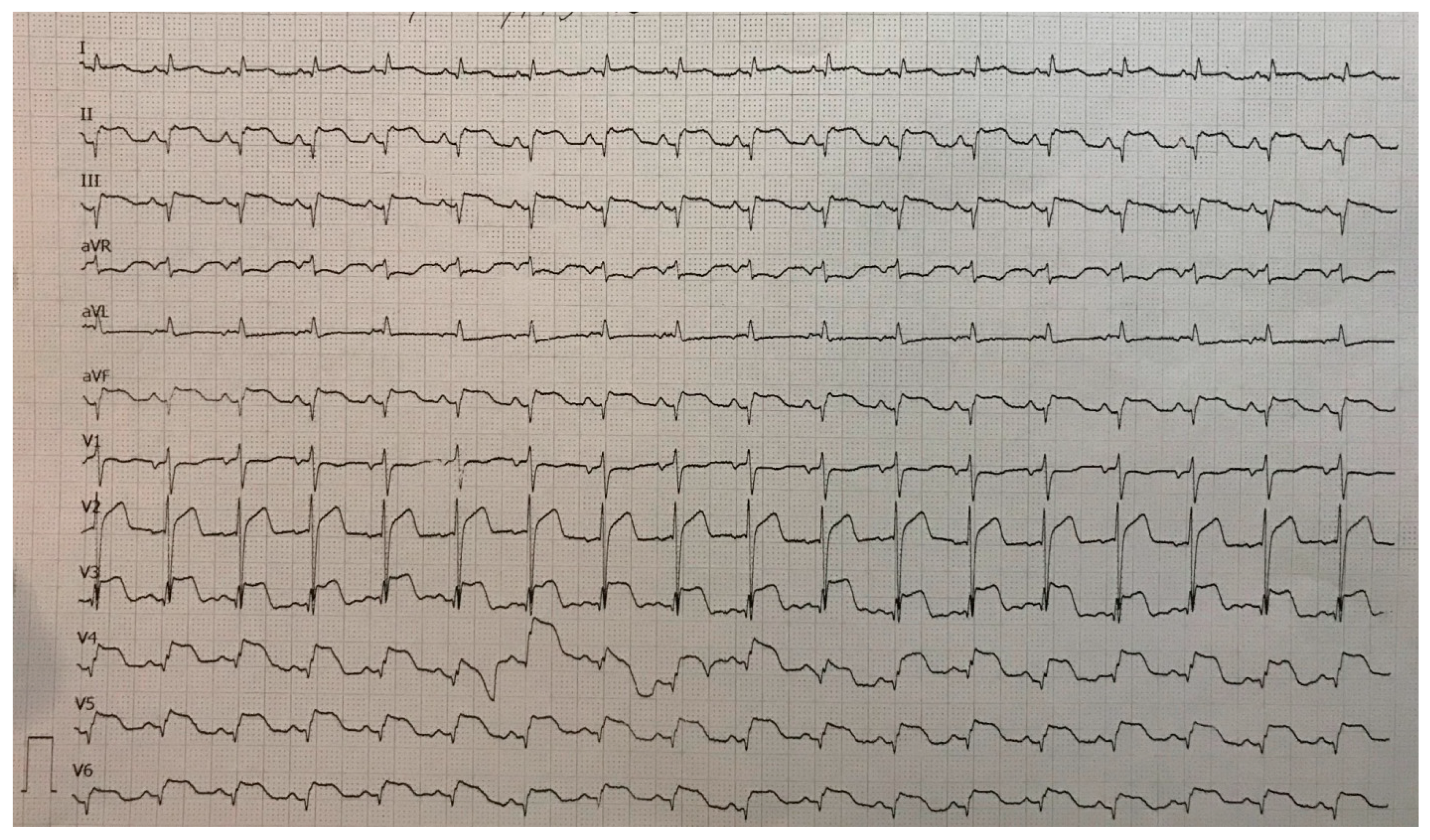
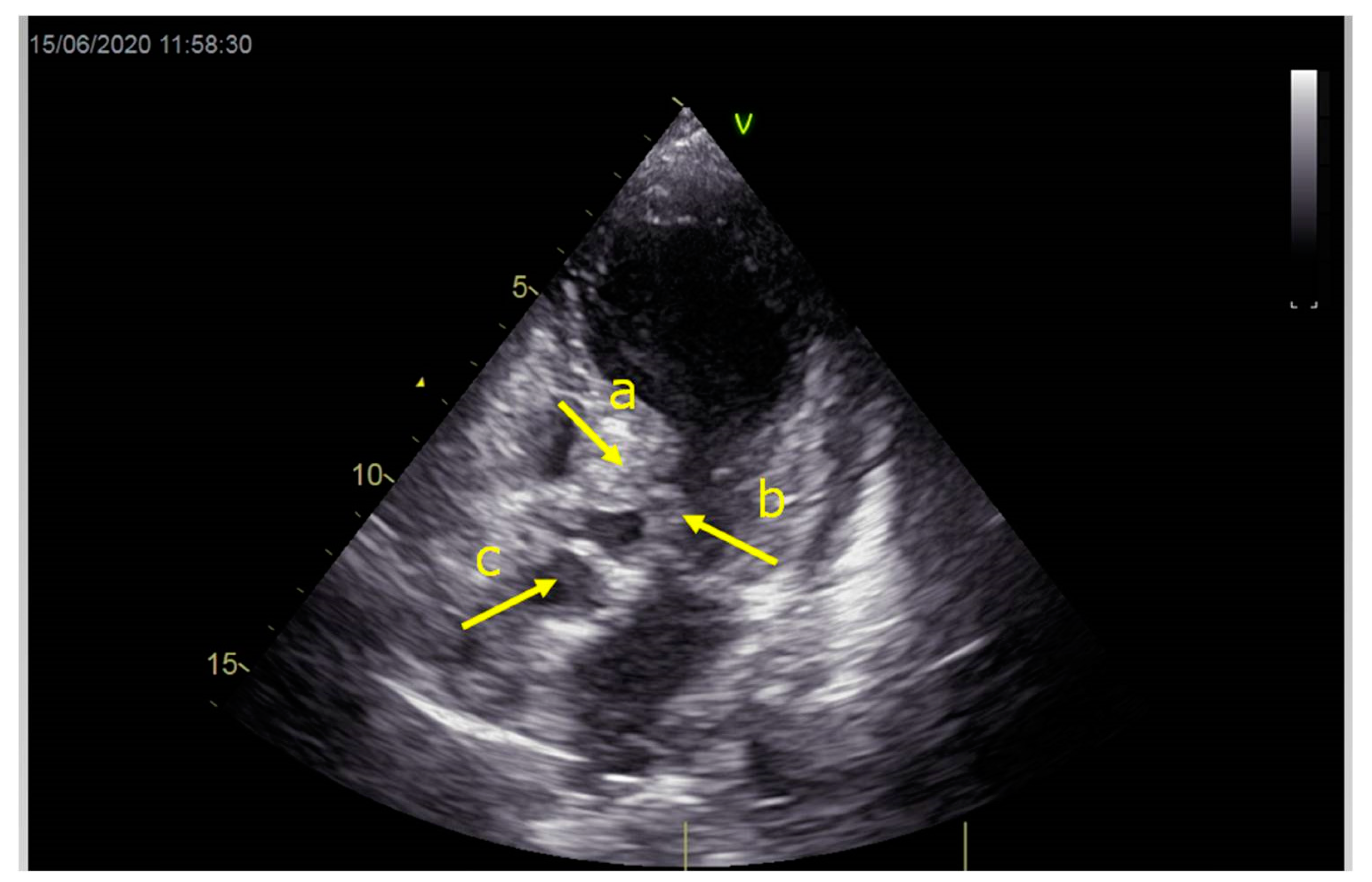
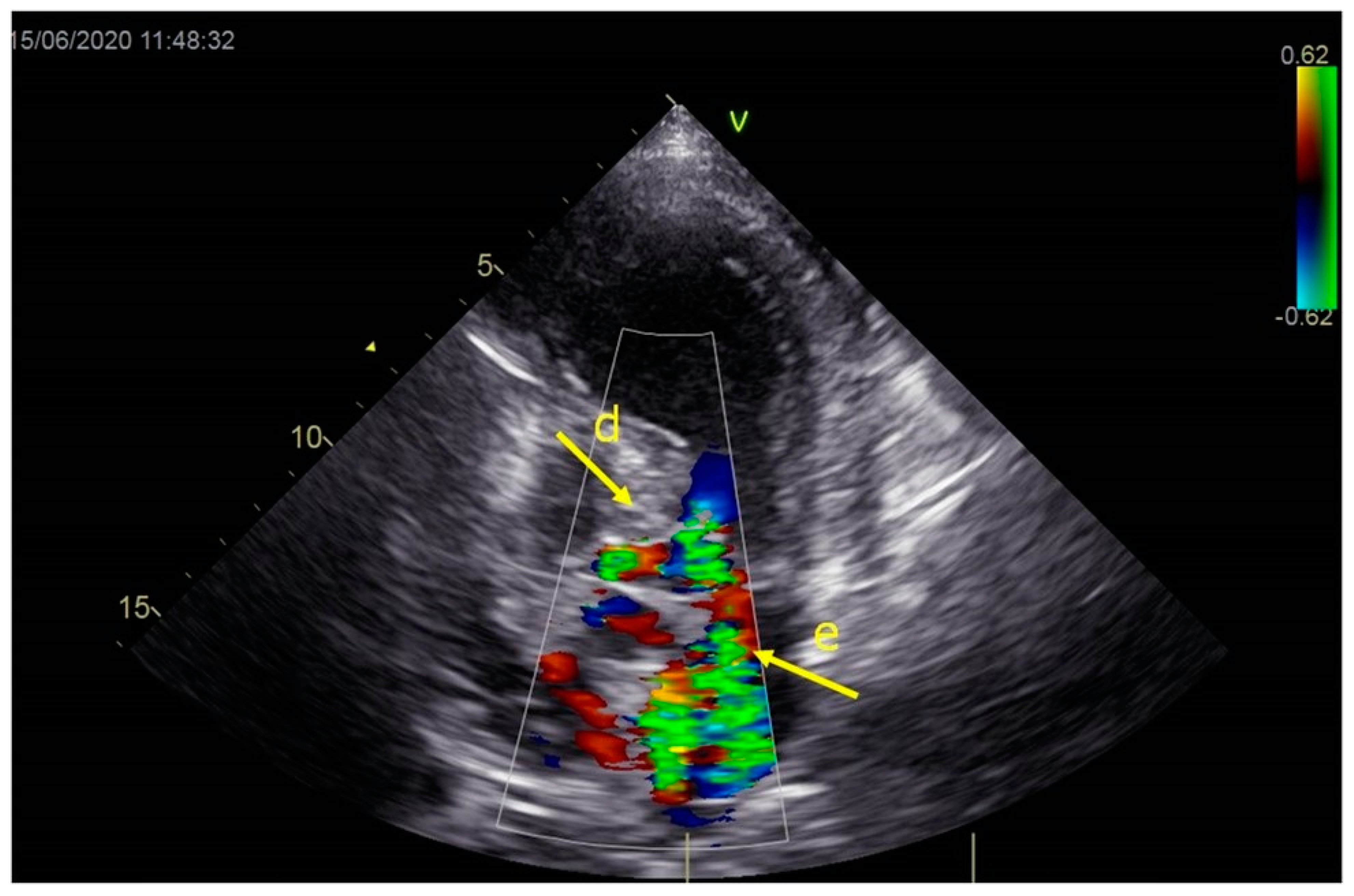
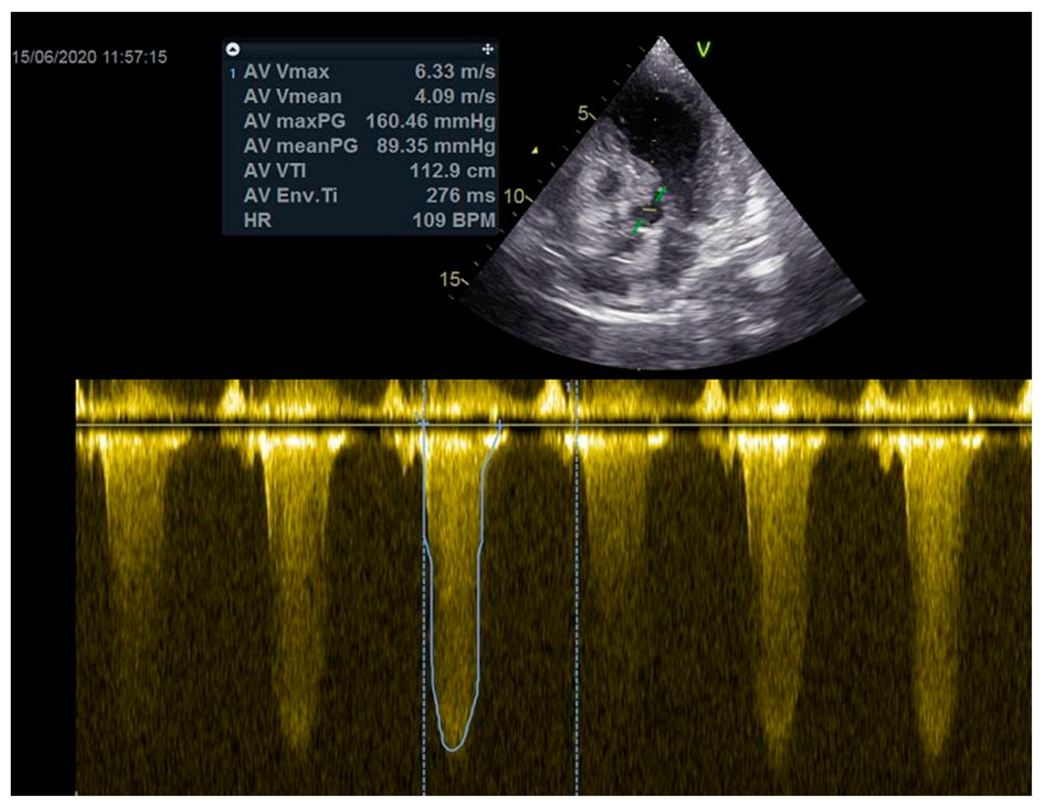
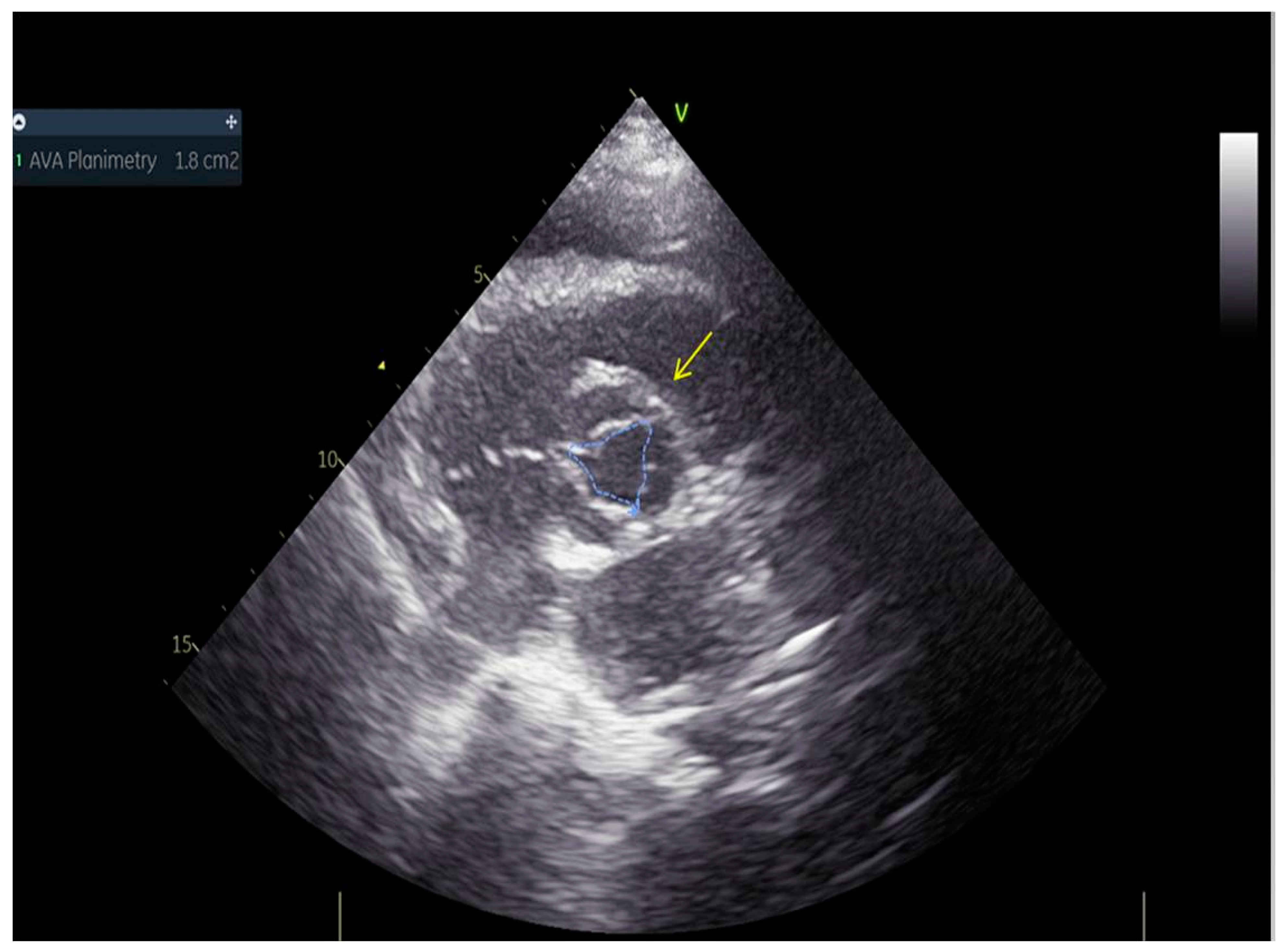
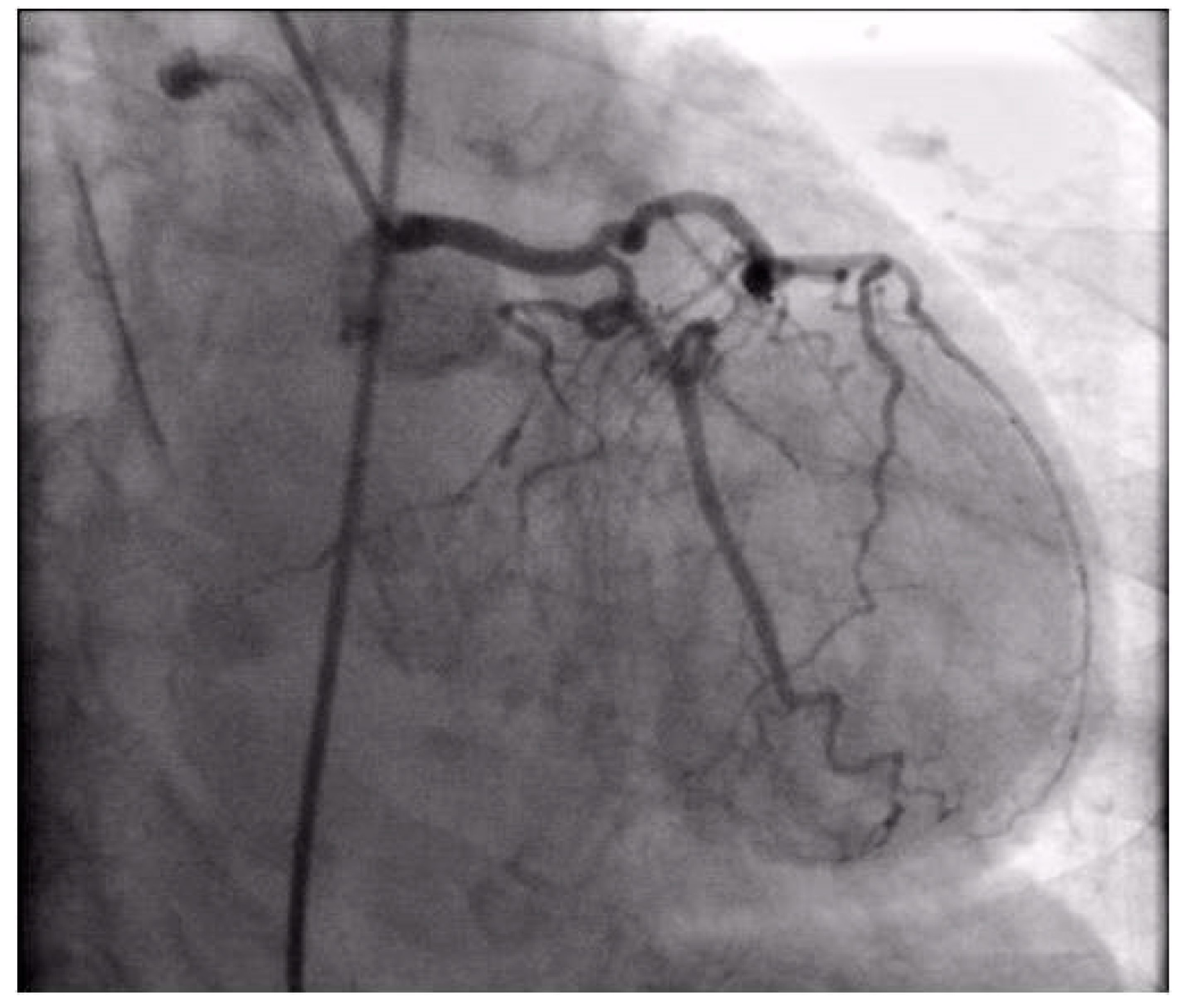
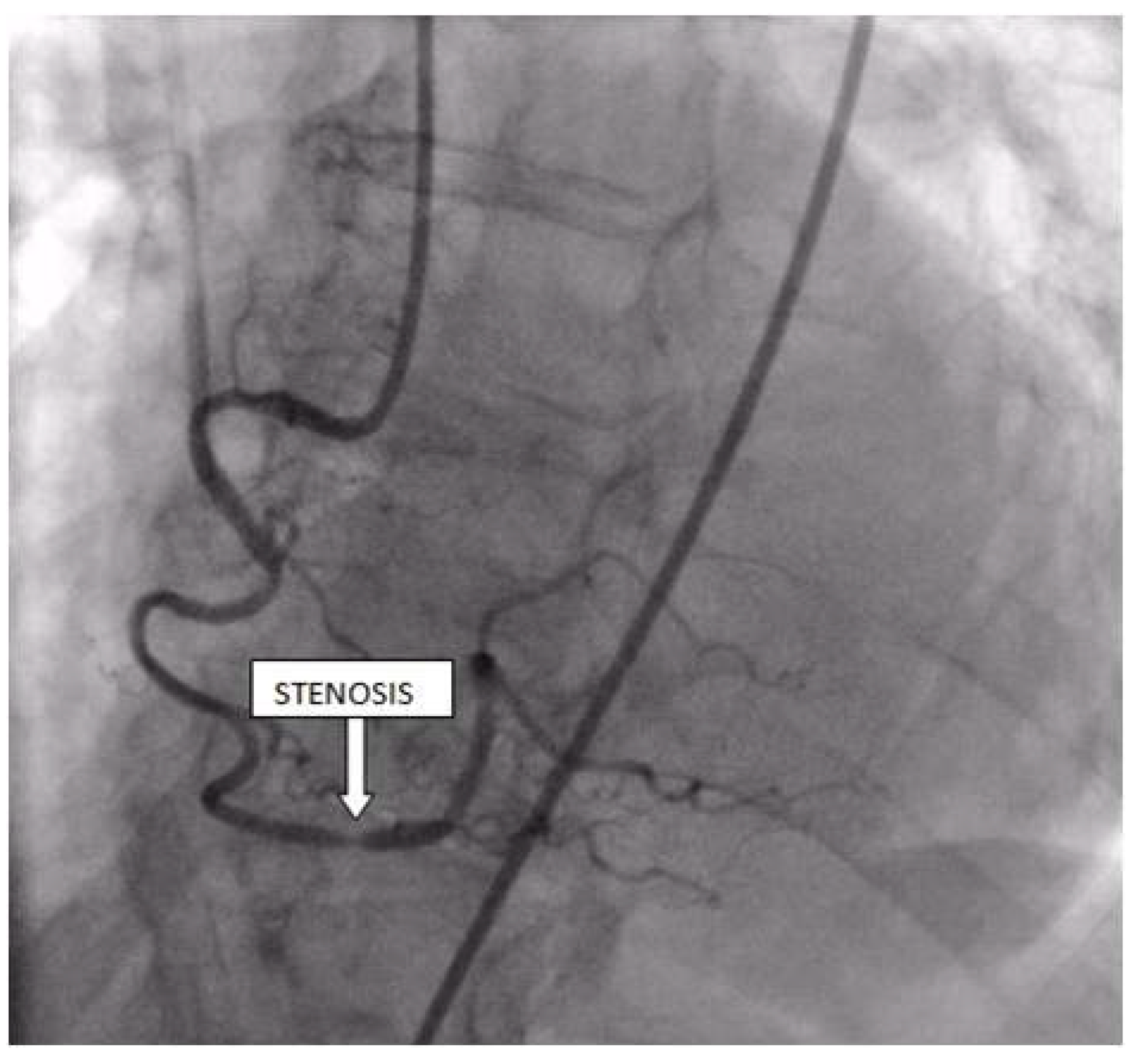
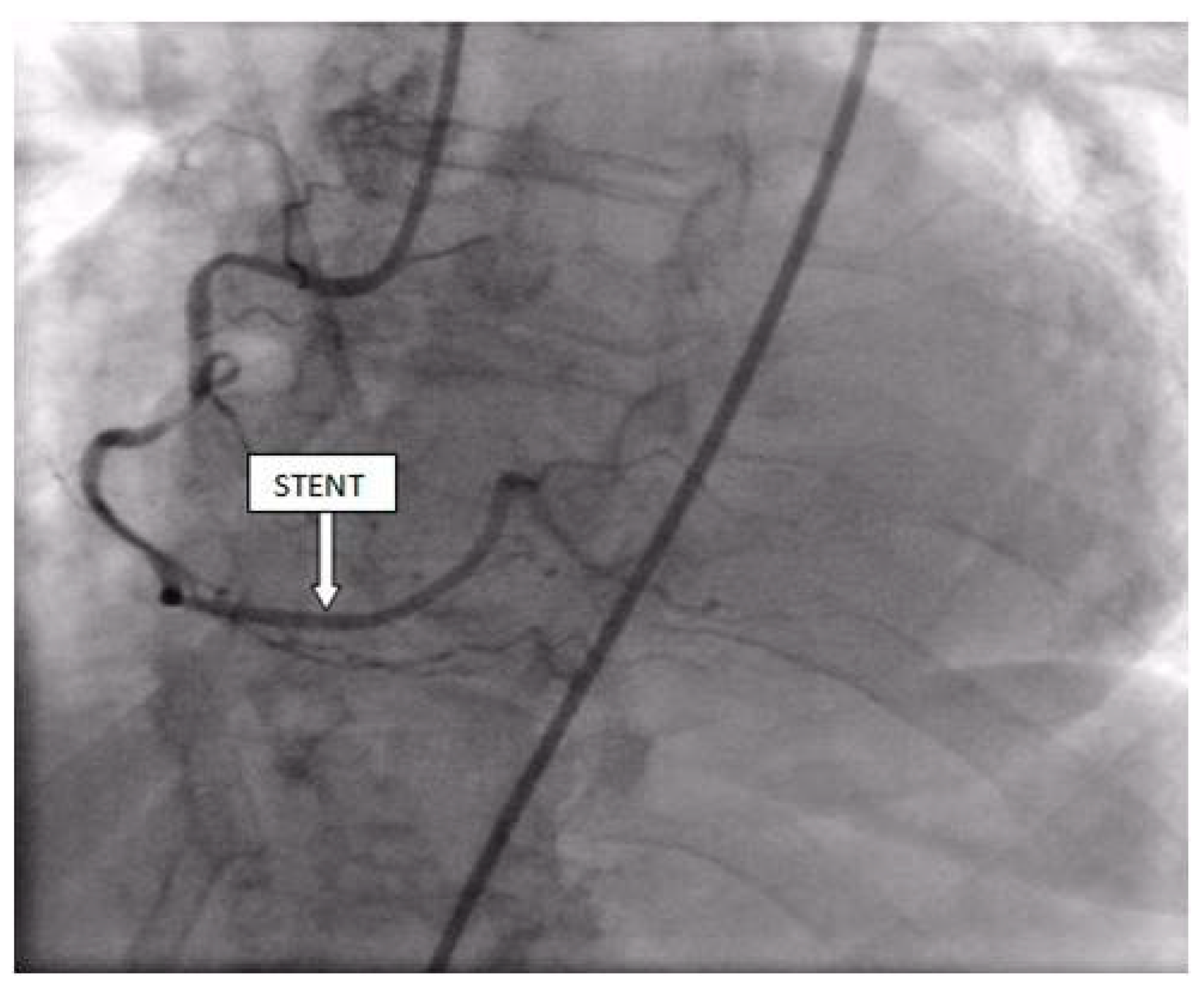
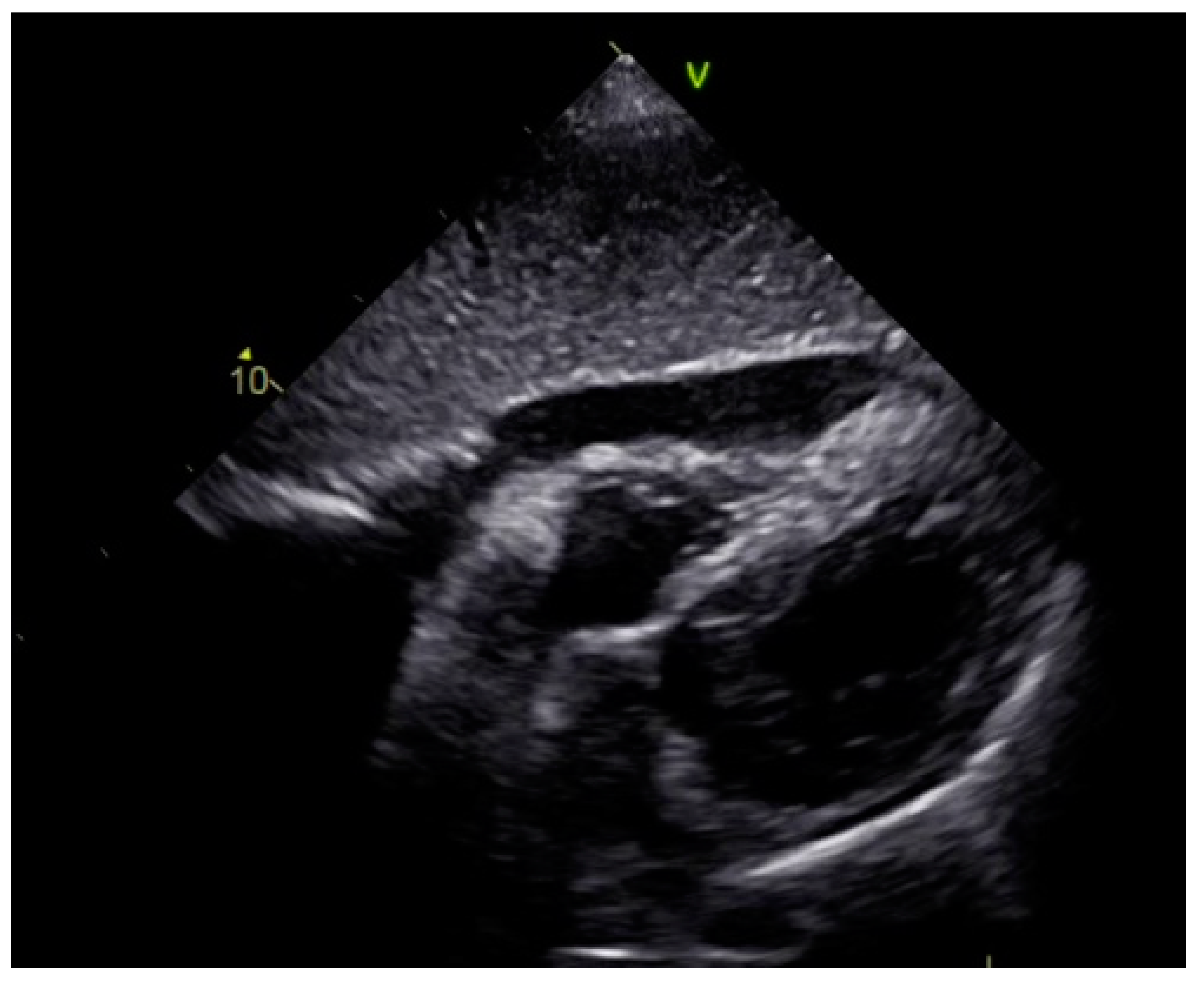
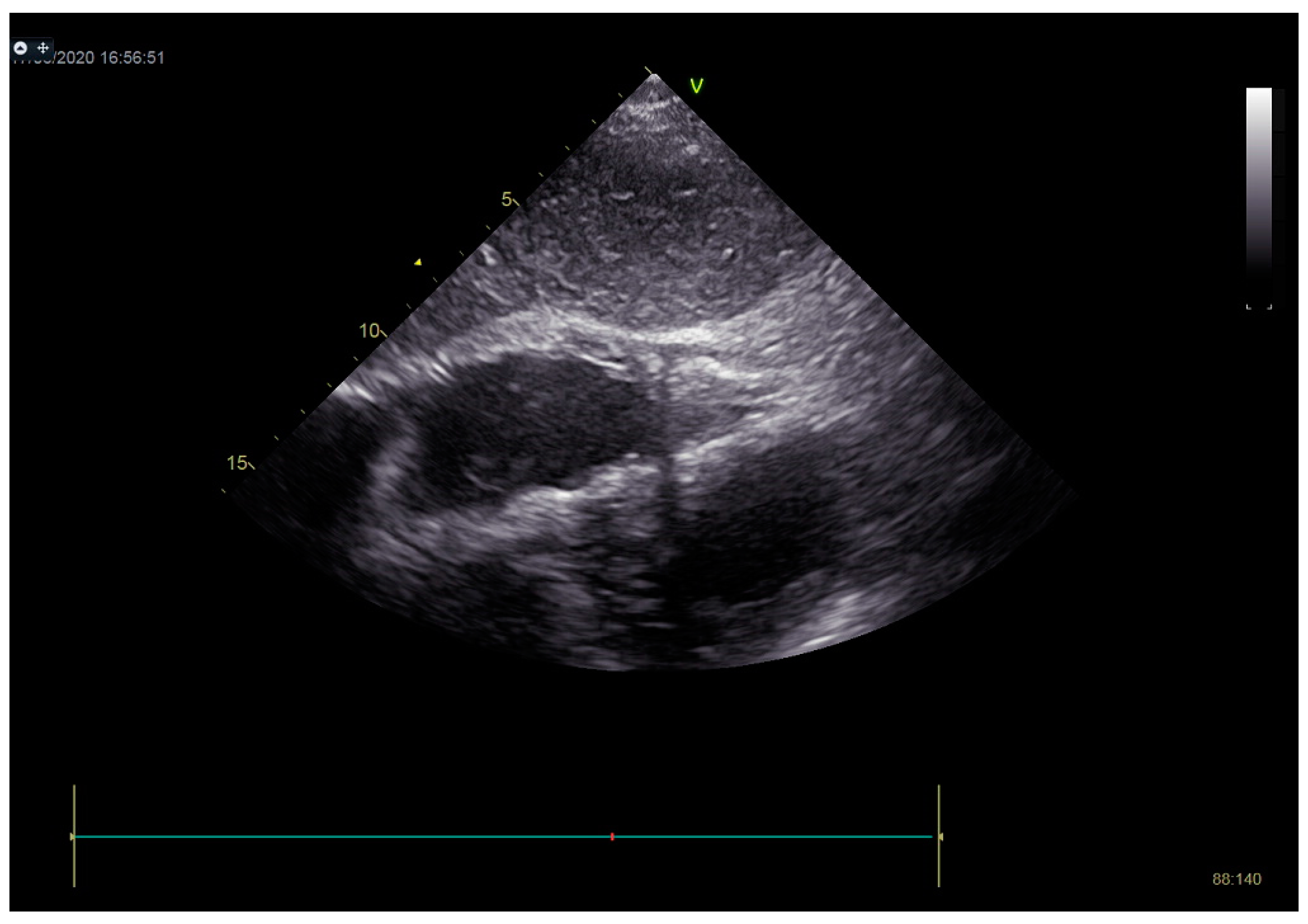
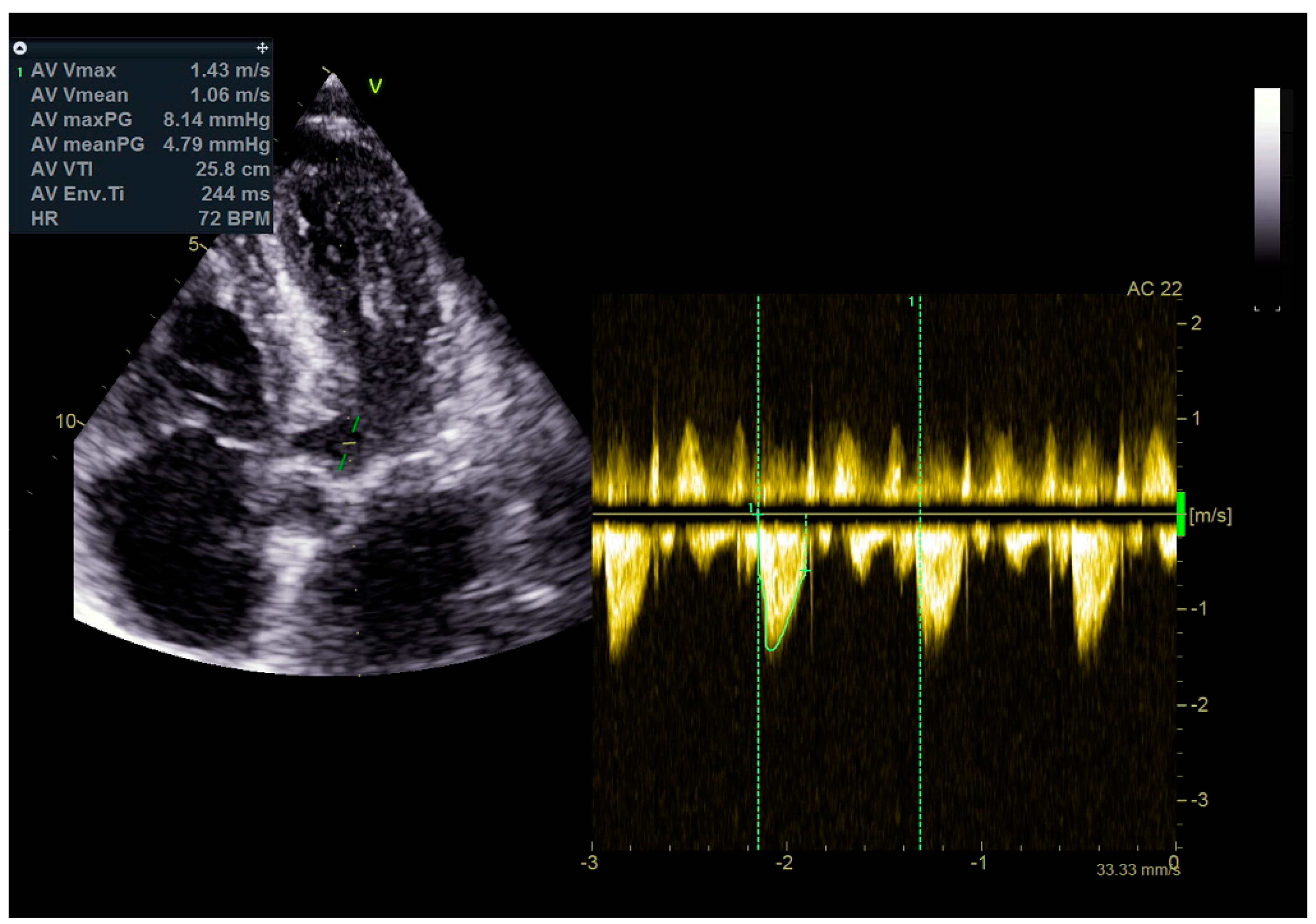
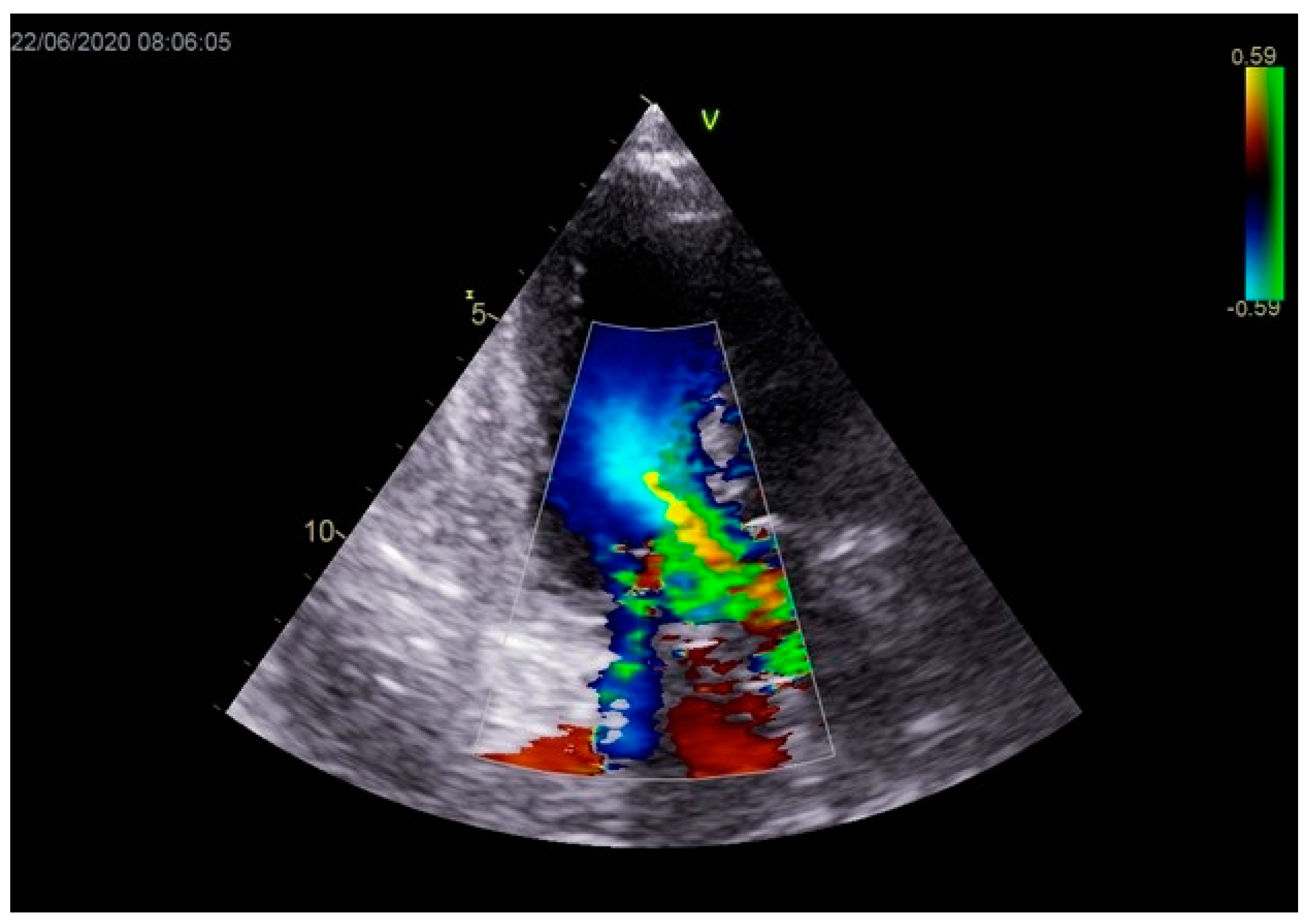
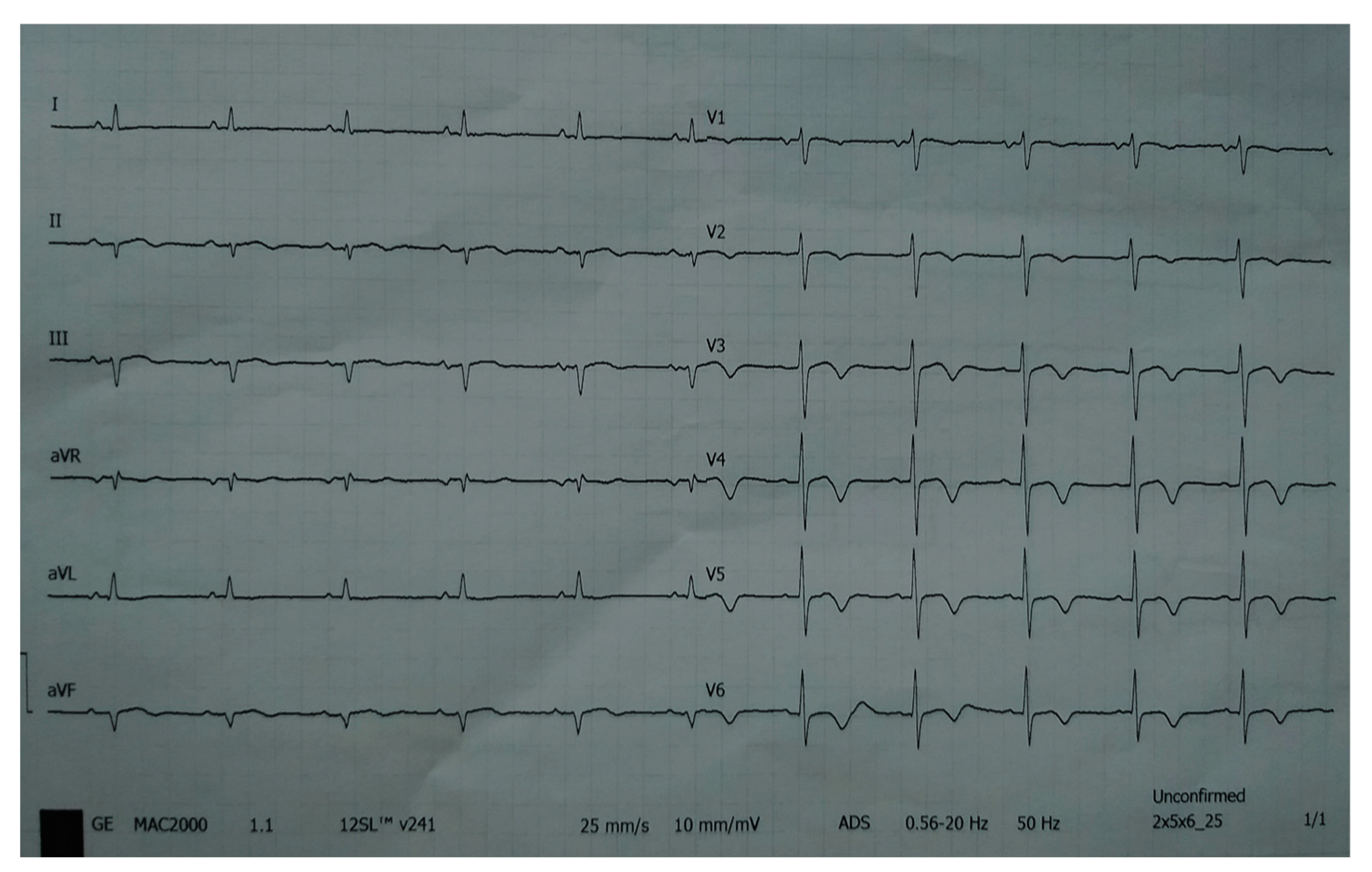
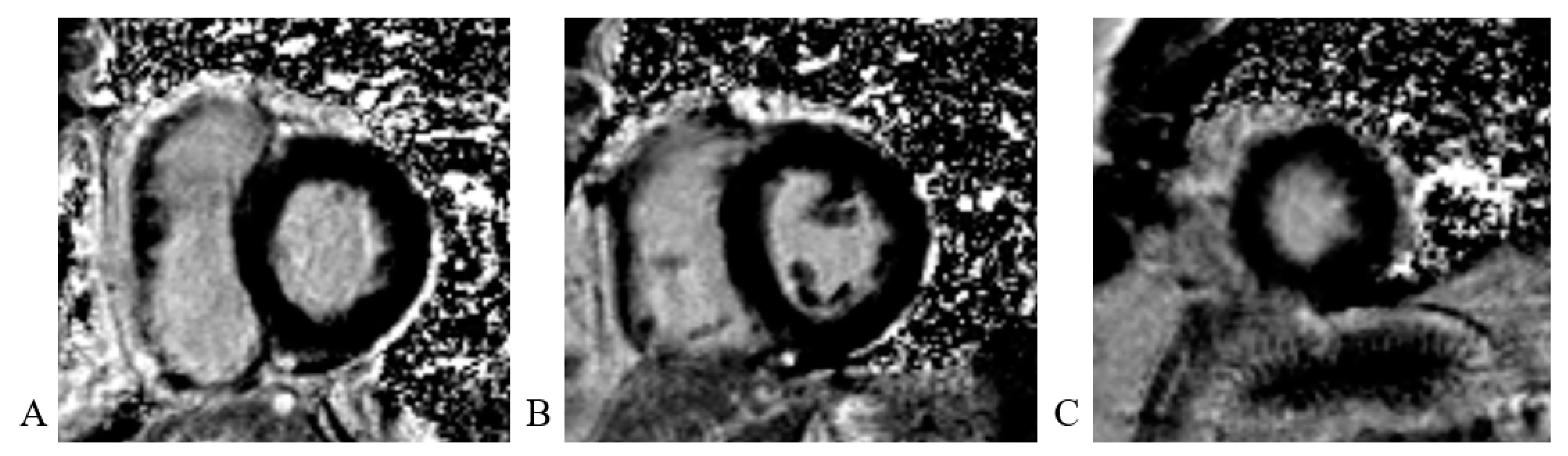
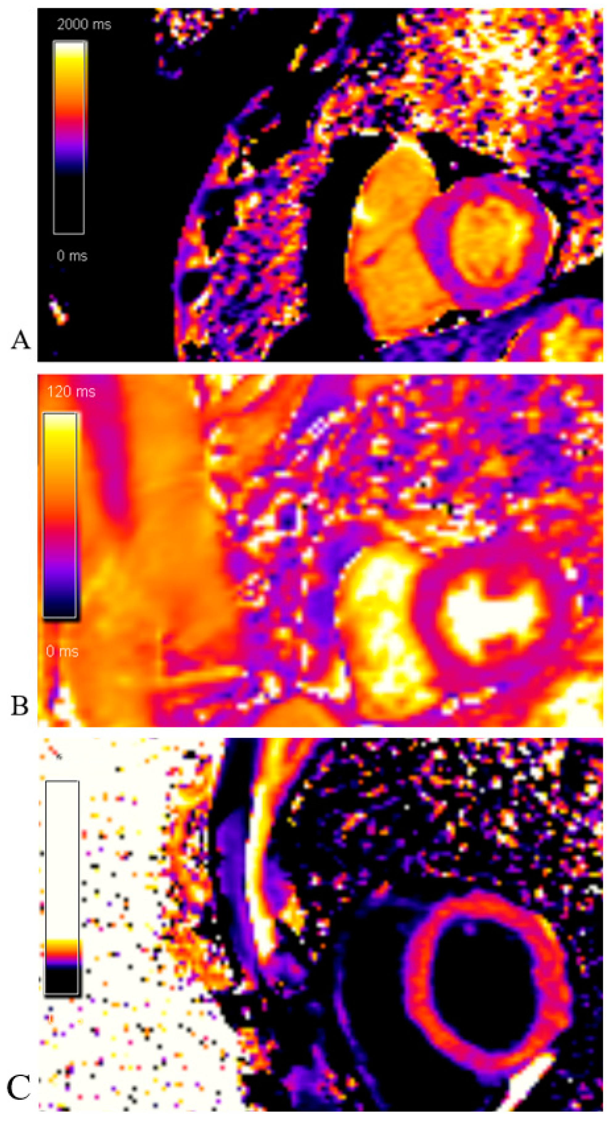
2. Case Report
3. Discussion
Author Contributions
Funding
Institutional Review Board Statement
Informed Consent Statement
Data Availability Statement
Conflicts of Interest
References
- Koeth, O.; Zeymer, U.; Schiele, R.; Zahn, R. Inferior ST elevation myocardial infarction associated with takotsubo cardiomyopathy. Case Rep. Med. 2010, 2, 467867. [Google Scholar] [CrossRef] [PubMed][Green Version]
- Prasad, A.; Dangas, G.; Srinivasan, M.; Yu, J.; Gersh, B.J.; Mehran, R.; Stone, G.W. Incidence and angiographic characteristics of patients with apical ballooning syndrome (takotsubo/stress cardiomyopathy) in the HORIZONS-AMI trial: An analysis from a multicenter, international study of ST-elevation myocardial infarction. Catheter. Cardiovasc. Interv. 2014, 83, 343–348. [Google Scholar] [CrossRef] [PubMed]
- Redfors, B.; Vedad, R.; Angerås, O.; Råmunddal, T.; Petursson, P.; Haraldsson, I.; Omerovic, E. Mortality in Takotsubo syndrome is similar to mortality in myocardial infarction—A report from the SWEDEHEART registry. Int. J. Cardiol. 2015, 185, 282–289. [Google Scholar] [CrossRef]
- Thygesen, K.; Alpert, J.S.; Jaffe, A.S.; Chaitman, B.R.; Bax, J.J.; Morrow, D.A.; White, H.D.; Executive Group on behalf of the Joint European Society of Cardiology (ESC)/American College of Cardiology (ACC)/American Heart Association (AHA)/World Heart Federation (WHF) Task Force for the Universal Definition of Myocardial Infarction. Fourth Universal Definition of Myocardial Infarction (2018). J. Am. Coll. Cardiol. 2018, 72, 2231–2264. [Google Scholar] [PubMed]
- Dullabh, D.; Zachariah, D. Takotsubo cardiomyopathy—An unusual manifestation of the COVID-19 pandemic. Wits J. Clin. Med. 2020, 2, 255–256. [Google Scholar] [CrossRef]
- Kurisu, S.; Inoue, I.; Kawagoe, T.; Ishihara, M.; Shimatani, Y.; Nakama, Y. Prevalence of incidental coronary artery disease in Takotsubo cardiomyopathy. Coron. Artery Dis. 2009, 20, 214–218. [Google Scholar] [CrossRef]
- Redfors, B.; Ramunddal, T.; Shao, Y.; Omerovic, E. Takotsubo triggered by acute myocardial infarction: A common but overlooked syndrome? J. Geriatr. Cardiol. 2014, 11, 171–173. [Google Scholar]
- Okura, H. Echocardiographic assessment of takotsubo cardiomyopathy: Beyond apical ballooning. J. Echocardiogr. 2016, 14, 13–20. [Google Scholar] [CrossRef] [PubMed]
- Lyon, A.R.; Citro, R.; Schneider, B.; Morel, O.; Ghadri, J.R.; Templin, C.; Omerovic, E. Pathophysiology of Takotsubo Syndrome: JACC State-of-the-Art Review. J. Am. Coll. Cardiol. 2021, 77, 902–921. [Google Scholar] [CrossRef]
- Gianni, M.; Dentali, F.; Grandi, A.M.; Sumner, G.; Hiralal, R.; Lonn, E. Apical ballooning syndrome or takotsubo cardiomyopathy: A systematic review. Eur. Heart J. 2006, 27, 1523–1529. [Google Scholar] [CrossRef] [PubMed]
- Wittstein, I.S.; Thiemann, D.R.; Lima, J.A.C.; Baughman, K.L.; Schulman, S.P.; Gerstenblith, G.; Wu, K.C.; Rade, J.J.; Bivalacqua, T.J.; Champion, H.C. Neurohumoral features of myocardial stunning due to sudden emotional stress. N. Engl. J. Med. 2005, 352, 539–548. [Google Scholar] [CrossRef] [PubMed]
- Y-Hassan, S.; Sörensson, P.; Ekenbäck, C.; Lundin, M.; Agewall, S.; Brolin, E.B.; Caidahl, K.; Cederlund, K.; Collste, O.; Daniel, M.; et al. Plasma catecholamine levels in the acute and subacute stages of takotsubo syndrome: Results from the Stockholm myocardial infarction with normal coronaries 2 study. Clin. Cardiol. 2021, 44, 1567–1574. [Google Scholar] [CrossRef]
- Redfors, B.; Shao, Y.; Omerovic, E. Stress-induced cardiomyopathy in the critically ill. Why inotropes fail to improve outcome. Int. J. Cardiol. 2013, 168, 4489–4490. [Google Scholar] [CrossRef] [PubMed]
- Prasad, A.; Lerman, A.; Rihal, C.S. Apical ballooning syndrome (tako-tsubo or stress cardiomyopathy): A mimic of acute myocardial infarction. Am. Heart J. 2008, 155, 408–417. [Google Scholar] [CrossRef]
- Ghadri, J.R.; Wittstein, I.S.; Prasad, A.; Sharkey, S.; Dote, K.; Akashi, Y.J. International Expert Consensus Document on Takotsubo Syndrome (Part I): Clinical characteristics, diagnostic criteria, and pathophysiology. Eur. Heart J. 2018, 39, 2032–2046. [Google Scholar] [CrossRef] [PubMed]
- Okura, H.; Nakada, Y.; Ishihara, S.; Nogi, M.; Onoue, K.; Soeda, T. “Hidden & quot; takotsubo cardiomyopathy in cardiac care unit. J. Echocardiogr. 2020, 18, 113–116. [Google Scholar]
- Citro, R.; Okura, H.; Ghadri, J.R.; Izumi, C.; Meimoun, P.; Izumo, M. Multimodality imaging in takotsubo syndrome: A joint consensus document of the European Association of Cardiovascular Imaging (EACVI) and the Japanese Society of Echocardiography (JSE). J. Echocardiogr. 2020, 18, 199–224. [Google Scholar] [CrossRef]
- Friedrich, M.G.; Sechtem, U.; Schulz-Menger, J.; Fausto, J.P.; Brito, D. Cardio-vascular magnetic resonance in myocarditis: A JACC White Paper. J. Am. Coll. Cardiol. 2009, 53, 1475–1487. [Google Scholar] [CrossRef]
- Ghadri, J.R.; Cammann, V.L.; Jurisic, S.; Seifert, B.; Napp, L.C.; Diekmann, J. A novel clinical score (InterTAK Diagnostic Score) to differentiate takotsubo syndrome from acute coronary syndrome: Results from the International Takotsubo Registry. Eur. J. Heart Fail. 2017, 19, 1036–1042. [Google Scholar] [CrossRef]
- Santoro, F.; Ieva, R.; Ferraretti, A.; Ienco, V.; Carpagnano, G.; Lodispoto, M.; Di Biase, L.; Di Biase, M.; Brunetti, N.D. Safety and feasibility of levosimendan administration in Takotsubo cardiomyopathy: A case series. Cardiovasc. Ther. 2013, 31, e133–e137. [Google Scholar] [CrossRef]
- Meyer, P.; Degrauwe, S.; Van Delden, C.; Ghadri, J.R.; Templin, C. Typical takotsubo syndrome triggered by SARS-CoV-2 infection. Eur. Heart J. 2020, 41, 1860. [Google Scholar] [CrossRef] [PubMed]
- Aldujeli, A.; Hamadeh, A.; Briedis, K.; Tecson, K.M.; Rutland, J.; Krivickas, Z.; Stiklioraitis, S.; Briede, K.; Aldujeili, M.; Unikas, R.; et al. Delays in presentation in patients with acute myocardial infarction during the COVID-19 pandemic. Cardiol. Res. 2020, 11, 386–391. [Google Scholar] [CrossRef] [PubMed]


Disclaimer/Publisher’s Note: The statements, opinions and data contained in all publications are solely those of the individual author(s) and contributor(s) and not of MDPI and/or the editor(s). MDPI and/or the editor(s) disclaim responsibility for any injury to people or property resulting from any ideas, methods, instructions or products referred to in the content. |
© 2023 by the authors. Licensee MDPI, Basel, Switzerland. This article is an open access article distributed under the terms and conditions of the Creative Commons Attribution (CC BY) license (https://creativecommons.org/licenses/by/4.0/).
Share and Cite
Srdanović, I.; Dabović, D.; Ivanović, V.; Čanković, M.; Pantić, T.; Stefanović, M.; Dimić, S.; Crnomarković, B.; Bjelobrk, M.; Govedarica, M.; et al. Takotsubo Cardiomyopathy Occurring Simultaneously with Acute Myocardial Infarction. Life 2023, 13, 1770. https://doi.org/10.3390/life13081770
Srdanović I, Dabović D, Ivanović V, Čanković M, Pantić T, Stefanović M, Dimić S, Crnomarković B, Bjelobrk M, Govedarica M, et al. Takotsubo Cardiomyopathy Occurring Simultaneously with Acute Myocardial Infarction. Life. 2023; 13(8):1770. https://doi.org/10.3390/life13081770
Chicago/Turabian StyleSrdanović, Ilija, Dragana Dabović, Vladimir Ivanović, Milenko Čanković, Teodora Pantić, Maja Stefanović, Sonja Dimić, Branislav Crnomarković, Marija Bjelobrk, Miljana Govedarica, and et al. 2023. "Takotsubo Cardiomyopathy Occurring Simultaneously with Acute Myocardial Infarction" Life 13, no. 8: 1770. https://doi.org/10.3390/life13081770
APA StyleSrdanović, I., Dabović, D., Ivanović, V., Čanković, M., Pantić, T., Stefanović, M., Dimić, S., Crnomarković, B., Bjelobrk, M., Govedarica, M., & Zdravković, M. (2023). Takotsubo Cardiomyopathy Occurring Simultaneously with Acute Myocardial Infarction. Life, 13(8), 1770. https://doi.org/10.3390/life13081770