
The Late Effects of Cancer Treatment on Female Fertility and the Current Status of Fertility Preservation—A Narrative Review


Abstract
1. Introduction
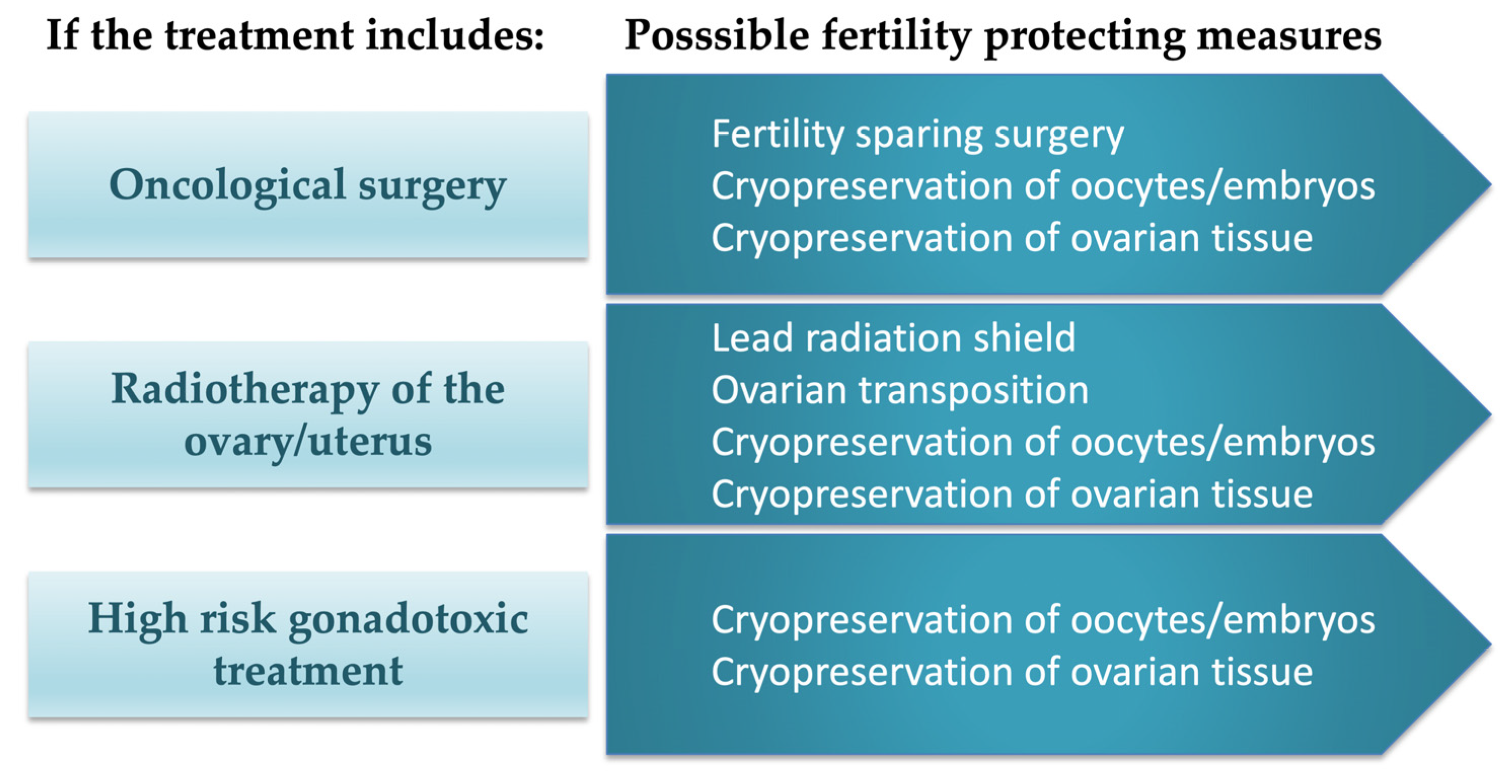
Indications for Fertility Preservation
2. Protecting the Ovarian Reserve In Vivo
GnRH Analogues
3. Fertility Preservation as an Acute Measure
3.1. Cryopreservation of Oocytes and Embryos
3.2. Hormone-Sensitive Breast Cancer
3.3. The BRCA Mutation
3.4. Cryopreservation of Ovarian Tissue for Later Re-Transplantation
3.5. In Vitro Maturation
4. Quality of Life and Late Effects
4.1. Premature Ovarian Insufficiency
4.2. Sexual Function
4.3. Psychological Effects
4.4. Pregnancy after Cancer
5. Future Developments of Fertility Preservation
5.1. In Vitro Activation and Maturation of Primordial Follicles
5.2. Protecting Ovarian Tissue In Vitro
5.3. Stem Cells
6. Summary
Author Contributions
Funding
Institutional Review Board Statement
Informed Consent Statement
Data Availability Statement
Conflicts of Interest
References
- You, L.; Lv, Z.; Li, C.; Ye, W.; Zhou, Y.; Jin, J.; Han, Q. Worldwide cancer statistics of adolescents and young adults in 2019: A systematic analysis of the Global Burden of Disease Study 2019. ESMO Open 2021, 6, 100255. [Google Scholar] [CrossRef] [PubMed]
- SIGN. Long Term Follow Up of Survivors of Childhood Cancer A National Clinical Guideline. Available online: http://wwwsignacuk/pdf/sign132pdf (accessed on 21 February 2022).
- van der Meer, D.J.; Karim-Kos, H.E.; van der Mark, M.; Aben, K.K.H.; Bijlsma, R.M.; Rijneveld, A.W.; van der Graaf, W.T.A.; Husson, O. Incidence, Survival, and Mortality Trends of Cancers Diagnosed in Adolescents and Young Adults (15–39 Years): A Population-Based Study in The Netherlands 1990–2016. Cancers 2020, 12, 3421. [Google Scholar] [CrossRef] [PubMed]
- Rosen, A.; Rodriguez-Wallberg, K.A.; Rosenzweig, L. Psychosocial Distress in Young Cancer Survivors. Semin. Oncol. Nurs. 2009, 25, 268–277. [Google Scholar] [CrossRef]
- Howard-Anderson, J.; Ganz, P.A.; Bower, J.E.; Stanton, A.L. Quality of Life, Fertility Concerns, and Behavioral Health Outcomes in Younger Breast Cancer Survivors: A Systematic Review. Gynecol. Oncol. 2012, 104, 386–405. [Google Scholar] [CrossRef] [PubMed]
- Rodriguez-Wallberg, K.A.; Borgström, B.; Petersen, C.; Thurin-Kjellberg, A.; Mörse, H.; Giwercman, A.; Jarfelt, M.; Work Group UNGA (YOUNG) for the Swedish Association of Local Authorities and Regions, SALAR (Sveriges Kommuner och Landsting, SKL). National guidelines and multilingual age-adapted patient brochures and videos as decision aids for fertility preservation (FP) of children and teenagers with cancer-A multidisciplinary effort to improve children’s information and access to FP in Sweden. Acta Obstet. Gynecol. Scand. 2019, 98, 679–680. [Google Scholar] [CrossRef] [PubMed]
- ESHRE Guideline Female Fertility Preservation. Available online: https://www.eshre.eu/FFPguideline (accessed on 3 March 2023).
- ESHRE Guideline Group on Female Fertility Preservation; Anderson, R.A.; Amant, F.; Braat, D.; D’Angelo, A.; Chuva de Sousa Lopes, S.M.; Demeestere, I.; Dwek, S.; Frith, L.; Lambertini, M.; et al. ESHRE guideline: Female fertility preservation. Hum. Reprod. Open 2020, 2020, hoaa052. [Google Scholar] [CrossRef]
- Oktay, K.; Harvey, B.; Partridge, A.H.; Quinn, G.; Reinecke, J.; Taylor, H.S.; Wallace, W.H.; Wang, E.T.; Loren, A.W. Fertility Preservation in Patients with Cancer: ASCO Clinical Practice Guideline Update. J. Clin. Oncol. 2018, 36, 1994–2001. [Google Scholar] [CrossRef]
- Lambertini, M.; Peccatori, F.; Demeestere, I.; Amant, F.; Wyns, C.; Stukenborg, J.-B.; Paluch-Shimon, S.; Halaska, M.; Uzan, C.; Meissner, J.; et al. Fertility preservation and post-treatment pregnancies in post-pubertal cancer patients: ESMO Clinical Practice Guidelines. Ann. Oncol. 2020, 31, 1664–1678. [Google Scholar] [CrossRef]
- Kieu, V.; Stern, C.; Harris, J.; Jayasinghe, Y.; Bradford, N.; Cui, W.; Deans, R.; Hunter, T.; Allingham, C.; Kane, S.C.; et al. Australian fertility preservation guidelines for people with cancer 2022: Review and recommendations. Med. J. Aust. 2022, 217, 564–569. [Google Scholar] [CrossRef]
- Rodriguez-Wallberg, K.A. Principles of cancer treatment: Impact on reproduction. Adv. Exp. Med. Biol. 2012, 732, 1–8. [Google Scholar] [CrossRef]
- Duffy, C.; Allen, S. Medical and Psychosocial Aspects of Fertility after Cancer. Cancer J. 2009, 15, 27–33. [Google Scholar] [CrossRef] [PubMed]
- Oktay, K.; Rodriguez-Wallberg, K. Fertility preservation during cancer treatment: Clinical guidelines. Cancer Manag. Res. 2014, 6, 105–117. [Google Scholar] [CrossRef] [PubMed]
- Aznar, J.; Martínez Peris, M. Gestational Surrogacy: Current View. Linacre Q. 2019, 86, 56–67. [Google Scholar] [CrossRef]
- Smietana, M.; Rudrappa, S.; Weis, C. Moral frameworks of commercial surrogacy within the US, India and Russia. Sex. Reprod. Health Matters 2021, 29, 377–393. [Google Scholar] [CrossRef]
- Jones, B.P.; Ranaei-Zamani, N.; Vali, S.; Williams, N.; Saso, S.; Thum, M.; Al-Memar, M.; Dixon, N.; Rose, G.; Testa, G.; et al. Options for acquiring motherhood in absolute uterine factor infertility; adoption, surrogacy and uterine transplantation. Obstet. Gynaecol. 2021, 23, 138–147. [Google Scholar] [CrossRef] [PubMed]
- Wu, C.; Wu, T.; Chen, D.; Wei, S.; Tang, W.; Xue, L.; Xiong, J.; Huang, Y.; Guo, Y.; Chen, Y.; et al. The effects and mechanism of taxanes on chemotherapy-associated ovarian damage: A review of current evidence. Front. Endocrinol. 2022, 13, 1025018. [Google Scholar] [CrossRef]
- Hao, X.; Anastácio, A.; Liu, K.; Rodriguez-Wallberg, K.A. Ovarian Follicle Depletion Induced by Chemotherapy and the Investigational Stages of Potential Fertility-Protective Treatments—A Review. Int. J. Mol. Sci. 2019, 20, 4720. [Google Scholar] [CrossRef]
- Zhao, J.; Liu, J.; Chen, K.; Li, S.; Wang, Y.; Yang, Y.; Deng, H.; Jia, W.; Rao, N.; Liu, Q.; et al. What lies behind chemotherapy-induced amenorrhea for breast cancer patients: A meta-analysis. Breast Cancer Res. Treat. 2014, 145, 113–128. [Google Scholar] [CrossRef]
- Oktem, O.; Oktay, K. A Novel Ovarian Xenografting Model to Characterize the Impact of Chemotherapy Agents on Human Primordial Follicle Reserve. Cancer Res 2007, 67, 10159–10162. [Google Scholar] [CrossRef]
- Lloyd, M.; Carr, M.; McElhatton, P.; Hall, G.; Hughes, R. The effects of methotrexate on pregnancy, fertility and lactation. Qjm Int. J. Med. 1999, 92, 551–563. [Google Scholar] [CrossRef]
- Lambertini, M.; Ceppi, M.; Anderson, R.A.; Cameron, D.A.; Bruzzone, M.; Franzoi, M.A.; Massarotti, C.; El-Abed, S.; Wang, Y.; Lecocq, C.; et al. Impact of Anti-HER2 Therapy Alone and with Weekly Paclitaxel on the Ovarian Reserve of Young Women with HER2-Positive Breast Cancer. J. Natl. Compr. Cancer Netw. 2023, 21, 33–41.e16. [Google Scholar] [CrossRef] [PubMed]
- Rodriguez-Wallberg, K.A.; Oktay, K. Fertility Preservation and Pregnancy in Women with and Without BRCA Mutation–Positive Breast Cancer. Oncologist 2012, 17, 1409–1417. [Google Scholar] [CrossRef] [PubMed]
- van Wagenen, G.; Simpson, M.E. Embryology of the Ovary and Testis. In Homo Sapiens and Macaca Mulatta; Yale University Press: New Haven, CT, USA, 1965. [Google Scholar]
- Barnett, K.R.; Schilling, C.; Greenfeld, C.R.; Tomic, D.; Flaws, J.A. Ovarian follicle development and transgenic mouse models. Hum. Reprod. Updat. 2006, 12, 537–555. [Google Scholar] [CrossRef]
- Fortune, J.E.; Yang, M.Y.; Muruvi, W. The earliest stages of follicular development: Follicle formation and activation. Reprod. Fertil. 2010, 67, 203–216. [Google Scholar] [CrossRef]
- Winship, A.L.; Stringer, J.M.; Liew, S.H.; Hutt, K.J. The importance of DNA repair for maintaining oocyte quality in response to anti-cancer treatments, environmental toxins and maternal ageing. Hum. Reprod. Updat. 2018, 24, 119–134. [Google Scholar] [CrossRef] [PubMed]
- Bedoschi, G.; Navarro, P.A.; Oktay, K. Chemotherapy-induced damage to ovary: Mechanisms and clinical impact. Futur. Oncol. 2016, 12, 2333–2344. [Google Scholar] [CrossRef]
- Dolmans, M.-M.; Taylor, H.S.; Rodriguez-Wallberg, K.A.; Blumenfeld, Z.; Lambertini, M.; von Wolff, M.; Donnez, J. Utility of gonadotropin-releasing hormone agonists for fertility preservation in women receiving chemotherapy: Pros and cons. Fertil. Steril. 2020, 114, 725–738. [Google Scholar] [CrossRef]
- Lambertini, M.; Moore, H.C.; Leonard, R.C.; Loibl, S.; Munster, P.; Bruzzone, M.; Boni, L.; Unger, J.M.; Anderson, R.A.; Mehta, K.; et al. Gonadotropin-Releasing Hormone Agonists during Chemotherapy for Preservation of Ovarian Function and Fertility in Premenopausal Patients with Early Breast Cancer: A Systematic Review and Meta-Analysis of Individual Patient–Level Data. J. Clin. Oncol. 2018, 36, 1981–1990. [Google Scholar] [CrossRef]
- Leonard, R.C.F.; Adamson, D.J.A.; Bertelli, G.; Mansi, J.; Yellowlees, A.; Dunlop, J.; Thomas, G.A.; Coleman, R.E.; Anderson, R.A.; Anglo Celtic Collaborative Oncology Group and National Cancer Research Institute Trialists. GnRH agonist for protection against ovarian toxicity during chemotherapy for early breast cancer: The Anglo Celtic Group OPTION trial. Ann. Oncol. 2017, 28, 1811–1816. [Google Scholar] [CrossRef]
- Gerber, B.; Von Minckwitz, G.; Stehle, H.; Reimer, T.; Felberbaum, R.; Maass, N.; Fischer, D.; Sommer, H.L.; Conrad, B.; Ortmann, O.; et al. Effect of Luteinizing Hormone–Releasing Hormone Agonist on Ovarian Function after Modern Adjuvant Breast Cancer Chemotherapy: The GBG 37 ZORO Study. J. Clin. Oncol. 2011, 29, 2334–2341. [Google Scholar] [CrossRef]
- Elgindy, E.; Sibai, H.; Abdelghani, A.; Mostafa, M. Protecting Ovaries during Chemotherapy through Gonad Suppression: A Systematic Review and Meta-analysis. Obstet. Gynecol. 2015, 126, 187–195. [Google Scholar] [CrossRef] [PubMed]
- Demeestere, I.; Brice, P.; Peccatori, F.; Kentos, A.; Dupuis, J.J.; Zachee, P.; Casasnovas, R.-O.; Van Den Neste, E.; DeChene, J.; De Maertelaer, V.; et al. No Evidence for the Benefit of Gonadotropin-Releasing Hormone Agonist in Preserving Ovarian Function and Fertility in Lymphoma Survivors Treated with Chemotherapy: Final Long-Term Report of a Prospective Randomized Trial. J. Clin. Oncol. 2016, 34, 2568–2574. [Google Scholar] [CrossRef] [PubMed]
- Calhaz-Jorge, C.; De Geyter, C.H.; Kupka, M.S.; Wyns, C.; Mocanu, E.; Motrenko, T.; Scaravelli, G.; Smeenk, J.; Vidakovic, S.; Goossens, V. Survey on ART and IUI: Legislation, regulation, funding and registries in European countries: The European IVF-monitoring Consortium (EIM) for the European Society of Human Reproduction and Embryology (ESHRE). Hum. Reprod. Open 2020, 2020, hoz044. [Google Scholar] [CrossRef]
- Blondeaux, E.; Massarotti, C.; Fontana, V.; Poggio, F.; Arecco, L.; Fregatti, P.; Bighin, C.; Giannubilo, I.; Ruelle, T.; Razeti, M.G.; et al. The PREgnancy and FERtility (PREFER) Study Investigating the Need for Ovarian Function and/or Fertility Preservation Strategies in Premenopausal Women with Early Breast Cancer. Front. Oncol. 2021, 11, 690320. [Google Scholar] [CrossRef] [PubMed]
- Rodriguez-Wallberg, K.A.; Eloranta, S.; Krawiec, K.; Lissmats, A.; Bergh, J.; Liljegren, A. Safety of fertility preservation in breast cancer patients in a register-based matched cohort study. Breast Cancer Res. Treat. 2018, 167, 761–769. [Google Scholar] [CrossRef]
- Rodriguez-Wallberg, K.A.; Tanbo, T.; Tinkanen, H.; Thurin-Kjellberg, A.; Nedstrand, E.; Kitlinski, M.L.; Macklon, K.T.; Ernst, E.; Fedder, J.; Tiitinen, A.; et al. Ovarian tissue cryopreservation and transplantation among alternatives for fertility preservation in the Nordic countries—Compilation of 20 years of multicenter experience. Acta Obstet. Gynecol. Scand. 2016, 95, 1015–1026. [Google Scholar] [CrossRef]
- Rodriguez-Wallberg, K.A.; Marklund, A.; Lundberg, F.; Wikander, I.; Milenkovic, M.; Anastacio, A.; Sergouniotis, F.; Wånggren, K.; Ekengren, J.; Lind, T.; et al. A prospective study of women and girls undergoing fertility preservation due to oncologic and non-oncologic indications in Sweden–Trends in patients’ choices and benefit of the chosen methods after long-term follow up. Acta Obstet. Gynecol. Scand. 2019, 98, 604–615. [Google Scholar] [CrossRef]
- Rodriguez-Wallberg, K.A.; Anastacio, A.; Vonheim, E.; Deen, S.; Malmros, J.; Borgström, B. Fertility preservation for young adults, adolescents, and children with cancer. Upsala J. Med. Sci. 2020, 125, 112–120. [Google Scholar] [CrossRef]
- Marklund, A.; Eloranta, S.; Wikander, I.; Kitlinski, M.L.; Lood, M.; Nedstrand, E.; Thurin-Kjellberg, A.; Zhang, P.; Bergh, J.; A Rodriguez-Wallberg, K. Efficacy and safety of controlled ovarian stimulation using GnRH antagonist protocols for emergency fertility preservation in young women with breast cancer—A prospective nationwide Swedish multicenter study. Hum. Reprod. 2020, 35, 929–938. [Google Scholar] [CrossRef]
- Marklund, A.; Lekberg, T.; Hedayati, E.; Liljegren, A.; Bergh, J.; Lundberg, F.E.; Rodriguez-Wallberg, K.A. Relapse Rates and Disease-Specific Mortality Following Procedures for Fertility Preservation at Time of Breast Cancer Diagnosis. JAMA Oncol. 2022, 8, 1438. [Google Scholar] [CrossRef]
- Marklund, A.; Lundberg, F.E.; Eloranta, S.; Hedayati, E.; Pettersson, K.; Rodriguez-Wallberg, K.A. Reproductive Outcomes after Breast Cancer in Women with vs without Fertility Preservation. JAMA Oncol. 2020, 7, 86. [Google Scholar] [CrossRef] [PubMed]
- Arecco, L.; Blondeaux, E.; Bruzzone, M.; Ceppi, M.; Latocca, M.M.; Marrocco, C.; Boutros, A.; Spagnolo, F.; Razeti, M.G.; Favero, D.; et al. Safety of fertility preservation techniques before and after anticancer treatments in young women with breast cancer: A systematic review and meta-analysis. Hum. Reprod. 2022, 37, 954–968. [Google Scholar] [CrossRef] [PubMed]
- Cakmak, H.; Rosen, M.P. Random-start ovarian stimulation in patients with cancer. Curr. Opin. Obstet. Gynecol. 2015, 27, 215–221. [Google Scholar] [CrossRef] [PubMed]
- Kim, J.H.; Kim, S.K.; Lee, H.J.; Lee, J.R.; Jee, B.C.; Suh, C.S.; Kim, S.H. Efficacy of Random-start Controlled Ovarian Stimulation in Cancer Patients. J. Korean Med. Sci. 2015, 30, 290–295. [Google Scholar] [CrossRef]
- Brougham, M.F.H.; Crofton, P.M.; Johnson, E.J.; Evans, N.; Anderson, R.A.; Wallace, W.H.B. Anti-Müllerian Hormone Is a Marker of Gonadotoxicity in Pre- and Postpubertal Girls Treated for Cancer: A Prospective Study. J. Clin. Endocrinol. Metab. 2012, 97, 2059–2067. [Google Scholar] [CrossRef]
- Hansen, K.R.; Hodnett, G.M.; Knowlton, N.; Craig, L.B. Correlation of ovarian reserve tests with histologically determined primordial follicle number. Fertil. Steril. 2011, 95, 170–175. [Google Scholar] [CrossRef]
- Cardozo, E.R.; Thomson, A.P.; Karmon, A.E.; Dickinson, K.A.; Wright, D.L.; Sabatini, M.E. Ovarian stimulation and in-vitro fertilization outcomes of cancer patients undergoing fertility preservation compared to age matched controls: A 17-year experience. J. Assist. Reprod. Genet. 2015, 32, 587–596. [Google Scholar] [CrossRef]
- Oktay, K.; Buyuk, E.; Libertella, N.; Akar, M.E.; Rosenwaks, Z. Fertility Preservation in Breast Cancer Patients: A Prospective Controlled Comparison of Ovarian Stimulation with Tamoxifen and Letrozole for Embryo Cryopreservation. J. Clin. Oncol. 2005, 23, 4347–4353. [Google Scholar] [CrossRef]
- Oktay, K.; Hourvitz, A.; Sahin, G.; Oktem, O.; Safro, B.; Cil, A.; Bang, H. Letrozole Reduces Estrogen and Gonadotropin Exposure in Women with Breast Cancer Undergoing Ovarian Stimulation before Chemotherapy. J. Clin. Endocrinol. Metab. 2006, 91, 3885–3890. [Google Scholar] [CrossRef]
- Oktay, K.; Türkçüoğlu, I.; Rodriguez-Wallberg, K.A. GnRH agonist trigger for women with breast cancer undergoing fertility preservation by aromatase inhibitor/FSH stimulation. Reprod. Biomed. Online 2010, 20, 783–788. [Google Scholar] [CrossRef]
- Bonardi, B.; Massarotti, C.; Bruzzone, M.; Goldrat, O.; Mangili, G.; Anserini, P.; Spinaci, S.; Arecco, L.; Del Mastro, L.; Ceppi, M.; et al. Efficacy and Safety of Controlled Ovarian Stimulation with or without Letrozole Co-administration for Fertility Preservation: A Systematic Review and Meta-Analysis. Front. Oncol. 2020, 10, 574669. [Google Scholar] [CrossRef] [PubMed]
- Francis, P.A.; Pagani, O.; Fleming, G.F.; Walley, B.A.; Colleoni, M.; Láng, I.; Gómez, H.L.; Tondini, C.; Ciruelos, E.; Burstein, H.J.; et al. Tailoring Adjuvant Endocrine Therapy for Premenopausal Breast Cancer. N. Engl. J. Med. 2018, 379, 122–137. [Google Scholar] [CrossRef] [PubMed]
- Davies, C.; Pan, H.; Godwin, J.; Gray, R.; Arriagada, R.; Raina, V.; Abraham, M.; Medeiros Alencar, V.H.; Badran, A.; Bonfill, X.; et al. Long-term effects of continuing adjuvant tamoxifen to 10 years versus stopping at 5 years after diagnosis of oestrogen receptor-positive breast cancer: ATLAS, a randomised trial. Lancet 2013, 381, 805–816. [Google Scholar] [CrossRef]
- Montagna, E.; Zagami, P.; Masiero, M.; Mazzocco, K.; Pravettoni, G.; Munzone, E. Assessing Predictors of Tamoxifen Nonadherence in Patients with Early Breast Cancer. Patient Prefer. Adherence 2021, 15, 2051–2061. [Google Scholar] [CrossRef]
- Partridge, A.; Niman, S.M.; Ruggeri, M.; Peccatori, F.A.A.; Azim, H.A.; Colleoni, M.; Saura, C.; Shimizu, C.; Saetersdal, A.; Kroep, J. Pregnancy outcome and safety of interrupting therapy for women with endocrine responsIVE breast cancer: Primary results from the POSITIVE trial (IBCSG 48-14/BIG 8-13). In Proceedings of the 2022 San Antonio Breast Cancer Symposium, San Antonio, TX, USA, 6–10 December 2022. [Google Scholar]
- Mavaddat, N.; Peock, S.; Frost, D.; Ellis, S.; Platte, R.; Fineberg, E.; Evans, D.G.; Izatt, L.; Eeles, R.A.; Adlard, J.; et al. Cancer Risks for BRCA1 and BRCA2 Mutation Carriers: Results from Prospective Analysis of EMBRACE. Gynecol. Oncol. 2013, 105, 812–822. [Google Scholar] [CrossRef] [PubMed]
- Turan, V.; Oktay, K. BRCA-related ATM-mediated DNA double-strand break repair and ovarian aging. Hum. Reprod. Updat. 2020, 26, 43–57. [Google Scholar] [CrossRef]
- Oktay, K.; Kim, J.Y.; Barad, D.; Babayev, S.N. Association of BRCA1 Mutations with Occult Primary Ovarian Insufficiency: A Possible Explanation for the Link between Infertility and Breast/Ovarian Cancer Risks. J. Clin. Oncol. 2010, 28, 240–244, Erratum in J. Clin. Oncol. 2010, 28, 4664. [Google Scholar] [CrossRef]
- Giordano, S.; Garrett-Mayer, E.; Mittal, N.; Smith, K.; Shulman, L.; Passaglia, C.; Gradishar, W.; Pavone, M.E. Association of BRCA1 Mutations with Impaired Ovarian Reserve: Connection between Infertility and Breast/Ovarian Cancer Risk. J. Adolesc. Young Adult Oncol. 2016, 5, 337–343. [Google Scholar] [CrossRef]
- Lambertini, M.; Goldrat, O.; Ferreira, A.R.; Dechene, J.; Azim, H.A., Jr.; Desir, J.; Delbaere, A.; t’Kint de Roodenbeke, M.D.; de Azambuja, E.; Ignatiadis, M.; et al. Reproductive potential and performance of fertility preservation strategies in BRCA-mutated breast cancer patients. Ann. Oncol. 2018, 29, 237–243. [Google Scholar] [CrossRef]
- Arav, A.; Patrizio, P. Techniques of Cryopreservation for Ovarian Tissue and Whole Ovary. Clin. Med. Insights Reprod. Health 2019, 13, 1179558119884945. [Google Scholar] [CrossRef]
- Rowell, E.E.; Corkum, K.S.; Lautz, T.B.; Laronda, M.M.; Walz, A.L.; Madonna, M.B.; Lockart, B.A.; Reynolds, M. Laparoscopic unilateral oophorectomy for ovarian tissue cryopreservation in children. J. Pediatr. Surg. 2018, 54, 543–549. [Google Scholar] [CrossRef] [PubMed]
- Dinikina, Y.; Belogurova, M.; Zaritskey, A.; Govorov, I.; Tsibizova, V.; Gamzatova, Z.; Pervunina, T.; Komlichenko, E. Ovarian tissue cryopreservation in prepubertal patients with oncological diseases: Multidisciplinary approach and outcomes. J. Matern. Neonatal Med. 2021, 34, 2391–2398. [Google Scholar] [CrossRef] [PubMed]
- Rodriguez-Wallberg, K.A.; Oktay, K. Fertility Preservation Medicine: Options for Young Adults and Children with Cancer. J. Pediatr. Hematol. 2010, 32, 390–396. [Google Scholar] [CrossRef]
- Kim, S.; Lee, Y.; Lee, S.; Kim, T. Ovarian tissue cryopreservation and transplantation in patients with cancer. Obstet. Gynecol. Sci. 2018, 61, 431–442. [Google Scholar] [CrossRef] [PubMed]
- Donnez, J.; Dolmans, M.-M. Fertility Preservation in Women. N. Engl. J. Med. 2017, 377, 1657–1665. [Google Scholar] [CrossRef]
- Rodriguez-Wallberg, K.A.; Karlström, P.; Rezapour, M.; Castellanos, E.; Hreinsson, J.; Rasmussen, C.; Sheikhi, M.; Ouvrier, B.; Bozóky, B.; Olofsson, J.I.; et al. Full-term newborn after repeated ovarian tissue transplants in a patient treated for Ewing sarcoma by sterilizing pelvic irradiation and chemotherapy. Acta Obstet. Gynecol. Scand. 2015, 94, 324–328. [Google Scholar] [CrossRef]
- Milenkovic, M.; Brännström, M.; Diaz-Garcia, C.; Lundin, K.; Selleskog, U.; Söderlund, B.; Khatibi, A.; Gull, B.; Bokström, H.; Mateoiu, C.; et al. Spontaneous twin pregnancy with live births after cryopreservation and re-implantation of ovarian tissue. Gynecol. Surg. 2017, 14, 9. [Google Scholar] [CrossRef]
- Dolmans, M.-M.; von Wolff, M.; Poirot, C.; Diaz-Garcia, C.; Cacciottola, L.; Boissel, N.; Liebenthron, J.; Pellicer, A.; Donnez, J.; Andersen, C.Y. Transplantation of cryopreserved ovarian tissue in a series of 285 women: A review of five leading European centers. Fertil. Steril. 2021, 115, 1102–1115. [Google Scholar] [CrossRef]
- Wallace, W.H.; Smith, A.G.; Kelsey, T.W.; Edgar, A.E.; Anderson, R.A. Fertility preservation for girls and young women with cancer: Population-based validation of criteria for ovarian tissue cryopreservation. Lancet Oncol. 2014, 15, 1129–1136. [Google Scholar] [CrossRef]
- Raimondo, D.; Giaquinto, I.; Maletta, M.; Vicenti, R.; Iodice, R.; Arena, A.; Del Forno, S.; Raffone, A.; Lenzi, J.; Casadio, P.; et al. Cost-effectiveness analysis of ovarian tissue cryopreservation and transplantation for preservation of fertility in post-pubertal oncological women submitted to high-risk gonadotoxic chemotherapy. Int. J. Gynecol. Obstet. 2022, 159, 116–121. [Google Scholar] [CrossRef]
- Fraison, E.; Huberlant, S.; Labrune, E.; Cavalieri, M.; Montagut, M.; Brugnon, F.; Courbiere, B. Live birth rate after female fertility preservation for cancer or haematopoietic stem cell transplantation: A systematic review and meta-analysis of the three main techniques; embryo, oocyte and ovarian tissue cryopreservation. Hum. Reprod. 2023, 38, 489–502. [Google Scholar] [CrossRef] [PubMed]
- Rodriguez-Wallberg, K.A.; Milenkovic, M.; Papaikonomou, K.; Keros, V.; Gustafsson, B.; Sergouniotis, F.; Wikander, I.; Perot, R.; Borgström, B.; Ljungman, P.; et al. Successful pregnancies after transplantation of ovarian tissue retrieved and cryopreserved at time of childhood acute lymphoblastic leukemia—A case report. Haematologica 2021, 106, 2783–2787. [Google Scholar] [CrossRef] [PubMed]
- Practice Committees of the American Society for Reproductive Medicine, the Society of Reproductive Biologists and Technologists, and the Society for Assisted Reproductive Technology. In Vitro maturation: A committee opinion. Fertil. Steril. 2021, 115, 298–304. [Google Scholar] [CrossRef] [PubMed]
- Roesner, S.; Dietrich, J.E.; Weigert, J.; Montag, M.; Toth, B.; Strowitzki, T. Time-lapse imaging reveals differences in growth dynamics of embryos after in vitro maturation compared with conventional stimulation. Fertil. Steril. 2017, 107, 606–612.e3. [Google Scholar] [CrossRef]
- Walls, M.L.; Hunter, T.; Ryan, J.P.; Keelan, J.; Nathan, E.; Hart, R.J. In vitro maturation as an alternative to standard in vitro fertilization for patients diagnosed with polycystic ovaries: A comparative analysis of fresh, frozen and cumulative cycle outcomes. Hum. Reprod. 2015, 30, 88–96. [Google Scholar] [CrossRef] [PubMed]
- Suikkari, A.M. In-Vitro maturation: Its role in fertility treatment. Curr. Opin. Obstet. Gynecol. 2008, 20, 242–248. [Google Scholar] [CrossRef]
- Yu, E.J.; Yoon, T.K.; Lee, W.S.; Park, E.A.; Heo, J.Y.; Ko, Y.K.; Kim, J. Obstetrical, neonatal, and long-term outcomes of children conceived from in vitro matured oocytes. Fertil. Steril. 2019, 112, 691–699. [Google Scholar] [CrossRef]
- Belva, F.; Roelants, M.; Vermaning, S.; Desmyttere, S.; De Schepper, J.; Bonduelle, M.; Tournaye, H.; Hes, F.; De Vos, M. Growth and other health outcomes of 2-year-old singletons born after IVM versus controlled ovarian stimulation in mothers with polycystic ovary syndrome. Hum. Reprod. Open 2020, 2020, hoz043. [Google Scholar]
- Sonigo, C.; Bajeux, J.; Boubaya, M.; Eustache, F.; Sifer, C.; Lévy, V.; Grynberg, M.; Sermondade, N. In Vitro maturation is a viable option for urgent fertility preservation in young women with hematological conditions. Hematol. Oncol. 2020, 38, 560–564. [Google Scholar] [CrossRef]
- Lakatos, E.; Szigeti, J.F.; Ujma, P.P.; Sexty, R.; Balog, P. Anxiety and depression among infertile women: A cross-sectional survey from Hungary. BMC Women’s Health 2017, 17, 48. [Google Scholar] [CrossRef]
- Jansen, N.A.; Saint Onge, J.M. An internet forum analysis of stigma power perceptions among women seeking fertility treatment in the United States. Soc. Sci. Med. 2015, 147, 184–189. [Google Scholar] [CrossRef] [PubMed]
- Namavar Jahromi, B.; Mansouri, M.; Forouhari, S.; Poordast, T.; Salehi, A. Quality of Life and Its Influencing Factors of Couples Referred to An Infertility Center in Shiraz, Iran. Int. J. Fertil. Steril. 2018, 11, 293–297. [Google Scholar] [CrossRef] [PubMed]
- Peate, M.; Meiser, B.; Hickey, M.; Friedlander, M. The fertility-related concerns, needs and preferences of younger women with breast cancer: A systematic review. Breast Cancer Res. Treat. 2009, 116, 215–223. [Google Scholar] [CrossRef]
- Gorman, J.R.; Usita, P.M.; Madlensky, L.; Pierce, J.P. Young Breast Cancer Survivors. Cancer Nurs. 2011, 34, 32–40. [Google Scholar] [CrossRef]
- Ruddy, K.J.; Gelber, S.I.; Tamimi, R.M.; Ginsburg, E.S.; Schapira, L.; Come, S.E.; Borges, V.F.; Meyer, M.E.; Partridge, A.H. Prospective Study of Fertility Concerns and Preservation Strategies in Young Women with Breast Cancer. J. Clin. Oncol. 2014, 32, 1151–1156. [Google Scholar] [CrossRef]
- Ruggeri, M.; Pagan, E.; Bagnardi, V.; Bianco, N.; Gallerani, E.; Buser, K.; Giordano, M.; Gianni, L.; Rabaglio, M.; Freschi, A.; et al. Fertility concerns, preservation strategies and quality of life in young women with breast cancer: Baseline results from an ongoing prospective cohort study in selected European Centers. Breast 2019, 47, 85–92. [Google Scholar] [CrossRef] [PubMed]
- Oktem, O.; Oktay, K. Quantitative assessment of the impact of chemotherapy on ovarian follicle reserve and stromal function. Cancer 2007, 110, 2222–2229. [Google Scholar] [CrossRef]
- Wo, J.Y.; Viswanathan, A.N. Impact of Radiotherapy on Fertility, Pregnancy, and Neonatal Outcomes in Female Cancer Patients. Int. J. Radiat. Oncol. 2009, 73, 1304–1312. [Google Scholar] [CrossRef]
- Molina, J.R.; Barton, D.L.; Loprinzi, C.L. Chemotherapy-induced ovarian failure: Manifestations and management. Drug Saf. 2005, 28, 401–416. [Google Scholar] [CrossRef]
- Ahlborg, H.G.; Johnell, O.; Nilsson, B.E.; Jeppsson, S.; Rannevik, G.; Karlsson, M. Bone loss in relation to menopause: A prospective study during 16 years. Bone 2001, 28, 327–331. [Google Scholar] [CrossRef]
- Svejme, O.; Ahlborg, H.G.; Nilsson, J.-Å.; Karlsson, M. Early menopause and risk of osteoporosis, fracture and mortality: A 34-year prospective observational study in 390 women. BJOG Int. J. Obstet. Gynaecol. 2012, 119, 810–816. [Google Scholar] [CrossRef] [PubMed]
- Shuster, L.T.; Rhodes, D.J.; Gostout, B.S.; Grossardt, B.R.; Rocca, W.A. Premature menopause or early menopause: Long-term health consequences. Maturitas 2010, 65, 161–166. [Google Scholar] [CrossRef] [PubMed]
- Rocca, W.A.; Bower, J.H.; Maraganore, D.M.; Ahlskog, J.E.; Grossardt, B.R.; de Andrade, M.; Melton, L.J., 3rd. Increased risk of cognitive impairment or dementia in women who underwent oophorectomy before menopause. Neurology 2007, 69, 1074–1083. [Google Scholar] [CrossRef] [PubMed]
- Rocca, W.A.; Grossardt, B.R.; Shuster, L.T. Oophorectomy, Menopause, Estrogen, and Cognitive Aging: The Timing Hypothesis. Neurodegener. Dis. 2010, 7, 163–166. [Google Scholar] [CrossRef]
- Jacobsen, B.K.; Knutsen, S.F.; Fraser, G.E. Age at Natural Menopause and Total Mortality and Mortality from Ischemic Heart Disease: The Adventist Health Study. J. Clin. Epidemiol. 1999, 52, 303–307. [Google Scholar] [CrossRef]
- Early Breast Cancer Trialists’ Collaborative Group (EBCTCG). Adjuvant bisphosphonate treatment in early breast cancer: Meta-analyses of individual patient data from randomised trials. Lancet 2015, 386, 1353–1361. [Google Scholar] [CrossRef]
- Sadovsky, R.; Basson, R.; Krychman, M.; Morales, A.M.; Schover, L.; Wang, R.; Incrocci, L. Cancer and Sexual Problems. J. Sex. Med. 2010, 7, 349–373. [Google Scholar] [CrossRef]
- Bober, S.L.; Varela, V.S. Sexuality in Adult Cancer Survivors: Challenges and Intervention. J. Clin. Oncol. 2012, 30, 3712–3719. [Google Scholar] [CrossRef]
- Bober, S.L.; Zhou, E.S.; Chen, B.; Manley, P.E.; Kenney, L.B.; Recklitis, C.J. Sexual Function in Childhood Cancer Survivors: A Report from Project REACH. J. Sex. Med. 2013, 10, 2084–2093. [Google Scholar] [CrossRef]
- Chung, C.P.; Sargent, R.E.; Chung, N.T.; Lacey, J.V.; Wakabayashi, M.T. Graft-versus-Host Disease–Associated Vulvovaginal Symptoms after Bone Marrow Transplantation. Biol. Blood Marrow Transplant. 2016, 22, 378–379. [Google Scholar] [CrossRef]
- Ochsenkühn, R.; Hermelink, K.; Clayton, A.H.; von Schönfeldt, V.; Gallwas, J.; Ditsch, N.; Rogenhofer, N.; Kahlert, S. Menopausal Status in Breast Cancer Patients with Past Chemotherapy Determines Long-Term Hypoactive Sexual Desire Disorder. J. Sex. Med. 2011, 8, 1486–1494. [Google Scholar] [CrossRef] [PubMed]
- Chachamovich, J.R.; Chachamovich, E.; Ezer, H.; Fleck, M.P.; Knauth, D.; Passos, E.P. Investigating quality of life and health-related quality of life in infertility: A systematic review. J. Psychosom. Obstet. Gynecol. 2010, 31, 101–110. [Google Scholar] [CrossRef] [PubMed]
- Rashidi, B.; Montazeri, A.; Ramezanzadeh, F.; Shariat, M.; Abedinia, N.; Ashrafi, M. Health-related quality of life in infertile couples receiving IVF or ICSI treatment. BMC Health Serv. Res. 2008, 8, 186. [Google Scholar] [CrossRef]
- Cousineau, T.M.; Domar, A.D. Psychological impact of infertility. Best Pract. Res. Clin. Obstet. Gynaecol. 2007, 21, 293–308. [Google Scholar] [CrossRef] [PubMed]
- Luk, B.H.-K.; Loke, A.Y. The Impact of Infertility on the Psychological Well-Being, Marital Relationships, Sexual Relationships, and Quality of Life of Couples: A Systematic Review. J. Sex Marital. Ther. 2015, 41, 610–625. [Google Scholar] [CrossRef]
- Onat, G.; Beji, N.K. Marital Relationship and Quality of Life Among Couples with Infertility. Sex. Disabil. 2012, 30, 39–52. [Google Scholar] [CrossRef]
- Keramat, A.; Masoomi, S.Z.; Mousavi, S.A.; Poorolajal, J.; Shobeiri, F.; Hazavhei, S.M.M. Quality of life and its related factors in infertile couples. J. Res. Health Sci. 2014, 14, 57–63. [Google Scholar]
- Ramezanzadeh, F.; Aghssa, M.M.; Jafarabadi, M.; Zayeri, F. Alterations of sexual desire and satisfaction in male partners of infertile couples. Fertil. Steril. 2006, 85, 139–143. [Google Scholar] [CrossRef]
- Quant, H.S.; Zapantis, A.; Nihsen, M.; Bevilacqua, K.; Jindal, S.; Pal, L. Reproductive implications of psychological distress for couples undergoing IVF. J. Assist. Reprod. Genet. 2013, 30, 1451–1458. [Google Scholar] [CrossRef]
- Galhardo, A.; Cunha, M.; Pinto-Gouveia, J. Psychological aspects in couples with infertility. Sexologies 2011, 20, 224–228. [Google Scholar] [CrossRef]
- Afshari, P.; Houshyar, Z.; Javadifar, N.; Pourmotahari, F.; Jorfi, M. The Relationship Between Body Image and Sexual Function in Middle-Aged Women. Electron. Physician 2016, 8, 3302–3308. [Google Scholar] [CrossRef] [PubMed]
- Olsson, M.; Enskär, K.; Steineck, G.; Wilderäng, U.; Jarfelt, M. Self-Perceived Physical Attractiveness in Relation to Scars Among Adolescent and Young Adult Cancer Survivors: A Population-Based Study. J. Adolesc. Young Adult Oncol. 2018, 7, 358–366. [Google Scholar] [CrossRef] [PubMed]
- Azim, H.A., Jr.; Kroman, N.; Paesmans, M.; Gelber, S.; Rotmensz, N.; Ameye, L.; De Mattos-Arruda, L.; Pistilli, B.; Pinto, A.; Jensen, M.-B.; et al. Prognostic Impact of Pregnancy after Breast Cancer According to Estrogen Receptor Status: A Multicenter Retrospective Study. J. Clin. Oncol. 2013, 31, 73–79. [Google Scholar] [CrossRef] [PubMed]
- Lambertini, M.; Blondeaux, E.; Bruzzone, M.; Perachino, M.; Anderson, R.A.; de Azambuja, E.; Poorvu, P.D.; Kim, H.J.; Villarreal-Garza, C.; Pistilli, B.; et al. Pregnancy after Breast Cancer: A Systematic Review and Meta-Analysis. J. Clin. Oncol. 2021, 39, 3293–3305. [Google Scholar] [CrossRef] [PubMed]
- Marklund, A.; Nasiell, J.; Berger, A.-S.; Fagerberg, A.; Rodriguez-Wallberg, K.A. Pregnancy Achieved Using Donor Eggs in Cancer Survivors with Treatment-Induced Ovarian Failure: Obstetric and Perinatal Outcome. J. Women’s Health 2018, 27, 939–945. [Google Scholar] [CrossRef]
- Chow, E.J.; Stratton, K.L.; Leisenring, W.M.; Oeffinger, K.C.; Sklar, C.A.; Donaldson, S.S.; Ginsberg, J.P.; Kenney, L.B.; Levine, J.M.; Robison, L.L.; et al. Pregnancy after chemotherapy in male and female survivors of childhood cancer treated between 1970 and 1999: A report from the Childhood Cancer Survivor Study cohort. Lancet Oncol. 2016, 17, 567–576. [Google Scholar] [CrossRef]
- Stensheim, H.; Cvancarova, M.; Møller, B.; Fosså, S.D. Pregnancy after adolescent and adult cancer: A population-based matched cohort study. Int. J. Cancer 2011, 129, 1225–1236. [Google Scholar] [CrossRef]
- Anderson, R.A.; Brewster, D.H.; Wood, R.; Nowell, S.; Fischbacher, C.; Kelsey, T.W.; Wallace, W.H.B. The impact of cancer on subsequent chance of pregnancy: A population-based analysis. Hum. Reprod. 2018, 33, 1281–1290. [Google Scholar] [CrossRef]
- Peccatori, F.A.; Azim, J.H.A.; Orecchia, R.; Hoekstra, H.J.; Pavlidis, N.; Kesic, V.; Pentheroudakis, G. Cancer, pregnancy and fertility: ESMO Clinical Practice Guidelines for diagnosis, treatment and follow-up. Ann. Oncol. 2013, 24, vi160–vi170. [Google Scholar] [CrossRef]
- Sauerbrun-Cutler, M.-T.; Vega, M.; Keltz, M.; McGovern, P.G. In Vitro Maturation and Its Role in Clinical Assisted Reproductive Technology. Obstet. Gynecol. Surv. 2015, 70, 45–57. [Google Scholar] [CrossRef]
- Yang, Q.; Zhu, L.; Jin, L. Human Follicle in vitro Culture Including Activation, Growth, and Maturation: A Review of Research Progress. Front. Endocrinol. 2020, 11, 548. [Google Scholar] [CrossRef] [PubMed]
- Kedem, A.; Yerushalmi, G.M.; Brengauz, M.; Raanani, H.; Orvieto, R.; Hourvitz, A.; Meirow, D. Outcome of immature oocytes collection of 119 cancer patients during ovarian tissue harvesting for fertility preservation. J. Assist. Reprod. Genet. 2018, 35, 851–856. [Google Scholar] [CrossRef] [PubMed]
- Abir, R.; Ben-Aharon, I.; Garor, R.; Yaniv, I.; Ash, S.; Stemmer, S.; Ben-Haroush, A.; Freud, E.; Kravarusic, D.; Sapir, O.; et al. Cryopreservation of In Vitro matured oocytes in addition to ovarian tissue freezing for fertility preservation in paediatric female cancer patients before and after cancer therapy. Hum. Reprod. 2016, 31, 750–762. [Google Scholar] [CrossRef]
- Segers, I.; Bardhi, E.; Mateizel, I.; Van Moer, E.; Schots, R.; Verheyen, G.; Tournaye, H.; De Vos, M. Live births following fertility preservation using in-vitro maturation of ovarian tissue oocytes. Hum. Reprod. 2020, 35, 2026–2036. [Google Scholar] [CrossRef] [PubMed]
- McLaughlin, M.; Albertini, D.F.; Wallace, W.H.B.; Anderson, R.A.; Telfer, E.E. Metaphase II oocytes from human unilaminar follicles grown in a multi-step culture system. Mol. Hum. Reprod. 2018, 24, 135–142. [Google Scholar] [CrossRef]
- Roesner, S.; von Wolff, M.; Elsaesser, M.; Roesner, K.; Reuner, G.; Pietz, J.; Bruckner, T.; Strowitzki, T. Two-year development of children conceived by IVM: A prospective controlled single-blinded study. Hum. Reprod. 2017, 32, 1341–1350. [Google Scholar] [CrossRef]
- Fouks, Y.; Hamilton, E.; Cohen, Y.; Hasson, J.; Kalma, Y.; Azem, F. In-vitro maturation of oocytes recovered during cryopreservation of pre-pubertal girls undergoing fertility preservation. Reprod. Biomed. Online 2020, 41, 869–873. [Google Scholar] [CrossRef]
- Vuong, L.N.; Nguyen, M.H.N.; Nguyen, N.A.; Ly, T.T.; Tran, V.T.T.; Nguyen, N.T.; Hoang, H.L.T.; Le, X.T.H.; Pham, T.D.; Smitz, J.E.J.; et al. Development of children born from IVM versus IVF: 2-year follow-up of a randomized controlled trial. Hum. Reprod. 2022, 37, 1871–1879. [Google Scholar] [CrossRef]
- De Vos, M.; Grynberg, M.; Ho, T.M.; Yuan, Y.; Albertini, D.F.; Gilchrist, R.B. Perspectives on the development and future of oocyte IVM in clinical practice. J. Assist. Reprod. Genet. 2021, 38, 1265–1280. [Google Scholar] [CrossRef]
- Reddy, P.; Zheng, W.; Liu, K. Mechanisms maintaining the dormancy and survival of mammalian primordial follicles. Trends Endocrinol. Metab. 2010, 21, 96–103. [Google Scholar] [CrossRef]
- Maidarti, M.; Anderson, R.A.; Telfer, E.E. Crosstalk between PTEN/PI3K/Akt Signalling and DNA Damage in the Oocyte: Implications for Primordial Follicle Activation, Oocyte Quality and Ageing. Cells 2020, 9, 200. [Google Scholar] [CrossRef] [PubMed]
- Zhao, B.; Tumaneng, K.; Guan, K.-L. The Hippo pathway in organ size control, tissue regeneration and stem cell self-renewal. Nat. Cell Biol. 2011, 13, 877–883. [Google Scholar] [CrossRef] [PubMed]
- Borreguero-Muñoz, N.; Fletcher, G.C.; Aguilar-Aragon, M.; Elbediwy, A.; Vincent-Mistiaen, Z.I.; Thompson, B.J. The Hippo pathway integrates PI3K–Akt signals with mechanical and polarity cues to control tissue growth. PLoS Biol. 2019, 17, e3000509. [Google Scholar] [CrossRef] [PubMed]
- Bernabé, B.P.; Woodruff, T.; Broadbelt, L.J.; Shea, L.D. Ligands, Receptors, and Transcription Factors that Mediate Inter-Cellular and Intra-Cellular Communication during Ovarian Follicle Development. Reprod. Sci. 2020, 27, 690–703. [Google Scholar] [CrossRef]
- Fabbri, R.; Zamboni, C.; Vicenti, R.; Macciocca, M.; Paradisi, R.; Seracchioli, R. Update on oogenesis in vitro. Minerva Obstet. Gynecol. 2018, 70, 588–608. [Google Scholar] [CrossRef] [PubMed]
- Candelaria, J.I.; Rabaglino, M.B.; Denicol, A.C. Ovarian preantral follicles are responsive to FSH as early as the primary stage of development. J. Endocrinol. 2020, 247, 153–168. [Google Scholar] [CrossRef] [PubMed]
- Vo, K.C.T.; Kawamura, K. In Vitro Activation Early Follicles: From the Basic Science to the Clinical Perspectives. Int. J. Mol. Sci. 2021, 22, 3785. [Google Scholar] [CrossRef]
- Suzuki, N.; Yoshioka, N.; Takae, S.; Sugishita, Y.; Tamura, M.; Hashimoto, S.; Morimoto, Y.; Kawamura, K. Successful fertility preservation following ovarian tissue vitrification in patients with primary ovarian insufficiency. Hum. Reprod. 2015, 30, 608–615. [Google Scholar] [CrossRef]
- Kawamura, K.; Cheng, Y.; Suzuki, N.; Deguchi, M.; Sato, Y.; Takae, S.; Ho, C.-H.; Kawamura, N.; Tamura, M.; Hashimoto, S.; et al. Hippo signaling disruption and Akt stimulation of ovarian follicles for infertility treatment. Proc. Natl. Acad. Sci. USA 2013, 110, 17474–17479. [Google Scholar] [CrossRef]
- Gavish, Z.; Spector, I.; Peer, G.; Schlatt, S.; Wistuba, J.; Roness, H.; Meirow, D. Follicle activation is a significant and immediate cause of follicle loss after ovarian tissue transplantation. J. Assist. Reprod. Genet. 2018, 35, 61–69. [Google Scholar] [CrossRef]
- Meirow, D.; Roness, H.; Kristensen, S.G.; Andersen, C.Y. Optimizing outcomes from ovarian tissue cryopreservation and transplantation; activation versus preservation. Hum. Reprod. 2015, 30, 2453–2456. [Google Scholar] [CrossRef] [PubMed]
- Dolmans, M.M.; Martinez-Madrid, B.; Gadisseux, E.; Guiot, Y.; Yuan, W.Y.; Torre, A.; Camboni, A.; Van Langendonckt, A.; Donnez, J. Short-term transplantation of isolated human ovarian follicles and cortical tissue into nude mice. Reproduction 2007, 134, 253–262. [Google Scholar] [CrossRef]
- Delgado-Rosas, F.; Gaytan, M.; Morales, C.; Gomez, R.; Gaytan, F. Superficial ovarian cortex vascularization is inversely related to the follicle reserve in normal cycling ovaries and is increased in polycystic ovary syndrome. Hum. Reprod. 2009, 24, 1142–1151. [Google Scholar] [CrossRef]
- Dath, C.; Dethy, A.; Van Langendonckt, A.; Van Eyck, A.; Amorim, C.; Luyckx, V.; Donnez, J.; Dolmans, M.M. Endothelial cells are essential for ovarian stromal tissue restructuring after xenotransplantation of isolated ovarian stromal cells. Hum. Reprod. 2011, 26, 1431–1439. [Google Scholar] [CrossRef] [PubMed]
- Lee, S.; Cho, H.-W.; Kim, B.; Lee, J.K.; Kim, T. The Effectiveness of Anti-Apoptotic Agents to Preserve Primordial Follicles and Prevent Tissue Damage during Ovarian Tissue Cryopreservation and Xenotransplantation. Int. J. Mol. Sci. 2021, 22, 2534. [Google Scholar] [CrossRef]
- Soleimani, R.; Heytens, E.; Oktay, K. Enhancement of Neoangiogenesis and Follicle Survival by Sphingosine-1-Phosphate in Human Ovarian Tissue Xenotransplants. PLoS ONE 2011, 6, e19475. [Google Scholar] [CrossRef] [PubMed]
- Zhao, Y.; Zhang, Y.; Liu, D.; Feng, H.; Wang, X.; Su, J.; Yao, Y.; Ng, E.H.Y.; Yeung, W.S.B.; Li, R.H.W.; et al. Identification of curcumin as a novel potential drug for promoting the development of small ovarian follicles for infertility treatment. PNAS Nexus 2023, 1, pgac108, Erratum in PNAS Nexus. 2023, 2, pgad010. [Google Scholar] [CrossRef] [PubMed]
- Johnson, J.; Canning, J.; Kaneko, T.; Pru, J.K.; Tilly, J.L. Germline stem cells and follicular renewal in the postnatal mammalian ovary. Nature 2004, 428, 145–150. [Google Scholar] [CrossRef] [PubMed]
- White, Y.A.R.; Woods, D.C.; Takai, Y.; Ishihara, O.; Seki, H.; Tilly, J.L. Oocyte formation by mitotically active germ cells purified from ovaries of reproductive-age women. Nat. Med. 2012, 18, 413–421. [Google Scholar] [CrossRef]
- Hayashi, K.; Ogushi, S.; Kurimoto, K.; Shimamoto, S.; Ohta, H.; Saitou, M. Offspring from Oocytes Derived from in Vitro Primordial Germ Cell–like Cells in Mice. Science 2012, 338, 971–975. [Google Scholar] [CrossRef]
- Wagner, M.; Yoshihara, M.; Douagi, I.; Damdimopoulos, A.; Panula, S.; Petropoulos, S.; Lu, H.; Pettersson, K.; Palm, K.; Katayama, S.; et al. Single-cell analysis of human ovarian cortex identifies distinct cell populations but no oogonial stem cells. Nat. Commun. 2020, 11, 1147. [Google Scholar] [CrossRef] [PubMed]
- Yoshihara, M.; Wagner, M.; Damdimopoulos, A.; Zhao, C.; Petropoulos, S.; Katayama, S.; Kere, J.; Lanner, F.; Damdimopoulou, P. The Continued Absence of Functional Germline Stem Cells in Adult Ovaries. Stem Cells 2023, 41, 105–110. [Google Scholar] [CrossRef] [PubMed]
- Wu, M.; Lu, Z.; Zhu, Q.; Ma, L.; Xue, L.; Li, Y.; Zhou, S.; Yan, W.; Ye, W.; Zhang, J.; et al. DDX04+ Stem Cells in the Ovaries of Postmenopausal Women: Existence and Differentiation Potential. Stem Cells 2022, 40, 88–101. [Google Scholar] [CrossRef] [PubMed]
- Martin, J.J.; Woods, D.C.; Tilly, J.L. Implications and Current Limitations of Oogenesis from Female Germline or Oogonial Stem Cells in Adult Mammalian Ovaries. Cells 2019, 8, 93. [Google Scholar] [CrossRef]
- Wang, N.; Satirapod, C.; Ohguchi, Y.; Park, E.-S.; Woods, D.C.; Tilly, J.L. Genetic studies in mice directly link oocytes produced during adulthood to ovarian function and natural fertility. Sci. Rep. 2017, 7, 10011. [Google Scholar] [CrossRef]
- Xia, X.; Yin, T.; Yan, J.; Yan, L.; Jin, C.; Lu, C.; Wang, T.; Zhu, X.; Zhi, X.; Wang, J.; et al. Mesenchymal Stem Cells Enhance Angiogenesis and Follicle Survival in Human Cryopreserved Ovarian Cortex Transplantation. Cell Transplant. 2015, 24, 1999–2010. [Google Scholar] [CrossRef]
- Lafosse, A.; Desmet, C.; Aouassar, N.; André, W.; Hanet, M.-S.; Beauloye, C.; Vanwijck, R.; Poirel, H.A.; Gallez, B.; Dufrane, D. Autologous Adipose Stromal Cells Seeded onto a Human Collagen Matrix for Dermal Regeneration in Chronic Wounds: Clinical Proof of Concept. Plast. Reconstr. Surg. 2015, 136, 279–295. [Google Scholar] [CrossRef]
- Li, S.H.; Hwu, Y.M.; Lu, C.H.; Chang, H.H.; Hsieh, C.-E.; Lee, R.K.-K. VEGF and FGF2 Improve Revascularization, Survival, and Oocyte Quality of Cryopreserved, Subcutaneously-Transplanted Mouse Ovarian Tissues. Int. J. Mol. Sci. 2016, 17, 1237. [Google Scholar] [CrossRef]
- Manavella, D.D.; Cacciottola, L.; Pommé, S.; Desmet, C.M.; Jordan, B.F.; Donnez, J.; Amorim, C.A.; Dolmans, M.M. Two-step transplantation with adipose tissue-derived stem cells increases follicle survival by enhancing vascularization in xenografted frozen–thawed human ovarian tissue. Hum. Reprod. 2018, 33, 1107–1116. [Google Scholar] [CrossRef]
- Rajabi, Z.; Yazdekhasti, H.; Noori Mugahi, S.M.H.; Abbasi, M.; Kazemnejad, S.; Shirazi, A.; Majidi, M.; Zarnani, A.H. Mouse preantral follicle growth in 3D co-culture system using human menstrual blood mesenchymal stem cell. Reprod. Biol. 2018, 18, 122–131. [Google Scholar] [CrossRef]


| High Risk | Medium Risk | Low Risk | Unknown Risk |
|---|---|---|---|
| Cyclophosphamide | Cisplatin 1 | Methotrexate | Paclitaxel 4 |
| Melphalan | Carboplatin 1 | Bleomycin | Docetaxel 4 |
| Busulfan | Adriamycin | Actinomycin D | Irinotecan |
| Procarbazine | Doxorubicin | Vincristine | Trastuzumab |
| Mustard gas derivatives | HL treatment 2 | Imatinib | |
| 5-flourouracil | Erlotinib | ||
| Radioactive-iodine 3 | Bevacizumab |
Disclaimer/Publisher’s Note: The statements, opinions and data contained in all publications are solely those of the individual author(s) and contributor(s) and not of MDPI and/or the editor(s). MDPI and/or the editor(s) disclaim responsibility for any injury to people or property resulting from any ideas, methods, instructions or products referred to in the content. |
© 2023 by the authors. Licensee MDPI, Basel, Switzerland. This article is an open access article distributed under the terms and conditions of the Creative Commons Attribution (CC BY) license (https://creativecommons.org/licenses/by/4.0/).
Share and Cite
Rodriguez-Wallberg, K.A.; Jiang, Y.; Lekberg, T.; Nilsson, H.P. The Late Effects of Cancer Treatment on Female Fertility and the Current Status of Fertility Preservation—A Narrative Review. Life 2023, 13, 1195. https://doi.org/10.3390/life13051195
Rodriguez-Wallberg KA, Jiang Y, Lekberg T, Nilsson HP. The Late Effects of Cancer Treatment on Female Fertility and the Current Status of Fertility Preservation—A Narrative Review. Life. 2023; 13(5):1195. https://doi.org/10.3390/life13051195
Chicago/Turabian StyleRodriguez-Wallberg, Kenny A., Yanyu Jiang, Tobias Lekberg, and Hanna P. Nilsson. 2023. "The Late Effects of Cancer Treatment on Female Fertility and the Current Status of Fertility Preservation—A Narrative Review" Life 13, no. 5: 1195. https://doi.org/10.3390/life13051195
APA StyleRodriguez-Wallberg, K. A., Jiang, Y., Lekberg, T., & Nilsson, H. P. (2023). The Late Effects of Cancer Treatment on Female Fertility and the Current Status of Fertility Preservation—A Narrative Review. Life, 13(5), 1195. https://doi.org/10.3390/life13051195