Panfolliculoma: Report of the Youngest Case and Literature Review of Its Histopathologic Variants
Abstract
1. Introduction
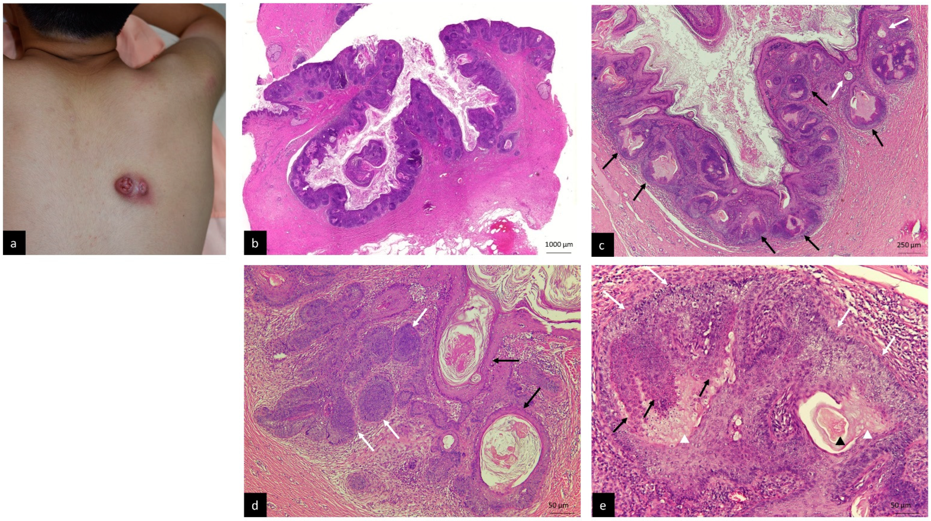
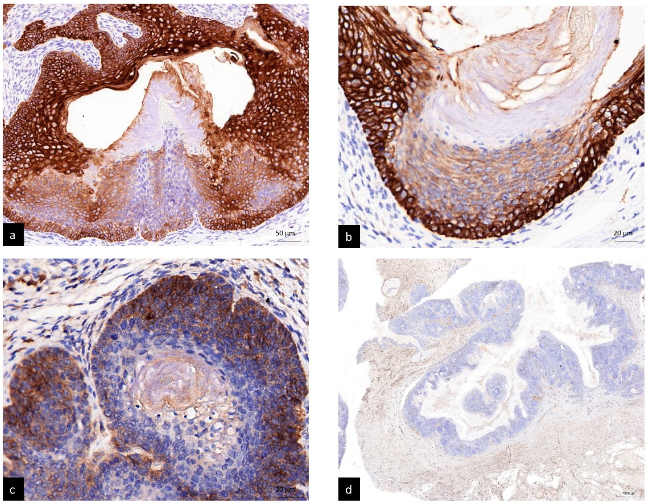
2. Case Report
3. Methods
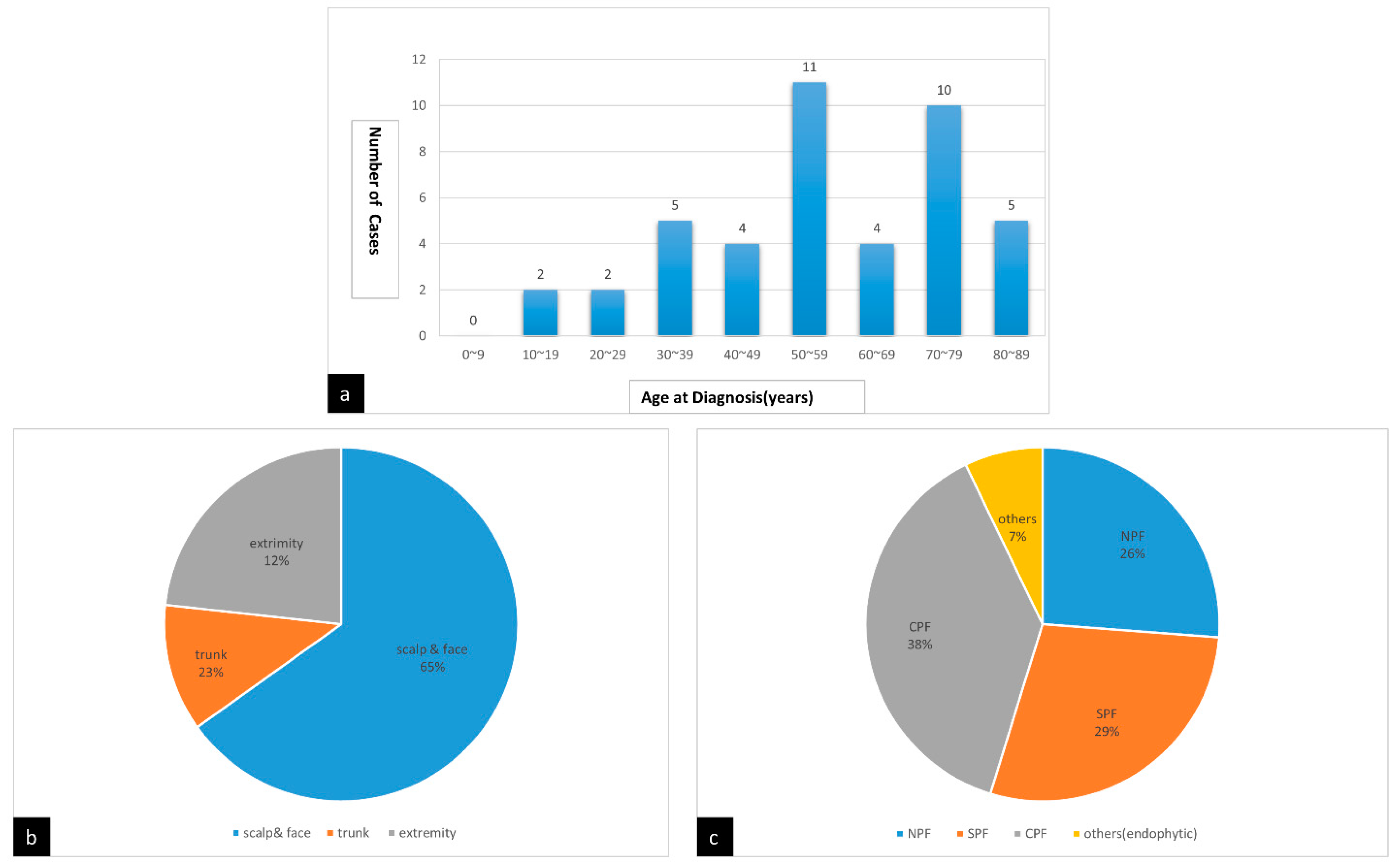
4. Results
5. Discussion
Author Contributions
Funding
Institutional Review Board Statement
Informed Consent Statement
Conflicts of Interest
References
- Ackerman, A.B.; de Viragh, P.A.; Chongchitnant, N. Neoplasms with Follicular Differentiation; Lea & Febiger: London, UK, 1993. [Google Scholar]
- Schirren, C.G.; Rutten, A.; Plewig, G. Panfolliculoma. Clinical and immunohistochemical findings in 4 cases. Hautarzt 1996, 47, 610–615. (In German) [Google Scholar] [CrossRef] [Scilit] [PubMed]
- Marini, M.A.; Starck, F.; Duhm, G.; Baldrich, M.A.; Magariños, G. Panfolliculoma: Case presentation. Arch. Argent. Dermatol. 2006, 56, 71–73. (In Spanish) [Google Scholar]
- Hoang, M.P.; Levenson, B.M. Cystic panfolliculoma. Arch. Pathol. Lab. Med. 2006, 130, 389–392. [Google Scholar] [CrossRef] [Scilit] [PubMed]
- Huang, C.Y.; Wu, Y.H. Panfolliculoma: Report of two cases. Dermatol. Sín. 2010, 28, 73–76. [Google Scholar] [CrossRef] [Scilit]
- Harris, A.; Faulkner-Jones, B.; Zimarowski, M.J. Epidermal panfolliculoma: A report of 2 cases. Am. J. Dermatopathol. 2011, 33, e7–e10. [Google Scholar] [CrossRef] [Scilit] [PubMed]
- Ruiz-Villaverde, R.; Elosegui-Martinez, F.; Luque-Barona, R. Panfolliculoma, an unusual cutaneous adnexal neoplasm. Eur. J. Dermatol. 2011, 21, 450–451. [Google Scholar] [CrossRef] [Scilit] [PubMed]
- Kacerovska, D.; Michal, M.; Kazakov, D.V. Panfolliculoma with sebaceous differentiation: A case report. Am. J. Dermatopathol. 2012, 34, e90–e93. [Google Scholar] [CrossRef] [Scilit] [PubMed]
- Idriss, M.H.; Khalil, A.; Long, W.; Elston, D.M. Epidermal panfolliculoma: An adnexal proliferation with advanced follicular differentiation confined to the epidermis. J. Cutan. Pathol. 2013, 40, 409–412. [Google Scholar] [CrossRef] [Scilit] [PubMed]
- Alkhalidi, H.S.; Alhumaidy, A.A. Cystic panfolliculoma of the scalp: Report of a very rare case and brief review. Indian J. Pathol. Microbiol. 2013, 56, 437–439. [Google Scholar] [CrossRef] [Scilit] [PubMed]
- Shan, S.J.; Guo, Y. Panfolliculoma and histopathologic variants: A study of 19 cases. Am. J. Dermatopathol. 2014, 36, 965. [Google Scholar] [CrossRef] [Scilit] [PubMed]
- Patel, M.B.S.; Jacobson-Dunlop, E.; Elston, D.M. An unusual cystic lesion on the helix. Indian Dermatol. Online J. 2014, 5, 326–327. [Google Scholar] [PubMed]
- Neill, B.; Bingham, C.; Braudis, K.; Zurowski, S. A rare cutaneous adnexal neoplasm: Cystic panfolliculoma. J. Cutan. Pathol. 2016, 43, 1183–1185. [Google Scholar] [CrossRef] [Scilit] [PubMed]
- Nishikawa, S.; Hamasaki, Y.; Otani, T.; Izumi, M. A Case of Panfolliculoma. Nishi. Nihon. Hifuka. 2016, 78, 621–624. (In Japanese) [Google Scholar] [CrossRef] [Scilit]
- Terushkin, V.; Meehan, S.; Shahabi, L.; Brinster, N. Panfolliculoma with an endophytic architecture resembling a hair follicle: A report of three cases. Cutan. Pathol. 2016, 43, 1074–1076. [Google Scholar] [CrossRef] [Scilit] [PubMed]
- Parvinnejad, N.; Orr, C.; Wang, A.; Hurlbut, D. Panfolliculoma: Report of a rare cutaneous neoplasm and literature review. Cana. Dian. J. Pathol. 2017, 9 (Suppl. 9), 26. [Google Scholar]
- Estrada-Castañón, R.; Vega-Memije, M.E.; Cuevas-González, J.C.; Chávez-López, M.G. Panfolliculoma: A Clinical Case Report. Int. J. Trichol. 2017, 9, 187–189. [Google Scholar]
- Fukuyama, M.; Sato, Y.; Yamazaki, Y.; Ohyama, M. Immunohistochemical dissection of cystic panfolliculoma focusing on the ex pression of multiple hair follicle lineage markers with an insight into the pathogenesis. J. Cutan. Pathol. 2017, 44, 861–866. [Google Scholar] [CrossRef] [Scilit] [PubMed]
- Rivera, V.; Campos, M. Cystic panfolliculoma: Case report. Med. Leg. Costa Rica 2018, 35, n.2. (In Spanish) [Google Scholar]
- Poblet, E.; Jiménez, F.; Godínez, J.M.; Pascual-Martín, A.; Izeta, A. The immunohistochemical expression of CD34 in human hair follicles: A comparative study with the bulge marker CK15. Clin. Exp. Dermatol. 2006, 31, 807–812. [Google Scholar] [CrossRef] [Scilit] [PubMed]



| Author, Year | Case Number | Age | Sex | Tumor Location | Clinical Impression | Lesion Size (cm) | Histopathology Variants |
|---|---|---|---|---|---|---|---|
| Schirren et al. 1996 [2] | 1 | 53 | M | infraclavicular | follicular cyst | 2~3 | NPF |
| 2 | N/A | N/A | N/A | N/A | N/A | N/A | |
| 3 | 22 | F | scalp | cylindroma | 2~3 | NPF | |
| 4 | 49 | F | right eyebrow | BCC | 2~3 | NPF | |
| Marini et al. 2006 [3] | 5 | 46 | F | nostril | N/A | N/A | N/A |
| Hoang et al. 2006 [4] | 6 | 33 | F | scalp | cyst | 3 × 2 × 2 | CPF |
| Huang et al. 2010 [5] | 7 | 41 | F | scalp | EC, pilar cyst | N/A | NPF |
| 8 | 51 | M | left eyebrow | ALHE, leiomyoma | N/A | NPF | |
| Harris et al. 2011 [6] | 9 | 81 | M | right medial thigh | inflammed SK | 0.6 | SPF |
| 10 | 61 | F | rightl lateral thigh | SCC | 0.3 | SPF | |
| Ruiz-Villaverde et al. 2011 [7] | 11 | 56 | M | Right tibia | EC | <3 | NPF |
| Kacerovska et al. 2012 [8] | 12 | 53 | M | occipital scalp | atheroma | N/A | NPF with sebaceous differentiation |
| Idriss et al. 2013 [9] | 13 | 55 | M | right leg | N/A | 0.8 | SPF |
| Alkhalidi et al. 2013 [10] | 14 | 19 | F | scalp | pilar cyst, lipoma | 0.9 × 0.8 | CPF |
| Shan et al. 2014 [11] | 15 | 76 | F | Left forearm | BCC | 0.5 × 0.4 | SPF |
| 16 | 65 | M | Left cheek | SK, AK, BCC | 0.4 × 0.3 | SPF | |
| 17 | 79 | M | Left nasal ala | BCC, dermal cyst | 0.6 × 0.5 | CPF | |
| 18 | 32 | M | Left scalp | Sebaceous cyst | 0.6 × 0.5 | CPF | |
| 19 | 55 | M | Right leg | VV | 0.5 × 0.4 | SPF | |
| 20 | 82 | M | Upper back | BCC | 1.2 × 0.8 | SPF | |
| 21 | 53 | M | Right low lip | Cyst | 0.3 × 0.3 | CPF | |
| 22 | 70 | F | Right lateral forehead | SCC, BCC | 0.8 × 0.6 | CPF | |
| 23 | 22 | F | Scalp | Cyst | 0.7 × 0.6 | CPF | |
| 24 | 33 | F | Left parietal scalp | BCC, nevus | 0.8 × 0.8 | NPF | |
| 25 | 49 | M | Right nostril | Cyst, BCC | 0.5 × 0.4 | CPF | |
| 26 | 70 | M | Central forehead | SCC, BCC | 0.7 × 0.6 | SPF | |
| 27 | 72 | M | Left post auricular | Wart, cyst | 0.4 × 0.4 | SPF | |
| 28 | 59 | F | Right lateral leg | Wart | 0.4 × 0.3 | SPF | |
| 29 | 71 | M | Forehead | BCC | 0.4 × 0.3 | SPF | |
| 30 | 83 | F | Left scalp | SCC | 4 × 3 | CPF | |
| 31 | 87 | M | Right temple | SCC, BCC | 0.8 × 0.7 | NPF | |
| 32 | 70 | M | Nose | Cyst, BCC | 0.4 × 0.3 | NPF | |
| 33 | 67 | M | Right lower leg | SCC | 0.4 × 0.3 | SPF | |
| Patel et al. 2014 [12] | 34 | 73 | M | Left helix | EC | N/A | CPF |
| Neill et al. 2016 [13] | 35 | 64 | F | right forearm | SCC, amelanotic melanoma, poroma | 0.8 | CPF |
| Nishikawa et al. 2016 [14] | 36 | 36 | F | back | N/A | 2.3 × 3 | NPF |
| Terushkin et al. 2016 [15] | 37 | 56 | F | Left cheek | BCC | N/A | endophytic |
| 38 | 88 | M | right anti-helix | cyst | N/A | endophytic | |
| 39 | 53 | F | right pretibial leg | foreign body | N/A | endophytic | |
| Parvinnejad et al.2017 [16] | 40 | 74 | M | abdomen | BCC | N/A | CPF |
| Estrada-Castañón et al. 2017 [17] | 41 | 55 | F | eyelid | comedone | N/A | CPF |
| Fukuyama et al. 2017 [18] | 42 | 70 | M | occipital scalp | trichilemmal cyst | 2.4 | CPF |
| Rivera et al.2018 [19] | 43 | 35 | M | right occipital scalp | pilar cyst | 1.3 | CPF |
| Present case | 44 | 14 | M | back | follicular cyst | 6 × 4 | CPF |
Publisher’s Note: MDPI stays neutral with regard to jurisdictional claims in published maps and institutional affiliations. |
© 2022 by the authors. Licensee MDPI, Basel, Switzerland. This article is an open access article distributed under the terms and conditions of the Creative Commons Attribution (CC BY) license (https://creativecommons.org/licenses/by/4.0/).
Share and Cite
Juang, S.-J.; Win, K.-T.; Lai, F.-J. Panfolliculoma: Report of the Youngest Case and Literature Review of Its Histopathologic Variants. Life 2022, 12, 881. https://doi.org/10.3390/life12060881
Juang S-J, Win K-T, Lai F-J. Panfolliculoma: Report of the Youngest Case and Literature Review of Its Histopathologic Variants. Life. 2022; 12(6):881. https://doi.org/10.3390/life12060881
Chicago/Turabian StyleJuang, Shiow-Jen, Khin-Than Win, and Feng-Jie Lai. 2022. "Panfolliculoma: Report of the Youngest Case and Literature Review of Its Histopathologic Variants" Life 12, no. 6: 881. https://doi.org/10.3390/life12060881
APA StyleJuang, S.-J., Win, K.-T., & Lai, F.-J. (2022). Panfolliculoma: Report of the Youngest Case and Literature Review of Its Histopathologic Variants. Life, 12(6), 881. https://doi.org/10.3390/life12060881