Article

Differential Expression of Duplicate Insulin-like Growth Factor-1 Receptors (igf1rs) in Medaka Gonads

1
Key Laboratory of Exploration and Utilization of Aquatic Genetic Resources, Ministry of Education, Shanghai Ocean University, Shanghai 201306, China
2
Eco-Environmental Protection Research Institute, Shanghai Academy of Agricultural Sciences, 1000 Jinqi Road, Shanghai 201403, China
3
Yeasen Biotechnology Co., Ltd., 800, Qingdai Road, Pudong New Area, Shanghai 201318, China
4
Shanghai Xihua Scientific Co., Ltd., Building 6-118, Furonghua Road, Pudong District, Shanghai 201318, China
*
Authors to whom correspondence should be addressed.
These authors contributed equally to this work.
Life 2022, 12(6), 859; https://doi.org/10.3390/life12060859
Submission received: 8 May 2022 / Revised: 4 June 2022 / Accepted: 5 June 2022 / Published: 8 June 2022

Abstract

:
Insulin-like growth factor-1 receptors (igf1rs) play important roles in regulating development, differentiation, and proliferation in diverse organisms. In the present study, subtypes of medaka igf1r, igf1ra, and igf1rb were isolated and characterized. RT-PCR results showed that igf1ra and igf1rb mRNA were expressed in all tissues and throughout embryogenesis. Using real-time PCR, the differential expression of igf1ra and igf1rb mRNA during folliculogenesis was observed. The results of in situ hybridization (ISH) revealed that both of them were expressed in ovarian follicles at different stages, and igf1rb was also expressed in theca cells and granulosa cells. In the testis, both igf1ra and igf1rb mRNA were highly expressed in sperm, while igf1rb mRNA was also obviously detected in spermatogonia. In addition, igf1ra mRNA was also present in Leydig cells in contrast to the distribution of igf1rb mRNA in Sertoli cells. Collectively, we demonstrated that differential igf1rs RNA expression identifies medaka meiotic germ cells and somatic cells of both sexes. These findings highlight the importance of the igf system in the development of fish gonads.

1. Introduction

The igf signaling system is a growth factor complex containing ligands, receptors, and binding proteins, which exists in all vertebrates [1]. The role of the igf signaling system in growth regulation has been well established, and its important roles in gonadal development has been gradually explored [2]. Igf1 mRNA is expressed in spermatogonia and spermatocytes as well as Leydig cells in Nile tilapia (Oreochromis niloticus) [3]. Igf2 could influence germ cell proliferation in the testis of Hypostomus garmani [4]. Furthermore, the gonad-specific igf3 can regulate spermatogenesis and reproduction in teleosts, such as Nile tilapia and zebrafish (Danio rerio) [5,6]. Igf2bp3 deletion leads to abnormal germ plasm assembly and a reduction in the number of germ cells in zebrafish [7]. Furthermore, igf1r is also expressed in the testis and ovary, which indicates the important roles of igf1r in gonadal development and gametogenesis [8,9].
Igf1r is a cell surface receptor that belongs to the tyrosine kinase receptor superfamily, which is expressed in diverse tissues of organisms [10]. In mammals, there is only one igf1r. Igf1r has been detected in sperm of humans, and its levels is positively correlated with sperm concentration [11]. Besides, igf1r is also expressed in ovaries of alpaca (Vicugna pacos), including follicles, granulosa, and theca interna cells [12]. In the cultured testicular cells, igf1r is highly expressed in Sertoli cells but also in spermatogonia and primary spermatocytes [13].
Due to whole genome duplication, fish have two subtypes of igf1r, igf1ra and igf1rb [14], and both of them play important biological functions. Igf1ra and igf1rb mRNA are highly expressed in gonads during vitellogenesis and spermatogonia proliferation in Pampus argenteus [15]. In adult zebrafish, igf1ra and igf1rb have distinct expression patterns, and the relative abundance of igf1ra and igf1rb is different in tissues [16]. It has been shown that inhibition of the igf signal pathway by knocking down igf1rb in the embryo of zebrafish can result in mis-migration and apoptosis of primordial germ cells (PGCs) [17]. Therefore, more attention has been focused on their cellular localization and biological activity in fish gonads. However, the role of igf1r in reproduction and gonadal development has rarely been studied, especially in fish.
Medaka (Oryzias latipes) is a good model and has been widely used in investigating developmental biology [18] and stem cell biology [19,20]. In addition, the primordial germ cells specification [21], migration [22], and sex-determination mechanism [23,24] have been systematically explored. Previously, we have investigated that igf1 is present in meiotic germ cells and somatic cells of both sexes in medaka [25]. Igf2 is associated with self-renewal of the embryonic stem cell [26]. Igf3 expression occurs both in germ cells and somatic cells in the ovary [27]. In the present study, to figure out the distribution and differences between igf1ra and igf1rb, the temporal and spatial expression patterns of their mRNAs in gonads, developing embryos, and follicles at different developmental stages were investigated. The expression patterns between igf1ra and igf1rb mRNAs in the gonads were also carried out by in situ hybridization studies. Our findings highlight the potential roles of the igf system in the reproduction and development of medaka and teleosts.

2. Materials and Methods

2.1. Fish and Embryos

Animal experiments were conducted strictly following the requirements of the Committee for Laboratory Animal Research at Shanghai Ocean University. Medaka was maintained in glass tanks with a water temperature of 26 °C, and an automatic photoperiod of 14 h light/10 h dark cycle was set. The developmental stage of medaka embryos has been previously described [28].

2.2. Isolation of Ovarian Follicles

The developing stages of the ovary were determined based on the original definition as described previously [25]. The ovaries were dissected from the anesthetized female medaka. As follicles have different stages, the same developmental stages were manually collected together and used for subsequent experiments: primary growth (stage I, below 0.1 mm in diameter), pre-vitellogenic (stage II, about 0.30 mm), early vitellogenic (stage III, about 0.40 mm), mid-vitellogenic (stage IV, about 0.50 mm), and fully-grown (stage V, about 0.65 mm).

2.3. RNA Extraction

Total RNA of adult tissues and embryos at different developmental stages of medaka was extracted by a TRIzol reagent (Invitrogen, Carlsbad, CA, USA). The NanoDrop 2000 Spectrophotometer (Thermo Fisher Scientific, Waltham, MA, USA) was used to detect the RNA quality and quantity. Furthermore, the integrity of RNA was verified by 1% agarose gel stained with nucleic acid dyes. Then, the cDNA was synthesized according to M-MLV reverse transcriptase (Takara, Shiga, Kusatsu, Japan) with an oligo(dT) 18 primer.

2.4. Cloning and Sequence Analysis of Medaka Igf1r

By searching the NCBI Gene database (https://www.ncbi.nlm.nih.gov/gene/ accessed on 1 March 2020), two computational predicted cDNAs encoding medaka igf1ra (Gene ID: 101173298) and igf1rb (Gene ID: 101163560) were obtained, respectively. To verify the accuracy of the two sequences, the Open Reading Frame (ORF) of igf1ra and igf1rb was cloned and sequenced. Afterwards, the igf1ra and igf1rb putative proteins were aligned with the igf1r orthologs from other examined organisms by using Vector NTI Advance 11.5 software (Thermo Fisher Scientific). The phylogenetic tree was based on the MEGA X program with the neighbor-joining (NJ) method [29]. All the primers used in the present study are listed in Table 1.

2.5. RT-PCR and Real-Time PCR Analysis

The expression of igf1r isoforms was detected by RT-PCR amplification using the primers in Table 1, and β-actin was used for calibration. PCR was performed for 35 cycles, and the reaction system contained 1 μL of cDNA template, 12.5 μL of 5 U/μL Premix Taq (EX Taq version) (Takara), and 0.5 μL of 10 mM each of the forward and reverse primers, and deionized water was added to replenish the total volume to 25 μL. The reaction procedures were as follows: 95 °C for 10 s, annealing at 58 °C for 10 s, and extension at 72 °C for 1 min. The PCR products were then detected on a 1.5% agarose gel stained with nucleic acid dyes and analyzed by a bio-imaging system (Bio-Rad, Hercules, CA, USA).
Real-time PCR (qPCR) was performed under the CFX96 TM Real-Time System (Bio-Rad) using the SYBR Green PCR Master Mix Kit (Takara). Using β-actin as calibration, the relative abundance of igf1ra and igf1rb mRNA was determined using the 2−ΔΔct, as described previously [30]. The data were presented as the mean ± SEM (n = 3). Statistical analyses were evaluated using a one-way ANOVA (p < 0.05) in GraphPad Prism 7 software.

2.6. RNA In Situ Hybridization

Sections in situ hybridization (SISH) were carried out as described previously [31]. Briefly, the gonads were fixed in 4% paraformaldehyde and then dehydrated gradiently with 20% and 30% sucrose. Next, the gonads were soaked with an embedding agent, Optimal Cutting Temperature (O.C.T., Sakura, Torrance, CA, USA), and subjected for sections with a freezing microtome (Leica, Wetzlar, Germany). To synthesize probes, the partial cDNA sequences of igf1ra and igf1rb obtained by PCR were inserted into the pGEM-T vector and sequenced for verification. The plasmids were then linearized with an appropriate restriction enzyme for the synthesis of probes by using the DIG or FITC RNA Labelling Kit (Roche, Basel, Switzerland). RNA of SISH was stained with BCIP/NBT and Fluorecent in situ hybridization (FISH) was carried out by using the (TSA TM) Plus Fluorescence Systems according to the product manual (Life Technologies, Carlsbad, CA, USA). The nucleus was stained with DAPI.

2.7. Microscopic Observation

Microscopy was performed as described [32]. In brief, microscopic observation and micrographs of gonadal sections were taken on a Nikon Ds-Ri2 camera (Nikon, Tokyo, Japan).

3. Results

3.1. Cloning and Sequence Analysis of Medaka Igf1r

PCR was employed to amplify the sequences of igf1r derived from tissues and embryo samples. The igf1ra ORF was obtained by TA cloning, which was 4197 nt and encoded 1398 amino acid residues (GenBank accession no. BK061359) (Figure S1). However, the igf1rb ORF was 4224 nt for 1407 amino acid residues (GenBank accession no. BK061360) (Figure S2). The IGF1Rs’ alignment showed the intracellular protein tyrosine kinase domain on the β-subunit of Igf1r (Figure 1). Igf1ra protein and Igf1rb protein of medaka are highly similar to those of zebrafish, with 74% and 71% identity, respectively, according to the Igf1rs multiple sequence alignment (Figure S3). Besides, the phylogenetic tree showed that compared with medaka Igf1ra, zebrafish Igf1rb is much closer to medaka Igf1rb (Figure 2). Such a divergence was also observed for Takifugu rubripes and Salmo salar (data not shown). Furthermore, although both Igf1ra and Igf1rb existed in different regions of different chromosomes in other species, they all showed strong chromosome synteny (Figure S4).

3.2. RT-PCR Analysis of Igf1r RNA Expression

The results of the RT-PCR showed that igf1ra and igf1rb were expressed in all adult tissues and embryos. Evidently, the expression of igf1ra and igf1rb was lower in the kidney, liver, and gut, in comparison with the eye, brain, ovary, and testis (Figure 3A). Besides, these two genes were similarly expressed during embryogenesis (Figure 3B). Furthermore, qPCR was performed to further analyze the expression profiles of the two igf1r types at different stages of folliculogenesis. The level of igf1ra increased from PG (stage I) to PV (stage II) and then decreased slowly, and it was hardly expressed in FG (stage V) (Figure 3G). The expression of igf1rb increased from the PG stage, peaked in EV (stage III), and weakened afterward (Figure 3H). These data indicate that igf1r was dynamically and differentially expressed during folliculogenesis in medaka.

3.3. Gonadal Expression of Igf1ra RNA and Igf1rb RNA by ISH

To better understand the subcellular distributions of igf1r, a chromogenic SISH was performed on cryosections. In the ovary, igf1ra RNA and igf1rb RNA were found in the cytoplasm of oocytes from stages I–IV (Figure 3C,D). Furthermore, igf1rb was also found in theca cells and granulosa cells, while igf1ra was absent (Figure 3D). In the testis, the igf1ra mRNA was highly expressed in sperm at the later stage of spermatogenesis (Figure 3E). Remarkably, igf1rb mRNA was detected in spermatogonia and sperm and was weakly detected in spermatocytes as well as spermatids, which was significantly different from the expression of igf1ra mRNA (Figure 3F). Furthermore, a positive signal for igf1ra was also found around Leydig cells, whereas the signal of igf1rb existed around Sertoli cells (Figure 3E,F). On the contrary, both of the sense probes, as controls, got no signal above the background (data not shown).

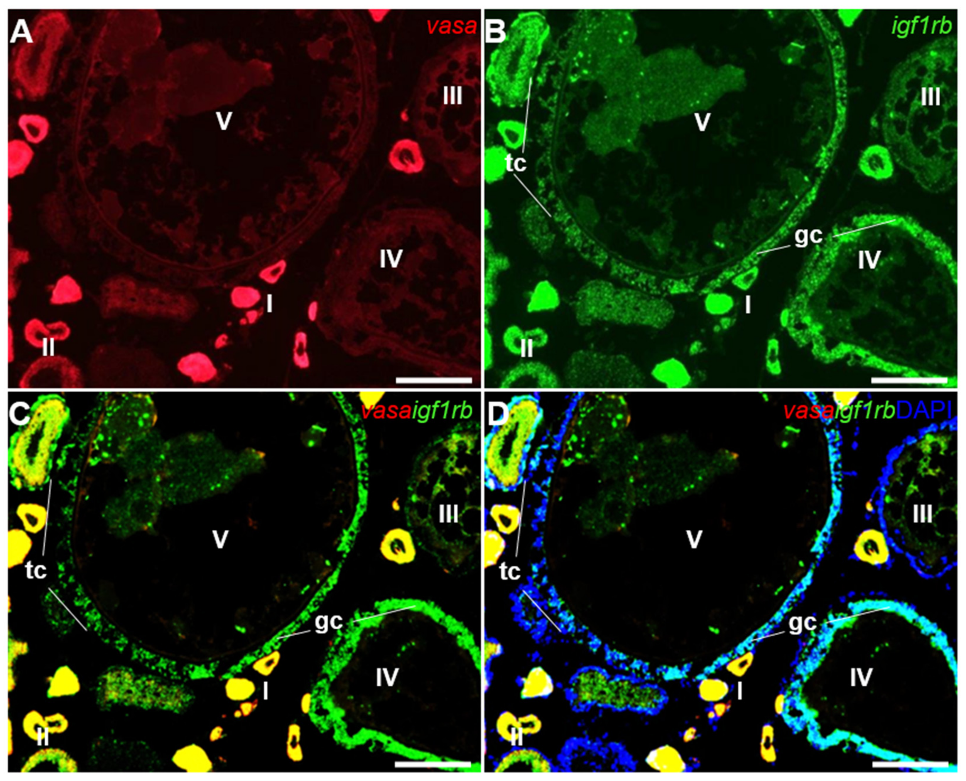
3.4. Ovarian Differential Expression of Igf1ra and Igf1rb RNAs by Fluorescence ISH

To further accurately identify the RNA expression of igf1r, the co-distribution of igf1rb and vasa was carried out by FISH. Vasa is a well-studied gene in medaka and other species, which is restrictively expressed in the germ cells of both sexes [33]. In the ovary, the vasa signal was expressed obviously in pre-meiotic oocytes and was decreased with the process of oogenesis (Figure 4A). Conversely, the igf1rb signal was easily found in oocytes from stages I–IV (Figure 4B). Notably, the igf1rb positive signal was also obvious in the granulosa cells and theca cells at later folliculogenesis (Figure 4C,D).
We compared the RNA expression of igf1ra with igf1rb by dual-color FISH. Conforming to their observations by chromogenic staining, both igf1ra and igf1rb RNAs were expressed in oocytes from stages I to IV (Figure 5A,B). Furthermore, igf1rb was detected abundantly in somatic cells, including granulosa cells, as well as theca cells at later developmental oocytes, while igf1ra was not detected in these somatic cells (Figure 5C,D)

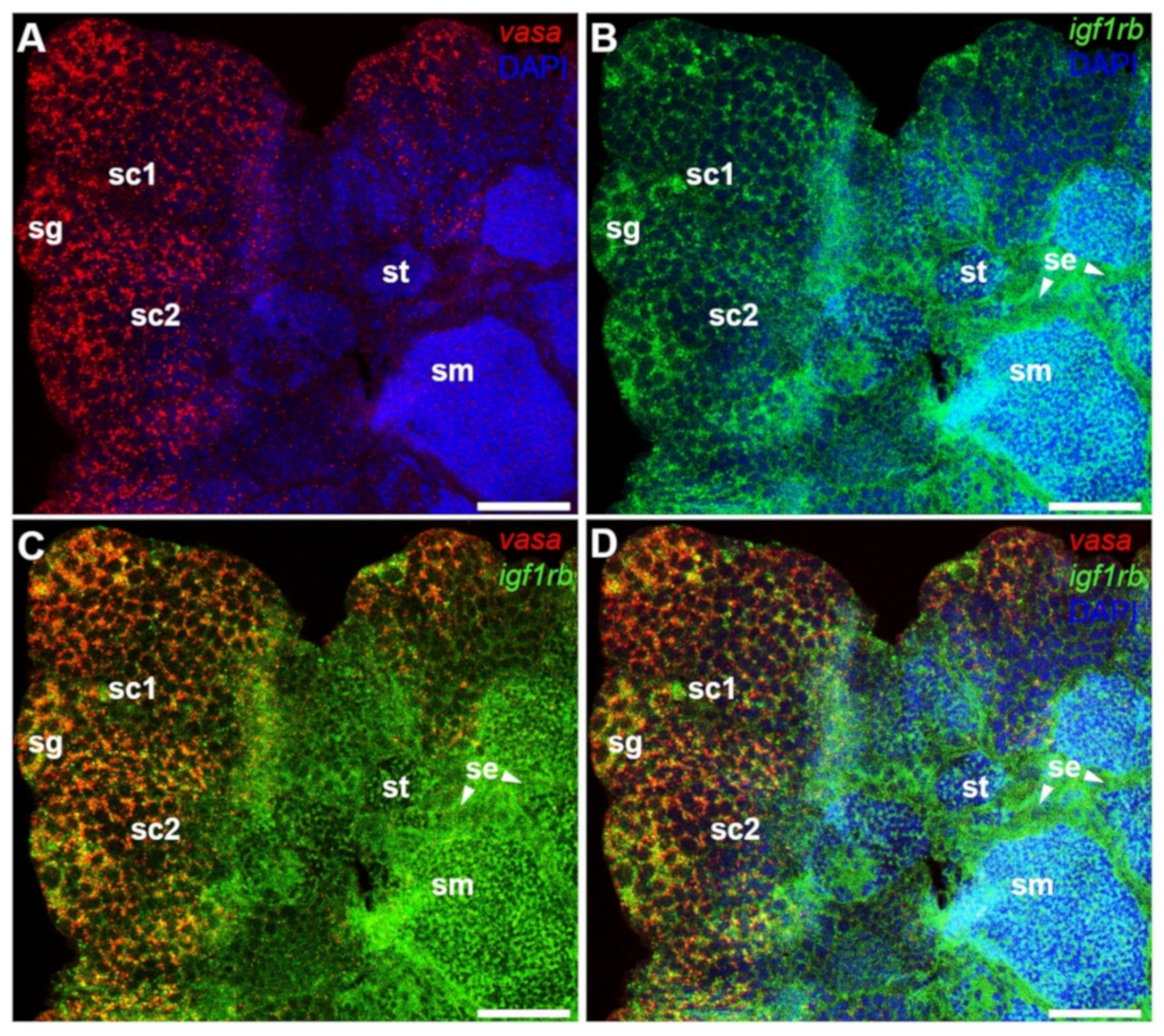
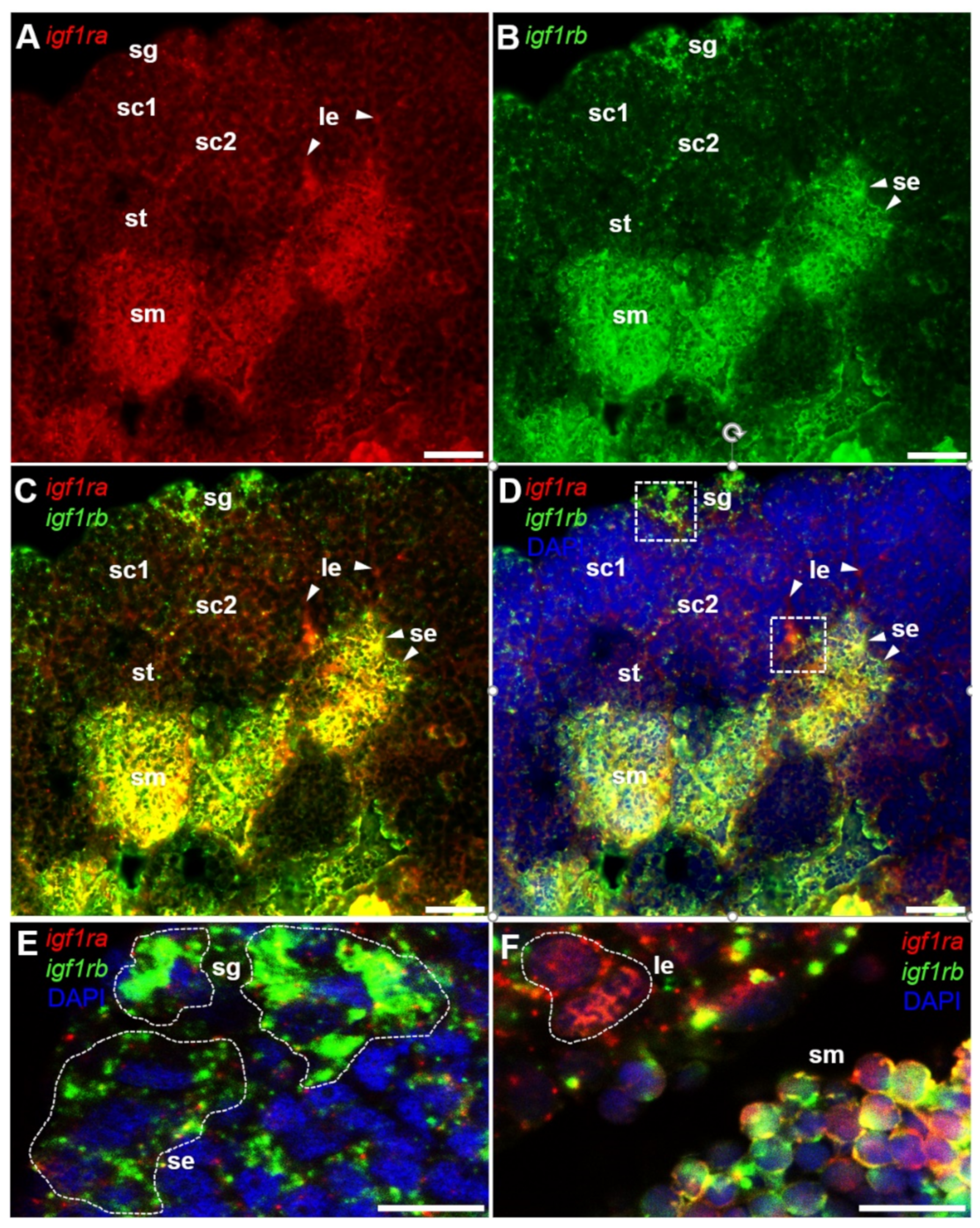
3.5. Testicular Differential Expression of Igf1ra and Igf1rb RNAs by Fluorescence ISH

In the next step, we compared the RNA expression of igf1rb with vasa by dual color FISH. In the testis, the vasa signal was strong in spermatogonia, and with the progress of spermatogenesis, the intensity of the vasa signal was decreased until it disappeared in sperm (Figure 6A). In contrast, the igf1rb mRNA was abundantly expressed in the spermatogonia and sperm, and it was relatively low in spermatocytes as well as in spermatids (Figure 6B). Surprisingly, a positive signal for igf1rb was also detected between the cyst and the cyst of the testis structure (Figure 6C,D), which is generally thought to be the location of Sertoli cells [34].
Then, a dual-color FISH was performed to precisely compare the expression of igf1ra and igf1rb RNA. Results indicated that igf1ra mRNA was richly expressed in sperm, but it was not in other stages of spermatogenic cells (Figure 7A,C). In contrast, igf1rb mRNA was expressed in almost all stages of spermatogenesis, especially spermatogonia and sperm. (Figure 7B–E). Furthermore, igf1ra mRNA and igf1rb mRNA were also found in somatic cells, with igf1ra expressed in the intertubular space where the Leydig cells usually located, while igf1rb mRNA delineated the germinal cysts called Sertoli cells (Figure 7A–F).

4. Discussion

In contrast to a single igf1r in mammals, many fish species have two igf1r paralogous isoforms, igf1ra and igf1rb, such as zebrafish [16], gilthead seabream (Sparus aurata) [9], and Epinephelus coioides [35]. According to the present study, medaka also has two igf1r subtypes. A sequence comparison and phylogenetic tree of the medaka two Igf1rs protein sequences with other vertebrates indicated that the receptors were highly conservative in the process of vertebrate evolution. The structure of the medaka igf1rs is most similar to that of zebrafish igf1rs [16]. Not only is their sequence identity of medaka igf1rs and zebrafish igf1rs greater than 60%, but structural motifs are nearly conserved in the two medaka igf1rs, such as the ATP-binding site, ligand-binding region, tyrosine kinase domain, autophosphorylation site, and IRS-I docking site. Igf1r is composed of extracellular α-subunits and transmembrane-spanning β-subunits, which contain cytoplasmic tyrosine kinase activity [36]. Activated igf1r phosphorylates specific reaction components, including IRS-1, IRS-2, and SRC homology collagen, and it regulates downstream responses through the phosphatidylinositol 3-kinase (PI3K)/AKT and mitogen-activated protein kinase (MAPK) pathways [1,37].
Both igf1r mRNAs were extensively expressed in different tissues in the adult medaka, which was consistent with reports from other teleosts [38]. The rich expression of two igf1r mRNAs in the eye, brain, and gonad generally agreed with the previous reports of igf1 mRNA expression levels in these tissues of medaka [25]. Similar to Paralichthys olivaceus [39], the igf1r involvement in reproduction and development was consistent with the functions of the igf system, such as igf1 promoted spermatogenesis, oocyte maturation, and steroidogenesis [40,41], and igf3 maintained the differentiation of ovary [42]. Furthermore, the expression of igf1rs mRNAs in the embryos at different developmental stages was demonstrated. Our present study further supports the potential functions of the igf system in fish.
In adult medaka, igf1ra and igf1rb mRNAs were detected in the oocytes at different stages; however, the expression levels were significantly different. During folliculogenesis, the level of igf1ra mRNA in mature follicles was extremely low in comparison with igf1rb, suggesting that igf1ra may play a minor role in mature follicles, while igf1rb plays a dominant role. Furthermore, through co-localization with the germ cell marker gene vasa [43], it was found that igf1rb RNA was highly expressed in many somatic cells, including theca cells and granulosa cells at later folliculogenesis, while igf1ra was absent. Similarly, igf1r was also found in theca cells and granulosa cells from the ovary of Oncorhynchus kisutch [44], gilthead seabream [9], alpaca [12], and mice [45]. Based on these findings, it indicates that the two igf1rs of medaka share some similar biological functions, which means that they play the same roles in many body activities. Meanwhile, it predicts that both of them also have their own unique roles in promoting individual growth and gonadal development. Igf1rb may also be involved in hormone production in the ovary of medaka according to previous studies that theca cells and granulosa cells regulate steroidal hormone production [44].
In this study, the two subtypes of igf1r were differentially expressed in the ovary as well as in the testis. Through the co-localization of vasa and igf1rb, as well as igf1ra and igf1rb, we found that both types of igf1r were expressed in sperm. In addition, igf1rb was also expressed in spermatogonia. Furthermore, it seemed that igf1ra preferred to express in Leydig cells, whereas igf1rb preferred to express in Sertoli cells, suggesting that the two isoforms may have different functions during spermatogenesis. These results agreed with the reported presence of igf1 receptors in somatic cells of zebrafish [46], rainbow trout (Oncorhynchus mykiss) [13], and gilthead seabream [9]. In recent research on zebrafish, it has been found that igf1rb was expressed in spermatogonia and could mediate igf3 to activate the β-catenin-dependent signal pathway to regulate spermatogenesis [46]. Moreover, it was shown that igf1 and igf2 interact with igf1r, causing the receptor activation and regulating organism growth, development, and reproduction [36]. Therefore, we speculate that the two subtypes of igf1r are essential for spermatogenesis, while igf1rb is also involved in the growth and proliferation of spermatogonia in the early stage. Overall, igf1rs play critical roles in the development of fish gonads, which is worthy of further research and provides a reference for other fish. Besides, the igf system is conserved in diverse species, and the interaction among ligands, receptors, and binding proteins of the system ensures that various life activities are carried out on the normal track, so the follow-up study of their co-localization is necessary for the further investigation of the igf system.
In summary, we demonstrated the differential expression of two igf1r subtypes in the adult gonads and embryos of medaka. The distinct expression patterns of the two subtypes of igf1r indicate that they play different roles in gonadal and embryonic development. Overall, the present study provides conclusive evidence for the potential roles of igf1r in gonadal development and gametogenesis in fish, as well as a reference for further research.

Supplementary Materials

The following supporting information can be downloaded at: https://www.mdpi.com/article/10.3390/life12060859/s1. Figure S1: Nucleotide and deduced amino acid sequence of medaka igf1ra; Figure S2: Nucleotide and deduced amino acid sequence of medaka igf1rb; Figure S3: Multiple sequence alignment of IGF1 receptors; Figure S4: Chromosome synteny diagrams for Igf1rs.

Author Contributions

Conceptualization, Experimental designing, and Project administration: M.L.; W.W.; Y.Z. (Yuli Zhao); C.Y.; Methodology: W.W.; Y.Z. (Yefei Zhu); C.Y.; Supervision: M.L.; W.W.; Writing—original draft: W.W.; Y.Z. (Yuli Zhao); Writing—review and editing: W.W.; M.L.; W.Z. All authors have read and agreed to the published version of the manuscript.

Funding

This research was funded by the National Key R&D Program of China (2018YFD0901205), the National Natural Science Foundation of China (31672700), and the China Agriculture Research System of MOF and MARA (CARS-46).

Institutional Review Board Statement

The study was conducted according to the guidelines of the Declaration of Helsinki, and it was approved by the Shanghai Ocean University Animal Care and Use Committee, with approval number SHOU-2021-118.

Informed Consent Statement

Informed consent was obtained from all subjects involved in the study.

Data Availability Statement

Not applicable.

Acknowledgments

We are grateful to all workers at the experimental sites.

Conflicts of Interest

The authors declare no conflict of interest. The company had no role in the design of the study; in the collection, analyses, or interpretation of data; in the writing of the manuscript, and in the decision to publish the results.

Abbreviations

Igf, insulin-like growth factor; RT-PCR, reverse transcription-polymerase chain reaction; ISH, in situ hybridization; FISH, fluorescence in situ hybridization; P13K, phosphoinositide 3 kinase; aa, amino acid residues; nt, nucleotide.

References

  1. Wood, A.W.; Duan, C.; Bern, H.A. Insulin-like growth factor signaling in fish. Int. Rev. Cytol. 2005, 243, 215–285. [Google Scholar] [PubMed]
  2. Liu, J.P.; Baker, J.; Perkins, A.S.; Robertson, E.J.; Efstratiadis, A. Mice carrying null mutations of the genes encoding insulin-like growth factor I (Igf-1) and type 1 IGF receptor (Igf1r). Cell 1993, 75, 59–72. [Google Scholar] [CrossRef]
  3. Berishvili, G.; D’Cotta, H.; Baroiller, J.F.; Segner, H.; Reinecke, M. Differential expression of IGF-I mRNA and peptide in the male and female gonad during early development of a bony fish, the tilapia Oreochromis niloticus. Gen. Comp. Endocrinol. 2006, 146, 204–210. [Google Scholar] [CrossRef] [PubMed]
  4. Moreira, D.P.; Melo, R.M.C.; Weber, A.A.; Rizzo, E. Insulin-like growth factors 1 and 2 are associated with testicular germ cell proliferation and apoptosis during fish reproduction. Reprod. Fertil. Dev. 2020, 32, 988–998. [Google Scholar] [CrossRef]
  5. Li, J.; Liu, Z.; Kang, T.; Li, M.; Wang, D.; Cheng, C.H.K. Igf3: A novel player in fish reproductiondagger. Biol. Reprod. 2021, 104, 1194–1204. [Google Scholar] [CrossRef]
  6. Li, M.; Liu, X.; Dai, S.; Xiao, H.; Qi, S.; Li, Y.; Zheng, Q.; Jie, M.; Cheng, C.H.K.; Wang, D. Regulation of spermatogenesis and reproductive capacity by Igf3 in tilapia. Cell. Mol. Life Sci. 2020, 77, 4921–4938. [Google Scholar] [CrossRef]
  7. Fan, R.; Ran, M.; Rui, X.; Jie, M. m6A reader Igf2bp3 enables germ plasm assembly by m6A-dependent regulation of gene expression in zebrafish. Sci. Bull. 2021, 66, 1119–1128. [Google Scholar]
  8. Mei, J.; Yan, W.; Fang, J.; Yuan, G.; Chen, N.; He, Y. Identification of a gonad-expression differential gene insulin-like growth factor-1 receptor (Igf1r) in the swamp eel (Monopterus albus). Fish Physiol. Biochem. 2014, 40, 1181–1190. [Google Scholar] [CrossRef]
  9. Perrot, V.; Moiseeva, E.B.; Gozes, Y.; Chan, S.J.; Funkenstein, B. Insulin-like growth factor receptors and their ligands in gonads of a hermaphroditic species, the gilthead seabream (Sparus aurata): Expression and cellular localization. Biol. Reprod. 2000, 63, 229–241. [Google Scholar] [CrossRef] [Green Version]
  10. Kineman, R.D.; Del Rio-Moreno, M.; Sarmento-Cabral, A. 40 YEARS of IGF1: Understanding the tissue-specific roles of IGF1/IGF1R in regulating metabolism using the Cre/loxP system. J. Mol. Endocrinol. 2018, 61, T187–T198. [Google Scholar] [CrossRef] [Green Version]
  11. Cannarella, R.; Condorelli, R.A.; La Vignera, S.; Bellucci, C.; Luca, G.; Calafiore, R.; Calogero, A.E. IGF2 and IGF1R mRNAs are Detectable in Human Spermatozoa. World J. Mens Health 2020, 38, 545–551. [Google Scholar] [CrossRef] [PubMed] [Green Version]
  12. Gallelli, M.F.; Bianchi, C.; Lombardo, D.; Rey, F.; Rodriguez, F.M.; Castillo, V.A.; Miragaya, M. Leptin and IGF1 receptors in alpaca (Vicugna pacos) ovaries. Anim. Reprod. Sci. 2019, 200, 96–104. [Google Scholar] [CrossRef] [PubMed]
  13. Le Gac, F.; Loir, M.; le Bail, P.Y.; Ollitrault, M. Insulin-like growth factor (IGF-I) mRNA and IGF-I receptor in trout testis and in isolated spermatogenic and Sertoli cells. Mol. Reprod. Dev. 1996, 44, 23–35. [Google Scholar] [CrossRef]
  14. Schlueter, P.J.; Royer, T.; Farah, M.H.; Laser, B.; Chan, S.J.; Steiner, D.F.; Duan, C. Gene duplication and functional divergence of the zebrafish insulin-like growth factor 1 receptors. FASEB J. 2006, 20, 1230–1232. [Google Scholar] [CrossRef] [Green Version]
  15. Gu, W.; Yang, Y.; Ning, C.; Wang, Y.; Hu, J.; Zhang, M.; Kuang, S.; Sun, Y.; Li, Y.; Zhang, Y.; et al. Identification and characteristics of insulin-like growth factor system in the brain, liver, and gonad during development of a seasonal breeding teleost, Pampus argenteus. Gen. Comp. Endocrinol. 2021, 300, 113645. [Google Scholar] [CrossRef]
  16. Maures, T.; Chan, S.J.; Xu, B.; Sun, H.; Ding, J.; Duan, C. Structural, biochemical, and expression analysis of two distinct insulin-like growth factor I receptors and their ligands in zebrafish. Endocrinology 2002, 143, 1858–1871. [Google Scholar] [CrossRef]
  17. Schlueter, P.J.; Sang, X.; Duan, C.; Wood, A.W. Insulin-like growth factor receptor 1b is required for zebrafish primordial germ cell migration and survival. Dev. Biol. 2007, 305, 377–387. [Google Scholar] [CrossRef] [Green Version]
  18. Wittbrodt, J.; Shima, A.; Schartl, M. Medaka—A model organism from the far East. Nat. Rev. Genet. 2002, 3, 53–64. [Google Scholar] [CrossRef]
  19. Yi, M.; Hong, N.; Hong, Y. Generation of medaka fish haploid embryonic stem cells. Science 2009, 326, 430–433. [Google Scholar] [CrossRef]
  20. Hong, Y.; Liu, T.; Zhao, H.; Xu, H.; Wang, W.; Liu, R.; Chen, T.; Deng, J.; Gui, J. Establishment of a normal medakafish spermatogonial cell line capable of sperm production in vitro. Proc. Natl. Acad. Sci. USA 2004, 101, 8011–8016. [Google Scholar] [CrossRef] [Green Version]
  21. Hong, N.; Li, M.; Yuan, Y.; Wang, T.; Yi, M.; Xu, H.; Zeng, H.; Song, J.; Hong, Y. Dnd is a critical specifier of primordial germ cells in the medaka fish. Stem Cell Rep. 2016, 6, 411–421. [Google Scholar] [CrossRef] [Green Version]
  22. Li, M.; Hong, N.; Gui, J.; Hong, Y. Medaka piwi is essential for primordial germ cell migration. Curr. Mol. Med. 2012, 12, 1040–1049. [Google Scholar] [CrossRef] [Green Version]
  23. Matsuda, M.; Nagahama, Y.; Shinomiya, A.; Sato, T.; Matsuda, C.; Kobayashi, T.; Morrey, C.E.; Shibata, N.; Asakawa, S.; Shimizu, N.; et al. DMY is a Y-specific DM-domain gene required for male development in the medaka fish. Nature 2002, 417, 559–563. [Google Scholar] [CrossRef]
  24. Nishimura, T.; Sato, T.; Yamamoto, Y.; Watakabe, I.; Ohkawa, Y.; Suyama, M.; Kobayashi, S.; Tanaka, M. Sex determination. foxl3 is a germ cell-intrinsic factor involved in sperm-egg fate decision in medaka. Science 2015, 349, 328–331. [Google Scholar]
  25. Yuan, C.; Chen, K.; Zhu, Y.; Yuan, Y.; Li, M. Medaka igf1 identifies somatic cells and meiotic germ cells of both sexes. Gene 2018, 642, 423–429. [Google Scholar] [CrossRef]
  26. Yuan, Y.; Hong, Y. Medaka insulin-like growth factor-2 supports self-renewal of the embryonic stem cell line and blastomeres in vitro. Sci. Rep. 2017, 7, 78. [Google Scholar] [CrossRef] [Green Version]
  27. Xie, J.; Zhong, Y.; Zhao, Y.; Xie, W.; Guo, J.; Gui, L.; Li, M. Characterization and expression analysis of gonad specific igf3 in the medaka ovary. Aquac. Fish. 2020, 7, 259–268. [Google Scholar] [CrossRef]
  28. Iwamatsu, T. Stages of normal development in the medaka Oryzias latipes. Mech. Dev. 2004, 121, 605–618. [Google Scholar] [CrossRef]
  29. Tamura, K.; Stecher, G.; Peterson, D.; Filipski, A.; Kumar, S. MEGA6: Molecular evolutionary genetics analysis version 6.0. Mol. Biol. Evol. 2013, 30, 2725–2729. [Google Scholar] [CrossRef] [Green Version]
  30. Shved, N.; Berishvili, G.; D’Cotta, H.; Baroiller, J.F.; Segner, H.; Eppler, E.; Reinecke, M. Ethinylestradiol differentially interferes with IGF-I in liver and extrahepatic sites during development of male and female bony fish. J. Endocrinol. 2007, 195, 513–523. [Google Scholar] [CrossRef] [Green Version]
  31. Chen, X.; Zhu, Y.; Zhu, T.; Song, P.; Guo, J.; Zhong, Y.; Gui, L.; Li, M. Vasa identifies germ cells in embryos and gonads of Oryzias celebensis. Gene 2022, 823, 146369. [Google Scholar] [CrossRef]
  32. Song, P.; Sun, B.; Zhu, Y.; Zhong, Y.; Guo, J.; Gui, L.; Li, M. Bucky ball induces primordial germ cell increase in medaka. Gene 2021, 768, 145317. [Google Scholar] [CrossRef]
  33. Li, M.; Zhao, H.; Wei, J.; Zhang, J.; Hong, Y. Medaka vasa gene has an exonic enhancer for germline expression. Gene 2015, 555, 403–408. [Google Scholar] [CrossRef]
  34. Schulz, R.W.; de Franca, L.R.; Lareyre, J.J.; Le Gac, F.; Chiarini-Garcia, H.; Nobrega, R.H.; Miura, T. Spermatogenesis in fish. Gen. Comp. Endocrinol. 2010, 165, 390–411. [Google Scholar] [CrossRef]
  35. Guo, L.; Yang, S.; Li, M.M.; Meng, Z.N.; Lin, H.R. Divergence and polymorphism analysis of IGF1Ra and IGF1Rb from orange-spotted grouper, Epinephelus coioides (Hamilton). Genet. Mol. Res. 2016, 15, gmr15048768. [Google Scholar] [CrossRef]
  36. Wu, J.; Li, W.; Craddock, B.P.; Foreman, K.W.; Mulvihill, M.J.; Ji, Q.S.; Miller, W.T.; Hubbard, S.R. Small-molecule inhibition and activation-loop trans-phosphorylation of the IGF1 receptor. EMBO J. 2008, 27, 1985–1994. [Google Scholar] [CrossRef] [Green Version]
  37. Hakuno, F.; Takahashi, S.I. IGF1 receptor signaling pathways. J. Mol. Endocrinol. 2018, 61, T69–T86. [Google Scholar] [CrossRef] [Green Version]
  38. Kuang, Y.M.; Li, W.S.; Lin, H.R. Molecular cloning and mRNA profile of insulin-like growth factor type 1 receptor in orange-spotted grouper, Epinephelus coioides. Acta Biochim. Biophys. Sin. 2005, 37, 327–334. [Google Scholar] [CrossRef] [Green Version]
  39. Zhang, J.; Shi, Z.; Cheng, Q.; Chen, X. Expression of insulin-like growth factor I receptors at mRNA and protein levels during metamorphosis of Japanese flounder (Paralichthys olivaceus). Gen. Comp. Endocrinol. 2011, 173, 78–85. [Google Scholar] [CrossRef]
  40. Weber, G.M.; Sullivan, C.V. Effects of insulin-like growth factor-I on in vitro final oocyte maturation and ovarian steroidogenesis in striped bass, Morone saxatilis. Biol. Reprod. 2000, 63, 1049–1057. [Google Scholar] [CrossRef] [Green Version]
  41. Neirijnck, Y.; Calvel, P.; Kilcoyne, K.R.; Kuhne, F.; Stevant, I.; Griffeth, R.J.; Pitetti, J.L.; Andric, S.A.; Hu, M.C.; Pralong, F.; et al. Insulin and IGF1 receptors are essential for the development and steroidogenic function of adult Leydig cells. FASEB J. 2018, 32, 3321–3335. [Google Scholar] [CrossRef] [PubMed] [Green Version]
  42. Xie, Y.X.; Huang, D.; Chu, L.H.; Liu, Y.; Sun, X.; Li, J.Z.; Cheng, C.H.K. Igf3 is essential for ovary differentiation in zebrafish. Biol. Reprod. 2021, 104, 589–601. [Google Scholar] [CrossRef] [PubMed]
  43. Li, M.; Hong, N.; Xu, H.; Yi, M.; Li, C.; Gui, J.; Hong, Y. Medaka vasa is required for migration but not survival of primordial germ cells. Mech. Dev. 2009, 126, 366–381. [Google Scholar] [CrossRef] [PubMed]
  44. Maestro, M.A.; Planas, J.V.; Moriyama, S.; Gutierrez, J.; Planas, J.; Swanson, P. Ovarian receptors for insulin and insulin-like growth factor I (IGF-I) and effects of IGF-I on steroid production by isolated follicular layers of the preovulatory coho salmon ovarian follicle. Gen. Comp. Endocrinol. 1997, 106, 189–201. [Google Scholar] [CrossRef]
  45. Baumgarten, S.C.; Armouti, M.; Ko, C.; Stocco, C. IGF1R expression in ovarian granulosa cells is essential for steroidogenesis, follicle survival, and fertility in female mice. Endocrinology 2017, 158, 2309–2318. [Google Scholar] [CrossRef] [Green Version]
  46. Safian, D.; Bogerd, J.; Schulz, R.W. Igf3 activates beta-catenin signaling to stimulate spermatogonial differentiation in zebrafish. J. Endocrinol. 2018, 238, 245–257. [Google Scholar] [CrossRef]
Figure 1. Multiple sequence alignment of medaka Igf1rs. The alignment sequences show the intracellular protein tyrosine kinase domain. Conserved regions between species are highlighted. The length and percentage identity values of Igf1rs homologs are given at the end of the alignment.
Life 12 00859 g001
Figure 2. Phylogenetic tree of Igf1r. The insulin receptor (InsR) served as the out-group. Bootstrap values are given, and the bar indicates number of substitutions per site. Accession numbers are after the organism. Igf1rs from different species are clustered together, indicating that generation of igf1r and insr took place in early vertebrate evolution.
Life 12 00859 g002
Figure 3. Expression of igf1ra RNA and igf1rb RNA. (A,B) RT-PCR analysis of medaka igf1ra and igf1rb in adult tissues (A) and developing embryos (B). (CF) Ovarian and testicular cryosections using antisense igf1ra and igf1rb probes and the signals were visualized by chromogenic staining. (G,H) qPCR results of igf1ra and igf1rb at different stages of follicles. I–V, stages of oocytes; sg, spermatogonia; sc, spermatocytes; st, spermatids; sm, sperm; gc, granulosa cells; tc, theca cells; se, Sertoli cells; le, Leydig cells. Scale bars, 100 µm.
Life 12 00859 g003
Figure 4. Expression of igf1rb RNA and vasa RNA in the ovary. FISH on ovarian cryosections using antisense RNA probes and the signals were visualized by fluorescence staining. The vasa RNA was stained in red, and the igf1rb was stained in green. Nuclei were stained in blue with DAPI. (A,B) Different stages of oocytes (I–V), granulosa cells and theca cells were indicated by sticks. (C,D) The merges of vasa with igf1rb and vasa with igf1rb and DAPI. I–V, stages of oocytes; gc, granulosa cells; tc, theca cells. Scale bars, 25 µm.
Life 12 00859 g004
Figure 5. Expression of igf1ra RNA and igf1rb RNA in the ovary. FISH on ovarian cryosections using antisense RNA probes and the signals were visualized by fluorescence staining. The igf1ra RNA was stained in red, and the igf1rb was stained in green. Nuclei were stained in blue with DAPI. (A,B) Different stages of oocytes (I–V), granulosa cells and theca cells were indicated by sticks. (C,D) Merges of igf1ra with igf1rb and igf1ra with igf1rb and DAPI. I–V, stages of oocytes; gc, granulosa cells; tc, theca cells. Scale bars, 25 µm.
Life 12 00859 g005
Figure 6. Expression of vasa RNA and igf1rb RNA in the testis. After hybridization with antisense vasa and igf1rb RNA probes, the signals were visualized by fluorescence staining. Nuclei were stained blue by DAPI. (AD) Merges of vasa with DAPI, igf1rb with DAPI, vasa with igf1rb, and vasa with igf1rb and DAPI. Sertoli cells were indicated by arrows. Vasa and igf1rb showed significantly different expression patterns. The vasa signal peaked in spermatogonia and then gradually decreased until it disappeared in sperm. Conversely, the igf1rb was obviously detected in spermatogonia, sperm, and Sertoli cells. sg, spermatogonia; sc, spermatocytes; st, spermatids; sm, sperm; se, Sertoli cells. Scale bars, 25 µm.
Life 12 00859 g006
Figure 7. Expression of igf1ra RNA and igf1rb RNA in the testis. Adult testicular cryosections were hybridized to antisense RNA probes and the signals were visualized by fluorescence staining. Nuclei were stained blue by DAPI. (A,B) Lower magnification view showing different stages of spermatogenesis. Sertoli cells and Leydig cells were indicated by arrows. (C,D) Merges of igf1ra with igf1rb and igf1ra with igf1rb and DAPI. (E,F) Larger Magnification of panels D (white frame), highlighting the different cells. The igf1ra signal exists in sperm and Leydig cells. Notably, the igf1rb signal exists in spermatogonia, sperm, as well as Sertoli cells. se, Sertoli cells; le, Leydig cells; sm, sperm; sc, spermatocytes; sg, spermatogonia. Scale bars, 25 µm.
Life 12 00859 g007
Table 1. Sequences of primers used in the present study.
PrimerSequence (5′ to 3′ Direction)Purpose
igf1ra-FATGGACATCCGAAACGACRT-PCR
igf1ra-RTCGTCTGTACAGGCCAGCTTG
igf1rb-FTGCTTCGGGAATGAGGCCTCCRT-PCR
igf1rb-RTGATGTGCAGGTTTCCTTTGAT
igf1ra-F1ATGGGCAGGCTGACCTTGTTTTGGCDS cloning
igf1ra-R1TTCGTCTGGAGTGTACATCTC
igf1ra-F2ATGTACACTCCAGACGAATGGCDS cloning
igf1ra-R2TCAGCAGGCCGACGACTGGGGCAG
igf1rb-F1ATGAGGCCTCCAGCGGAAACGAGGCDS cloning
igf1rb-R1GGCCACGCCCTCGTACACCAT
igf1rb-F2ATGGTGTACGAGGGCGTGGCCCDS cloning
igf1rb-R2CCCTTCAGCAGGCTGAG
igf1ra-QFCGCCTGCTTGGTGTAGTCTReal-time PCR
igf1ra-QRGGACCTGGCTGTTGGAGTT
igf1rb-QFGGTCTGATGCTGGCTCTGTReal-time PCR
igf1rb-QRACTTCCTGGTTGGCGTTGT
Actin-FTTCAACAGCCCTGCCATGTAInternal control
Actin-RCCTCCAATCCAGACAGTAT
Publisher’s Note: MDPI stays neutral with regard to jurisdictional claims in published maps and institutional affiliations.

