Article

The Cellular Immunological Responses and Developmental Differences between Two Hosts Parasitized by Asecodes hispinarum

1
State Key Laboratory of Ecological Pest Control for Fujian and Taiwan Crops, Fujian Agriculture and Forestry University, Fuzhou 350002, China
2
Integrated Technical Service Center of Rongcheng Customs, Fuzhou 350015, China
3
Fujian Province Key Laboratory of Insect Ecology, Department of Plant Protection, Fujian Agriculture and Forestry University, Fuzhou 350002, China
4
Key Lab of Biopesticide and Chemical Biology, Ministerial and Provincial Joint Innovation Centre for Safety Production of Cross-Strait Crops, Fujian Agriculture and Forestry University, Fuzhou 350002, China
*
Author to whom correspondence should be addressed.
These authors contributed equally to this work.
Life 2022, 12(12), 2025; https://doi.org/10.3390/life12122025
Submission received: 26 October 2022 / Revised: 20 November 2022 / Accepted: 1 December 2022 / Published: 4 December 2022
(This article belongs to the Special Issue Advances in Plant Defensive Biochemical Activity)

Abstract

:
This study aims to investigate the developmental interactions of Asecodes hispinarum Bouček on Brontispa longissima Gestro and Octodonta nipae Maulik, as well as the cellular immune responses of B. longissima and O. nipae larvae in response to parasitism by A. hispinarum, with the hope of determining the reason for the difference in larval breeding of A. hispinarum in B. longissima and O. nipae. The effects of parasitism by A. hispinarum on the larval development, hemocyte count, and proportion of the hemocyte composition of the two hosts were carried out through selective assay and non-selective assay using statistical analysis and anatomical imaging. There was no significant difference in parasitic selection for A. hispinarum on the larvae of these two beetles; however, more eggs were laid to B. longissima larvae than to O. nipae larvae after parasitism by A. hispinarum. The eggs of A. hispinarum were able to grow and develop normally inside the larvae of B. longissima, and the parasitism caused the larvae of B. longissima become rigid within 7 d, with a high larval mortality rate of 98.88%. In contrast, the eggs of A. hispinarum were not able to develop normally inside the O. nipae larvae, with a high encapsulation rate of 99.05%. In addition, the parasitism by A. hispinarum caused a 15.31% mortality rate in O. nipae larvae and prolonged the larval stage by 5 d and the pupal stage by 1 d. The number of hemocytes during the 12, 24, 48, 72, and 96 h of the four instars from O. nipae larvae was 6.08 times higher than from B. longissima larvae of the same age. After 24 h of being parasitized by A. hispinarum, the total number of hemocytes and granulocyte proportion of B. longissima larvae increased significantly. However, the total number of hemocytes and plasmatocyte proportion of O. nipae increased significantly after 24, 72, and 96 h, and the proportion of granulocytes increased significantly after 12 h post-parasitism. The results in the present study indicated that A. hispinarum was unable to successfully reproduce offspring in O. nipae, but its spawning behavior had an adverse effect on the larval development of its host. In addition, the adequate number of hemocytes and more pronounced changes in the hemocyte count and hemocyte composition ratio in the larvae after parasitization may be important factors for the successful encapsulation in O. nipae larvae.

1. Introduction

The coconut leaf beetle, Brontispa longissima Gestro (Coleoptera: Chrysomelidae), is thought to be native to Indonesia and Papua New Guinea. The nipa palm hispid beetle, Octodonta nipae Maulik (Coleoptera: Chrysomelidae), is native to Malaysia. These species have invaded China with the international trade in seedlings and are currently wreaking havoc in southern China [1,2,3,4]. The two beetles attack young leaf fronds of different palm plants, leaving behind small brown spots that run parallel to veins and can even kill the entire plant [3,5]. As a result, the decorative palm sector in China suffers large palm losses every year [3,6].
For these two beetles, chemical and biological control are currently significant control methods. The high stems of palm plants combined with the characteristics of these two beetles, such as feeding and lodging in the tightly furled fronds and trunk fibers, render typical chemical control ineffectual [7]. There have been numerous successful reports of using local natural enemies or introducing natural enemies to manage these two invasive beetles, which is why many nations and areas focus on this approach [8].
Different insects can be parasitized by parasitic wasps; however, not all parasitized insects can produce parasitoid offspring. Adaptive hosts are insects that parasitic wasps may effectively parasitize and breed in, whereas non-adaptive hosts are insects that are incapable of producing progeny [9,10,11,12]. After being parasitized, insect immune systems react right away [13,14]. There are two types of immunological responses in insects: cellular immunity and humoral immunity. Cellular immunity uses hemocytes to enclose, phagocytose, and nodulate foreign compounds [15,16]. The encapsulation in host cellular immunity is the initial immunological response that the parasitic wasp encounters [12,17]. The encapsulation response requires a sufficient number of immunological hemocytes [18,19]. The type, quantity, and composition of hemocytes are significant indicators in depicting the strength of host cell immunity [20,21,22,23].
A gregarious and koinobiont endoparasitoid native to Western Samoa and Papua New Guinea [24], Asecodes hispinarum Bouček (Hymenoptera: Eulophidae) demonstrates an enhanced potential in the biocontrol of B. longissima larvae [8,25,26,27]. Understanding how A. hispinarum manipulates the physiology and biochemistry of B. longissima larvae to generate an environment favorable for the development of its progeny is important for developing an efficient pest-management approach [23,28,29,30,31].
We observed that O. nipae larvae were also parasitized, but the eggs of A. hispinarum that were encapsulated cannot develop normally. Invasive pests can be managed with the help of parasitic wasps in a long-lasting and environmentally responsible manner. The parasitic wasp and the host engage in interaction, and the parasitic aspect of the parasitoid engages the immune system of the host. The focus of most research has always been on the aforementioned biological phenomena and mechanisms [32,33,34,35].
This study compared the interactions between parasitoid-adaptive host and parasitoid-non-adaptive host from the perspectives of biological phenomenon and the function of immune hemocytes in order to better understand the developmental and immune interactions between parasitoids and their hosts. A. hispinarum, its adaptive host B. longissima, and its non-adaptive host O. nipae were used as research objects.

2. Materials and Methods

2.1. Experimental Insects

B. longissima and O. nipae samples were obtained in July 2017 from the diseased Phoenix canariensis Hort.ex Chabaud host tree in Xiamen City (Fujian Province) (24.52° N, 118.18° E) and were introduced alive with a natural food source into the laboratory (fresh leaves of P. canariensis). The F4 laboratory generations were used for the experiment. The Chinese Academy of Tropical Agricultural Sciences provided the A. hispinarum. All insects were kept at (25 ± 1) °C, (70 ± 5)% RH, and 12 h:12 h photoperiod (light:dark).

2.2. A. hispinarum Parasite Selection on B. longissima and O. nipae Larvae

2.2.1. Non-Selective Parasitism

In a plastic box measuring 15 cm long, 6 cm wide, and 4.5 cm high, 30 B. longissima larvae in their fourth instar were chosen and fed on fresh P. canariensis leaves. After 0.5 days of rearing, the larvae were parasitized with 30 mated one-day-old females of A. hispinarum, and the parasitic wasps were fed cotton dipped in 10% sucrose in plastic boxes. After 24 h, the parasitized larvae were dissected. The average parasitism rate and fecundity per female were obtained by counting the number of parasitized larvae and eggs per larva. The experiment was repeated in 40 groups, but only 10 groups had the A. hispinarum eggs counted.
O. nipae used the same experimental procedure as B. longissima.

2.2.2. Selective Parasitism

In the same plastic box, thirty 4th instar larvae of B. longissima and thirty 4th instar larvae of O. nipae were chosen and fed on fresh P. canariensis leaves, respectively. Sixty mated one-day-old female A. hispinarum were introduced to the parasitoid wasps after 0.5 days of upbringing, and the parasitic wasps were fed cotton dipped in 10% sucrose in plastic containers. After 24 h, the parasitized larvae were dissected. The average parasitism rate and fecundity per female were obtained by counting the number of parasitized larvae and eggs per larva. The experiment was repeated in 40 groups, but only 10 groups had the A. hispinarum eggs counted.

2.3. Developmental Interaction of A. hispinarum with B. longissima and O. nipae Larvae

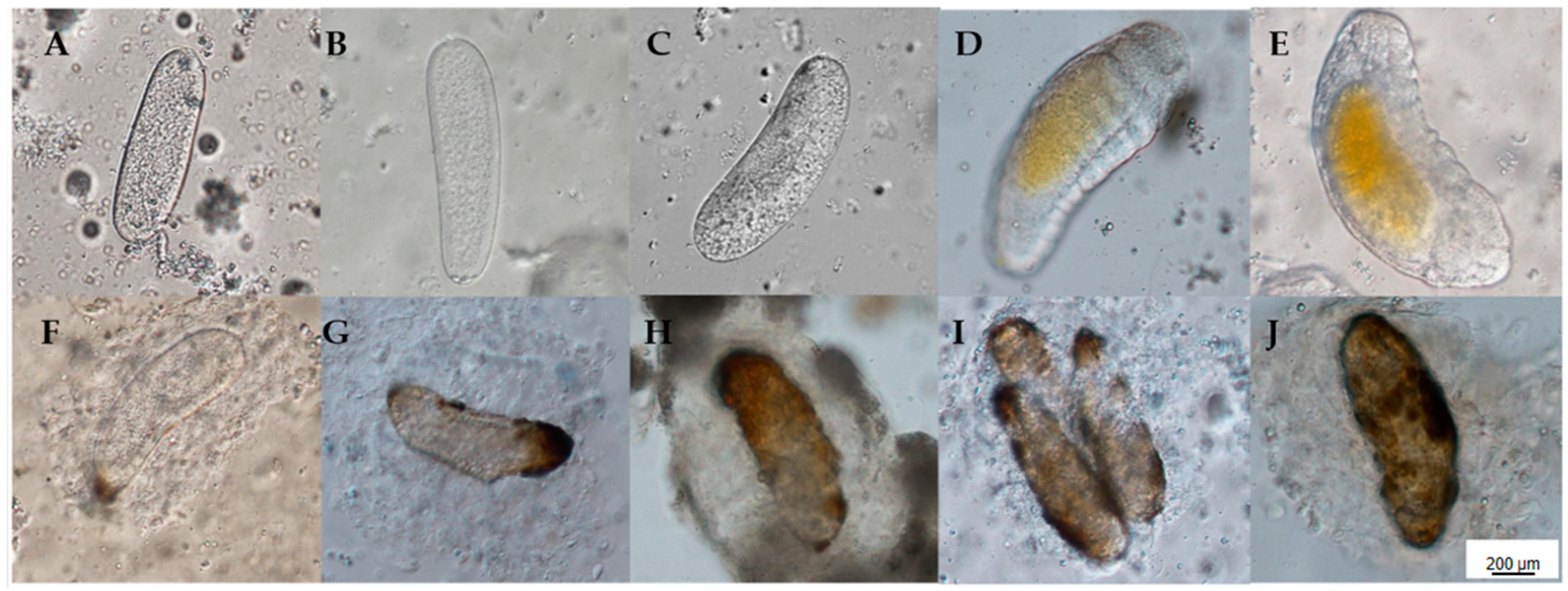
2.3.1. The Development of A. hispinarum Eggs in the Body of B. longissima and O. nipae Larvae

B. longissima larvae in their fourth instar were selected, placed in separate 9 cm petri dishes, and parasitized by fifteen A. hispinarum mating females in each dish. We dissected B. longissima larvae that were infected by A. hispinarum, using a differential interference microscope. We also observed their progress and captured images at 12, 24, 48, 72, and 96 h after the two insects lay their eggs. Each time point involved the repetition of three groups. The same experimental protocol as B. longissima was followed by O. nipae.
The eggs were stained with rhodamine phalloidin and DAPI and photographed using a differential interference microscope within 24 h. These are the precise steps: The eggs were transferred into a slide coated with poly-L-lysine after 10 μL of PBS was poured upon it. Remove the liquid, absorb 10 μL of PBS rinse, repeat three times, add 10 μL of 4% paraformaldehyde, and then place the wet box in a fixed-temperature environment for 15 min. Rinse three times with 10 μL of PBS, then add 10 μL of 0.1% Triton X-100 and incubate for five minutes in a moist box. Following three rinses with 10 μL of PBS, 10 μL of 1% BSA was added to the wet box for one hour. After three rinses with 10 μL PBS, 10 μL Rhodamine phalloidin and 5 μL (1 μg/μL) DAPI were added and stained in a wet box for 45 min. Rinse three times with 10 μL PBS, then add another 10 μL PBS, cover with a coverslip, and photograph with a differential interference microscope.

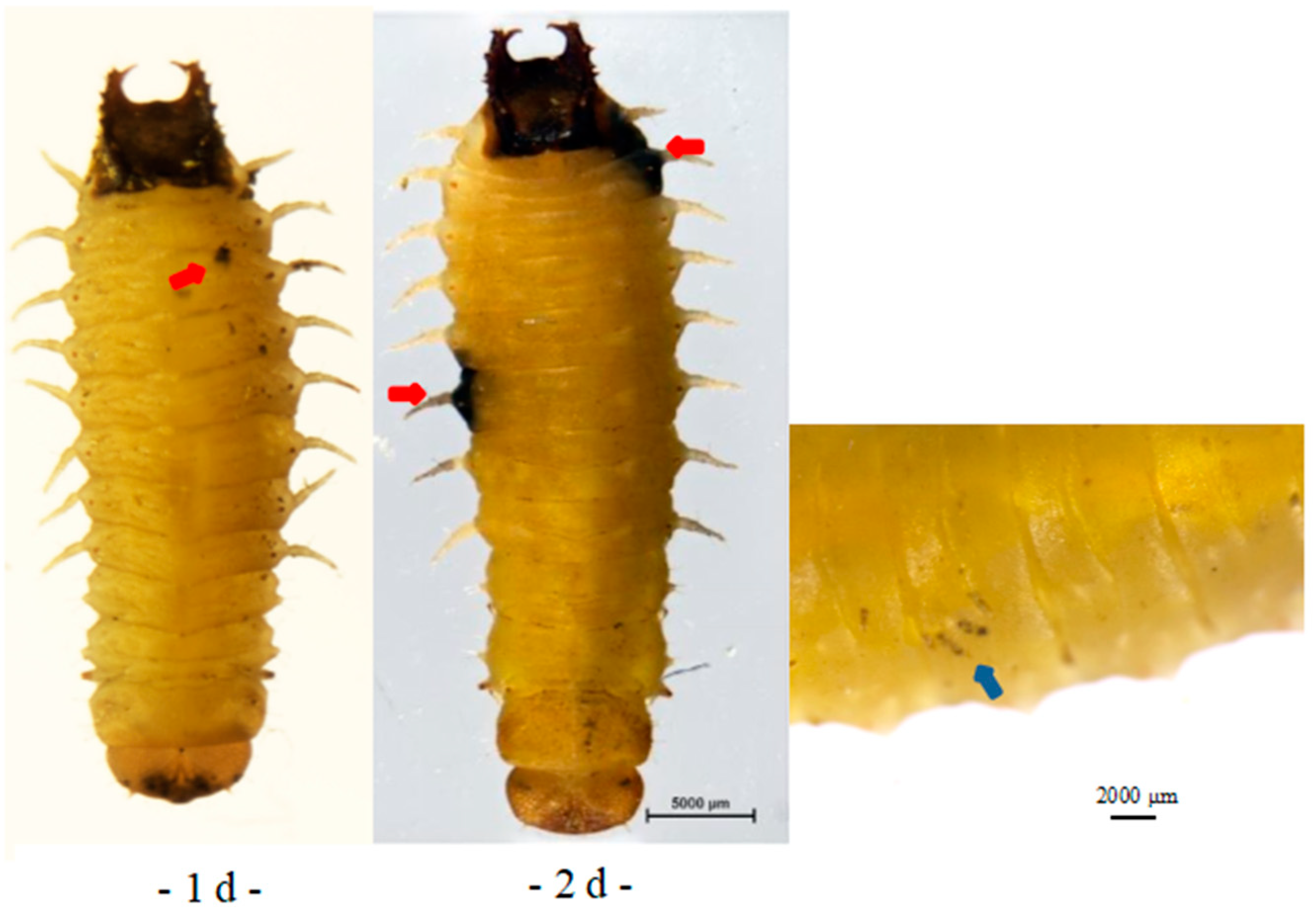
2.3.2. Effects of A. hispinarum Parasitism on B. longissima and O. nipae Larvae Development

B. longissima and O. nipae larvae that were parasitized by A. hispinarum were observed and captured under a stereo microscope; 30 B. longissima 4th instar larvae and 30 O. nipae 4th instar larvae were chosen and placed in separate 9 cm petri dishes. One-day-old, mated female A. hispinarum parasitized each larva one at a time. A. hispinarum was removed 24 h later. The parasitized larvae were constantly fed until they either pupated or became stiff. To count the number of larval deaths, B. longissima and O. nipae fourth instar larvae that were not parasitized throughout the same period were utilized as controls. The experiment was carried out in 30 groups. Ten fourth-instar O. nipae larvae were chosen, and 30 mated female A. hispinarum were inserted to perform one-for-one parasitism. The larvae were used as a control, and the pupal and fourth instar larvae of the palmar anise were counted. The experiment was carried out in thirty groups.

2.4. Comparison of the Encapsulation Rates of A. hispinarum Eggs with B. longissima and O. nipae Larvae

Thirty B. longissima 4th instar larvae and thirty O. nipae 4th instar larvae were dissected, and the total number of eggs in each larva and the number of eggs encapsulated were counted.

2.5. The Effect of A. hispinarum Parasitism on the Number and Proportion of B. longissima and O. nipae Larvae Hemocytes

The hemolymph of B. longissima fourth instar larvae was collected at 12, 24, 48, 72, and 96 h after parasitization, and the hemolymph of non-parasitic B. longissima larvae was used as a control at each time. The hemocytes were counted with a blood cell counter (25 × 16) under an optical microscope, including the number of cells and blood cell types; this was repeated five times. At each time point, 30 larvae were taken as replicates, and the experimental method was the same for O. nipae larvae.

2.6. Satistical Analysis

The mortality of of B. longissima and O. nipae larvae under non-parasitization and parasitization, the selective parasitism rate of A. hispinarum to B. longissima and O. nipae larvae, and the egg encapsulation rate of these two hosts after parasitization were analyzed using the Chi-square test. The fecundity of B. longissima and O. nipae larvae after parasitization, the duration of the larvae’s fourth instar and pupal stage, the hemocyte count, and the proportion of different hemocyte types under parasitization and non-parasitization were compared using an independent sample t-test. The changes in the proportion of hemocyte types at different time points post-parasitization and non-parasitization were analyzed using ANOVA. All statistical analysis mentioned above were performed on SPSS 21, and the graphs were drawn using GraphPad Prism 7.

3. Results

3.1. Parasitic Selection of A. hispinarum to B. longissima and O. nipae Larvae

There was no difference in the parasitism selection of A. hispinarum to B. longissima and O. nipae larvae in the non-selective parasitism experiment (p = 0.859), although there was a significant difference in the number of eggs laid (p < 0.001). The number of eggs produced in the B. longissima larvae was more than that in the O. nipae larvae (Table 1).
There was no difference in the parasitism selection of A. hispinarum to B. longissima and O. nipae larvae in the selective parasitism experiment (p = 0.568). In terms of oviposition, there was a significant difference in the quantity of eggs laid (p < 0.001). The quantity of eggs deposited in the B. longissima larvae was more than that in the O. nipae larvae (Table 1).

3.2. The Developmental Interaction of A. hispinarum with B. longissima and O. nipae Larvae

3.2.1. Development of A. hispinarum Egg in B. longissima and O. nipae Larvae

A. hispinarum eggs laid in B. longissima larvae can grow normally and mature into larvae 72 h later, according to ongoing observations of eggs laid in B. longissima and O. nipae larvae (Figure 1A–E). The eggs were encapsulated in O. nipae larvae from 12 h on, but as the encapsulation grew stronger over time, the eggs were unable to develop normally and eventually died (Figure 1F–J).
It can be seen that the eggs could develop normally in the B. longissima larvae (Figure 2B), and they formed a blastoderm within 24 h, after staining to observe the eggs in fallopian tubes of A. hispinarum and those laid in B. longissima and O. nipae larvae 24 h later (Figure 2A) (Figure 2B Phalloidin). The eggs laid in the larvae of O. nipae can also develop, but they asphyxiate until they die, because the egg surface is covered in thick hemocytes, in contrast to the eggs in the fallopian tubes of A. hispinarum. (Figure 2C).

3.2.2. The Effect of A. hispinarum Parasitism on the Development of B. longissima and O. nipae Larvae

A. hispinarum parasitized B. longissima larva, leaving behind black spots on the body surface where the parasites lay their eggs. The parasitized B. longissima larva gradually turned black over a period of seven days (see Figure 3: the red arrow is where the parasite wasp spawned). O. nipae larva showed localized blackening around the spawning position after being parasitized by A. hispinarum (Figure 4, red arrow), and the eggs enclosed in the larva could be seen through the body surface (Figure 4, blue arrow). The parasitized larvae can still be observed with the encapsulated eggs until they become adults after extended observation.
A. hispinarum parasitized B. longissima, O. nipae larvae were raised, and the uninfected B. longissima and O. nipae larvae served as the control to measure the mortality of the two insects (Table 2). When B. longissima larvae were parasitized by A. hispinarum, their death rate was 98.88% ± 0.33%, which was substantially greater than the unparasitized rate of (1.11% ± 0.33%) (χ2 = 1716.890, df = 1, p < 0.001). O. nipae larvae infected with A. hispinarum had a mortality rate of 15.31% ± 1.62% (Table 2), which was significantly greater than the rate of uninfected larvae (1.44% ± 0.35%) (χ2 = 112.768, df = 1, p < 0.001). The death rates of unparasitized B. longissima and O. nipae larvae are identical (χ2 = 0.184, df = 1, p = 0.668). The death rate of B. longissima larvae was considerably higher than that of O. nipae larvae following parasitism by A. hispinarum (χ2 = 1275.909, df = 1, merged DAPI, merged phalloidin DAPIDAPIABC8 p < 0.001).
The statistics of the larval and pupal stages of O. nipae larvae surviving after being parasitized by A. hispinarum (Table 3) showed that the fourth instar larval stage is 12.55 ± 0.09 d, which was 5 d longer than the unparasitized one (7.17 ± 0.08 d); the pupal stage of parasitized was 10.64 ± 0.08 d, which was 1 d longer than the unparasitized one (8.96 ± 0.06 d).

3.3. Comparison of Encapsulation Rate of B. longissima and O. nipae Larvae to A. hispinarum Eggs

O. nipae larval hemocytes promptly encapsulated the A. hispinarum eggs when they were placed in B. longissima and O. nipae larvae. The encapsulation rate reached 99.05%, which was substantially greater than that of B. longissima larval hemocytes (1.14%) (χ2 = 1352.977, df = 1, p < 0.001) (Figure 5). Therefore, it stands to reason that eggs cannot survive in O. nipae larvae.

3.4. The Effect of the Hemocytes in B. longissima and O. nipae Larvae and the Proportion of Various Types of Hemocytes after Parasitization by A. hispinarum

3.4.1. The Effect of Hemocytes in B. longissima and O. nipae Larvae after Parasitization by A. hispinarum

The number of hemocytes in the fourth instar larva of B. longissima and O. nipae was maintained at a largely constant level at 12, 24, 48, 72, and 96 h after feeding (Table 4). The statistics show that O. nipae larvae in their fourth instar have 6.08 more hemocytes than B. longissima larvae (t = −21.496, df = 292, p < 0.001).
The number of hemocytes increased considerably in B. longissima larvae 24 h after being parasitized by A. hispinarum (t = −3.763, df = 58, p < 0.001) (Table 4). In O. nipae larvae, the number of hemocytes dramatically increased 48, 72, and 96 h following A. hispinarum parasitism (48 h: t = −4.190, df = 58, p < 0.001; 72 h: t = −3.199, df = 58, p = 0.002; 96 h: t = −2.730, df = 58, p = 0.008) (Table 4). It can be seen that the change in blood cell number in O. nipae larvae following parasitization by A. hispinarum was more pronounced than it was in B. longissima larvae.

3.4.2. The Effect of Parasitism of A. hispinarum on the Proportion of Various Hemocytes in Larvae of B. longissima and O. nipae

Blood cell composition and fluctuation, in addition to number, are additional indicators of the degree of encapsulation reaction. The results demonstrated that plasma cells, granular cells, oenocytoid, primitive cells, and beaded cells were the most prevalent blood cell types and morphologies in the larvae of B. longissima and O. nipae. The largest proportions were seen in plasma cells (Table 5) and granular cells (Table 6).
The proportion of plasma hemocytes in B. longissima larvae parasitized by A. hispinarum did not change significantly after 12, 24, 48, 72, and 96 h (12 h: t = 0.543, df = 58, p = 0.589; 24 h: t = −1.301, df = 54, p = 0.198; 48 h: t = −0.317, df = 58, p = 0.753; 72 h: t = 1.110, df = 58, p = 0.272; 96 h: t =1.728, df = 52, p = 0.090) (Table 5); the proportion of granulocytes increased and decreased significantly, at 24 and 96 h after parasitization (24 h: t = −2.569, df = 54, p= 0.013; 96 h: t = 2.552, df = 58, p = 0.013) (Table 6); the proportion of oenocytoid increased significantly at 12 h after being parasitized (t = −2.623, df = 54, p = 0.011) (Table 7); The proportion of primary hemocytes decreased significantly 48 h, 72 h (48 h: t = 3.380, df = 58, p = 0.001; 72 h: t = 3.866, df = 52, p < 0.001), but increased significantly 96 h after parasitism (t = −3.855, df = 58, p < 0.001) (Table 8).
The proportion of plasmacytes in the larvae of O. nipae decreased significantly at 12 h after parasitism by A. hispinarum (t = 2.043, df = 58, p = 0.046) and increased significantly at 24, 72, and 96 h (24 h: t = −2.479, df = 58, p = 0.016; 72 h: t = −2.525, df = 58, p = 0.014; 96 h: t = −2.419, df = 58, p = 0.019) (Table 5); the proportion of granulocytes increased significantly at 12 h after parasitization (t = −2.406, df = 58, p = 0.019) (Table 6); the proportion of oenocytoids gradually decreased after parasitization, and there was a significant difference at 72 h (t = 2.970, df = 58, p = 0.004) (Table 7); the proportion of prohemocytes increased significantly at 24 h after parasitization (t = 5.636, df = 58, p < 0.001) (Table 8). When compared to B. longissima, the changes in the proportion of hemocyte composition of the larvae after parasitization by A. hispinarum were more significant in O. nipae.

4. Discussion

In nature, parasitoid wasps and hosts have a balanced struggle and restraint interaction [35,36]. Researchers from all around the world have been investigating the immunological and developmental interactions between parasitoids and hosts in recent decades [37,38,39,40,41]. Depending on whether the parasitoid can develop and enclose successfully in the host, the parasitoid’s host can be classified as adaptive or non-adaptive [42]. In contrast to non-adaptive hosts, in which the host is unable to overcome the host’s immune system, adaptable hosts allow the host to suppress or avoid the immune system. During the parasitic process, the host must first contend with cellular immunity, with the host hemocytes’ envelope reaction serving as the initial hurdle [16,18].
Studies have been conducted on the selection of A. hispinarum as a parasite, the development interaction between A. hispinarum and the hosts B. longissima and O. nipae, and the hemocyte response of these hosts. The findings demonstrated that A. hispinarum exhibited no preference for B. longissima or O. nipae parasitic selection, but that it preferred to deposit more eggs in B. longissima larvae. This might be connected to how big B. longissima and O. nipae are physically. The body size of the B. longissima is larger than that of the O. nipae in all life stages, including larvae, pupa, and adults. During the parasitism process, parasitic wasps typically choose the most suitable host in which to lay eggs and tend to lay more eggs into the most suitable hosts [43]. Host density, body size, nutritional status, and developmental stage are the main influencing factors [44,45,46,47].
However, the eggs laid by A. hispinarum into its adaptive host B. longissima larvae could develop normally, whereas the eggs laid into its non-adaptive host O. nipae larvae were encapsulated to death and could not develop normally. In fact, in the O. nipae larvae, there are some A. hispinarum eggs that can develop into A. hispinarum, but this phenomenon is extremely rare; the current experiment found only one case. Non-adaptive hosts will still be affected by this process, even though parasitic wasps cannot successfully parasitize them. The parasitism of A. hispinarum in this experiment can cause a death rate of 15.31% of the O. nipae larvae and extend the larval stage by 5 days and the pupal stage by 1 day. O. nipae damages the young leaves of palm trees by feeding adults and larvae [48]. Field chemical control often uses beta-cypermethrin, imidacloprid, and other drugs to spray leaves [49,50]. Thus, the extension of the larval stage may allow the insect to absorb the drug more fully, thereby achieving a good control effect. Additionally, we discovered through ongoing field research that though B. longissima and O. nipae can coexist in one area, they rarely feed on the same plants. As a result, utilizing A. hispinarum to regulate B. longissima can have some negative consequences on O. nipae.
The encapsulation response of the host is the first thing the A. hispinarum eggs encounter after being deposited into the host. In this experiment, hemolymph from B. longissima larvae could only encapsulate 1.14% of A. hispinarum eggs, but hemolymph from O. nipae larvae could contain 99.05% of A. hispinarum eggs. Studies have shown a strong correlation between the overall number of host hemocytes and the number of differential hemocytes and the success of parasitic wasps [51]. Statistics show that O. nipae larvae had 6.08 times as many total hemocytes than B. longissima larvae. The amount of circulating hemocytes in the host hemolymph may have a significant role in controlling the capacity to trigger cyst response [52].
The immune system of the host is also suppressed or avoided by parasitic wasps, ensuring the normal development of the offspring [22,29,30]. The hemocytes in the B. longissima larvae did not react significantly to the attack by A. hispinarum, and only a small increase in the total number of hemocytes and an increase in the proportion of granulocytes and oenocytoids were visible in the early stages of the parasitization. This demonstrates that the cell’s immune system can act quickly, but it may be slowed down by A. hispinarum’s parasitic elements or rendered ineffective by the parasite’s eggs, which prevent the hemocytes from encasing the foreign substance. However, after O. nipae larvae were attacked by A. hispinarum, the total number of hemocytes grew, and the proportion of plasma and granular hemocytes increased, which formed the basis for the success of O. nipae larvae’s cellular immunity. According to research, the capacity of hemocytes is positively correlated with the number of circulating hemocytes in Drosophila melanogaster [20]. Plasmatocytes and granulocytes are key players in the encapsulation reaction, and oenocytoid cells can produce phenoloxidase to help blacken and kill bee eggs. Additionally, the original hemocytes can change into different types of hemocytes. Since the likelihood of A. hispinarum eggs being encapsulated by the two hosts differ significantly, it may be concluded that A. hispinarum eggs are capable of passive escape, which can only happen in B. longissima larvae.

5. Conclusions

The goal of this research was to look into A. hispinarum’s developmental interactions with B. longissima and O. nipae, as well as the hemocyte immune responses of B. longissima and O. nipae larvae to A. hispinarum parasitism, so that the breeding differences between B. longissima and O. nipae could be explained. The results showed that A. hispinarum had a preference for oviposition over the parasitism of B. longissima and O. nipae. The number of eggs laid to B. longissima larvae was significantly greater than that to O. nipae larvae, which led to a 98.95% mortality rate for B. longissima larvae and only a 15.31% mortality rate for O. nipae larvae. Another important finding is that the sufficient amount of hemocytes in O. nipae larvae served as the foundation for cellular immunity; the number of hemocytes in O. nipae larvae was 6.08 times that of B. longissima larvae. This study’s limitation is that it does not take humoral immunity into consideration. The fact that melanization takes place during A. hispinarum egg encapsulation suggests that parasitism will also cause variations in terms of how humoral immunity responds. As a result, we may further investigate various immunological effects on the two beetles from the perspective of humoral immunity.

Author Contributions

Methodology, Z.C. and T.F.; validation, Z.C. and T.F.; formal analysis, Z.C., B.T. and L.F.; data curation, Z.C., B.L. and Y.L.; writing—original draft preparation, Z.C. and T.F.; writing—review and editing, Z.C.; funding acquisition, Y.H. All authors have read and agreed to the published version of the manuscript.

Funding

We are grateful to the Natural Science Foundation of Fujian Province (2021J01448), the National Key Research and Development Program of China (2017YFC1200605), the Fujian Science and Technology Special Project (2017NZ0003-1-6), the Project of the General Administration of Customs of the People’s Republic of China (2020HK144), and the Project of the Fuzhou Customs District of the People’s Republic of China (FK2020-02).

Institutional Review Board Statement

Not applicable.

Informed Consent Statement

Not applicable.

Acknowledgments

All of the authors sincerely thank Mubasher Hussain (Institute of Zoology, Guangdong Academy of Sciences, Guangzhou) for insightful revisions of our manuscript and Zhengqiang Peng (the Environment and Plant Protection Institute, Chinese Academy of Tropical Agricultural Sciences, Haikou 571101, China) for providing samples of A. hispinarum for the experiment.

Conflicts of Interest

The authors declare no conflict of interest.

References

  1. Maulik, S. A new hispid beetle injurious to nipa palm. Ann. Mag. Nat. Hist. 1921, 7, 451–452. [Google Scholar] [CrossRef] [Green Version]
  2. Waterhouse, D.F.; Norris, K.R. Biological Control Pacific Prospects: Brontispa longissima (Gestro); Inkata Press: Melbourne, Australia, 1987; pp. 134–141, 211–218. [Google Scholar]
  3. Hou, Y.M.; Weng, Z.Q. Temperature-Dependent Development and Life Table Parameters of Octodonta nipae (Coleoptera: Chrysomelidae). Environ. Entomol. 2010, 39, 1676–1684. [Google Scholar] [CrossRef] [PubMed]
  4. Liu, K.; Fu, B.L.; Lin, J.R.; Fu, Y.G.; Peng, Z.Q.; Tang, L.D. Parasitism Performance of Tetrastichus brontispae Ferriere over the Coconut Hispine Beetle, Brontispa longissima (Gestro). Neotrop. Entomol. 2016, 45, 420–426. [Google Scholar] [CrossRef] [PubMed]
  5. Stapley, J.H. Coconut leaf beetle (Brontispa longissima) in the Solomons. Alafua Agr. Bull. 1980, 5, 17–22. [Google Scholar]
  6. Chen, Z.M.; Wang, G.H.; Li, M.; Peng, Z.Q.; Ali, H.; Xu, L.N.; Geoff, G.M.; Hou, Y.M. Development of Single Nucleotide Polymorphism (SNP) Markers for Analysis of Population Structure and Invasion Pathway in the Coconut Leaf Beetle Brontispa longissima (Gestro) Using Restriction Site-Associated DNA (RAD) Genotyping in Southern China. Insects 2020, 11, 230. [Google Scholar] [CrossRef] [Green Version]
  7. Lv, B.Q.; Chen, Y.Q.; Bao, Y.; Han, R.D.; Peng, Z.Q. The feasibility of the controlling coconut leaf beetle (Brontispa longissima) with introducing natural enemies Asecodes hispinarum. Chin. Bull. Entomol. 2005, 42, 254–258. [Google Scholar]
  8. Lv, B.Q.; Peng, Z.Q.; Tang, C.; Wen, H.B.; Jin, Q.A.; Fu, Y.G.; Du, Y.Z. Biological characteristics of Asecodes hispinarum Bouček (Hymenoptera: Eulophidae), a parasitoid of Brontispa longissima (Gestro) (Coleoptera: Hispidae). Acta Entomol. Sin. 2005, 48, 943–948. [Google Scholar]
  9. Minchella, D.J. Host life-history variation in response to parasitism. Parasitology 1985, 90, 205–216. [Google Scholar] [CrossRef]
  10. Strand, M.R.; Pech, L.L. Immunological Basis for Compatibility in Parasitoid-Host Relationships. Annu. Rev. Entomol. 1995, 40, 31–56. [Google Scholar] [CrossRef]
  11. Poyet, M.; Havard, S.; Prevost, G.; Chabrerie, O.; Doury, G.; Gibert, P.; Eslin, P. Resistance of Drosophila suzukii to the larvae parasitoids Leptopilina heterotoma and Asobara japonica is related to haemocyte load. Physiol. Entomol. 2013, 38, 45–53. [Google Scholar] [CrossRef]
  12. Bitra, K.; Burke, G.R.; Strand, M.R. Permissiveness of lepidopteran hosts is linked to differential expression of bracovirus genes. Virology 2016, 492, 259–272. [Google Scholar] [CrossRef] [PubMed]
  13. Li, L.F.; Xu, Z.W.; Liu, N.Y.; Wu, G.X.; Ren, X.M.; Zhu, J.Y. Parasitism and venom of ectoparasitoid Scleroderma guani impairs host cellular immunity. Arch. Insect Biochem. 2018, 98, e21451. [Google Scholar] [CrossRef]
  14. Darsouei, R.; Karimi, J.; Dunphy, G.B. Functional Characterization of Outer Membrane Proteins (OMPs) in Xenorhabdus nematophila and Photorhabdus luminescens through Insect Immune Defense Reactions. Insects 2019, 10, 352. [Google Scholar] [CrossRef] [PubMed]
  15. Sorrentino, R.P.; Carton, Y.; Govind, S. Cellular Immune Response to Parasite Infection in the Drosophila Lymph Gland Is Developmentally Regulated. Dev. Biol. 2002, 243, 65–80. [Google Scholar] [CrossRef] [PubMed] [Green Version]
  16. Parsons, P.; Foley, E. Cellular immune defenses of Drosophila melanogaster. Dev. Comp. Immunol. 2016, 58, 95–101. [Google Scholar] [CrossRef]
  17. Hillyer, J.F.; Strand, M.R. Mosquito hemocyte-mediated immune responses. Curr. Opin. Insect Sci. 2014, 3, 14–21. [Google Scholar] [CrossRef] [Green Version]
  18. Tsuzuki, S.; Matsumoto, H.; Furihata, S.; Ryuda, M.; Tanaka, H.; Sung, E.J.; Bird, G.S.; Zhou, Y.X.; Shears, S.B.; Hayakawa, Y. Switching between humoral and cellular immune responses in Drosophila is guided by the cytokine GBP. Nat. Commun. 2014, 5, 4628. [Google Scholar] [CrossRef] [Green Version]
  19. McGonigle, J.E.; Leitão, A.B.; Ommeslag, S.; Smith, S.; Day, J.P.; Jiggins, F.M. Parallel and costly changes to cellular immunity underlie the evolution of parasitoid resistance in three Drosophila species. PLoS Pathog. 2017, 13, e1006683. [Google Scholar] [CrossRef] [Green Version]
  20. Eslin, P.; Prévost, G. Hemocyte load and immune resistance to Asobara tabida are correlated in species of the Drosophila melanogaster subgroup. J. Insect Physiol. 1998, 44, 807–816. [Google Scholar] [CrossRef]
  21. Fors, L.; Robert, M.; Theopold, U.; Hambäck, P.A. Differences in cellular immune competence explain parasitoid resistance for two coleopteran species. PLoS ONE 2014, 9, e108795. [Google Scholar] [CrossRef] [Green Version]
  22. Shi, M.; Chen, X.X. Progress in Study on Regulation of Insect Host Physiology by Parasitoids in China. Chin. J. Biol. Control 2015, 31, 620–637. [Google Scholar]
  23. Meng, E.; Qiao, T.; Tang, B.Z.; Hou, Y.M.; Yu, W.Z.; Chen, Z.M. Effects of ovarian fluid, venom and egg surface characteristics of Tetrastichus brontispae (Hymenoptera: Eulophidae) on the immune response of Octodonta nipae (Coleoptera: Chrysomelidae). J. Insect Physiol. 2018, 109, 125–137. [Google Scholar] [CrossRef] [PubMed]
  24. Voegele, J.M. Biological control of in Western Samoa: An ecological and economic evaluation. Agr. Ecosyst. Environ. 1989, 27, 315–329. [Google Scholar] [CrossRef]
  25. Lv, B.Q.; Tang, C.; Peng, Z.Q.; John, L.S.; Fanghao, W. Biological assessment in quarantine of Asecodes hispinarum Bouček (Hymenoptera: Eulophidae) as an imported biological control agent of Brontispa longissima (Gestro) (Coleoptera: Hispidae) in Hainan, China. Chin. J. Biol. Control 2008, 45, 29–35. [Google Scholar]
  26. Jin, T.; Jin, Q.A.; Wen, H.B.; Lv, B.Q.; Lin, Y.Y.; Peng, Z.Q. Research Progress and Perspective Outlook of Using Parasitic Wasps to Control Brontispa longissima. Chin. J. Trop. Agric. 2012, 32, 67–74. [Google Scholar]
  27. He, L.S.; Zurian, M.D.; Yap, M.L. A review of the status of the larvae parasitoid, Asecodes hispinarum Bouček, and of the pupal parasitoid, Tetrastichus brontispae Ferriere (Hymenoptera: Eulophidae), as biological control agents of the coconut leaf beetle, Brontispa longissima (Gestro) (Coleoptera: Chrysomelidae: Cassidinae), in the Asia-Pacific Region. Life Excit. Biol. 2014, 2, 42–62. [Google Scholar]
  28. Poirié, M.; Carton, Y.; Dubuffet, A. Virulence strategies in parasitoid Hymenoptera as an example of adaptive diversity. Comptes Rendus Biol. 2009, 332, 311–320. [Google Scholar] [CrossRef]
  29. Yin, C.L.; Li, M.Z.; Hu, J.; Lang, K.; Chen, Q.M.; Liu, J.D.; Guo, D.H.; He, K.; Dong, Y.P.; Luo, J.P.; et al. The genomic features of parasitism, Polyembryony and immune evasion in the endoparasitic wasp Macrocentrus cingulum. BMC Genom. 2018, 19, 420–438. [Google Scholar] [CrossRef] [Green Version]
  30. Kaiser, M.; Arvidson, R.; Zarivach, R.; Adams, M.E.; Libersat, F. Molecular cross-talk in a unique parasitoid manipulation strategy. Insect Biochem. Mol. Biol. 2019, 106, 64–78. [Google Scholar] [CrossRef]
  31. Ye, G.Y.; Hu, J.; Zhu, J.Y.; Fang, Q.; Yan, Z.C.; Wang, L. Recent advances in research on the mechanisms through which parasitoid wasps regulate host immunity and development. Chin. J. Appl. Entomol. 2019, 56, 382–400. [Google Scholar]
  32. Kati, A.; Hardie, J. Regulation of wing formation and adult development in an aphid host, Aphis fabae, by the parasitoid Aphidius colemani. J. Insect Physiol. 2010, 56, 14–20. [Google Scholar] [CrossRef] [PubMed]
  33. Charles, H.; Godfray, J. Four decades of parasitoid science. Entomol. Exp. Appl. 2016, 159, 135–146. [Google Scholar]
  34. Kim, Y.; Hepat, R. Baculoviral p94 homologs encoded in Cotesia plutellae bracovirus suppress both immunity and development of the diamondback moth, Plutellae xylostella. Insect Sci. 2016, 23, 235–244. [Google Scholar] [CrossRef] [PubMed]
  35. Aya, V.M.; Montoya-Lerma, J.; Echeverri-Rubiano, C.; Michaud, J.P.; Vargas, G. Host resistance to two parasitoids (Diptera: Tachinidae) helps explain a regional outbreak of novel Diatraea spp. stem borers (Lepidoptera: Crambidae) in Colombia sugarcane. Biol. Control 2019, 129, 18–23. [Google Scholar] [CrossRef]
  36. Mathiron, A.G.E.; Pottier, P.; Goubault, M. Let the most motivated win: Resource value components affect contest outcome in a parasitoid wasp. Behav. Ecol. 2018, 29, 1088–1095. [Google Scholar] [CrossRef] [Green Version]
  37. Cai, J.; Ye, G.Y.; Hu, C. Parasitism of Pieris rapae (Lepidoptera: Pieridae) by a pupal endoparasitold, Pteromalus puparum (Hymenoptera: Pteromalidae): Effects of parasitization and venom on host hemocytes. J. Insect Physiol. 2004, 50, 315–322. [Google Scholar] [CrossRef] [PubMed]
  38. Gandon, S.; Buckling, A.; Decaestecker, E.; Day, T. Host–parasite coevolution and patterns of adaptation across time and space. J. Evol. Biol. 2008, 21, 1861–1866. [Google Scholar] [CrossRef] [PubMed] [Green Version]
  39. Yan, Z.; Qi, F.; Liu, Y.; Xiao, S.; Yang, L.; Wang, F.; An, C.; Werren, J.H.; Ye, G.Y. A Venom Serpin Splicing Isoform of the Endoparasitoid Wasp, Pteromalus puparum, Suppresses Host Prophenoloxidase Cascade by Forming Complexes with Host Hemolymph Proteinases. J. Biol. Chem. 2016, 292, 1038–1051. [Google Scholar] [CrossRef] [Green Version]
  40. Ye, Z.; Vollhardt, I.M.G.; Parth, N.; Rubbmark, O.; Traugott, M. Facultative bacterial endosymbionts shape parasitoid food webs in natural host populations: A correlative analysis. J. Anim. Ecol. 2018, 87, 1440–1451. [Google Scholar] [CrossRef]
  41. Cepeda, A.S.; Lotta-Arévalo, I.A.; Pinto-Osorio, D.; Macías-Zacipa, J.; Valkiūnas, G.; Barato, P.; Matta, N.E. Experimental characterization of the complete life cycle of Haemoproteus columbae, with a description of a natural host-parasite system used to study this infection. Int. J. Parasitol. 2019, 49, 975–984. [Google Scholar] [CrossRef]
  42. Chen, Y.F. A Study on Developmental and Immunological Interactions between Parasitoids and Their Nonpermissive Host. Doctoral Dissertation, Zhejiang University, Hangzhou, China, 2009. [Google Scholar]
  43. Vieira, N.F.; Pomari-Fernandes, A.; Lemes, A.A.F.; Vacari, A.M.; De, B.S.A.; De, F.B.A. Cost of Production of Telenomus remus (Hymenoptera: Platygastridae) Grown in Natural and Alternative Hosts. J. Econ. Entomol. 2017, 110, 2724–2726. [Google Scholar] [CrossRef] [PubMed]
  44. Barzman, M.S.; Daane, K.M. Host-handling behaviours in parasitoids of the black scale: A case for ant-mediated evolution. J. Anim. Ecol. 2001, 70, 237–247. [Google Scholar]
  45. Lebreton, S.; Darrouzet, E.; Chevrier, C. Could hosts considered as low quality for egg-laying be considered as high quality for host-feeding? J. Insect Physiol. 2009, 55, 694–699. [Google Scholar] [CrossRef] [PubMed]
  46. Pan, M.Z.; Liu, T.X.; Nansen, C. Avoidance of parasitized host by female wasps of Aphidius gifuensis (Hymenoptera: Braconidae): The role of natal rearing effects and host availability. Insect Sci. 2018, 25, 1035–1044. [Google Scholar] [CrossRef] [Green Version]
  47. Uy, F.M.K.; Espinoza, A.M. Differential Host Handling Behavior between Feeding and Oviposition in the Parasitic Wasp Haplogonatopus hernandezae. J. Insect. Behav. 2018, 31, 569–584. [Google Scholar] [CrossRef]
  48. Sun, J.H.; Yu, P.Y.; Zhang, Y.Z.; Wang, X.J. A new invasive coconut pest in Hainan Province. Entomol. Knowl. 2003, 40, 286–287. [Google Scholar]
  49. Huang, S.C.; Tan, W.Q.; Ma, Z.L.; Li, Z.X. Main Invasive Pests of Palm Plants in China and Their Control. Xiandai Nongye Keji 2007, 9, 91–92. [Google Scholar]
  50. Wei, Y.H.; Tan, W.Q.; Huang, S.C.; Yu, F.Y. Risk Analysis of Octodonta nipae in Hainan Province. Chin. J. Trop. Agr. 2017, 37, 46–49, 71. [Google Scholar]
  51. Gerritsma, S.; de Haan, A.; van de Zande, L.; Wertheim, B. Natural variation in differentiated hemocytes is related to parasitoid resistance in Drosophila melanogaster. J. Insect Physiol. 2013, 59, 148–158. [Google Scholar] [CrossRef]
  52. Salt, G. Experimental studies in insect parasitism-XII. The reactions of six exopterygote insects to an alien parasite. J. Insect Physiol. 1963, 9, 647–650. [Google Scholar] [CrossRef]
Figure 1. Development of A. hispinarum eggs in B. longissima and O. nipae larvae. (AE), A. hispinarum eggs in the fourth instar larvae of B. longissima parasitized by A. hispinarum after 12/24/48/72/96 h; (FJ), A. hispinarum eggs in the fourth instar larvae of O. nipae parasitized by A. hispinarum after 12/24/48/72/96 h. Scale bar = 200 μm.
Life 12 02025 g001
Figure 2. Encapsulation of A. hispinarum eggs in different sites. (A) The fallopian tube of A. hispinarum; (B) The fourth instar larvae of B. longissima parasitized by A. hispinarum after 24 h; (C) The fourth instar larvae of O. nipae parasitized by A. hispinarum after 24 h. Nuclei are stained with DAPI (blue) and the hemocytes stained with Phalloidin (red). Merged is the merged picture of blue and red channels. Scale bar = 200 μm.
Life 12 02025 g002
Figure 3. The growth and development of B. longissima larvae parasitized by A. hispinarum. (The red arrow is where the parasite wasp spawned. Scale bar = 5000 μm.).
Life 12 02025 g003
Figure 4. The growth and development of O. nipae larvae parasitized by A. hispinarum. (The red arrow is where the parasite wasp spawned. The blue arrow is the egg of A. hispinarum. Scale bar = 5000 μm/2000 μm.).
Life 12 02025 g004
Figure 5. Encapsulation rate of B. longissima and O. nipae larvae on A. hispinarum eggs.
Life 12 02025 g005
Table 1. The rate of parasitism of A. hispinarum on B. longissima and O. nipae and the amount of oviposition of A. hispinarum.
TreatmentSpeciesParasitism Rate (%)Oviposition Amountχ2tdfp
No-choice testB. longissima71.70 ± 1.75-0.032-10.859
O. nipae72.33 ± 2.36-
B. longissima-18.81 ± 0.31-7.61598<0.001
O. nipae-15.52 ± 0.30
Dural-choice testB. longissima58.26 ± 1.55-0.326-10.568
O. nipae55.87 ± 1.52-
B. longissima-17.31 ± 0.28-6.36583<0.001
O. nipae-15.00 ± 0.24
Table 2. Mortality of the fourth instar larvae of B. longissimi and O. nipae under unparasitization and parasitization by A. hispinarum.
SpeciesMortality Rate (%)
UnparasitizedParasitized
B. longissima1.11 ± 0.33 Ba98.88 ± 0.33 Aa
O. nipae1.44 ± 0.35 Ba15.31 ± 1.62 Ab
Note: The different uppercase letters within the same row represents significant difference between non-parasitization and parasitization by A. hispinarum at p < 0.05 level. The different lowercase letters within the same column represent significant difference between different hosts at p < 0.05 level.
Table 3. Comparison of duration of the fourth instar stage or pupae stage of O. nipae between non-parasitization and parasitizaiton by A. hispinarum.
Developmental DurationTreatmentDays (d)tdfp
Fourth instar larvaeUnparasitized7.17 ± 0.08−17.244453<0.001
Parasitized12.55 ± 0.09
PupaeUnparasitized8.96 ± 0.06−44.194577<0.001
Parasitized10.64 ± 0.08
Table 4. Effects of the total hemocyte counts of B. longissima larvae and O. nipae larvae parasitization by A. hispinarum.
Time (h)Total Hemocyte Counts (×2.5 × 10⁵)
B. longissimaO. nipae
UnparasitizedParasitizedUnparasitizedParasitized
1213.33 ± 1.33 Aa15.30 ± 1.45 Ab79.43 ± 9.67 Aa86.17 ± 8.22 Ab
2415.67 ± 2.07 Ba28.73 ± 2.97 Aa78.07 ± 6.87 Aa94.60 ± 8.09 Ab
4814.63 ± 1.94 Aa17.50 ± 2.83 Ab75.57 ± 8.40 Ba125.00 ± 8.28 Aab
7212.37 ± 1.49 Aa15.43 ± 2.03 Ab92.13 ± 12.71 Ba150.43 ± 13.05 Aa
9612.87 ± 1.56 Aa13.90 ± 1.54 Ab84.73 ± 12.87 Ba132.03 ± 11.60 Aab
Note: The different uppercase letters within the same row represent significant difference between non-parasitization and parasitization by A. hispinarum at p < 0.05 level. The different lowercase letters within the same column represent significant difference between different parasitization time at p < 0.05 level.
Table 5. Effects of the percentage of plasmatocytes of B. longissima larvae and O. nipae larvae parasitized by A. hispinarum.
Time (h)Percentage of Plasmatocytes (%)
B. longissimaO. nipae
UnparasitizedParasitizedUnparasitizedParasitized
1238.33 ± 2.07 Ac36.78 ± 1.97 Ab56.49 ± 1.91 Aa51.69 ± 1.09 Bab
2440.72 ± 2.00 Abc44.98 ± 2.59 Aa49.61 ± 1.61 Babc55.06 ± 1.50 Aa
4847.37 ± 2.05 Aab48.24 ± 1.79 Aa53.03 ± 1.80 Aab52.92 ± 1.32 Aab
7252.81 ± 1.88 Aa49.84 ± 1.90 Aa43.58 ± 2.01 Bc49.17 ± 0.99 Ab
9654.14 ± 1.42 Aa49.84 ± 2.04 Aa46.70 ± 1.58 Bbc51.67 ± 1.32 Aab
Note: The different uppercase letters within the same row represent significant difference between non-parasitization and parasitization by A. hispinarum at p < 0.05 level. The different lowercase letters within the same column represent significant difference between different parasitization time at p < 0.05 level.
Table 6. Effects of the percentage of granulocytes of B. longissima larvae and O. nipae larvae parasitized by A. hispinarum.
Time (h)Percentage of Granulocytes (%)
B. longissimaO. nipae
UnparasitizedParasitizedUnparasitizedParasitized
1240.24 ± 2.00 Aa38.14 ± 2.18 Aa27.80 ± 1.76 Bc31.91 ± 0.86 Ab
2435.91 ± 1.56 Ba43.14 ± 2.19 Aa30.90 ± 1.73 Ac32.26 ± 0.84 Ab
4827.72 ± 1.76 Ab22.75 ± 2.11 Ab33.83 ± 1.31 Abc33.82 ± 0.92 Ab
7224.73 ± 1.89 Ab26.09 ± 2.10 Ab40.62 ± 2.09 Aa39.42 ± 0.94 Aa
9626.87 ± 1.90 Ab20.16 ± 1.82 Bb38.86 ± 1.00 Aab37.93 ± 1.22 Aa
Note: The different uppercase letters within the same row represent significant difference between non-parasitization and parasitization by A. hispinarum at p < 0.05 level. The different lowercase letters within the same column represent significant difference between different parasitization time at p < 0.05 level.
Table 7. Effects of the percentage of oenocytoids of B. longissima larvae and O. nipae larvae parasitized by A. hispinarum.
Time (h)Percentage of Oenocytoids (%)
B. longissimaO. nipae
UnparasitizedParasitizedUnparasitizedParasitized
124.23 ± 0.38 Bc5.91 ± 0.52 Ab6.57 ± 0.79 Aa7.37 ± 0.63 Aa
244.73 ± 0.53 Ac6.49 ± 0.83 Ab6.50 ± 0.65 Aa6.97 ± 0.77 Aa
4812.73 ± 1.40 Aa15.36 ± 1.43 Aa4.26 ± 0.42 Aa5.16 ± 0.43 Abc
727.59 ± 0.71 Ab6.44 ± 0.79 Ab6.08 ± 0.57 Aa4.01 ± 0.31 Bc
968.66 ± 0.76 Aab11.00 ± 0.94 Aa4.77 ± 0.56 Aa3.54 ± 0.34 Ac
Note: The different uppercase letters within the same row represent significant difference between non-parasitization and parasitization by A. hispinarum at p < 0.05 level. The different lowercase letters within the same column represent significant difference between different parasitization time at p < 0.05 level.
Table 8. Effects of the percentage of prohemocytes of B. longissima larvae and O. nipae larvae parasitized by A. hispinarum.
Time (h)Percentage of Prohemocytes (%)
B. longissimaO. nipae
UnparasitizedParasitizedUnparasitizedParasitized
126.03 ± 0.53 Ac5.37 ± 0.38 Ac7.80 ± 0.83 Ab6.91 ± 0.72 Aa
246.30 ± 0.46 Ac8.44 ± 0.95 Abc10.91 ± 0.86 Aa5.07 ± 0.56 Ba
488.10 ± 0.56 Abc5.77 ± 0.40 Bb8.35 ± 0.80 Aab7.40 ± 0.69 Aa
7212.83 ± 1.00 Aa8.10 ± 0.70 Bb6.56 ± 0.58 Ab6.28 ± 0.59 Aa
9610.03 ± 0.82 Bab16.14 ± 1.43 Aa7.15 ± 0.83 Ab5.45 ± 0.47 Aa
Note: The different uppercase letters within the same row represent significant difference between non-parasitization and parasitization by A. hispinarum at p < 0.05 level. The different lowercase letters within the same column represent significant difference between different parasitization time at p < 0.05 level.
Publisher’s Note: MDPI stays neutral with regard to jurisdictional claims in published maps and institutional affiliations.

