Article

Plasma Long Noncoding RNA LeXis is a Potential Diagnostic Marker for Non-Alcoholic Steatohepatitis

1
Department of Internal Medicine, College of Medicine, Yeungnam University, Daegu 42415, Korea
2
Department of Biochemistry and Cell Biology, School of Medicine, Kyungpook National University, Daegu 41944, Korea
3
Department of Internal Medicine, School of Medicine, Kyungpook National University, Kyungpook National University Hospital, Daegu 41944, Korea
4
Department of Pathology, Dongsan Medical Center, School of Medicine, Keimyung University, Daegu 42601, Korea
5
Department of Pathology, School of Medicine, Kyungpook National University, Kyungpook National University Hospital, Daegu 41944, Korea
6
Department of Surgery, School of Medicine, Kyungpook National University, Kyungpook National University Hospital, Daegu 41944, Korea
7
Department of Internal Medicine, Busan Paik Hospital, Inje University College of Medicine, Busan 74392, Korea
*
Authors to whom correspondence should be addressed.
These authors contributed equally to this study.
Life 2020, 10(10), 230; https://doi.org/10.3390/life10100230
Submission received: 6 August 2020 / Revised: 27 September 2020 / Accepted: 29 September 2020 / Published: 3 October 2020
(This article belongs to the Special Issue Fatty Liver Syndrome)

Abstract

:
Non-invasive diagnostic markers are needed to ease the diagnosis of non-alcoholic steatohepatitis (NASH) among patients with non-alcoholic fatty liver disease (NAFLD). The long noncoding RNA (lncRNA) LeXis is related to cholesterol metabolism and hepatic steatosis in mice, and its batch genome conversion in humans is TCONS_00016452. Here, we aimed to evaluate the potential of lncRNA LeXis as a non-invasive diagnostic marker for NASH. We analyzed a total of 44 NAFLD patients whose diagnosis was confirmed by a pathologist through analysis of a percutaneous liver biopsy. The expression of LeXis in the plasma of NAFLD patients with and without NASH was compared using quantitative real-time polymerase chain reaction. The expression of plasma LeXis was significantly higher in patients with NASH than in those with NAFL (8.2 (5.0–14.9); 4.6 (4.0–6.6), p = 0.025). The area under the receiver operating characteristic curve was 0.743 (95% CI 0.590–0.895, p < 0.001), and a sensitivity of 54.3% and specificity of 100% could be achieved for NASH diagnosis. Low LeXis was independently associated with NASH diagnosis in patients with NAFLD (p = 0.0349, odds ratio = 22.19 (5% CI, 1.25–395.22)). Therefore, circulating lncRNA LeXis could be a potential non-invasive diagnostic biomarker for NASH.

1. Introduction

Non-alcoholic fatty liver disease (NAFLD) is the most common chronic liver disease, affecting one-fourth of the population in western countries [1]. NAFLD prevalence has increased concomitantly with the increase in the number of obese individuals, thus representing elevated healthcare costs over time [2]. In the initial stages, NAFLD patients show excessive accumulation of fat in the liver without inflammation, which may later progress to an inflammation process, with cell damage and fibrosis [3]. The pathology of NAFLD ranges widely, from non-alcoholic fatty liver (NAFL) without liver cell injury to non-alcoholic steatohepatitis (NASH), leading to advanced fibrosis and cirrhosis, which can ultimately cause hepatocellular carcinoma [4]. Owing to its variable clinical progression, the identification of patients with poor prognosis among those with NAFLD is difficult. A recent study suggested that the presence of advanced fibrosis is a useful factor in predicting overall mortality in NAFLD patients; however, NASH is important for the pathogenesis of liver fibrosis [1,5,6,7]. Nevertheless, liver biopsy is the only confirmatory test available for the diagnosis of NASH and to ascertain NAFLD grading and staging. Non-invasive approaches for assessing liver fibrosis in NAFLD using demographic characteristics and biochemical markers were proposed in several studies [8], but no reliable biomarker to diagnose NASH has yet been identified. Thus, the identification of a diagnostic marker for the diagnosis of NASH via a non-invasive testing is necessary.
Long noncoding RNAs (lncRNAs) are a type of transcribed RNA molecule, longer than 200 nucleotides, that do not encode proteins [9]. They are known to be important regulators of gene expression and act via diverse mechanisms, including chromatin modification involving the conformation of the nuclear domain, the activation of transcriptional enhancers, and interference with transcriptional machinery [10,11]. Furthermore, some lncRNAs are involved in post-transcriptional processes including the regulation of splicing, the decay of mRNA and miRNA, and protein translation and stability [11]. Liver-expressed liver X receptors (LXR) induced sequence (LeXis) is a lncRNA that was recently identified in mice livers and was found to regulate lipid metabolism hemostasis in the liver and plasma by interacting with the ribonucleoprotein RALY [12]. Chronic inactivation of LeXis increases the hepatic cholesterol content without inflammation or endoplasmic reticulum (ER) stress. However, to date, the hepatic and plasma expression of LeXis in patients with NAFLD has not been not evaluated.
In the present study, we analyzed the expression of the lncRNA LeXis in both the liver tissue and plasma of patients with NAFLD by evaluating lncRNA TCONS_00016452, the corresponding human batch genome conversion of mouse lncRNA LeXis.

2. Methods

2.1. Patient Samples

Between January 2014 and June 2016, a total of 44 liver tissue biopsies and plasma samples were obtained from patients with pathologically confirmed NAFLD at Kyungpook National University Hospital. All tissue samples were obtained using ultrasound-guided percutaneous liver biopsy on the same day that biochemical parameters and a plasma sample were obtained from the patients, to minimize possible confounding factors. This study was approved by the ethical committees of the institute (KNUH 2018-05-025-003), and written informed consent was obtained from all patients.

2.2. NAFLD Diagnois and Pathological Evaluation

The diagnosis of NAFLD was made by a pathologist via the identification of at least 5% hepatic fat accumulation in pathology without excessive alcohol intake (men >140 g/week; women >70 g/week). Any other possible causes of chronic liver disease were excluded via analysis of serologic markers for viral hepatitis and the evaluation of medical records. All specimens were reviewed by a single experienced pathologist to avoid inter-observer variability. The diagnosis of NASH was made according to the current guidelines of the American Association for the Study of Liver Diseases [13]. Fibrosis staging and NAFLD activity score (NAS) were assessed according to recommendations of the Pathology Committee of the NASH Clinical Research Network (Figure 1) [14]. Severe NAFLD was defined as NAS above 4.

2.3. RNA Extraction

For total RNA extraction from clinical tissues, 1 mL of QIAzol Lysis Reagent (Qiagen, Hilden, Germany) was added to 100 mg of tissues. The tissue samples were homogenized and incubated for 5 min at room temperature (RT). Next, 0.2 mL chloroform was added to each tube, followed by vigorous stirring for 30 sec and additional 5-min incubation at RT. The samples were centrifuged at 12,000× g for 15 min at 4 °C. The upper aqueous phase was transferred to a new tube that was mixed by vortexing after added 0.5 mL isopropanol. After 10 min incubation at RT, the tube was centrifuged again at 12,000× g for 10 min at 4 °C, and the supernatant was discarded. The pellet was washed with 75% ethanol and centrifuged at 7500× g for 5 min at 4 °C. The pellet was air dried and resuspended in 20 μL of RNase-free water, and then stored at −80 °C until use.
The plasma sample was separated from whole peripheral blood by centrifugation at 3000 rpm for 10 min at 4 °C. Next, a miRNeasy Serum/Plasma Kit (Cat No./ID: 217204, Qiagen) was used to extract total RNA from the plasma, as described previously [15]. Briefly, 200 μL of plasma sample was mixed with 60 μL of Buffer RPL (Qiagen) and incubated for 3 min at RT. The sample was mixed with spike-in control (1.6 × 10⁸ copies/μL of cel-miR-39) and 20 μL of Buffer RPP; and the tube was mixed vigorously for 30 sec and incubated for 3 min at RT. After centrifuging at 12,000× g for 3 min at RT, the supernatant was transferred to a new tube, which was then mixed by vortexing after adding 0.2 mL of isopropanol. The mixture solution was transferred onto the column and was centrifuged at 8000× g for 15 min. After removing the flow-through, 700 μL of Buffer RWT (Qiagen) was added, and the column was centrifuged again at 8000× g for 15 min. This process was repeated with 500 μL of Buffer RPE (Qiagen) and then with 500 μL of 80% ethanol; the solution was then centrifuged at 10,000× g for 2 min. Lastly, the RNA captured in the column filter was eluted in 20 μL of RNase-free water.
The quantity and quality of the total extracted RNA was evaluated on a NanoDrop ND-1000 spectrophotometer (NanoDrop Technologies, Wilmington, DE, USA) was used.

2.4. Quantitative Real-Time PCR

For reverse transcription, a High-Capacity cDNA Reverse Transcription Kit (Thermo Fisher Scientific Inc., Waltham, MA, USA) was used with 800 µg of total RNA according to the manufacturer’s protocol for tissue samples. The protocol was performed with 1 µL of the solution, which involved the dilution of the resulting cDNA with 80 µL of distilled water. For reverse transcription of the plasma samples, 500 µg of total RNA was used which was diluted with 80 µL of distilled water. The protocol was performed with 4 µL of this diluted cDNA. Each sample was processed in triplicate via quantitative real-time PCR using SYBR Green PCR Master Mix (Thermo Fisher Scientific Inc.). The primer sequences of LeXis were used as follows: TCONS-00016452 (forward), 5′–TCACATCTCCTCCGTTTCAGAG–3′, and TCONS-00016452 (reverse), 5′–GTT ATGCGTCATGCCAAAGAAATC–3′. The PCR conditions were as follows: 2 min at 50 °C and 10 min at 95 °C, followed by 45 cycles of 15 s at 95 °C and 60 s at 60 °C. GAPDH (Glyceraldehyde-3-Phosphate Dehydrogenase) was used as the endogenous control, and the relative expression of LeXis was determined via the 2−ΔΔCt method.

2.5. Statistical Analysis

All continuous data are expressed as the means ± standard deviations or medians with interquartile ranges after testing for normality. All categorical data are expressed as numbers with percentages. Significant differences between patients were analyzed using the chi-square test, Student’s t-test, Mann-Whitney U test, or Fisher’s exact test. The analysis of the correlation between LeXis and steatosis was performed using the Spearman’s correlation coefficient. Receiver operating characteristic (ROC) analysis was conducted to assess the diagnostic performance of LeXis for NASH. All statistical analyses were performed using the R software (version 3.2.2; R Foundation for Statistical Computing, Vienna, Austria). A p value less than 0.05 was considered statistically significant.

3. Results

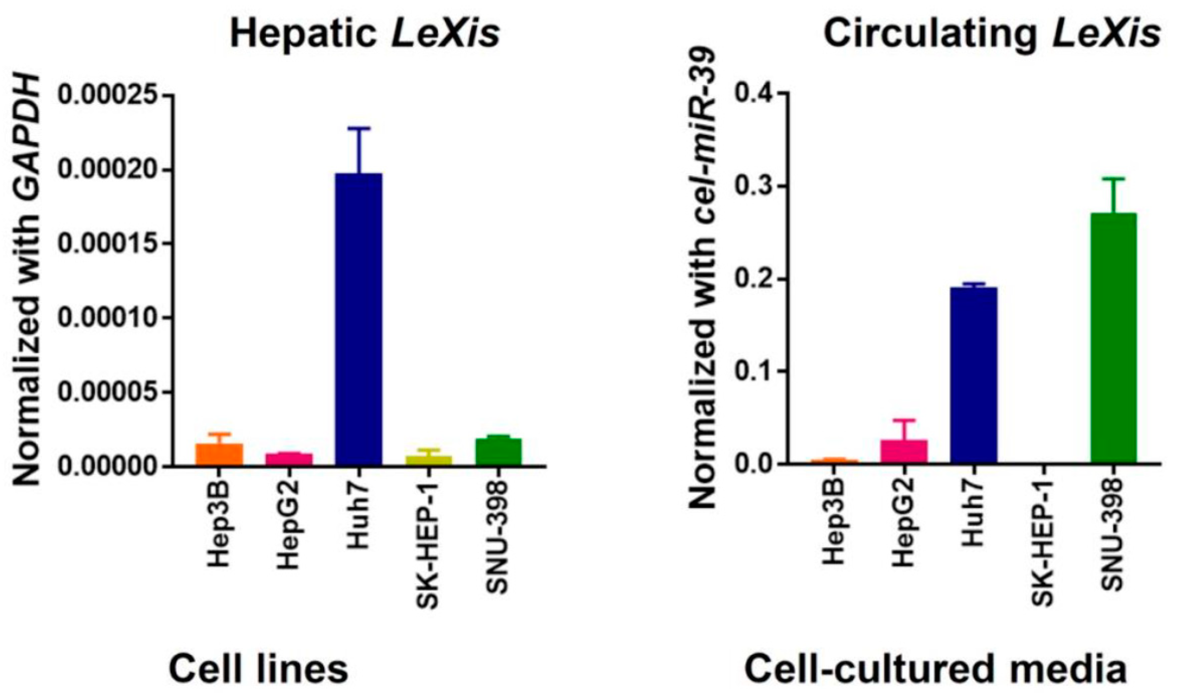
3.1. Hepatic lncRNA LeXis is Downregulated in Patients with Severe Hepatic Steatosis

As reported previously, the expression of hepatic LeXis was significantly downregulated in patients with severe steatosis compared with that in patients with mild-to-moderate steatosis (S = 1–2: 6.6 (3.7–12.0); S = 3: 3.2 (1.4–5.5); p = 0.017). However, the expression of plasma LeXis was not significantly different between the patient groups (S = 1–2: 7.7 (4.6–13.7), S = 3: 6.7 (4.2–7.9), p = 0.399; Figure 2). In addition, the degree of steatosis was negatively correlated with hepatic LeXis levels but not with plasma LeXis levels. (tissue, r = −0.327, p = 0.040; plasma, r = −0.131, p = 0.396; Figure 3).

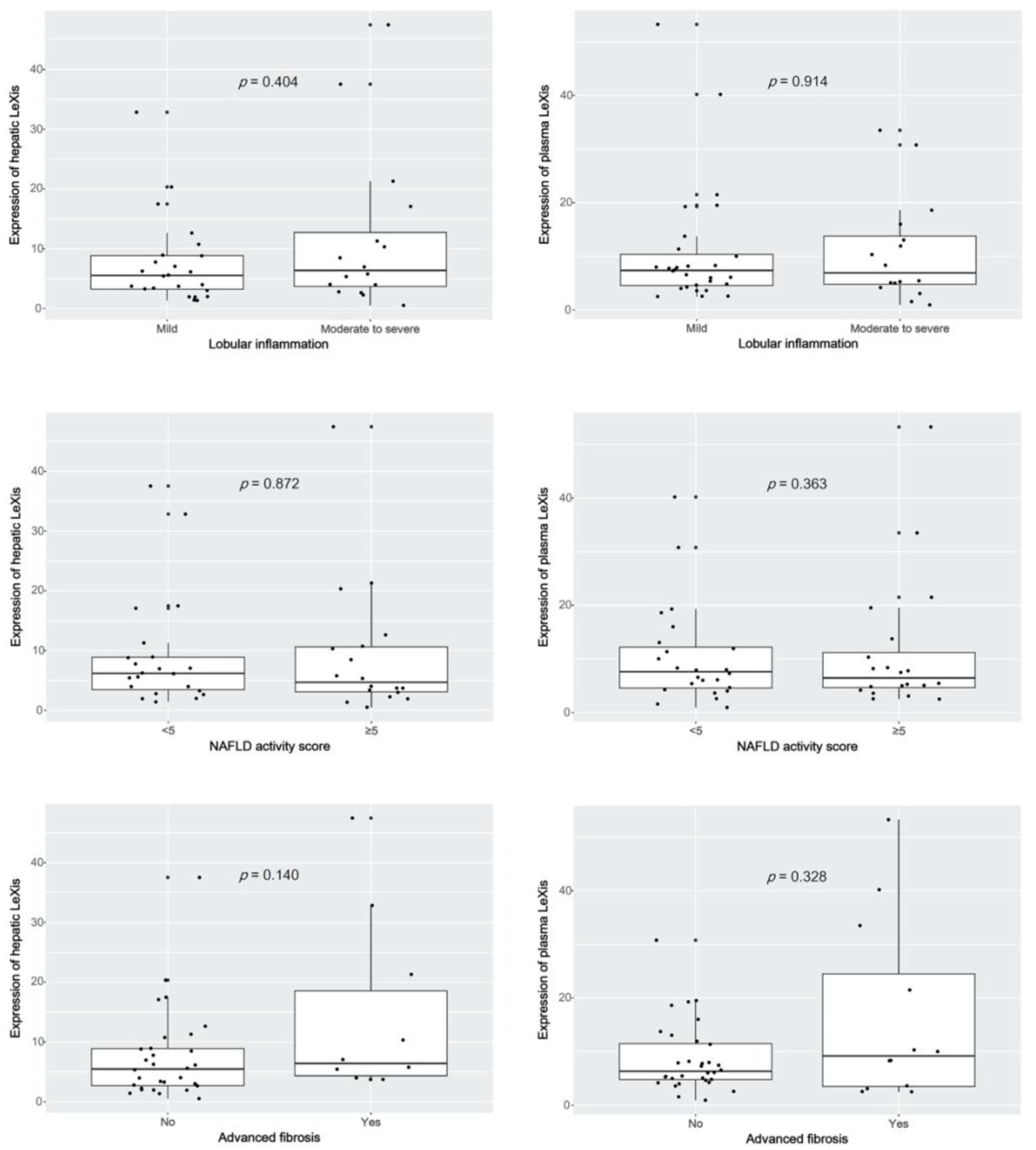
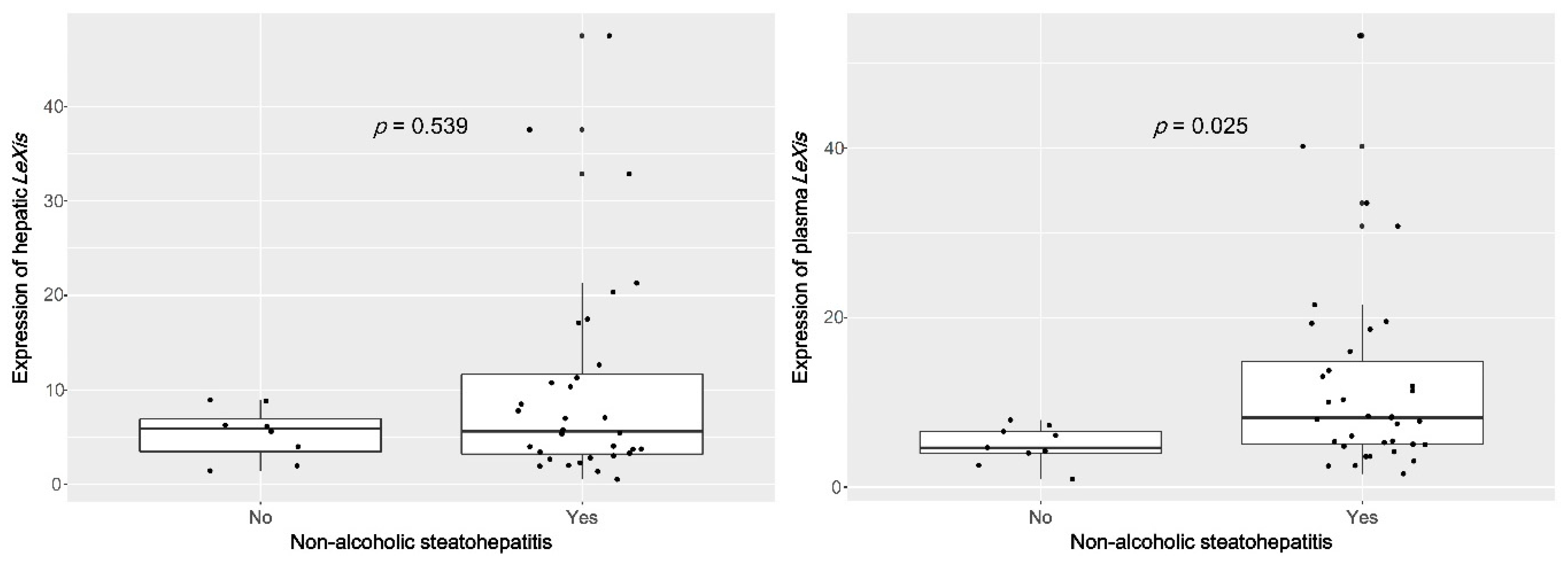
3.2. Plasma lncRNA LeXis Levels are Increased in Patients with NASH

In the pathology results, one-half of the NASH patients showed severe NAFLD (NAS ≥ 5), and significant fibrosis (F ≥ 2), whereas none of the non-alcoholic fatty liver (NAFL) patients exhibited high NASH or significant fibrosis (Table 1). The patients with NASH were significantly older (p = 0.020) and showed lower serum albumin levels (p = 0.043) than those without NASH. None of the other patient characteristics were significantly different between these groups and were thus not associated with the diagnosis of NASH. The expression of LeXis did not differ significantly with regard to the degree of inflammation, NASH severity, or stage of liver fibrosis in both tissue and plasma samples (Figure A1). Contrary to our expectation, the expression of plasma LeXis was significantly upregulated in patients with NASH compared with that in those without (4.6 (4.0–6.6) vs. 8.2 (5.0–14.9), p = 0.025; Figure 4). However, hepatic LeXis did not significantly differ between patients with or without NASH (5.9 (3.0–7.5) vs. 5.6 (3.2–12.0), p = 0.539). To confirm the stability of plasma LeXis, we evaluated the expression of LeXis in cell-cultured media. Among the five hepatocellular carcinoma cell lines tested, we could detect the expression of LeXis in four cultured media (Figure A2).

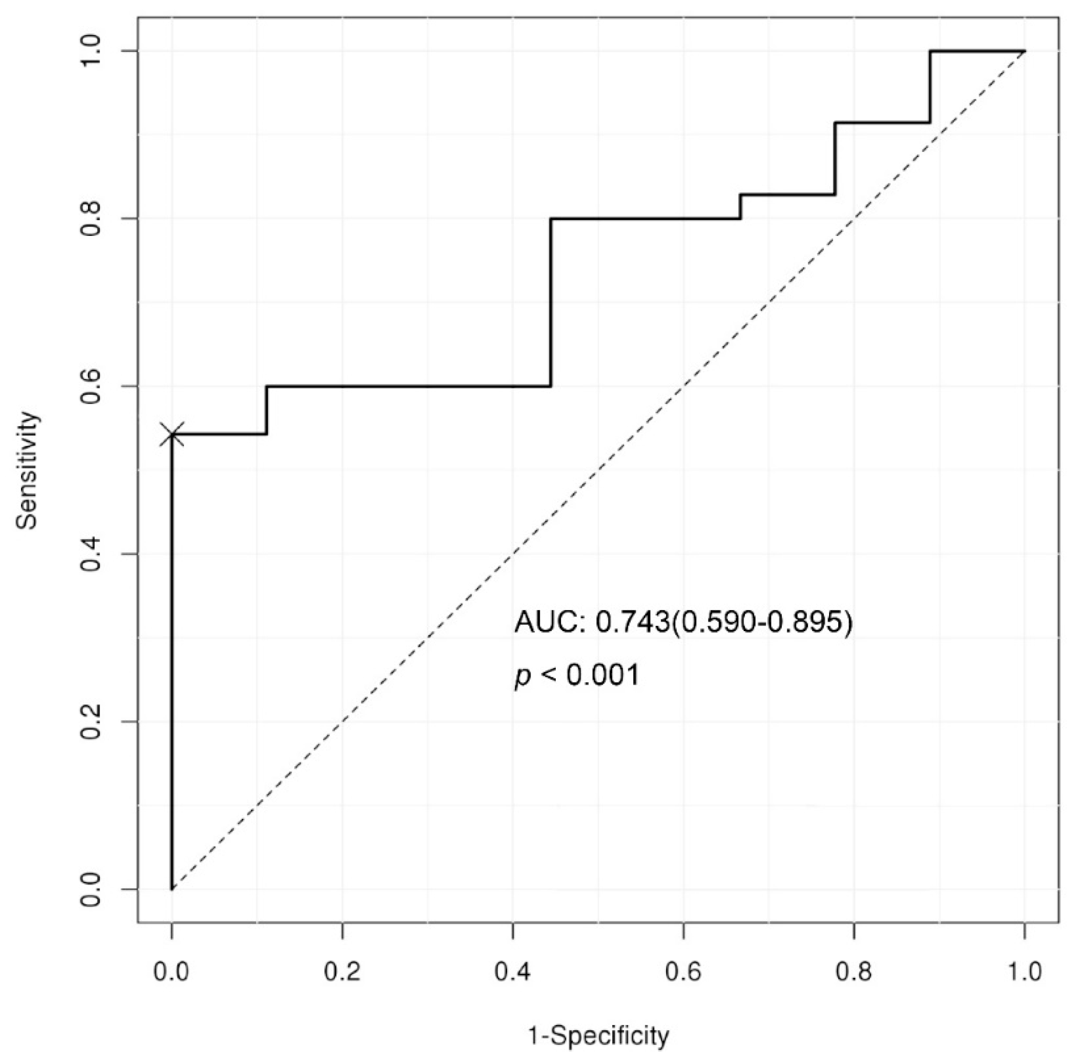
3.3. Predictive Cut-off Value and Diagnostic Performance of Plasma LeXis Levels for Diagnosing NASH.

The area under the ROC curve of the plasma LeXis for diagnosing NASH was 0.743 (95% CI 0.59–0.895, p < 0.001; Figure 5). The maximal sum of the sensitivity (54.3%) and specificity (100%) for diagnosing NASH could be achieved at a relative expression level of 7.92. Thus, all the patients with plasma LeXis levels above 7.92 were diagnosed with NASH. In addition, high plasma levels of LeXis, as defined by the cut-off value, were independently associated with NASH diagnosis in patients with NAFLD. (p = 0.0349, odds ratio = 22.19 (95% CI, 1.25–395.22); Table 2).

4. Discussion

In this study, we demonstrated that hepatic LeXis is negatively correlated with steatosis, and that patients with NASH have increased expression of plasma LeXis. In addition, plasma LeXis was independently associated with NASH with an acceptable diagnostic performance. The activation of LXR through pharmacologic intervention or a western diet induces the overexpression of hepatic LeXis. The interaction between LeXis and RALY inhibits the ability of the protein to bind to the cholesterol biosynthesis gene in the liver, which ameliorates hepatic steatosis. As shown in a previous report, downregulation of hepatic LeXis induces the vulnerable state of hepatic steatosis by altering the lipid metabolism. However, downregulated LeXis does not affect ER stress and inflammation, which are important pathological features of NASH [12]. Herein, we found that hepatic LeXis was downregulated as hepatic steatosis progressed to severe grades, which was not related to inflammation and ballooning, in accordance with previous reports. However, plasma LeXis was found not to reflect steatosis and inflammation, which are related to the ballooning degeneration of hepatocytes. As ballooned hepatocytes are considered a hallmark of steatohepatitis, decreased keratin 8/18 immunostaining in the cytoplasm was proposed as an objective marker for specifically identifying ballooned hepatocytes in patients with NAFLD [16]. Based on this study, the reciprocal elevation of plasma caspase-generated cytokeratin-18 fragments (CK-18) was proposed as a promising non-invasive biomarker for NASH diagnosis. However, a single CK-18 test revealed its lack of diagnostic potential for NASH [17,18]. In the present study, plasma LeXis was significantly upregulated in NASH and was also found to be a specific biomarker for NASH diagnosis in patients with NAFLD, regardless of other clinical factors such as hypertension and diabetes, or the levels of aminotransferase, creatinine, and platelets. In addition, plasma LeXis was also found to be independent of other pathological conditions, including steatosis, lobular inflammation, and fibrosis stage. Therefore, we believe that hepatocytes apoptosis followed by ballooning degeneration could induce the upregulation of plasma LeXis.
LeXis has been reported to promote the growth of osteosarcoma through the upregulation of CTNNB1 expression [19]. However, to date circulating LeXis has not been studied yet in patients with NAFLD or those with cancer.
Our study has certain limitations. First, although significant differences were shown, functional assessment of plasma LeXis in NASH was not performed. Nevertheless, the functional analysis of hepatic LeXis described in this study was consistent with the previous mouse data. Second, the number of patients with NAFL included in the study was relatively small. and the evaluated cohort lacked healthy controls. Third, no external validation was performed on the diagnostic performance of plasma LeXis for NASH. Nonetheless, this is the first study to suggest plasma LeXis as a potential diagnostic biomarker for NASH. We confirmed that hepatic LeXis is not associated with NASH, as previously described [12], whereas plasma LeXis was found to be independently associated with NASH. In the present study, however, the underlying mechanism of these findings could not be determined; thus, further evaluation is required in future investigations.
In conclusion, the overexpression of plasma lncRNA Lexis is associated with the ballooning degeneration of hepatocyte in patients with NAFLD. Moreover, circulating lncRNA LeXis could represent a useful non-invasive diagnostic biomarker for NASH.

Author Contributions

Conceptualization, S.Y.P., and K.H.; Formal analysis, J.G.P.; Funding acquisition, J.G.P., S.Y.P., G.K., S.Y.J., M.K.K., and K.H.; Investigation, J.G.P., G.K., S.Y.J., Y.R.L., E.L., H.W.L., M.-H.H., J.M.C., Y.S.H., J.S.Y., M.K.K., Y.O.K., W.Y.T., S.Y.P., and K.H.; Methodology, G.K.; Supervision, S.Y.P., and K.H.; Visualization, J.G.P.; Writing—original draft, J.G.P., and G.K.; Writing—review & editing, S.Y.P., and K.H. All authors have read and agree to the published version of the manuscript.

Funding

This research was supported by a grant of the Korea Health technology R&D Project through the Korea Health Industry Development Institute (KHIDI), funded by the Ministry of Health & Welfare [HI16C1501], and the Basic Science Research Program through the National Research Foundation of Korea (NRF), funded by the Ministry of Science, ICT and Future Planning [2017R1C1B5076851], and the Korean government [2014R1A5A2009242, 2017M3A9G8083382, 2019R1A2C1083892, 2019R1F1A1060878, 2019M3E5D1A02068089, and 2019R1A6A3A01095839].

Acknowledgments

We would like to thank Editage for English language editing.

Conflicts of Interest

The authors declare no conflict of interest about this work.

Appendix A

Figure A1. Expression of tissue and plasma LeXis according to the degree of inflammation, severity of NASH, and stage of fibrosis in patients with NAFLD.
Life 10 00230 g0a1
Figure A2. Expression of LeXis in hepatocellular carcinoma cell lines and their cultured media.
Life 10 00230 g0a2

References

  1. Williams, R.; Aspinall, R.; Bellis, M.; Camps-Walsh, G.; Cramp, M.; Dhawan, A.; Ferguson, J.; Forton, D.; Foster, G.; Gilmore, I.; et al. Addressing liver disease in the UK: A blueprint for attaining excellence in health care and reducing premature mortality from lifestyle issues of excess consumption of alcohol, obesity, and viral hepatitis. Lancet 2014, 384, 1953–1997. [Google Scholar] [CrossRef]
  2. NatCen Social Research, University College London Department of Epidemiology and Public Health. Health Survey for England, 2012; UK Data Archive: Colchester, UK, 2014. [Google Scholar]
  3. Brunt, E.M.; Wong, V.W.; Nobili, V.; Day, C.P.; Sookoian, S.; Maher, J.J.; Bugianesi, E.; Sirlin, C.B.; Neuschwander-Tetri, B.A.; Rinella, M.E. Nonalcoholic fatty liver disease. Nat. Rev. Dis. Primers 2015, 1, 15080. [Google Scholar] [CrossRef] [PubMed]
  4. Ascha, M.S.; Hanouneh, I.A.; Lopez, R.; Tamimi, T.A.; Feldstein, A.F.; Zein, N.N. The incidence and risk factors of hepatocellular carcinoma in patients with nonalcoholic steatohepatitis. Hepatology 2010, 51, 1972–1978. [Google Scholar] [CrossRef] [PubMed]
  5. Angulo, P.; Kleiner, D.E.; Dam-Larsen, S.; Adams, L.A.; Bjornsson, E.S.; Charatcharoenwitthaya, P.; Mills, P.R.; Keach, J.C.; Lafferty, H.D.; Stahler, A.; et al. Liver Fibrosis, but No Other Histologic Features, Is Associated With Long-term Outcomes of Patients With Nonalcoholic Fatty Liver Disease. Gastroenterology 2015, 149, 389–397. [Google Scholar] [CrossRef] [PubMed] [Green Version]
  6. Ekstedt, M.; Hagstrom, H.; Nasr, P.; Fredrikson, M.; Stal, P.; Kechagias, S.; Hultcrantz, R. Fibrosis stage is the strongest predictor for disease-specific mortality in NAFLD after up to 33 years of follow-up. Hepatology 2015, 61, 1547–1554. [Google Scholar] [CrossRef] [PubMed] [Green Version]
  7. Yoo, J.J.; Kim, W.; Kim, M.Y.; Jun, D.W.; Kim, S.G.; Yeon, J.E.; Lee, J.W.; Cho, Y.K.; Park, S.H.; Sohn, J.H.; et al. Recent research trends and updates on nonalcoholic fatty liver disease. Clin. Mol. Hepatol. 2019, 25, 1–11. [Google Scholar] [CrossRef] [PubMed] [Green Version]
  8. Oh, H.; Jun, D.W.; Saeed, W.K.; Nguyen, M.H. Non-alcoholic fatty liver diseases: Update on the challenge of diagnosis and treatment. Clin. Mol. Hepatol. 2016, 22, 327–335. [Google Scholar] [CrossRef] [PubMed] [Green Version]
  9. Mercer, T.R.; Dinger, M.E.; Mattick, J.S. Long non-coding RNAs: Insights into functions. Nat. Rev. Genet. 2009, 10, 155–159. [Google Scholar] [CrossRef] [PubMed]
  10. Smith, C.M.; Steitz, J.A. Classification of gas5 as a multi-small-nucleolar-RNA (snoRNA) host gene and a member of the 5’-terminal oligopyrimidine gene family reveals common features of snoRNA host genes. Mol. Cell. Biol. 1998, 18, 6897–6909. [Google Scholar] [CrossRef] [PubMed] [Green Version]
  11. Marchese, F.P.; Raimondi, I.; Huarte, M. The multidimensional mechanisms of long noncoding RNA function. Genome Biol. 2017, 18, 206. [Google Scholar] [CrossRef] [PubMed] [Green Version]
  12. Sallam, T.; Jones, M.C.; Gilliland, T.; Zhang, L.; Wu, X.; Eskin, A.; Sandhu, J.; Casero, D.; Vallim, T.Q.; Hong, C.; et al. Feedback modulation of cholesterol metabolism by the lipid-responsive non-coding RNA LeXis. Nature 2016, 534, 124–128. [Google Scholar] [CrossRef] [PubMed] [Green Version]
  13. Chalasani, N.; Younossi, Z.; Lavine, J.E.; Diehl, A.M.; Brunt, E.M.; Cusi, K.; Charlton, M.; Sanyal, A.J. The diagnosis and management of non-alcoholic fatty liver disease: Practice Guideline by the American Association for the Study of Liver Diseases, American College of Gastroenterology, and the American Gastroenterological Association. Hepatology 2012, 55, 2005–2023. [Google Scholar] [CrossRef] [PubMed]
  14. Kleiner, D.E.; Brunt, E.M.; Van Natta, M.; Behling, C.; Contos, M.J.; Cummings, O.W.; Ferrell, L.D.; Liu, Y.C.; Torbenson, M.S.; Unalp-Arida, A.; et al. Design and validation of a histological scoring system for nonalcoholic fatty liver disease. Hepatology 2005, 41, 1313–1321. [Google Scholar] [CrossRef] [PubMed]
  15. Hur, K.; Toiyama, Y.; Okugawa, Y.; Ide, S.; Imaoka, H.; Boland, C.R.; Goel, A. Circulating microRNA-203 predicts prognosis and metastasis in human colorectal cancer. Gut 2017, 66, 654–665. [Google Scholar] [CrossRef] [PubMed] [Green Version]
  16. Lackner, C.; Gogg-Kamerer, M.; Zatloukal, K.; Stumptner, C.; Brunt, E.M.; Denk, H. Ballooned hepatocytes in steatohepatitis: The value of keratin immunohistochemistry for diagnosis. J. Hepatol. 2008, 48, 821–828. [Google Scholar] [CrossRef] [PubMed]
  17. Kim, Y.S.; Jung, E.S.; Hur, W.; Bae, S.H.; Choi, J.Y.; Song, M.J.; Kim, C.W.; Jo, S.H.; Lee, C.D.; Lee, Y.S.; et al. Noninvasive predictors of nonalcoholic steatohepatitis in Korean patients with histologically proven nonalcoholic fatty liver disease. Clin. Mol. Hepatol. 2013, 19, 120–130. [Google Scholar] [CrossRef] [PubMed]
  18. Cusi, K.; Chang, Z.; Harrison, S.; Lomonaco, R.; Bril, F.; Orsak, B.; Ortiz-Lopez, C.; Hecht, J.; Feldstein, A.E.; Webb, A.; et al. Limited value of plasma cytokeratin-18 as a biomarker for NASH and fibrosis in patients with non-alcoholic fatty liver disease. J. Hepatol. 2014, 60, 167–174. [Google Scholar] [CrossRef] [PubMed]
  19. Wang, G.; Cui, T.; Sun, L.; Peng, N.; Yang, C. Long noncoding RNA LeXis promotes osteosarcoma growth through upregulation of CTNNB1 expression. Am. J. Cancer Res. 2017, 7, 1577–1587. [Google Scholar] [PubMed]
Figure 1. (a) Steatosis (black arrow), lobular inflammation (blue arrow), and ballooning degeneration (red arrow) were scored on 1–3, 0–3, and 0–2 scale (hematoxylin and eosin staining; amplification, 200×). (b) The fibrosis stage was determined using the Kleiner scoring system as F0–F4 (trichrome staining; amplification, 100×).
Life 10 00230 g001
Figure 2. The expression of hepatic LeXis is significantly upregulated in patients with severe steatosis compared with those without.
Life 10 00230 g002
Figure 3. The degree of steatosis is negatively correlated with hepatic lncRNA LeXis levels but not with plasma lncRNA LeXis levels.
Life 10 00230 g003
Figure 4. The expression of plasma LeXis is significantly upregulated in patients with NASH compared with those without.
Life 10 00230 g004
Figure 5. The area under the ROC curve of the plasma LeXis for diagnosing NASH was 0.743.
Life 10 00230 g005
Table 1. Characteristics of patients.
CharacteristicsNon-Alcoholic Fatty Liver Disease (NAFL) (n = 9)Non-Alcoholic Steatohepatitis (NASH) (n = 35)p Value
Male2 (22.2)20 (57.1)0.135
Age, year38.3 ± 12.051.9 ± 15.50.020 *
Steatosis 0.144
5-334 (44.4)16 (45.7)
>33–661 (11.1)13 (37.1)
>66%4 (44.4)6 (17.1)
Lobular inflammation 0.197
<2 foci per 200× field 8 (88.9)20 (57.1)
2–4 foci per 200× field1 (11.1)11 (31.4)
>4 foci per 200× field0 (0.0)4 (11.4)
Ballooning 0.000 *
None9 (100.0)0 (0.0)
Few ballooned cells0 (0.0)20 (57.1)
Many cells/prominent ballooning 0 (0.0)15 (42.9)
Advanced fibrosis0 (0.0)12 (34.3)0.101
NAS ≥ 50 (0.0)20 (57.1)0.007 *
Weight, kg83.3 ± 20.371.7 ± 11.60.182
BMI, kg/m231.0 (26.8–33.1)27.1 (25.9–30.4)0.348
Hypertension4 (44.4)17 (48.6)1.000
Diabetes1 (11.1)15 (42.9)0.168
Platelets, × 103/mm240.4 ± 51.3219.8 ± 71.50.426
AST, IU/L57.0 (43.0–95.0)78.0 (46.5–106.5)0.211
ALT, IU/L90.0 (68.0–147.0)98.0 (66.5–120.5)0.816
Bilirubin, mg/dL0.7 (0.5–0.8)0.6 (0.4–0.6)0.119
Albumin, g/dL4.7 ± 0.34.5 ± 0.30.043 *
γ-GTP, mg/dL55.0 (43.0–95.0)77.0 (55.0–124.0)0.366
Creatinine, mg/dL0.9 (0.8–1.0)0.8(0.6–0.9)0.134
Fasting blood glucose, mg/dL107.0 (102.0–113.5)117.5 (104.0–136.0)0.133
Total cholesterol, mg/dL179.0 (162.5–188.5)177.0 (159.0–210.0)0.835
HDL, mg/dL47.5 (35.5–57.5)41.5 (33.0–50.0)0.441
LDL, mg/dL117.0 ± 25.7117.7 ± 38.30.963
Triglyceride, mg/dL161.5 (115.5–194.5)142.5 (112.0–213.0)0.946
Values are presented as the mean ± standard deviation, median (interquartile ranges) or number (%). ALT, alanine aminotransferase; AST, aspartate aminotransferase; BMI, body mass index; GTP, guanosine triphosphate; HDL, high-density lipoprotein; LDL, low-density lipoprotein; NAS, non-alcoholic fatty liver disease activity score. * p < 0.05 was considered statistically significant.
Table 2. Factors associated with risk for non-alcoholic steatohepatitis.
VariableUnivariate AnalysisMultivariate Analysis
Odds Ratiop ValueOdds Ratiop Value
Sex4.67 (0.85–25.75)0.0771
Age1.06 (1.01–1.12)0.0298
Hypertension1.18 (0.27–5.15)0.8251
Diabetes6.00 (0.68–53.29)0.1078
Platelets1.00 (1.00–1.00)0.4175
AST1.02 (0.99–1.05)0.13101.04 (0.99–1.08)0.136
ALT1.00 (0.99–1.01)0.8114
Bilirubin0.17 (0.02–1.78)0.13850.02 (0.00–0.68)0.029 *
Albumin0.07 (0.00–1.09)0.0574
Creatinine0.10 (0.00–4.10)0.2263
High plasma LeXis9.50 (1.07–84.25)0.043222.19 (1.25–395.22)0.035 *
ALT, alanine aminotransferase; AST, aspartate aminotransferase; * p < 0.05 was considered statistically significant.

