1. Introduction
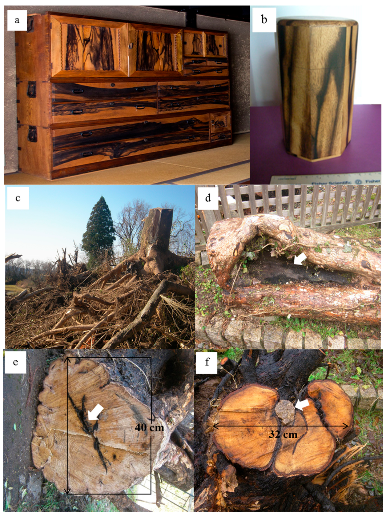
Patterned kurogaki is currently very rare and difficult to find. It is an important material for manufacturing furniture, tea ceremony goods, boxes, and other miscellaneous articles in Japan (
Figure 1a,b). Here, we report the characterization of kurogaki at Tawara, Kanazawa, Ishikawa Prefecture, Japan, based on radioactivity, mineral analyses, chemical analyses, H
2O
2 reactions, and biological observations. No report has yet described the results of electron microscopy observations and chemical analyses of kurogaki, limiting our ability to obtain insights into its nature. We thus studied the mineralogy, chemistry, and micro-morphology of kurogaki using a combination of micro-techniques.
The objective of this study was to illustrate the association of minerals with various microorganisms that are capable of absorbing these elements from weathered andesite soils. The H
2O
2 reaction produced oxygen bubbles within the black portions of the wood, but the beige portions were unreactive. Ultraviolet analysis indicated color in the purple part of the spectrum in the black “peacock pattern” portions and the black soils taken from around the roots [
1].
Kurogaki (black persimmon) grows very slowly, has very hard wood, and is known for a striking black and beige coloration, referred to as a “peacock pattern” (
Figure 1a,b). Patterned kurogaki occurs very rarely, being only one of every 1000–10,000 persimmon trees found in Japan and China. This tree was previously planted in Kanazawa, Japan, and is of historical and artistic importance in Kanazawa City, Ishikawa, Japan, particularly in the Edo period (1603–1868).
In this study, we analyzed the mineralogy, chemistry, micro-morphology, and microstructure of kurogaki wood, considering its association with microorganisms, through a combination of analytical data obtained from X-ray diffraction (XRD) and inductively coupled plasma-mass spectrometry (ICP-MS), X-ray fluorescence analyses (XRF), scanning electron microscopy equipped with energy-dispersive spectroscopy (SEM-EDS), and transmission electron microscopy (TEM). We investigated the distribution and location, identification and structure, as well as differentiation between the black “peacock pattern” and ordinary beige wood portions. We analyzed the chemical composition of black “peacock pattern” portions, the associated microstructure, and elemental distribution in order to clarify the influences of environmental soil and water quality on these properties. The results provided evidence of the microorganisms’ ability to grow in the black “peacock pattern” portions, immobilizing elements, and performing biomineralization and phytoremediation, in particular, within wood tissues of kurogaki.
2. Materials and Methods
2.1. Exploring Kurogaki Field and Investigated Specimens
Kurogaki samples were obtained from 20 trees from Tawara, Kanazawa, Japan. They were cut down in December 2016 and subjected to chemical, physical, biological, and mineralogical composition analyses. The kurogaki trees had grown for more than 100 years in a mixture of relatively fresh andesite rocks and weathered rocks (Tomuro andesite) associated with dark brown soils. The pH of the soils is slightly acidic (6.4–6.8) at the foothills of Tomuro volcano in Kanazawa. The andesite rocks contain large amounts of white plagioclase in 5-mm-sized, black small grains of hypersthene and amphibole, which originated from the eruptions of ash and lava 500,000 years ago.
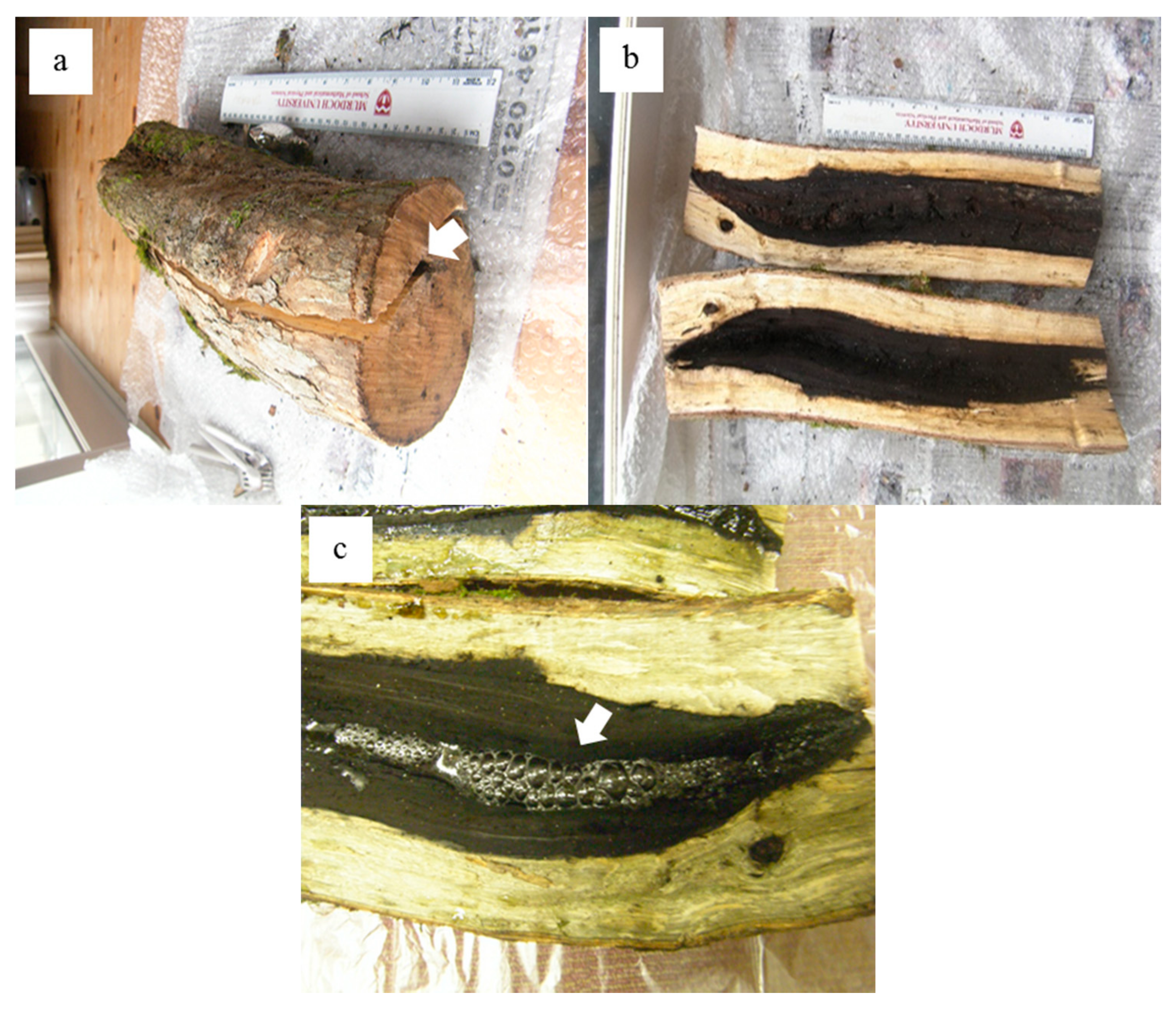
We identified and collected several trees with a black color on the branches, trunk, and roots and their associated andesitic soils, characterizing their structure and properties. At that time, the air radiation dose rate was 110–200 cpm, while BG was 70–80 cpm. The black “peacock pattern” portions showed a somewhat higher dosage, in the range of 150–200 cpm, based on the results from the Aloka β (γ) survey meter TSG-136. The black “peacock pattern” and beige portions from the taproot were compared with the black root parts for the samples collected from Tawara in January 2017 (
Figure 1 and
Figure 2).
Views of cut-down kurogaki trees in the hilly Tawara field are shown in the Figures. The thick and long roots hold the large stalk of kurogaki trees (
Figure 1c), resulting in hollowing-out of the trunk represented by a black color, which reflects silicification and lignification (arrow in
Figure 1d). There is also a black injury on a 40-cm cross section (arrow in
Figure 1e), and one andesitic rock is caught between large roots in a 32-cm cross section (arrow in
Figure 1f).
2.2. H2O2 Reaction Checks
We tested the black and beige portions of the “peacock pattern” wood with a 3.5% H
2O
2 solution. If oxygen is generated upon adding H
2O
2 to a black part, this indicates the presence of organics; enzyme catalyst; metals such as Fe, Cu, Mn, and Cr; and microorganisms (arrow in
Figure 2c).
Many organisms can decompose hydrogen peroxide (H
2O
2) enzymatically. Enzymes are globular proteins, responsible for most of the chemical activities of living organisms. They act as catalysts, substances that speed up chemical reactions without being destroyed or altered during the process. H
2O
2 is toxic to most living organisms. Many organisms are capable of enzymatically destroying the H
2O
2 before it can do much damage. H
2O
2 can be converted to oxygen and water, as follows:
2.3. X-ray Powder Diffraction
To identify minerals and analyze the structure of crystals and crystallinity of cellulose in the trunk and the black roots of kurogaki (
Figure 2b) associated with andesitic rocks (Figures 5 and 6), we used X-ray powder diffraction analysis. An X-ray powder diffractometer (XRD) (Rinto 2200, Rigaku, Tokyo, Japan; Cu-Kα radiation, 40 kV, 30 mA, scan speed of 2°/min) was used for identifying minerals in kurogaki black roots at Tawara, Kanazawa, Japan. The powder samples (bulk, <2 μm, and suspension) were used for X-ray powder diffraction analyses.
2.4. XRF Chemical Analyses
The kurogaki tree samples at Tawara were investigated using an X-ray fluorescence analyzer (Rigaku Primus II, Tokyo, Japan), which operated at an accelerating voltage of 60 kV, 150 mA (max.), 4 kW, under vacuum conditions, after heating (KDFS 80) at 600 °C for 30 min. The samples were pressed (
Table 1).
2.5. ICP-MS Analyses
The semi-quantitative analyses of black and beige portions of the kurogaki samples were conducted using Elementar Analysensysteme GmbH Vario MAX type (ICP-MS), Thermo Fisher Scientific (Bremen, Germany) iCAP, and compared with the kurogaki sample collected from Tawara and Makiyama, Kanazawa, Japan (
Table 2).
2.6. Scanning Electron Microscopy (SEM) Equipped with Energy-Dispersive Spectroscopy (EDS)
A scanning electron microscope (S-3400N. Hitachi, Ibaragi, Japan) equipped with an energy-dispersive X-ray analyzer (Horiba EMAX, Kyoto, Japan) was used to study the kurogaki samples collected from Tawara. We attempted to analyze the appearance, distribution, tissue structure, and inorganic and organic constituents of the wood, evaluating the morphology at the microscale. The conditions used were as follows: 15 kV accelerating voltage, 70–80 μA current, analytical time of 1000 s, and an area of 10 mm × 10 mm on a carbon double tape with C coating (Figures 7–13).
2.7. Transmission Electron Microscopy (TEM)
A transmission electron microscopy (Hitachi H7650, Ibaragi, Japan) was used to study the black particles suspend in the water of kurogaki powder samples collected from Tawara. We attempted to observe the appearance and morphology of grains, distribution, tissue structure with inorganics and microorganisms in the black parts, evaluating the morphology at the microscale. The conditions used were as follows: 80 kV accelerating voltage, on a micro grid without any coating (Figure 14).
3. Results
3.1. H2O2 Reaction
The black portion of the roots collected from Tawara reacted strongly with the 3.5% H
2O
2 solution, allows looking for organics (either dead organic materials or living one such as microbes) or manganese (arrow in
Figure 2c). The beige region did not react with the H
2O
2 solution, showing no bubbles. It was possible to divide catalysts into two portions, the black and beige parts, indicating biological catalysts of enzymes at black portions. Enzymes are proteins responsible for most of the chemical activities of living organisms at black portion of kurogaki.
3.2. Mineralogy of Kurogaki and Andesitic Rocks (Tomuro-ishi) around the Roots of Kurogaki
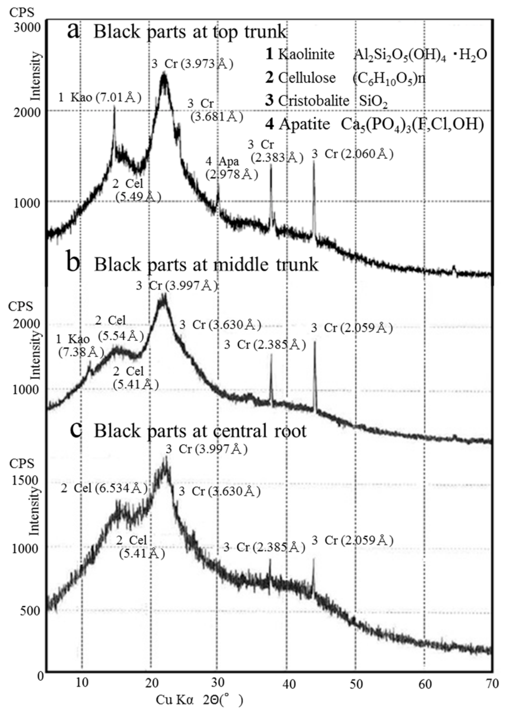
The mineralogy of the black parts at top trunk (
Figure 3a), black parts at middle trunk (
Figure 3b) and black parts at central root (
Figure 3c) of the kurogaki samples from Tawara was determined using XRD analyses. The black parts contained mainly crystalline cellulose ((C
6H
10O
5)n; 5.54 Å, 5.41 Å) associated with kaolinite (Al
2Si
2O
5·(OH)
4·H
2O (7.38–7.01 Å)), cristobalite (SiO
2 (3.997, 3.630, 2.385, 2.059 Å)) and apatite (Ca
5(PO
4)
3(F, Cl, OH) (2.978 Å)). The peaks of cellulose indicated wider and gradual reflections, whereas those of cristobalite indicated sharp and strong reflections (
Figure 3).
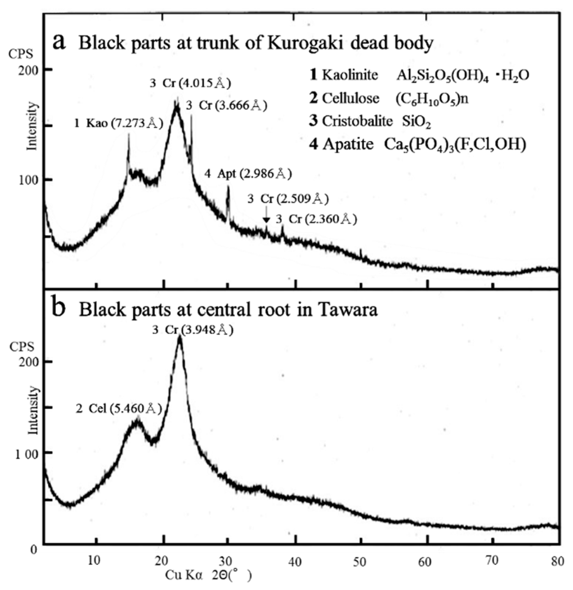
The mineralogy of the black parts at trunk of kurogaki dead body (
Figure 4a) and black parts at central root in Tawara (
Figure 4b) samples was determined using XRD analyses (
Figure 4b). The peaks of cellulose indicated wider and gradual reflections, whereas those of cristobalite, kaolinite (7.273 Å) and apatite (2.986 Å) indicated sharp and strong reflections (
Figure 4a). Especially the kurogaki dead body suggested mineralization of kaolinite, cristobalite and apatite in black parts of wood structure.
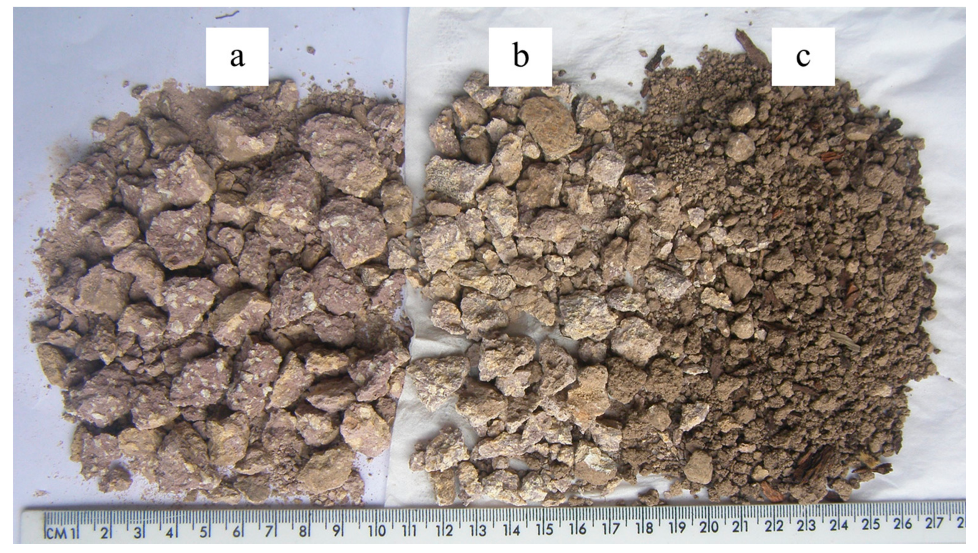
On the other hand, the relatively fresh andesite rocks (
Figure 5a), the weathered rocks (
Figure 5b), and soils with clays (
Figure 5c) around the roots of kurogaki, as analyzed by XRD, were subjected to mineralogical composition analyses during the weathering processes that affect kurogaki trees. The fresh andesite rocks (
Figure 5a), called “Tomuro-ishi”, were produced from Mt. Tomuro volcano (Quaternary period; about 600,000–500,000 years ago). The andesite fresh rocks (
Figure 5a), weathered rocks (
Figure 5b) and clayey soils (
Figure 5c) are distributed widely throughout Kanazawa (
Figure 5).
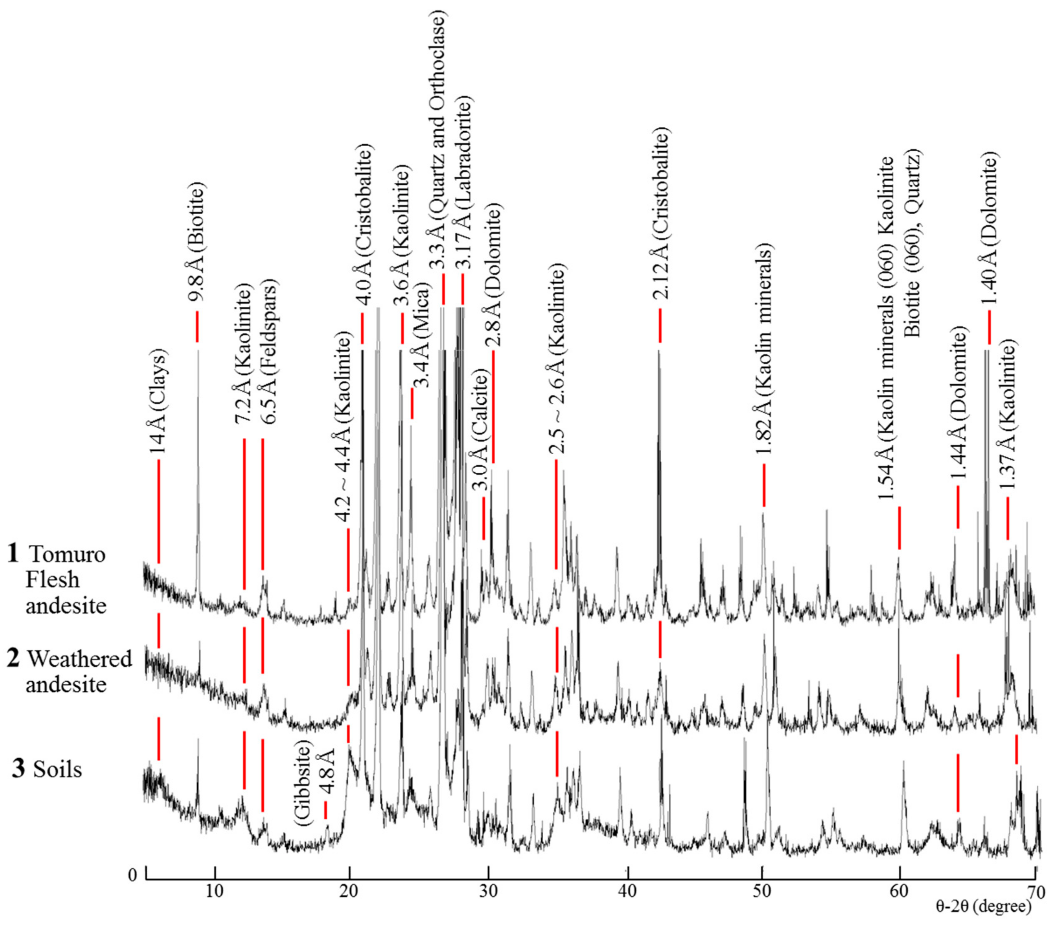
The Tomuro fresh andesite rocks are composed of mainly feldspars (Orthoclase, Labradorite), mica (biotite), quartz, calcite, dolomite, cristobalite, and a trace of kaolinite, whereas the weathered andesite is composed of higher contents of kaolinite and cristobalite than the fresh andesite. The soils of weathered andesite are rich in clay minerals, such as 14 Å clays, kaolinite (7.2 Å), gibbsite, and cristobalite. The 4.2–4.4 Å reflection peak in the soils has increased intensity compared to that of fresh and weathered andesite rocks, suggesting that the content of clay minerals is increased by weathering (
Figure 5 and
Figure 6). The Tomuro flesh andesite (1); weathered Tomuro andesite (2); and the soils (3) were composed of minerals, such as 14 Å clays, biotite (9.8 Å and 1.54 Å), feldspers (6.5 Å and 3.17 Å), kaolinite (4.2–4.4 Å, 2.5–2.6 Å, 1.82 Å, 1.54 Å, and 1.37 Å), cristobalite (4.0 Å and 2.12 Å), mica (3.4 Å), quartz (3.3 Å), calcite (3.0 Å), and dolomite (2.8 Å, 1.44 Å, and 1.40 Å), respectively (
Figure 6).
3.3. XRF Chemical Analyses
The chemical composition o: fresh Tomuro volcanic fresh andesite (1); weathered andesite (2); and soils (3) collected from near kurogaki roots at Tawara, Ishikawa (
Figure 5) showed the major and trace elements by XRF analyses (
Table 1). The elements of SiO
2, Al
2O
3, Fe
2O
3, CO
2, CaO, Na
2O, K
2O, and MgO are major elements of andesite, associated with trace elements of TiO
2, P
2O
5, MnO, WO
3, SrO, and SO
3. The trace elements are attributed to organics, such as microorganisms, showing SEM-EDS elemental content maps in
Figure 7,
Figure 8 and
Figure 9. Some of elements decreased by weathering, such as SiO
2, CaO, Na
2O, MgO, WO
3, SrO and CuO.
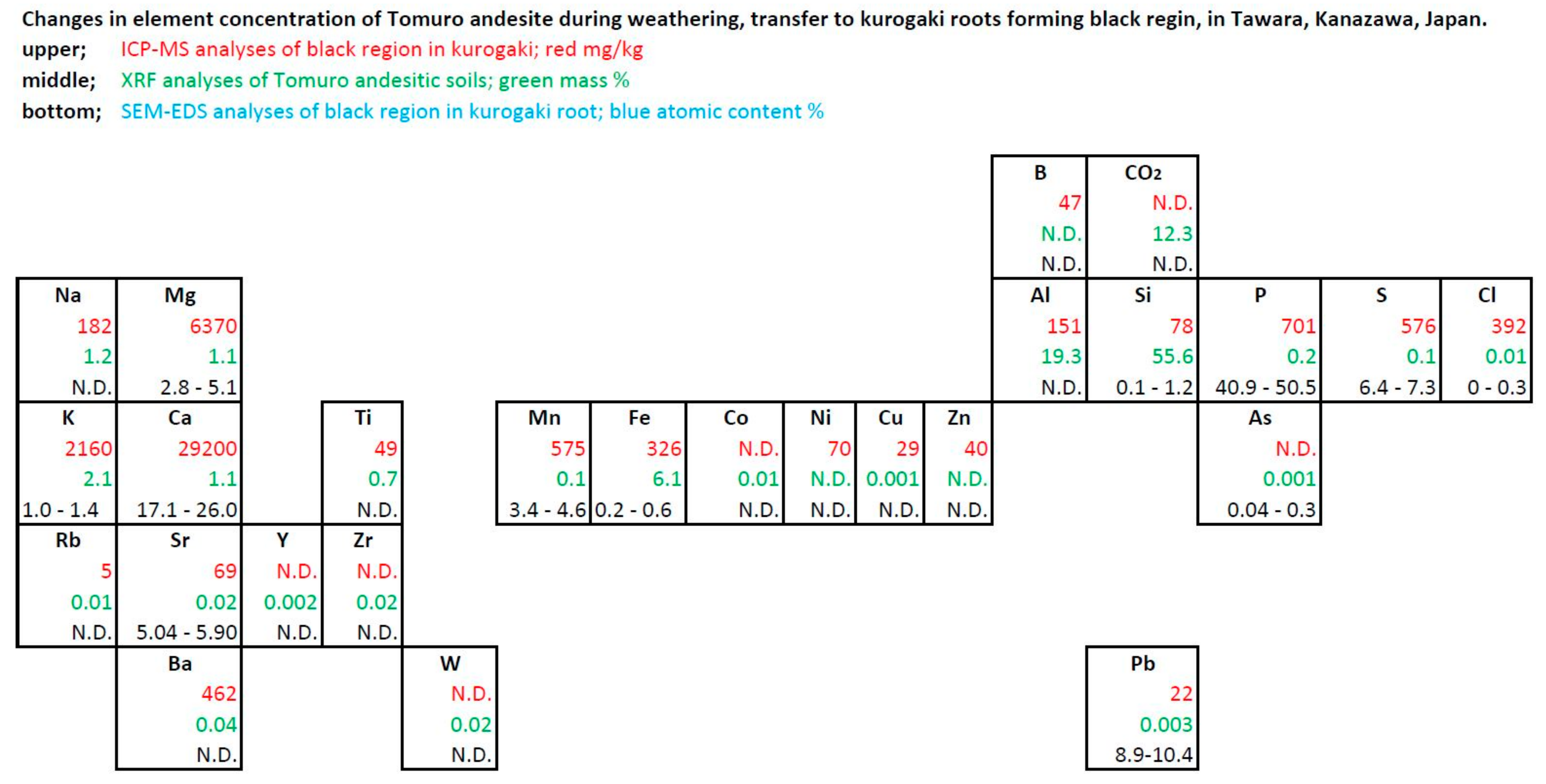
3.4. ICP-MS Analyses of Kurogaki
ICP-MS analyses indicated the presence of major and trace elements in the chemical composition of the black and beige portions of the kurogaki samples collected from Tawara, in comparison with the samples collected from Makiyama (
Table 2). Nearly all elements were more abundant in the black portions of the wood than the elements in the beige portions.
In particular, Ca (29,200 mg/kg), Mg (6370 mg/kg), K (2160 mg/kg), P (701 mg/kg), Mn (575 mg/kg), Ba (462 mg/kg), S (576 mg/kg), Cl (392 mg/kg), Fe (326 mg/kg), Na (182 mg/kg), and Al (151 mg/kg) contents were high in the black region. Trace elements such as B, Si, Ni, Sr, Ti, Zn, Cu, Pb, and Rb could also be seen in the black portions of kurogaki samples collected from Tawara. The chemical composition in the black region of the samples collected from Makiyama indicated a similar tendency for main elements, in that they were more abundant in the black portions than in the beige ones. Note that the concentrations of B and Ba are important characteristics in colored persimmon.
3.5. Scanning Electron Microscopy (SEM) Equipped with Energy-Dispersive Spectroscopy (EDS)
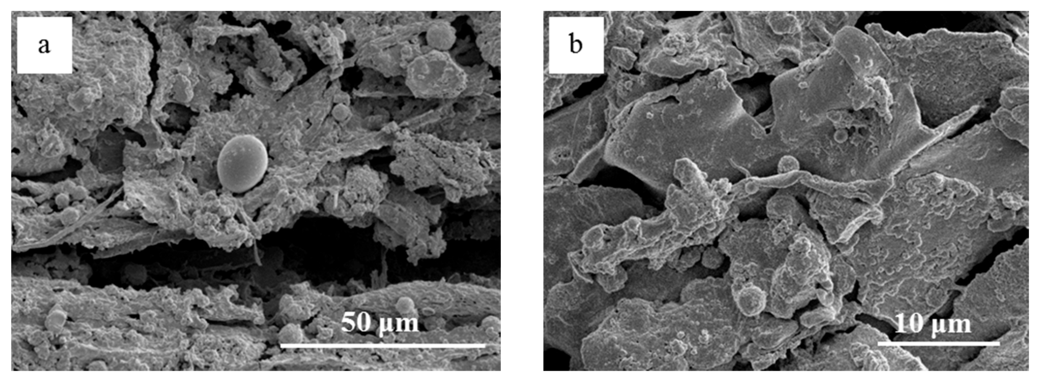
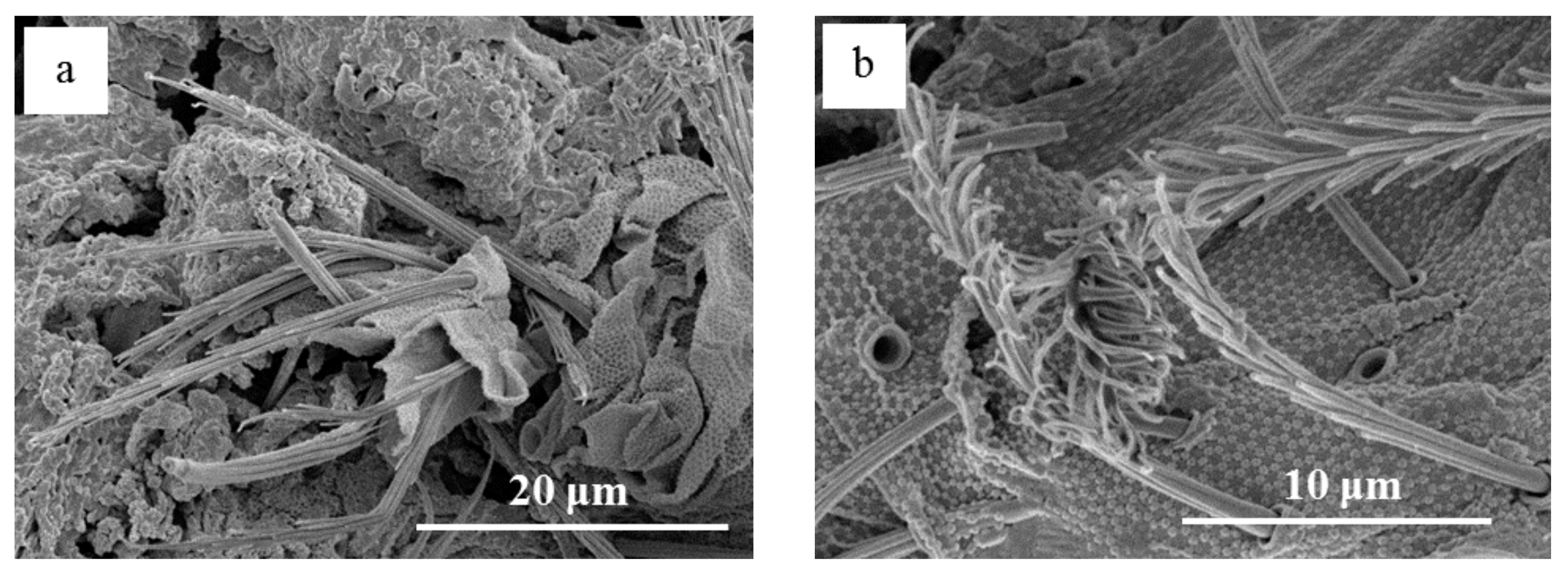
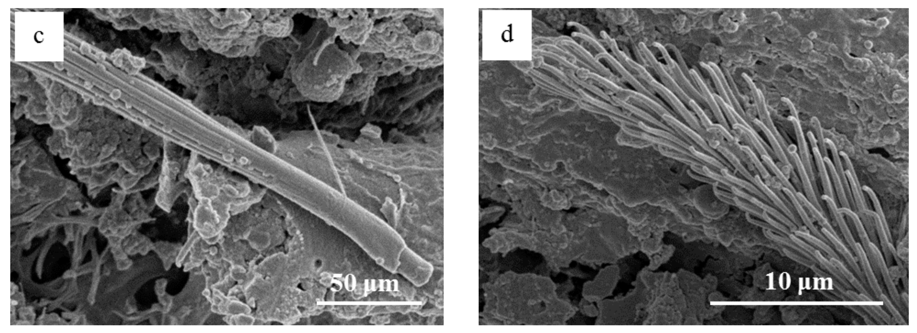
SEM observation of the black pith in the central root at Tawara showed various typed microorganisms with different sizes from several tens of micrometers to 500 μm in size (
Figure 7 and
Figure 8). The microorganisms showed different shapes, such as coccus-type and filamentous bacteria, indicating fission process. The fission of coccus-type bacteria can be seen in
Figure 7a–d. The bead-like microorganisms are commonly present in the black portion (
Figure 7e,f). Development processes of broomstick-like microorganisms from the root to the very top of the stick can be seen in
Figure 8c,d; some of them approach 300 μm or more in length.
3.6. SEM-EDS Analyses of Kurogaki
SEM-EDS semi-quantitative analyses of kurogaki for four different points of the black portions of the taproot collected from Tawara revealed the accumulation of particular elements at each point (
Table 3). The highest concentrations were of P (41–50 atomic content %) and Ca (17–26 atomic content %), suggesting the formation of phosphorus carbonate, such as apatite, which is agreed with XRD data (
Figure 3 and
Figure 4). Small amounts of S (6.4–7.3 atomic content %), Mn (3.4–4.6 atomic content %), Mg (3.5–5.1 atomic content %), and Si, K, Fe, and As were also detected. On the other hand, Sr (5.0–5.9 atomic content %) and Pb (9.0–10.2 atomic content %) were notable for their high content in the black portions (
Table 3).
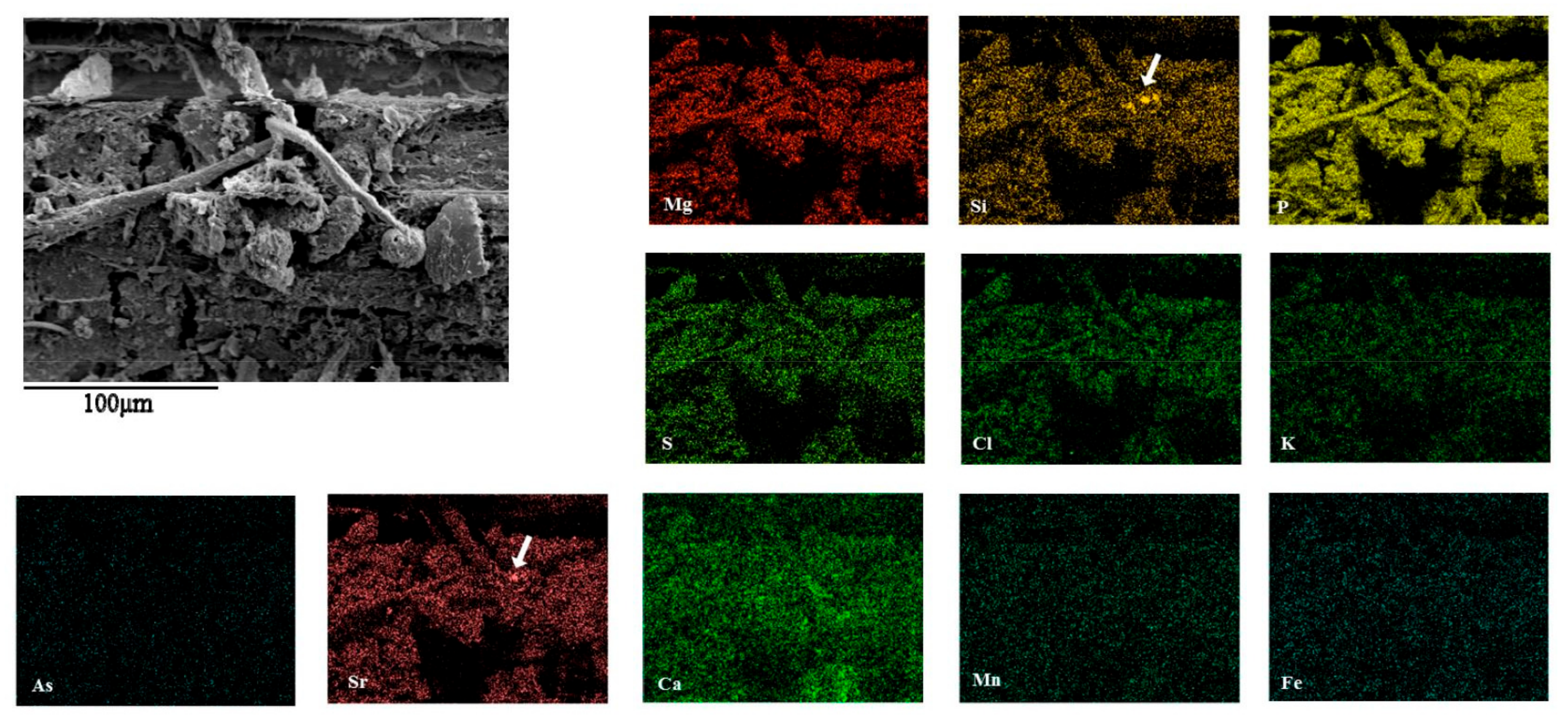
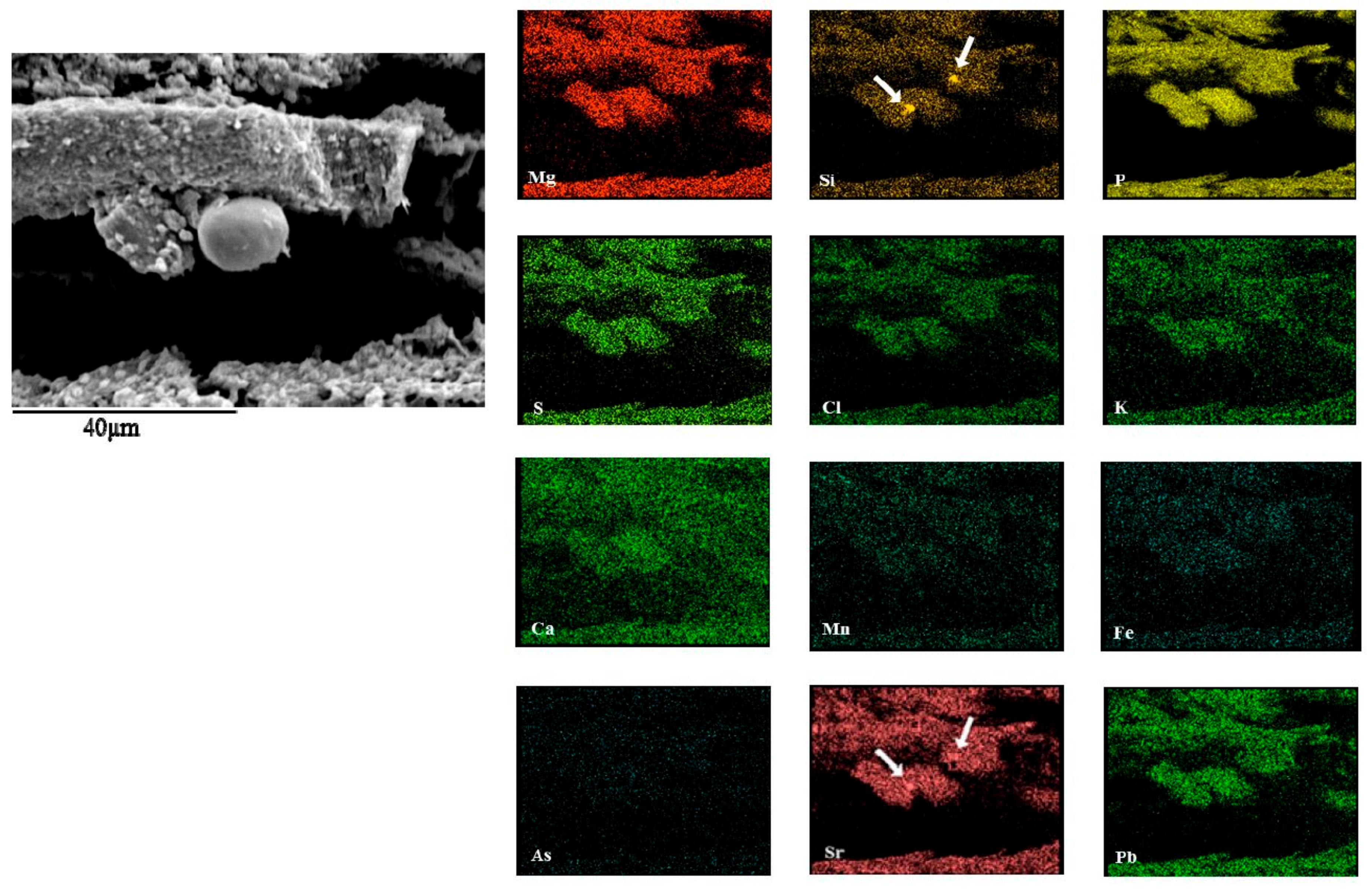
Elemental content maps of points 2–4 in
Table 3 are shown in
Figure 9,
Figure 10 and
Figure 11, respectively. The presence of various microorganisms, such as coccus-type bacteria, filamentous bacteria, broom-type bacteria, and a pair of beaded bacteria, was associated with high concentrations of Mg, Si, P, S, Cl, K, Ca, Mn, Fe, Sr, and Pb in the black portions of the taproot (
Figure 9,
Figure 10 and
Figure 11). However, the Cd, Sr, Mn, Fe, and Pb elements dotted among the map.
The filamentous and coccus-type bacteria exhibited high P and Ca concentrations, suggesting the formation of carbonates in the black parts of the taproot. In addition, the concentrations of Sr and Pb were extremely high at points 1–4, suggesting that the kurogaki samples obtained from Tawara are hyperaccumulators of these elements [
1,
2].
3.7. SEM-EDS Elemental Content Maps of Kurogaki
The elemental content maps of black portions at central root of kurogaki at Tawara, showed distribution of bio-minerals associated with microorganisms, such as silicate minerals and carbonate minerals (
Figure 12 and
Figure 13). The combination of elements such as Si, Sr and Ca accords with the other analytical results of XRF and IC-Mass.
3.8. TEM Observation of Black Parts of Kurogaki
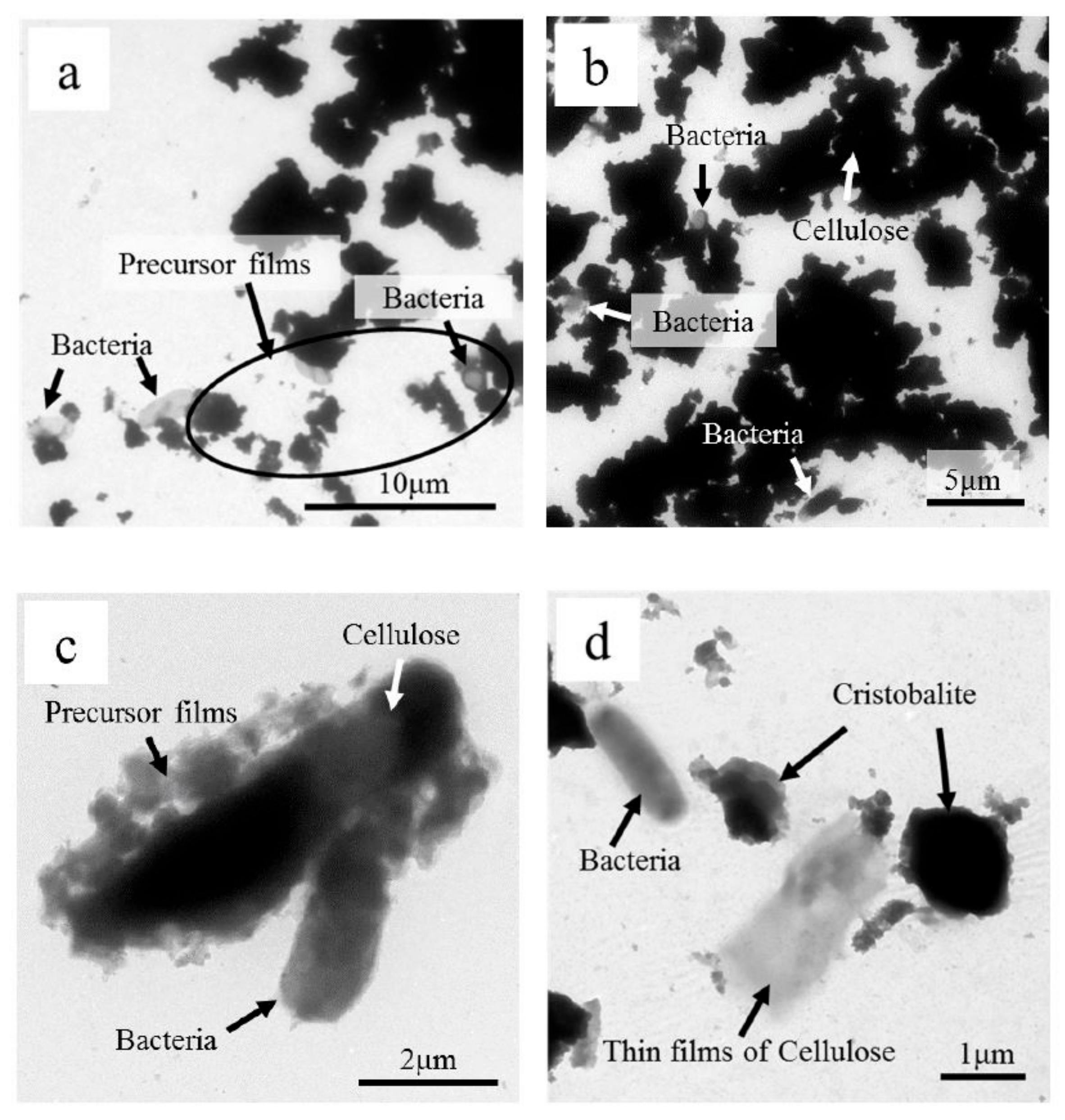
TEM observation of kurogaki black parts of roots in Tawara shows various kind of particles, such as thick chunks of grains (cellulose), precursor thin films, angular and square (cristobalite), and various sized and shaped microorganisms (coccus type,
Bacillus type, and filamentous bacteria, few microns in size) (
Figure 14a,b). Some of precursor thin films may be clay minerals associated with thick cellulose and bacteria (
Figure 14c,d). We did not obtain electron diffraction patterns of each particle, which might be future study.
4. Discussion
The bark tissues can be divided into two major regions: the inner bark and the outer bark. Physiologically, the inner bark transports the assimilates and serves as a storage organ for food reserves, while the outer bark is physiologically inactive and forms a protective layer against mechanical and chemical injury. Accordingly, basic chemical studies on bark are important not only to understand the physiology of trees but also to ensure better utilization of the bark [
1]. From this viewpoint, the chemical and physical properties of bark lignin and bark phenolic compounds have been investigated [
2,
3,
4,
5].
On the other hand, the heartwood of the Japanese persimmon tree (
Diospyros kaki) becomes black on rare occasions and has been highly sought after as a substitute for ebony. Previous research has attempted to clarify how the physical, mechanical, chemical, and biodegradation properties differ between lighter-colored sapwood and blackened heartwood in
D. kaki. The specific gravity, equilibrium moisture content, modulus of rupture, and modulus of elasticity in the blackened heartwood have been shown to be higher and the loss tangent lower than those in sapwood [
5]. However, these previous studies did not address biomineralization of the wood structure.
In this paper, we report the characterization of not only trunk tissue but also root tissue of kurogaki associated with the striking black “peacock pattern” at Kanazawa, Ishikawa, Japan, based on radioactivity, ultraviolet analysis, and H
2O
2 reactions in the field. We also report for the first time the results of biomineralization using ICP-MS, XRD, XRF, SEM-EDS and TEM. These results can provide insights into the simple identification of mineral species and characteristic structures (
Figure 15). We analyzed the mineralogy, chemistry, and micro-morphology of tissues by using a combination of micro-techniques. The black portions were found to consist mainly of cellulose, high-crystalline cristobalite and apatite, associated with kaolinite and many microorganisms (
Figure 16). Particular elements such as abundant Ca, associated with Mg, Si, P, S, Cl, and K, were accumulated at the same sites as microorganisms, as shown in the elemental content maps (
Figure 9,
Figure 10,
Figure 11,
Figure 12 and
Figure 13). The compositions of black and beige portions of the black persimmon tree were determined based on chemical data obtained from ICP-MS analyses (
Table 2,
Figure 15).
The objective of this study was to illustrate the potential role of various microorganisms in the mineral composition of black persimmon. Microorganisms have the ability to incorporate silicon into various organic compounds, forming C–O–Si linkages and more complicated hard structures.
4.1. Formation of Crystals and Amorfous Materials in the Trunk and Roots
The mineral materials in the trunk proved that they consist of cristobalite, apatite and kaolinite by XRD, whereas amorphous calcium carbonate and calcium oxalate existing in living cells at the black portions of central root, namely the bacteria by SEM-EDS. This is the factor attributed to the growth of high crystals from low crystals or amorphous calcium carbonate in the annual rings. The cristobalite and apatite, in particular, is formed within the living cells in association with bacteria; this results in increased hardness of the trunk. Microorganisms are thus an integral part of biomineralization within wood tissues of kurogaki. Microbial communities of phytostabilization are extremely complex, containing thousands of species associated with both light and heavy elements, and the exact function of most species, in a given environment, is simply illustrated the points with diagrams andesite weathering soils—kurogaki root tissue and trunk tissue with the striking black “peacock pattern” in kurogaki (
Figure 15 and
Figure 16).
In contrast to kurogaki, doronoki (
Populus maximowiczii) grows rapidly and the wood is soft and light. It has recently been planted in South Korea and Japan for use as pulpwood, lumber, boxes, matches, and other miscellaneous articles. However, when the wood is processed, the edge of the sawing machinery and tools is easily worn away and spoiled due to the crystals of inorganic matter contained in the wood [
6]. The crystals in the heartwood are primarily white. In the case of the discolored wet wood of
P. maximowiczii, crystals and certain anaerobic bacteria are observed. The mineral content of the sap is the highest during fall, and the crystals prove to be calcite of calcium carbonate and calcium oxalate existing within living cells of bacteria. This can be the reason why the crystals grow in a ring-like pattern in the late wet wood formed during the year [
7,
8,
9].
Strontium carbonate is more insoluble than calcium carbonate and precipitates under appropriate conditions. Ferris et al. (1995) observed the precipitation of strontium-rich calcite on a serpentine outcrop in a groundwater discharge zone near Rock Creek, British Columbia, Canada [
10]. These mineral precipitates nucleate around epilithic cyanobacteria, including
Colothrix, Synechococcus, and
Gloeocapsa. The strontium content of calcite is as high as 1 wt % and strontium carbonate forms a homogeneous solid solution in calcite. Intracellular Sr-Mg-Ca carbonate also precipitates in the cells of some cyanobacteria from an alkaline lake in Mexico [
10].
4.2. Formation of Minerals in the Soils near Kurogaki Roots
Weathered Tomuro andesite changes to fertile soils under conditions of pH 6.4–6.8, containing clay minerals of kaolinite and cristobalite. Weathering of Tomura andesite rocks dissolves the constituent parts of the rocks (SiO
2, CaO, Na
2O, MgO, TiO
2, WO
3, and SrO) (
Table 1). Microorganisms in the fertile soils mediate between soils and roots of kurogaki trees, associated with calcium oxalate (CaC
2O
4 or (COO)
2Ca), which does not mix with soils and water showing SEM-EDS crystals. Plants soak up the dissolved elements from rocks. At the rudiments of micron-scale biomineralization of kaolinite and cristobalite occurred in kurogaki root during Tomuro andesite weathering. The black and beige coloration, referred to as a “peacock pattern”, then emerges at the trunk of kurogaki after biomineralization around the roots (
Figure 14,
Figure 15 and
Figure 16).
The weathering process has generally been considered from only a chemical/physical perspective; however, recent observations of bacteria in weathered rocks have questioned the importance of microbial activity in this process. To examine this, an outdoor natural experiment was performed, where Tomuro andesite rocks were immersed in running groundwater at outside temperature for one year [
11,
12]. After three months of incubation in the running groundwater, biomineralization of carbonate minerals (calcite and aragonite) by cyanobacteria was found. After one year, bio-clays (smectite) and zeolite (heulandite and clinoptilolite) were found. Various microorganisms, such as cyanobacteria, diatoms, and bacteria, accelerated the weathering reactions of Tomuro andesite. Microorganisms play an important role in the change of soils from andesite within such a short period, showing the accumulation of elements such as Si and Ca, which could have been derived by dissolution of the rocks associated with rainwater.
In this study, the XRF, ICP-MS, XRD, SEM-EDS, and TEM data collectively demonstrate the microbial formation of the bio-clays kaolinite, apatite, and crystobalite near kurogaki roots. The microorganism activity near the roots may hence have a great influence on the clay mineral development commonly observed in naturally weathered rocks. Characteristics of soils, fertilizer, agriculture, and forestry are biological production using ecosystem services in natural and/or artificial fields. In these fields, the roles of clays and other inorganic materials involve providing essential nutrients to plants and animals, and to conserve the environmental conditions suitable for their growth and sustenance [
9].
Some plants that accumulate heavy metals have been reported. For example,
Thlaspi calaminare accumulates Zn,
Brassica juncea accumulates Pb, and
Populus accumulates TCE at the rhizosphere, which is the most active site of living microorganisms in the soil. Biomethylation can be performed with As, Hg, Cd, and Pb of remedial options for metal-contamination sites near plants [
13,
14]. In this study, some of the essential elements were transported to kurogaki roots via soils from weathered andesite rocks (Tomuro-ishi) by bacterial biomineralization, as shown by the XRF, ICP-MS, XRD, and SEM-EDS analyses. Especially in the SEM-EDS results, elemental content maps of the black portions of kurogaki indicated higher populations of microorganisms than in the beige areas, suggesting biomineralization processes (
Figure 7 and
Figure 16). Biomineralization started in the areas of kurogaki taproot, and developed minerals, such as cristobalite, apatite, strontinite, carbonate, and kaolinite clays, in the black parts of the root, which proceeded to substitute elements as follows:
The black portions were more resistant to fungal and termite attacks than the beige portions. The mineralization process is thus speculated to be a defense mechanism. These results will be of use not only in scientific research but also for the advancement of important materials for manufacturing furniture and miscellaneous articles in Japan, potentially contributing to the vitality of local economies.
4.3. Physical and Chemical Characteristics of the Black Portion
Wood color parameters showed differing relationships with soil chemical properties, ranging from no relationship to a weak or moderate relationship. For instance, soil pH decreased moderately with decreasing L* values (increasing darkness), and there was very little evidence that wood color was influenced by soil exchangeable cations (Ca
2+, Mg
2+, Na
+, and K
+). However, results from previous studies [
9] disagree with these findings. They showed that yellowness (b*) was more affected by soil or ecological zones. However, redness (a*) and yellowness (b*) are not good indicators of durability in teak wood because they are related to the species of fungi [
13]. Microcodium root calcification products of terrestrial plants on carbonate-rich substrates have been revisited elsewhere [
12,
15].
In this study, ICP-MS analytical data indicated higher concentrations of Ba (16 times) and B (6.7 times) in the black parts than in the beige parts (
Table 2). The findings of trace element analysis using ICP-MS also showed that the boron content was markedly higher in the black portions. This suggests that boron has antifungal properties associated with the blackening of Japanese persimmon [
3]. Although many techniques to standardize color in wood have been developed, such as drying schedules or heat treatment, as well as the application of chemical substances, studies on heartwood color control in teak are limited. For this reason, control of color of teakwood should be properly addressed in future research.
Matsushiro Hot Spring in Nagano, Japan, is known for its very high concentration of boron (854 mg B/kg). The microbial mat there is more than 2 m in depth and contains more than 50 wt % Ca, indicating the presence of calcite. Cyanobacteria and diatoms inhabit these black microbial mats. Cyanobacteria use an enzyme named carbonate anhydrase (CA), which breaks down the HCO
3− dissolved in the water into CO
2 and OH
− [
16].
4.4. Relationships among Microorganisms, Calcite, and Trace Elements in the Black Portion of Kurogaki
The traces of not only B and Ba but also Sr can be ascribed to an association of abundant microorganisms with the calcification and color of black persimmon in the severe environment, indicating a capacity for absorbing both radionuclides and stable isotope elements. Microbial precipitation of strontium calcite in hot springs and a groundwater discharge zone has been reported [
17,
18]. Filamentous fungi, yeasts, algae, bacteria, and diatoms have been evaluated for their ability to remove radioactive elements via bio-sorption. They can accumulate B, Ba, Sr, and radionuclides through precipitation and complexation on and within environments containing hydroxyl and carboxyl groups. Calcification and diagenesis of bacterial colonies, the role of fungi in the biomineralization of calcite, and an early-branching microbialite cyanobacterium forming intracellular carbonates were reported by many researchers [
18,
19,
20,
21]. These various microorganisms live in soil and water, producing distinct minerals such as barite (BaSO
4), celestite (SrSO
4), and gypsum (CaSO
4·2H
2O). However, biomineralization in living wood, such as kurogaki, has never been reported.
4.5. Bacterial Precipitation of Calcium Carbonate
The Ca in calcium oxalate (CaC
2O
4 or (COO)
2Ca) and Ca
5(PO
4)
3(F, Cl, OH) in the taproot of kurogaki can easily be substituted with Sr, Ba, Ce and Pb, whereas P can be substituted with As, V, and Si. In addition, CO
3 can be partially substituted with PO
4 in the roots (
Figure 9,
Figure 10,
Figure 11,
Figure 12 and
Figure 13). Moreover, the Sr in SrCO
3 can be substituted with Ca, Ba and Pb through precipitation and complexation on and within the environments containing hydroxyl and carboxyl groups with bacterial colonies in the hyper accumulator [
22,
23,
24,
25,
26,
27,
28]. Typical CaCO
3 minerals, such as calcite and aragonite were not found in trunk and root of kurogaki, using XRD method, whereas high concentration of Ca and P were found by SEM-EDS and XRD methods which is formation of apatite in black parts at trunk of kurogaki. Bacterial precipitation of amorphous Calcium Carbonate is quite common at the microbial mats of modern and ancient microorganisms in stratified systems [
29,
30,
31].
4.6. Biomineralization of Crystoballite Identification
Formation of crystoballite by biomineralization in kurogaki is a new discovery, confirmed by chemical, physical and biological data, using ICP-MS, XRF, XRD, SEM-EDS and TEM in this study. All the data put together, forming an idea of biomineralization between black parts sections, originated minerals and microorganisms of black persimmon tree (
Table 4). The exact nature of the natural crystobalite in kurogaki and their relationship between wood tissue and andesitic soils, however, requires further study.
On the other hand, XRD combined with TEM/HRTEM techniques confirmed crystalline character to gain insight into the structural nature of the crystoballite, tridymite, and opals materials. To obtain more direct evidence for the presence of two layer types in opal-CT, HRTEM images were reported for several samples [
26,
27,
28,
29,
30,
31].
5. Concluding Remarks
Although patterned kurogaki occurs in only one of every 1000–10,000 trees, we found several specimens at Tawara, Kanazawa, Japan, in 2016. We concluded that kurogaki microbiota are from microorganisms in the soil environment associated with silicification and carbonate precipitation. Patterned kurogaki thus consists of silicified wood.
This study is probably the first to identify that the minerals in the black portions of kurogaki taproot and trunk contain not only cellulose but also kaolinite, cristobalite and apatite associated with many microorganisms.
Particular elements such as abundant P and Ca were found to be associated with Mg, Si, S, K, Pb, and Sr with small amounts of Cl, Mn, and Fe elements concentrated in the black portions, accompanied by various microorganisms, as shown in SEM-EDS elemental content maps, associated with TEM and XRD analyses.
Various microorganisms responded with a metabolic reaction of the roots of kurogaki, forming not only cellulose but also CaC2O4 or (COO)2Ca in andesitic soils (pH 6.4–6.8).
The compositions of black and beige portions of the black persimmon tree were determined based on ICP-MS analyses. Almost all elemental contents in the black portions were higher than those in the beige parts.
The objective of this study was to illustrate the ability of various microorganisms associated with biominerals that are capable of absorbing these elements (Na, Mg, Al, Si, P, S, Cl, K, Ca, Mn, Fe, Sr, Ba, and Pb) from soils and weathered andesite, as revealed by XRD and XRF analyses of Tomuro andesite at Tawara, Kanazawa, Japan. SEM and TEM images showed various microorganisms propagating and undertaking cell division and a diffuse distribution of coccus-type bacteria, filamentous bacteria, and broom-type bacteria in the black portions of kurogaki taproot. H2O2 solution showed reactivity in the black portions, while the beige parts showed no reaction.
In conclusion, the crystals of kaolinite, cristobalite and apatite associated with cellulose grew chemically and biologically in the sap under the conditions found in the taproot and trunk. We describe the existence of various microorganisms and other crystals including calcium, sulfur, and phosphorus oxalate existing in the living cells. This could be the reason why the crystals that grew in kurogaki taproot formed late during the year. The minerals are thought to have formed antigenically as a result of the mineralization of organic matter by microbes.
