Article

Colour Stability of Light-Sensitive Minerals Under UVA340nm Irradiation: Implications for the Conservation of Cultural Heritage and Museum Display Conditions

HERCULES Laboratory & IN2PAST, University of Évora, Palácio do Vimioso, Largo Marquês de Marialva 8, 7000-809 Evora, Portugal
*
Author to whom correspondence should be addressed.
Minerals 2025, 15(9), 999; https://doi.org/10.3390/min15090999
Submission received: 24 July 2025 / Revised: 8 September 2025 / Accepted: 16 September 2025 / Published: 20 September 2025

Abstract

Several minerals are known to undergo chromatic variations when exposed to sunlight, particularly ultraviolet (UV) radiation. These phases, defined as photosensitive, exhibit colour change due to photochemical reactions. To understand this phenomenon, this study investigates the colour alteration in 26 common mineral phases, aiming to better understand their behaviour under artificial ageing conditions. Each mineral was firstly chemically characterised by SEM-EDS to identify common chromophoric impurities. Subsequently, samples were exposed to UV radiation in the climatic chamber. The colour shifts were semi-quantitatively assessed in the CIEL*a*b* colour space after ageing. The results indicate that just 4 minerals out of 26 display negligible colour variation, whereas 22 show evident colour changes. These findings highlight the vulnerability of photosensitive minerals to UV-induced colour changes and raise concerns regarding their behaviour in artworks, historical surfaces, and cultural heritage objects. As such minerals are frequently found in the heritage field, understanding their photochemical responses is essential for developing informed preventive conservation strategies, particularly concerning light exposure in both museum and outdoor contexts.

1. Introduction

Ultraviolet (UV) is a form of electromagnetic radiation with a wavelength (λ) ranging from 10 nm (corresponding to ca. 30 PHz frequency) to 400 nm (750 THz). Shorter than visible light but longer than X-rays, UV radiation can be further classified into the sub-categories UVA, UVB, and UBC by wavelength, as specified in the ISO-21348:2007 standard (Space environment—natural and artificial, Process for determining solar irradiances) [1].
UV radiation is an important spectral component of daylight. Sunlight in vacuum consists of about 50% infrared (IR), 40% visible light (Vis), and 10% UV in terms of energy, equivalent to a total intensity of about 1400 W/m2 [2]. To reach the sea-level, solar radiation must pass through multiple atmospheric layers, thus showing the relative amounts of these energetic components change from the values in vacuum. The layers of gas enveloping the Earth can block around 77% of the solar UV at its zenith [3]. In these latter conditions, sunlight consists of approximately 44% of Vis light (390 < λ < 700 nm), and 3% IR (700 nm < λ < 1 mm) and UV (280 < λ < 400 nm) [4]. As to the relative amount of each sub-component, above 95% of terrestrial UV radiation is UVA (the weakest energetic component), whereas UVB represents only 5%, with almost no UVC (the most energetic component).
The effects of UV exposure on a material depend largely on its chemical composition. In organic materials such as polymers, photodegradation induced by UV radiation in the 300–400 nm range can significantly impact their aesthetic properties, leading to discolouration, yellowing, and surface whitening [5,6,7]. Inorganic materials, while generally more resistant to photodegradation than organic compounds, can still undergo chemical alterations when exposed to UV radiation. As a result, even highly durable materials such as stones and minerals may experience reversible or irreversible colour changes under sunlight, particularly due to UV rays, a phenomenon known as photosensitivity.
Previous studies have examined mineral photosensitivity, revealing a direct correlation between UV irradiance over time, rock discolouration, and loss of gloss [8,9,10,11]. Further literature has documented instances of light-induced changes in mineral specimens. A notable case in point is proustite (Ag3AsS3), whose crystals showed chromatic alterations ever since they were first extracted and brought to the surface [12]. Intentional Vis-induced colour changes in a specific group of silver halides (AgBr, AgI, AgCl) represented the basis of film photography [13]. Accidental changes were reported for hackmanite (Na8Al6Si6O24(Cl,S)2) from Bancroft, Ontario, which exhibits a slow-bleaching blue coloration upon exposure to 1850 Å (185 nm) ultraviolet. The white hackmanite turns pink to purple/violet when exposed to UV radiation [14]. Finally, corderoite, (Hg3S2CI2) from Miocenic sediments in Nevada is reportedly photosensitive [15].
Certain colour changes can be reversed once the UV source is removed [16,17] without affecting the specimen’s physical or chemical properties, as observed in some quartz (SiO2) varieties [18]. For those minerals that manifest light-sensitivity, this behaviour forms three categories [16,19]:
(1)
Possibly reversible light-induced colour changes without any other physical or chemical alteration (e.g., the faded colour of blue celestine SrSO4 may return to its original colour if stored in the dark [16]).
(2)
Irreversible light-induced decompositions producing significant bulk physical or chemical changes. Good instances of this second class include red α-HgS (trigonal, cinnabar), where light causes a phase transition of its black polymorph β-HgS (cubic, metacinnabar) at room temperature [20]. Similarly, red–orange coloured realgar (AsS) converts permanently to yellow pararealgar (As4S4) at low temperature [21].
(3)
Irreversible light-accelerated surface reactions with air, moisture, and/or pollutants. Instances for this last class are vivianite (Fe2+3(PO4)2 8H2O) darkening with possible crystal decohesion upon light exposure [16].
Light may cause chemical rearrangement of the mineral structure by converting one phase into another (realgar to pararealgar, [22]) or releasing volatile species, even at room temperature, such as metallic mercury [23].
Colour variations upon light exposure become crucial when minerals are considered for industrial and economical purposes. This phenomenon is particularly important for cultural heritage artefacts, even though changes due to sunlight exposure, especially in inorganic materials, occur slowly and typically become noticeable only over extended periods. Discoloration was reported in ornamental stones, materials consisting of mineral assemblage, whose cumulative colour variations contribute to the overall colour change in the rock. Exposure to UV340nm light was found to have considerable effects on the optic properties of a wide variety of Portuguese limestones used in ancient architecture [24].
Ancient pigments are known to undergo significant chromatic alterations upon exposure to light, with the extent and characteristics of these changes largely determined by the pigment’s chemical composition and the spectral properties of the light source. For example, in traditional Chinese paintings, inorganic pigments such as azurite Cu3(CO3)2(OH)2, cinnabar (HgS), orpiment (As2S3), graphite (C), and clam shell powder (CaCO3) exhibit varying degrees of colour shift under prolonged exposure to artificial museum lighting [25]. These correspond to tungsten halogen, metal halide, white-light diodes, and other light sources with high-energy shortwave radiation, which has been identified as especially deleterious. All these types of lighting, especially metal halide, depending on the production technology, are responsible for an albeit small emission of ultraviolet rays.
Among artificial light sources, tungsten halogen and metal halide lamps are generally considered the most hazardous for mineral preservation, as they emit a significant fraction of shortwave UV radiation [26,27]. In contrast, modern white LEDs typically produce negligible UV emission, although their spectrum strongly depends on lamp design and quality [28]. For museum applications, the use of UV filters (e.g., acrylic sheets or films with a cut-off below 380–400 nm, or integrated optical filters in the lighting system) is recommended to minimise the risk of photo-induced alteration.
Azurite and malachite are notably susceptible to ultraviolet radiation; while azurite undergoes relatively minor compositional changes compared to malachite, it displays more pronounced visible colour shifts [29]. Organic pigments, including cochineal-based lakes employed for reds and purples, are even more light-sensitive. Certain shades, such as carmine and scarlet, are prone to almost complete fading, whereas others, like purple cochineal, demonstrate greater stability [30]. Prussian blue, a historically significant synthetic pigment, also exhibits a tendency to fade or decolorize, especially when contaminated with materials such as alumina hydrate Al(OH)3 or ferrihydrite Fe3+2O3·0.5H2O, or when combined with white pigments like lead carbonate PbCO3 or zinc oxide ZnO [31].
Given the above considerations, in this study, a group of 26 minerals (Figure 1) were analysed to obtain information about their mineralogic and elemental composition (μ-XRD and SEM-EDS). The phases were subsequently subjected to artificially induced alteration through UV irradiation in controlled conditions, and the ensuing colour changes were assessed after the ageing.
Since these phases are commonly encountered in the heritage field, gaining a thorough understanding of their photochemical behaviour is crucial. This knowledge plays a key role in formulating well-informed preventive conservation strategies, especially in relation to light exposure, both in controlled museum environments and in outdoor settings where heritage materials are often displayed or preserved.
The following paper enhances and extends the scientific work “Colour fading and changing in light-sensitive minerals exposed to UV rays” orally presented at the VII Global Stone international conference, Batalha (Portugal, June 2023) [32].

2. Materials and Methods

2.1. Materials

Table 1 provides an essential description of the classification, crystallographic properties and provenance of the 26 minerals selected for the study. These samples were sourced from a private collection and were selected according to their frequency of occurrence and susceptibility to colour changes, as determined through bibliographic research.

2.2. Methods

2.2.1. XRD Analyses

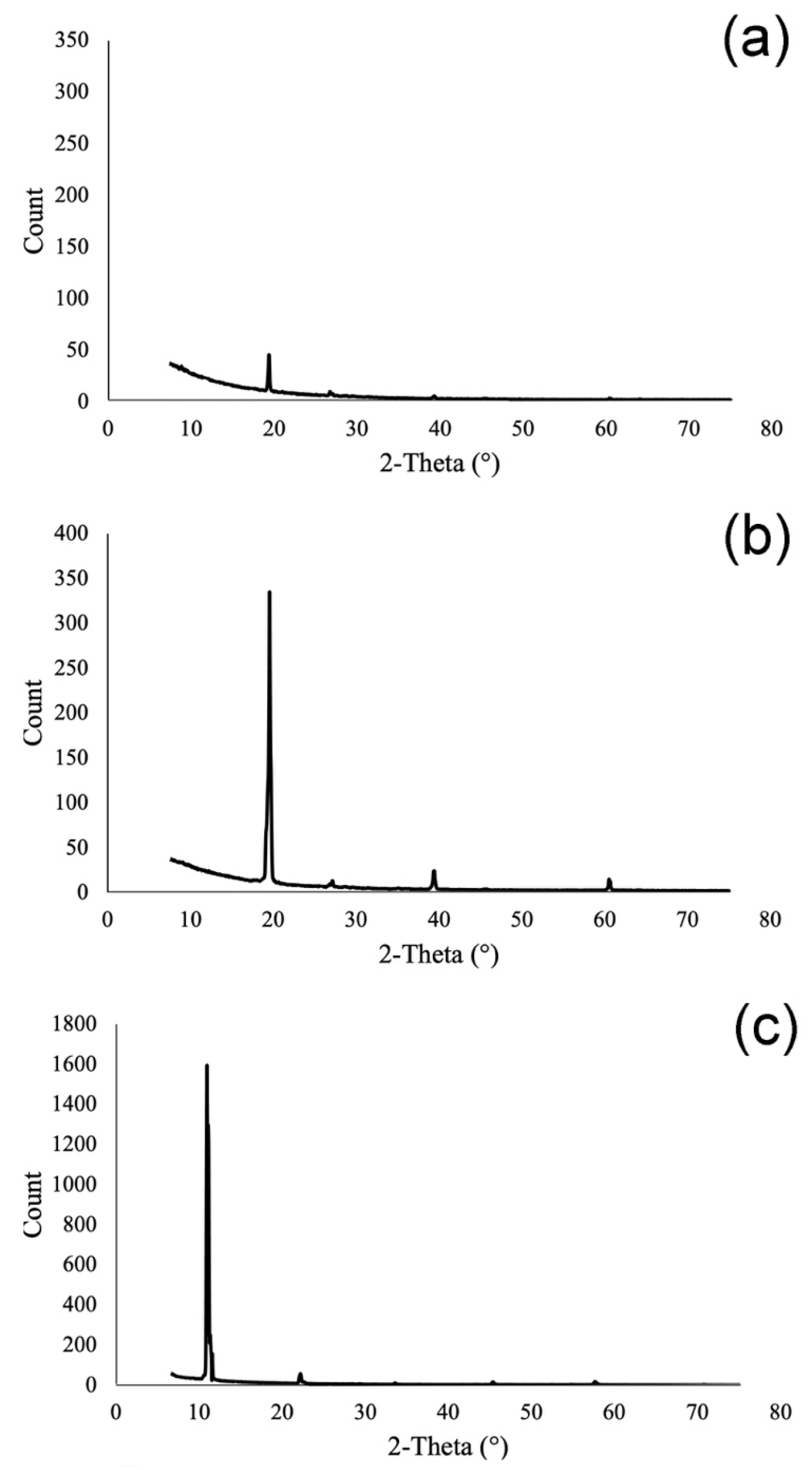
A Bruker AXS D8 Discover XRD with a Cu Kα source, operating at 40 kV and 40 mA, and a Lynxeye 1-dimensional detector were used for the diffraction analysis. Scans were performed from 3 to 75° 2θ, with 0.05° 2θ step and 1 s/step measuring time by point. Diffract-Eva software (version V7.5.0 32 bit) from Bruker with the PDF-2 mineralogical database (International Centre for Diffraction Data-ICDD) was utilised to interpret the scans. During the μ-XRD experiments, a Goebel mirror and 1 mm collimator were employed. The μ-XRD is a non-destructive technique that does not require grinding or powdering of the sample, which makes it particularly suitable for specimens originating from collections. However, despite its advantages, when applied to centimetre-sized single crystals, this technique often yields diffractograms with very few or even no observable peaks. This is due to the limited number of crystallographic planes intersecting the incident beam and the fixed orientation of the crystal, which restricts the generation of detectable reflections. As a result, the interpretation software may fail to match the pattern with the correct mineral phase. In some cases, such as for beryl, 3 diffractograms were collected by repositioning the crystal to different orientations, and the final pattern was obtained by summing the individual scans (Figure 2a–c). Conversely, for other samples, the diffraction produced enough peaks to allow for successful phase identification by the software.

2.2.2. SEM-EDS Analyses

To understand the elemental composition, minerals were analysed by a variable-pressure HITACHI S3700N SEM (Hitachi, Tokyo, Japan) coupled with a Quantax EDS (Bruker, Billerica, USA) microanalysis system. The Quantax system was equipped with a Bruker AXS XFlash Silicon Drift Detector (Bruker, Billerica, MA, USA) with (129 eV FWHM spectral resolution at Mn Kα.
Standardless PB/ZAF quantitative elemental analysis was performed using the Bruker ESPRIT software version 1.9. The operating conditions for EDS analysis were as follows: backscattering mode (BSEM), 20 kV accelerating voltage, 10 mm working distance, 120 μA emission current, and 40 Pa pressure in the chamber. The detection limits for major elements (>Na) were in the order of 0.1 wt.%. The instrumental statistical uncertainty was 1σ.

2.2.3. Colorimetric Analyses

To determine semi-quantitatively the colour coordinates, Vis-NIR HSI hypercubes were acquired with a Specim IQ® hyperspectral camera by Specim, Spectral Imaging Ltd. (Oulu, Finland). The camera is equipped with a Si-CMOS sensor serving as a photographic camera with a resolution of 512 × 512 pixels (5 Mpixel) and a spectral detector (400–1000 nm spectral range). The radiation is allowed into the camera through two distinct objectives, one for the spectral analysis and the other for the photographic recording. The hypercubes were acquired by placing the reference for white contextually to every acquisition, without spectral binning (204 channel/pixel), with spatial resolution in the 0.1–0.3 mm/pixel range and integration time in the 96–116 ms range. Spectral extraction and mapping were performed as single-pixel extraction of the irradiance values with the ENVI® 5.3 software by L3Harris Geospatial (Broomfield, CO, USA). For the illumination set, two air-cooled, dimmable VC-400HH lighting heads mounted on LS-8008K stands, both by VISICO (Yuyao, China), equipped with 1000 W, 240 V, 3200 K temperature colour GX9.5 64745 halogen lamps by Osram (Munich, Germany) were placed at ca. 1.5 m distance from the minerals and directly projected at 40° (lighting set–sample analyser angle) to avoid specular reflection. All measurements took place in a dark room to avoid interference from extraneous lighting and were placed on a black paperboard ground.
To ensure homogeneous diffuse illumination, a dedicated soft box was applied before each lighting head, and the incident ambient irradiation was checked with a portable light meter L-308S FLASHMATE (Sekonic, Denville, NJ, USA) to ensure 800 lux incidents to all samples, both before and after illumination. This choice for this value was based on previous experience with the device and the need to provide sufficient radiation to obtain a satisfactory result in a relatively short exposure time (<10 s). To make sure that the light target would be achieved, the dimmable sets were adjusted accordingly, and the lighting head-to-sample distance was measured with a digital distance meter GLM 40 by BOSCH (Gerlingen, Germany). The analytical conditions before UV exposure (sample-to-camera distance, field of view, sample positioning within the viewing field) were recorded for each sample and reproduced following UV exposure.
Before the UV exposure test (moment 0), the colour of the minerals was measured on 3–5 representative areas by using a routine based on HSI and MATLAB® (version R2023b) programming. The areas were selected from the hypercube to represent facets with different orientations as parallel as possible to the detector, while excluding undesired phases (such as inclusions, substrate, and macroscopic accessory phases) and spatially diverse regions of the analysed mineral. Each sampling area corresponds to a single pixel (1 × 1 kernel), and each CIELAB coordinate reported in the study is the average of 5 measurements, each being a spectrum converted into a set of CIELAB coordinates. The representative pixels for the post-treatment samples (moment 1) were selected based on the (m, n) location of the first hypercube (moment 0), which was also visually checked and adjusted where necessary to ensure geographic consistency. The exact number of spectra used for the averaging depended on the standard variation of each coordinate of the set: whenever it exceeded 1 unit of both/either the a* and b*, the outliers were not considered for the calculation.
The adopted procedure relies on the initial acquisition of irradiance values for the white standard, a black reference material and the areas to inspect with the HSI camera. Those values were directly fed into a custom-made function written in the MATLAB® environment. This function was written to perform automated data averaging, conversion to apparent reflectance (dark-subtracted) first, and then to the CIELAB colour space, as recommended in the ASTM E308-22 (Standard practice for computing the colors of objects by using the CIE system 2022) [33], stating the adopted chromaticity coordinates x, y, and z specific for the 10° Supplementary Standard Observer, 5 nm spectral step and D65 input Standard Illuminant. The final conversion to the perceptual space (CIELAB colour space, L*, a*, b*) adopted the Xn, Yn, and Zn values of the Standard Illuminant D65 (output illuminant) and CIE XYZ-to-CIELAB colour conversion formulas.
The process was computed with a custom-made, automated procedure by using an iterative MATLAB® code. For each mineral, 5 representative areas were chosen and sampled digitally with HSI before exposure (moment 0).
After 2 days of UV exposition performed by the QUV spray chamber (Q-LAB), mineral colours were measured.
Following the exposure, the colour coordinates were measured on the same spots as moment 0 and by adopting the same HSI-MATLAB routine as before.

2.2.4. UV Photoaging Tests

The QUV-spray Q-LAB climatic chamber, used for this experiment, was equipped with 8 UV lamps. These provide the best possible simulation of sunlight in the critical short wavelength region from 365 nm down to the solar cut-off of 295 nm. Its peak emission is at 340 nm.
The lamps’ intensity expressed in W/m2 can be regulated by the operator in the range 0.35 < I < 1.55 W/m2 to equalise, accelerate (up to 2 times) or even set an irradiance (I) lower than the natural one. For this purpose, it is necessary to specify the order of magnitude of the UV340nm radiation in the natural environment.
According to Q-LAB, an irradiance of 0.68 W/m2 340nm is equivalent to noon summer sunlight at the middle latitudes (30–40°). Further, 1.36 W/m2 340nm represents 2× solar maximum, an artificial enhancement whose value does not exist at 0 m above sea level. This value can, in fact, be measured only at tropical-height altitude. The irradiation of 0.35 W/m2 at 340 nm represents the so-called average optimal level and corresponds to the typical noon UV exposure in mid-latitudes during March and September under ‘average’- or low-UV conditions. For our test, the chamber was set by performing a new cycle “Sitzia standard” consisting of 48 h of UV exposure at 0.35 W/m2 340nm with 45 °C temperature. This cycle exposes the minerals to a non-accelerated test with a temperature low enough not to create mutations in the same chemical/physical features.
As mentioned above, the test lasted for 48 h, where minerals were exposed to the following irradiance energy: as 1 W/m2340nm corresponds to 1 J/m2·s, 0.35 W/m2340nm = 0.35 J/m2·s. In consideration of the above conversion factor, the irradiation of a 48 h photoaging test would amount to 0.06 MJ/m2. In fact, (0.35 J/m2·s)·(1.728·105 s) = 60,480 J/m2, or 0.06 MJ/m2.
It is important to note that the 48 h of UV exposure applied in this test (0.35 W/m2 at 340 nm, 45 °C) corresponds roughly to the UV dose in outdoor exposure. Therefore, these results cannot be directly extrapolated to long-term scenarios such as several years of exposure in a museum environment, where the UV intensity is generally much lower and other decay factors (fluctuating temperature, humidity, pollutants) interact over time. The present accelerated test is intended to highlight the relative sensitivity of the investigated minerals to UV radiation, providing a first indication of their stability, rather than a quantitative prediction of their long-term behaviour. Future work including longer-term artificial ageing and natural exposure tests would be required to establish a more robust correlation with museum environment.

3. Results and Discussion

Colour of minerals results from the interaction of light with a surface, where light can be reflected, transmitted, or absorbed. In a simplified sense, the portion that reaches the observer’s eye is the part that is not absorbed, that is, the scattered light. The absorbed portion, on the other hand, promotes electronic transitions within the valence shells of the mineral. Factors influencing the colour of minerals are as follows:
(1)
The particle size alters light scattering and absorption [34].
Normally, when a mineral is ground into smaller particles, thus reducing its grain size distribution, its colour changes compared to the cohesive mineral, usually accompanied by an increase in lightness (L).
(2)
The crystal structure, which affects electronic transitions and interference patterns [35]. In some minerals, the colour variations could be due to the oxidation of organic matter, iron (from Fe+2 to Fe+3) and manganese (from Mn+2 to Mn+3).
(3)
The surface properties (roughness) influence reflectance and adsorption [36]
This is a critical factor, as varying degrees of surface roughness have been shown to significantly influence the colour of minerals and stones [37,38].
(4)
Anisotropy/orientation changes polarisation and rotation of reflected light [39].
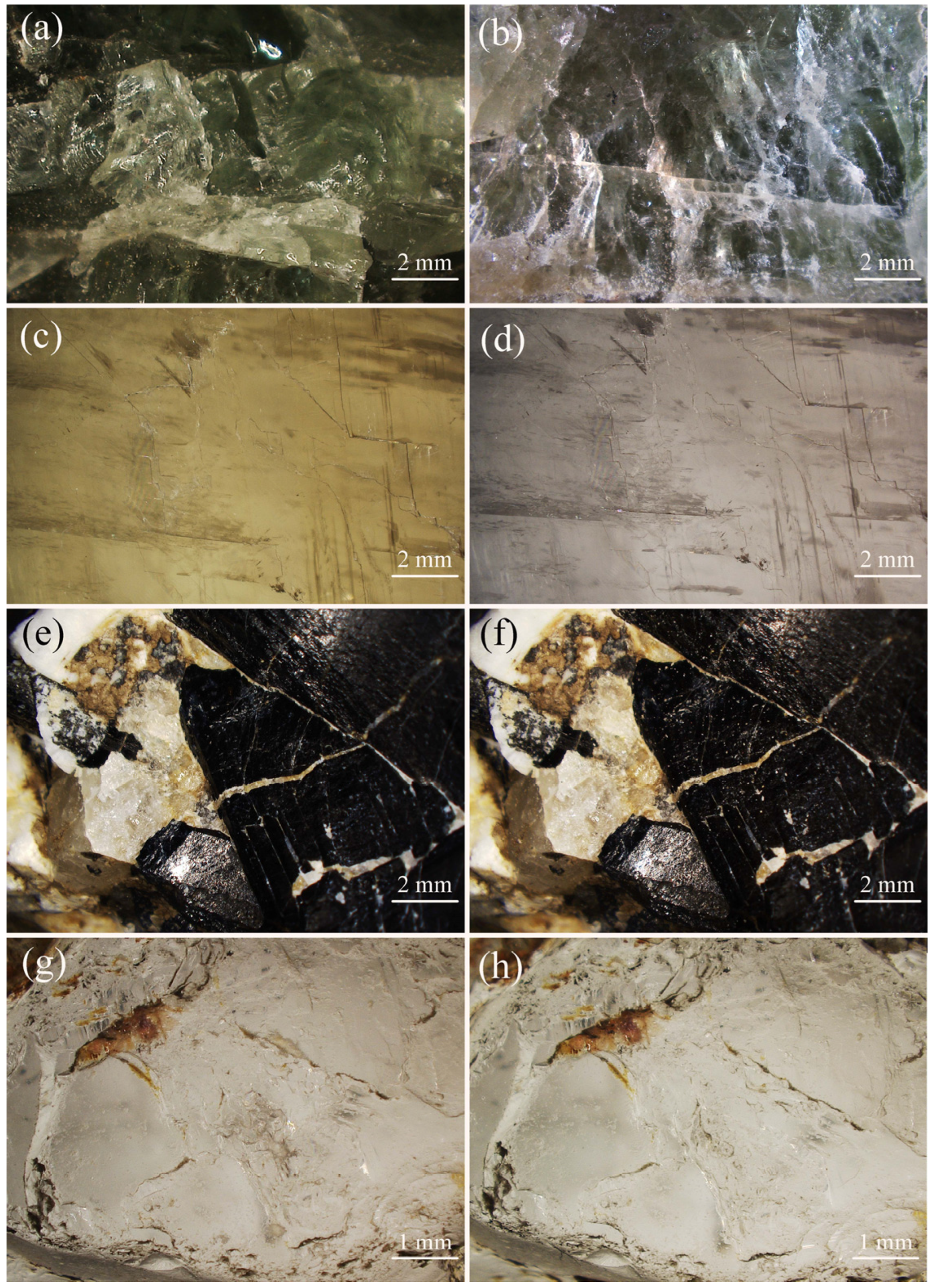
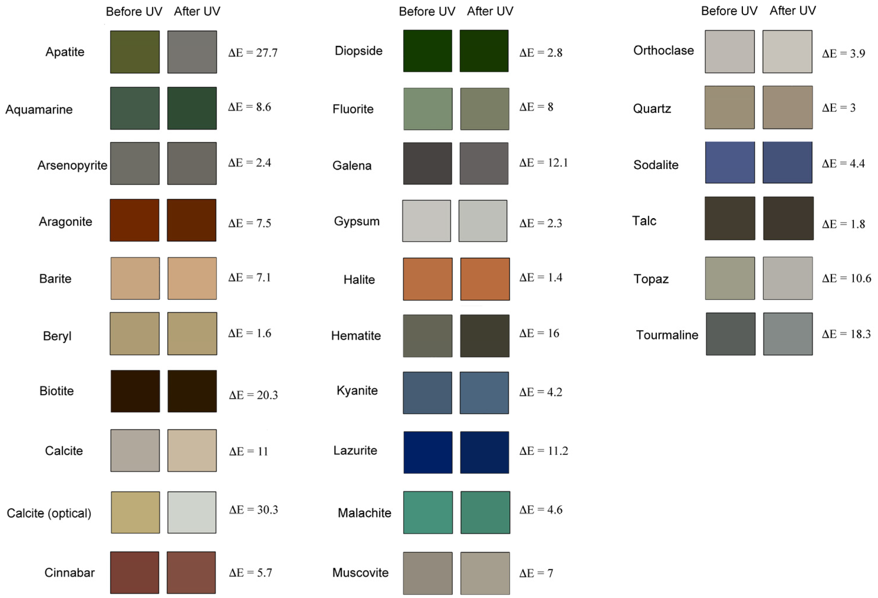
The chromatic effects of the UV ageing tests are shown in Figure 3, while the complete chart of discoloration is given in Figure 4. The latter reports, for each mineral phase, two squares filled with a solid colour, one corresponding to the original appearance and the other to the colour following the UV-photoaging tests. The relative colour distances are also given at the end of each row. This value is expressed as the distance metric ΔE of perceptual colour difference, calculated with the following formula:
ΔE = ((L*post − L*pre)2 + (a*post − a*pre)2 + (b*post − b*pre)2)0.5
When ΔE ≤ 2.3, the colour distance is not detectable; otherwise, when ΔE > 2.3, the human eye becomes capable of perceiving those differences. This value is defined as JND (Just Noticeable Difference) [40]. Another colour distances method, such as ΔE2000, would be the most suitable choice due to its uniformity and the fact it is the recommended parameter in ISO standards. However, its application conflicts with the scope of this study. This research is situated within the artistic and cultural heritage domain, where ΔELab (hereafter ΔE) remains widely adopted for tracking colour changes in materials over time, so this parameter offers comparability with existing conservation and heritage studies, ensures methodological continuity with established approaches in the field, and provides a common reference point for interpreting visible colour shifts in a way that is familiar to both scientific and heritage-focused audiences.
According to the results, only 4 samples out of 26 exhibit a ΔE ≤ 2.3. In these samples, namely beryl, gypsum, halite and talc, the colour changes are, indeed, so minimal that they could not be observed with the naked eye. As for the remaining 22 mineral samples, the colorimetric variations were so strong that they became perceptually noticeable. The colour distance ΔE found in this UV exposure ranges from 1.4 (halite) to 30.3 (optical calcite).
Excluding the samples with subperceptual colour changes, the data allow for drawing three clusters of minerals. The first includes minerals displaying a relatively low colour distance (2.4 ≤ ΔE ≤ 6), and it includes arsenopyrite, beryl, cinnabar, diopside, gypsum, malachite, orthoclase, quartz, sodalite and talc. The changes that interested this group involve slight to noticeable changes in chroma, mostly toward desaturation (arsenopyrite, cinnabar, diopside, gypsum, kyanite, malachite, quartz, sodalite and talc), occasionally involving a slight increase in saturation (beryl and kyanite). However, no mineral saw significant changes in hue, meaning the colour changes of this group are perceptually minimal, aside from a few instances (cinnabar, quartz, sodalite). In this latter case, the colour does not change (i.e., cinnabar stays red), and the change in chroma could be a sign of possible fading, since ΔC* tends toward negative values.
A second cluster gathers minerals with average colour shift (7 ≤ ΔE ≤ 20) included aquamarine, aragonite, barite, calcite, fluorite, galena, hematite, lazurite, muscovite, topaz and tourmaline. The situation of this cluster appears more heterogeneous in comparison with the respective changes in chroma and hue. Among these minerals, five subgroups emerge: (I) minerals with very slight negative ΔC* (<−0.5) and slight ΔH* (<1.0, Muscovite), (II) strong negative ΔC* (comprised between ca. −11 and −8), moderate ΔH* (in the 1–2 range, galena and lazurite), (III) strong negative ΔC* (between ca. −7 and −3) and obvious-to-strong ΔH* (between 2 and 6, aragonite and fluorite), (IV) slight positive ΔC* and strong ΔH* (tourmaline), (V) strong positive ΔC* and slight-to-noticeable ΔH* (aquamarine, barite and calcite). This means that the minerals in this cluster show very diverse patterns of colour shifts, and that only in a few cases are accompanied by substantial alterations in hue, namely in subgroup III and IV.
The last group is represented by minerals that manifest strong colour variation (ΔE ≥ 21), namely the cluster of apatite, biotite and optical calcite. Within this latter class, particularly notable are apatite (ΔE = 27.7) and optical calcite (ΔE = 30.3). These alterations are accompanied by extremely strong variations in chroma and hue, so further considerations must be drawn among these minerals with the greatest changes.
Considering the case of biotite, a colour distance of 20.3 can be observed (Table 2). In this mineral, the colour change involves a slight mutation of the L* and a* component; however, a significant difference is detected in the b* component, which changes from 26.59 to 46.77. The increase in this parameter indicates that the colour of the mineral has changed by increasing the amount of yellow. For biotite, there is no direct research evidence specifically documenting a colour change (photosensibility) when exposed to UV radiation. Biotite is known to undergo colour changes due to exposure to ionising radiation, such as alpha particles, which can induce coloration and subsequent reversal at specific energy thresholds. However, this process is distinct from UV-induced changes and is not directly related to UV photosensibility [41]. One possible explanation for this colour change in biotite is related to the oxidation of iron present in the mineral, which leads to the formation of new compounds consisting of oxides and hydroxides. However, this process could be linked to simple atmospheric oxidation, where UV rays did not play a decisive role.
Apatite, a group of phosphate minerals, can undergo notable colour changes and fluorescence when exposed to ultraviolet (UV) light. In the analysed sample of apatite, the initial green colour had scaled down to a dull grey with the UV irradiation. The colorimetric analysis in Table 2 shows that the apatite under study has undergone lightening, with the L* parameter changing from 37.79 to 48.84, a loss of its characteristic green colour due to the increase in a* (from −8.82 to −0.01) and a loss of yellow tones due to a drastic decrease in the b* component. The origin of colour in apatite minerals arises from a combination of factors, including the incorporation of transition metals (e.g., Fe2+, Mn2+ and Cd2+), rare earth elements (REEs, such as Ce3+, Nd3+, La3+, and Sm3+) and halogen impurities or substituents (F, Cl or O), which can act as luminescence activators or co-activators [42]. Additional contributions include specific conditions of formation and mineral association [42], while the formation of F-centres in halogenoapatites is considered relatively rare [42,43]. In the present study, the analysed apatite contained calcium and 0.21 wt.% sodium (Table 3), which generally act as divalent and monovalent cations respectively, [40,41,42] and initially exhibited a green colour. No other metals or REEs were identified. Regarding REEs, their concentration in apatite can vary significantly, yet it typically falls within the range of 0.8–1.0 wt.% [44,45,46]. These values are often below the detection limit (LOD) of SEM-EDS systems, so REE presence is generally investigated through extraction procedures [45], or with highly sensitive techniques such as LA-ICP-MS or EMPA [47].
A useful indirect indicator of REE presence of this study is the detection of Na. In the apatite structure, cation substitution can occur either between divalent cations or between a pair of monovalent and trivalent cations, following the charge-compensation mechanism A+ + C3+ = 2 B2+, where A, B, and C represent monovalent, divalent, and trivalent cations respectively, [41]. The presence of Na+ thus implies the need for a charge-compensating trivalent cation. Common trivalent substituents include Sc, Y, La, Ce, Tb, Eu, and Dy [44]. If charge compensation does not occur, F-centres may form, which are typically observable as a well-defined absorption band at 450 nm [43]. Colorimetrically, the un-weathered apatite of this study appeared green, shifting to a dull grey following UV irradiation. Previous studies have reported that UV exposure rarely induces structural changes in apatite [48], and its luminescence is more frequently associated with REEs [42]. The UV-Vis spectrum at t0 (prior to UV exposure) showed a steady increase in absorbance up to 500 nm (Figure 5a), with no intermediate absorption bands, suggesting the absence of F-centres. Between 500 and 900 nm, the untreated apatite exhibited four absorption bands at 522, 580, 738–747, and 802 nm (Figure 5b, spectrum t0). UV-Vis spectroscopy is a valuable technique for detecting REEs, as their characteristic electronic transitions often occur in this spectral range [42,43,44,46].
Previous studies have attributed these bands to Nd3+ and possibly Ce3+/Ce4+ impurities, which may be involved in photo-induced charge transfer processes acting as photochromic centres. Knutson et al. [46] performed a statistical analysis on natural Portuguese apatite samples and reported a correlation between REEs (detected via NAA) and Mn2+, suggesting that Mn2+ may function as a sensitiser for REE-related luminescence.
As referred above, following UV weathering, both the a* and b* colour coordinates shifted toward zero (Table 2), and the resulting colour appeared greyish overall (Figure 4), with the UV-Vis spectrum flattening out (Figure 5b, spectrum t2). This indicates a potential quenching effect. However, the mechanisms behind luminescence quenching in apatite remain under debate. Although Mn2+ and Fe2+ ions are frequently implicated in quenching processes, neither was detected in the present SEM-EDS analysis, preventing further discussion of their possible roles. The mechanisms underlying the light sensitivity of apatite are complex and can vary depending on the specific type of apatite and the surrounding environmental conditions. However, it is generally understood that its light sensitivity is linked to its crystalline structure ability to absorb and interact with light energy, leading to alterations in its structure, composition or physical properties [16]. It is worth noting that not all apatite minerals are light sensitive to the same degree, and the extent of their light sensitivity can vary depending on factors such as crystal size, impurity content, and the wavelength and intensity of the incident light. The light sensitivity of apatite can have various practical applications, such as in photoluminescent materials, optical devices, and geological dating techniques [19].
Another remarkable example of colour change is the optical calcite, which shows an initial yellowish tint that gradually fades to a light grey, retaining only a faint trace of yellow. UV exposure in calcite can cause bleaching or fading of certain calcite varieties [49]. Sunlight UV accelerates the transition from bluish-grey to yellow/beige calcites attributed to changes in iron oxidation states (Fe2+ to Fe3+) and the breakdown of colour centres [49].
Chromophores are responsible for absorbing light and causing the calcite crystal to undergo a change in its electronic structure, resulting in a change in colour. Calcite crystals may also contain such structural defects as vacancies or dislocations, which can trap and absorb radiative energy [19]. This can result in the excitation of electrons within the crystal lattice and subsequent changes in the crystalline optical properties, such as their colour or opacity. Previous studies showed that short exposure times to UV light can induce local relaxation or reordering of the crystalline lattice, inducing reduction in lattice defects or scattering centres [47,48]. At times, the magnitude of this effect is such that may even induce macroscopic reductions in surface smoothing and cracks [50]. In this case, such macroscopic effects were not observed (Figure 3) but flattening and an overall increase in reflectance was seen in the Vis-NIR spectrum of UV-weathered samples (Figure 5b, spectrum t0). At the same time, the presence of a vestigial broad band at 595 nm possibly suggests that some chromophores are part of the crystalline structure, and that the UV dose was insufficient to remove them completely.
Significant cases of colour loss also occur in the group with average colour shift (7 ≤ ΔE ≤ 20), including aquamarine, aragonite, barite, calcite, fluorite, galena, hematite, lazurite, muscovite, topaz and tourmaline.
Considering the case of tourmaline, a colour distance of 18.3 can be observed from Table 2. In this mineral phase, the colour change involves a considerable mutation of the L* (from 39.34 to 57.61). These changes in coordinates indicate that the mineral has undergone a colour lightening.
Current research confirms that tourmaline is photosensitive and enhances photocatalytic reactions under UV and visible light, but there is no scientific evidence that tourmaline changes colour when exposed to UV light [51,52].
Another mineral subject to significant colour variation is lazurite. It shows a colour shift (ΔE = 11.2) with an increase in the L* coordinate (from 13.06 to 14.96), a slight decrease in the a* component (from 10.30 to 9.38) and a decrease in b (from −48.11 to −37.10). In this case, the phase underwent to a lightening of colour accompanied by a shift towards green tones and a loss of intensity of the original blue colour. In general, colour changes in minerals belonging to the sodalite group, such as lazurite, and hackmanite is well documented [53]. The colour change in hackmanite is driven by the formation of colour centres (F-centres) when UV light excites electrons from anionic polysulfide species (like S22−) into chloride vacancies within the crystal structure. These trapped electrons alter the mineral’s absorption properties, resulting in visible colour. The process is reversible: exposure to visible light or heat releases the electrons, returning hackmanite to its original colourless state [54,55,56]. These impurities can absorb light energy and undergo photochemical reactions, resulting in changes to the crystal properties.
In hematite (ΔE = 16), the colour changes from a light grey to a dark grey tending to black with a noticeable decrease in the L* component.
In general, there is an average colour variation in the group of 26 samples of ΔE = 8.99, with a standard deviation of 7.81.
The causes of colour variation are quite complex and would require dedicated attention to elaborate hypotheses about their causes. However, this work attempts to advance some theories based on literature.
An important study on light-sensitive minerals was published in 2013 [16]. In that paper, the author identified 35 light-induced colour changes, 8 light-induced decompositions, and 78 light accelerated surface reactions. These changes were related to the 15 physical and chemical causes of colour [57].
One of the simplest cases of colour modification is the case of fluorite, whose colour arises from the occurrence of the so-called hole colour centres or F-centres. More specifically, the coloration in this class of minerals arises from electrons occupying vacancies left by missing halide ions (F) that were previously removed. The absence of the ion leaves unpaired electrons in the crystal lattice, which are free to interact with incoming radiation. When exposed to light, these free electrons can be excited by the incident energy. Under natural irradiation, the displacement of these electrons can disturb the electronic cloud responsible for the mineral’s original colour, thereby altering its appearance. When external energy, whether thermal or radiative, strikes these minerals, it may lead to the removal of their F-centres. As a result, their colour can fade or even disappear [16].
In our case, fluorite shows a ΔE = 8, where the b* component is subjected to an increase from −11.23 to −4.69.
In the groups of minerals with less colour change, namely (2.4 ≤ ΔE ≤ 6), an interesting case is represented by the cinnabar. It is known for displaying colour changes with UV irradiation by forming black metacinnabar. This latter phase owes its dark colour to an hypsochromic shift in the bandgap edge by 1.2 eV (380 nm) of the initial 2.0 eV edge (610 nm) of α-HgS, with the result of the black mineral absorbing radiation in the UV range and becoming invisible to the eye [58]. In our case, the observed colour shift was rather weak (ΔE = 5.7).

4. Conclusions

This study demonstrates that a significant majority of the tested mineral samples are sensitive to ultraviolet radiation, showing chromatic alterations exceeding the human perceptibility threshold after just 48 h of UVA340nm exposure. These results confirm the relevance of colour stability as a diagnostic and preventive parameter when dealing with mineral-based materials in cultural heritage, particularly those employed in artworks, architectural surfaces, pigments, and museum collections.
The observed chromatic variations span from subtle shifts to drastic alterations in lightness, hue, and chroma, and are linked to different mechanisms: from structural rearrangements and chromophore degradation to lattice defects and trace element oxidation. While only a minority of minerals displayed negligible sensitivity, others including apatite and optical calcite underwent pronounced discolouration, often resulting in irreversible aesthetic degradation.
From a conservation perspective, these findings reinforce the need to carefully assess and manage light exposure conditions in museum and exhibition environments, and they must not be directly exposed to sunlight but to artificial ones [59]. Light-sensitive minerals should be displayed under strictly controlled lighting conditions, preferably below 50 lux [17], with annual exposure limits for highly reactive specimens capped at 104 lux·hours, as established by conservation guidelines [60]. Moreover, this work highlights the importance of preliminary mineralogical and chemical characterisation, including the detection of chromophoric elements, as an essential step in the conservation planning process. These analytical approaches can help predict colour instability and guide the adoption of tailored preventive strategies.
Beyond the museum and heritage fields, the findings of this research have significant implications for architecture, especially regarding the use of natural stones in outdoor cladding and paving. Since many ornamental stones are composed of mineral phases that have proven sensitive to UV radiation, prolonged sunlight exposure can lead to progressive discolouration, surface whitening, or uneven chromatic alterations. These effects not only compromise the aesthetic appearance of buildings and monuments but may also lead to disputes between suppliers and clients due to unexpected changes in material tone after installation.
Recognising the light sensitivity of specific minerals, such as those rich in organic matter, transition metals, or structural defects, can inform the selection of more stable lithotypes for outdoor use or motivate the adoption of protective strategies (e.g., architectural shading, UV resistant hydrophobic coatings), or design features limiting direct exposure to sunlight).
Moreover, understanding colour instability is crucial for restoration projects, where material matching plays a key aesthetic and cultural role. This research therefore can support the integration of photostability assessments in the design and specification phase of architectural applications, helping to promote more durable, predictable, and visually consistent building envelopes over time.

Author Contributions

F.S.: conceptualization, data curation, formal analysis, investigation, project administration, resources, software, supervision, methodology, validation, visualisation, writing—original draft, writing—review and editing, C.L.: formal analysis, methodology, validation, visualisation, writing—original draft, writing—review and editing S.B.-S.: formal analysis, methodology, validation, writing—review and editing. P.M.: formal analysis. All authors have read and agreed to the published version of the manuscript.

Funding

The authors gratefully acknowledge the funder European Communion—Portugal, with number: Sustainable Stone by Portugal, Call: 2021-C05i0101-02—mobilising agendas/alliances for reindustrialization—PRR. Funding number: C644943391-00000051.

Data Availability Statement

Data are contained within the article.

Acknowledgments

The authors would like to thank the organising committee of the VII Global Stone Congress 2023 (Batalha, Portugal) for allowing the oral presentation ‘Colour fading and changing in light-sensitive minerals exposed to UV rays’, from which this paper was developed.

Conflicts of Interest

The authors declare no conflicts of interest.

References

  1. ISO 21348:2007; Space Environment (Natural and Artificial)-Process for Determining Solar Irradiances. ISO: Geneva, Switzerland, 2007.
  2. Kopp, G.; Lean, J.L. A New, Lower Value of Total Solar Irradiance: Evidence and Climate Significance. Geophys. Res. Lett. 2011, 38, 714. [Google Scholar] [CrossRef]
  3. El-Nouby Adam, M. Effect of The Atmosphere on UVB Radiation Reaching the Earth’s Surface: Dependence on Solar Zenith Angle. Atmos. Ocean. Sci. Lett. 2011, 4, 139–145. [Google Scholar] [CrossRef]
  4. Bennson, G. Handbook on Synchrotron Radiation. Opt. Acta Int. J. Opt. 1984, 31, 979–980. [Google Scholar] [CrossRef]
  5. Cogulet, A.; Blanchet, P.; Landry, V. Wood Degradation under UV Irradiation: A Lignin Characterization. J. Photochem. Photobiol. B Biol. 2016, 158, 184–191. [Google Scholar] [CrossRef] [PubMed]
  6. Rajagopalan, N.; Khanna, A.S. Effect of Methyltrimethoxy Silane Modification on Yellowing of Epoxy Coating on UV (B) Exposure. J. Coatings 2014, 2014, 22. [Google Scholar] [CrossRef]
  7. Rosu, D.; Rosu, L.; Cascaval, C.N. IR-Change and Yellowing of Polyurethane as a Result of UV Irradiation. Polym. Degrad. Stab. 2009, 94, 591–596. [Google Scholar] [CrossRef]
  8. Careddu, N.; Marras, G. The Effects of Solar UV Radiation on the Gloss Values of Polished Stone Surfaces. Constr. Build. Mater. 2013, 49, 828–834. [Google Scholar] [CrossRef]
  9. Navarro, R.; Catarino, L.; Pereira, D.; Paulo, F.; Campos, D.S. Effect of UV Radiation on Chromatic Parameters in Serpentinites Used as Dimension Stones. Bull. Eng. Geol. Environ. 2019, 78, 5345–5355. [Google Scholar] [CrossRef]
  10. Sitzia, F.; Lisci, C.; Mirão, J. Accelerate Ageing on Building Stone Materials by Simulating Daily, Seasonal Thermo-Hygrometric Conditions and Solar Radiation of Csa Mediterranean Climate. Constr. Build. Mater. 2021, 266, 121009. [Google Scholar] [CrossRef]
  11. Sitzia, F.; Lisci, C.; Mirão, J. Building Pathology and Environment: Weathering and Decay of Stone Construction Materials Subjected to a Csa Mediterranean Climate Laboratory Simulation. Constr. Build. Mater. 2021, 300, 124311. [Google Scholar] [CrossRef]
  12. Virgil Lueth Light Sensitive Minerals. In Proceedings of the 39th Annual New Mexico Mineral Symposium, Socorro, New Mexico, 10–11 November 2018; pp. 34–35.
  13. Bringley, J.F.; Rajeswaran, M.; Olson, L.P.; Liebert, N.M. Silver-Halide/Organic-Composite Structures: Toward Materials with Multiple Photographic Functionalities. J. Solid State Chem. 2005, 178, 3074–3089. [Google Scholar] [CrossRef]
  14. Kirk, R.D. The Luminescence and Tenebrescence of Natural and Synthetic Sodalite. Am Min. 1955, 40, 22–31. [Google Scholar]
  15. Foord, E.E.; Berendsen, P.; Storey, L.O. Corderoite, First Natural Occurrence of Alpha -Hg//3s//2cl//2, From The Cordero Mercury Deposit, Humboldt County, Nevada; American Mineralogist: Chantilly, VA, USA, 1985; ISBN 0895204444. [Google Scholar]
  16. Nassau, K. Conserving Light-Sensitive Minerals and Gems. In The Care and Conservation of Geological Material: Minerals, Rocks, Meteorites and Lunar Finds; Routledge: Abingdon, Oxfordshire, UK, 2013; ISBN 9781135385149. [Google Scholar]
  17. Horak., J.M. Environmental Effects on Geological Material: Light Induced Changes of Minerals. In Conservation of Geological Collections; Child, R.E., Ed.; Archetype Publications: London, UK, 1994; pp. 23–30. ISBN 1873132603. [Google Scholar]
  18. Rossman, G.R. Colored Varieties of the Silica Minerals. In Silica: Physical Behavior, Geochemistry, and Materials Applications; de Gruyter: Berlin, Germany, 2019; ISBN 9781501509698. [Google Scholar]
  19. Howie, F.M. The Care and Conservation of Geological Material: Minerals, Rocks, Meteorites and Lunar Finds; Routledge: Abingdon, Oxfordshire, UK, 1992. [Google Scholar] [CrossRef]
  20. Ballirano, P.; Botticelli, M.; Maras, A. Thermal Behaviour of Cinnabar, α-HgS, and the Kinetics of the β-HgS (Metacinnabar)—α-HgS Conversion at Room Temperature. Eur. J. Mineral. 2014, 25, 957–965. [Google Scholar] [CrossRef]
  21. Douglass, D.L.; Shing, C.; Wang, G. The Light-Induced Alteration of Realgar to Pararealgar. Am. Mineral. 1992, 77, 1266–1274. [Google Scholar]
  22. Brunton, C.H.C.; Besterman, T.P.; Cooper, J.A. Guidelines for the Curation of Geological Material; The Geological Society: London, UK, 1985. [Google Scholar]
  23. Howie, F.M.P. Museum Climatology and the Conservation of Palaeontological Material. In Curation of Palaeontological Collections: Special Papers in Palaeontology; The Palaeontological Association: Durham, UK, 1979. [Google Scholar]
  24. Lisci, C.; Sitzia, F.; Pires, V.; Mirão, J. Building Stones Durability by UVA Radiation, Moisture and Spray Accelerated Weathering. J. Build. Pathol. Rehabil. 2022, 7, 60. [Google Scholar] [CrossRef]
  25. Dang, R.; Yuan, Y.; Luo, C.; Liu, J. Chromaticity Shifts Due to Light Exposure of Inorganic Pigments Used in Traditional Chinese Painting. Light. Res. Technol. 2017, 49, 818–828. [Google Scholar] [CrossRef]
  26. Xu, H.; Zhang, Z.; Dong, X.; Huang, L.; Zeng, H.; Lin, Z.; Zou, G. Corrugated 1D Hybrid Metal Halide [C6H7ClN]CdCl3Exhibiting Broadband White-Light Emission. Inorg. Chem. 2022, 61, 4752–4759. [Google Scholar] [CrossRef] [PubMed]
  27. Mo, X.; Li, T.; Huang, F.; Li, Z.; Zhou, Y.; Lin, T.; Ouyang, Y.; Tao, X.; Pan, C. Highly-Efficient All-Inorganic Lead-Free 1D CsCu2I3 Single Crystal for White-Light Emitting Diodes and UV Photodetection. Nano Energy 2021, 81, 105570. [Google Scholar] [CrossRef]
  28. Worku, M.; Tian, Y.; Zhou, C.; Lee, S.; Meisner, Q.; Zhou, Y.; Ma, B. Sunlike White-Light-Emitting Diodes Based on Zero-Dimensional Organic Metal Halide Hybrids. ACS Appl. Mater. Interfaces 2018, 10, 30051–30057. [Google Scholar] [CrossRef] [PubMed]
  29. Dang, R.; Yuan, Y.; Liu, G.; Liu, J. Chromaticity Changes of Inorganic Pigments in Chinese Traditional Paintings Due to the Illumination of Frequently-Used Light Sources in Museum. Color Res. Appl. 2018, 43, 596–605. [Google Scholar] [CrossRef]
  30. Vitorino, T.; Picollo, M.; del Hoyo-Meléndez, J.M. An Investigation of the Photostability of 19th Century Artists’ Cochineal Lake Pigments Using MFT and Conventional Accelerated Ageing Tests. Nondestruct. Test. Eval. 2024, 39, 2530–2548. [Google Scholar] [CrossRef]
  31. Grandjean, F.; Samain, L.; Long, G.J. Characterization and Utilization of Prussian Blue and Its Pigments. Dalt. Trans. 2016, 45, 18018–18044. [Google Scholar] [CrossRef] [PubMed]
  32. Sitzia, F.; Moita, P.; Bottura-Scardina, S.; Lisci, C. Colour Fading and Changing in Light-Sensitive Minerals Exposed to UV Rays. In Proceedings of the Global Stone Congress 2023; Lopes, L., Peres, M., Marques, P., Eds.; University of Evora: Batalha, Portugal, 2023. [Google Scholar]
  33. ASTM E308-22: 2022; Standard Practice for Computing The Colors of Objects by Using the CIE System. ASTM International: West Conshohocken, PA, USA, 2022. [CrossRef]
  34. Yang, X.L.; Wan, X.X. Analysis of the Spectral Reflectance and Color of Mineral Pigments Affected by Their Particle Size. Color Res. Appl. 2020, 45, 246–261. [Google Scholar] [CrossRef]
  35. Zhang, A.; Mu, B.; Wang, X.; Wen, L.; Wang, A. Formation and Coloring Mechanism of Typical Aluminosilicate Clay Minerals for CoAl2O4 Hybrid Pigment Preparation. Front. Chem. 2018, 6, 125. [Google Scholar] [CrossRef]
  36. Lu, L.; Xu, B.; Zhang, Q.; Lu, T.; Farooq, U.; Chen, W.; Zhou, Q.; Qi, Z. Adsorption Features of Pigments onto Iron Oxides: Co-Effect of the Molecular Structure of Adsorbates and the Varied Surface Properties of Minerals. J. Mol. Liq. 2023, 388, 122766. [Google Scholar] [CrossRef]
  37. Sitzia, F.; Lisci, C.; Mirão, J. The Interaction between Rainwater and Polished Building Stones for Flooring and Cladding—Implications in Architecture. J. Build. Eng. 2022, 52, 104495. [Google Scholar] [CrossRef]
  38. Sitzia, F.; Lisci, C.; Pires, V.; Alves, T.; Mirão, J. Laboratorial Simulation for Assessing the Performance of Slates as Construction Materials in Cold Climates. Appl. Sci. 2023, 13, 2761. [Google Scholar] [CrossRef]
  39. Green, L.H. The Relationship between Polarization Colors and Rotation Properties of Anisotropic Minerals. Econ. Geol. 1952, 47, 451–458. [Google Scholar] [CrossRef]
  40. Witzel, R.F.; Burnham, R.W.; Onley, J.W. Threshold and Suprathreshold Perceptual Color Differences. J. Opt. Soc. Amer. 1973, 63, 615–625. [Google Scholar] [CrossRef]
  41. Moazed, C.; Overbey, R.; Spector, R.M. Alpha-Induced Coloration Reversal in Biotite. Nature 1977, 267, 818–819. [Google Scholar] [CrossRef]
  42. Waychunas, G.A. Apatite Luminescence. Rev. Mineral. Geochemistry 2002, 48, 701–742. [Google Scholar] [CrossRef]
  43. Jayaprakash, R.; Ratnam, V.V.; Durrani, S.A. Optical Bleaching of Radiation-Induced Colour Centres in Fluorapatite. Nucl. Tracks Radiat. Meas. 1985, 10, 601–604. [Google Scholar] [CrossRef]
  44. Liu, H.; Liao, L.; Pan, X.; Su, K.; Shuai, P.; Yan, Z.; Guo, Q.; Mei, L. Recent Research Progress of Luminescent Materials with Apatite Structure: A Review. Open Ceram. 2022, 10, 100251. [Google Scholar] [CrossRef]
  45. Anufrieva, A.V.; Andrienko, O.S.; Buynovskiy, A.S.; Makaseev, Y.N.; Mazov, I.N.; Nefedov, R.A.; Sachkov, V.I.; Stepanova, O.B.; Valkov, A.V. Rare Earth Elements Materials Production from Apatite Ores. IOP Conf. Ser. Mater. Sci. Eng. 2016, 112, 012002. [Google Scholar]
  46. Knutson, C.; Peacor, D.R.; Kelly, W.C. Luminescence, Color and Fission Track Zoning in Apatite Crystals of the Panasqueira Tin-Tungsten Deposit, Beira-Baixa, Portugal. Am. Mineral. 1985, 70, 829–837. [Google Scholar]
  47. Bačík, P.; Fridrichová, J.; Štubňa, J.; Bancík, T.; Illášová, L.; Pálková, H.; Škoda, R.; Mikuš, T.; Milovská, S.; Vaculovič, T.; et al. The Ree-Induced Absorption and Luminescence in Yellow Gem-Quality Durango-Type Hydroxylapatite from Muránska Dlhá Lúka, Slovakia. Minerals 2020, 10, 1001. [Google Scholar] [CrossRef]
  48. Clavé, E.; Beyssac, O.; Bernard, S.; Royer, C.; Lopez-Reyes, G.; Schröder, S.; Rammelkamp, K.; Forni, O.; Fau, A.; Cousin, A.; et al. Radiation-Induced Alteration of Apatite on the Surface of Mars: First in Situ Observations with SuperCam Raman Onboard Perseverance. Sci. Rep. 2024, 14, 11284. [Google Scholar] [CrossRef]
  49. Silva, T.P.; de Oliveira, D.; Veiga, J.P.; Lisboa, V.; Carvalho, J.; Barreiros, M.A.; Coutinho, M.L.; Salas-Colera, E.; Vigário, R. Contribution to the Understanding of the Colour Change in Bluish-Grey Limestones. Heritage 2022, 5, 1479–1503. [Google Scholar] [CrossRef]
  50. Kabacińska, Z.; Yate, L.; Wencka, M.; Krzyminiewski, R.; Tadyszak, K.; Coy, E. Nanoscale Effects of Radiation (UV, X-Ray, and γ) on Calcite Surfaces: Implications for Its Mechanical and Physico-Chemical Properties. J. Phys. Chem. C 2017, 121, 13357–13369. [Google Scholar] [CrossRef]
  51. Xiong, Y.; Chen, B.; Xu, T.; Tan, L.; Xin, W.; Du, J.; Bao, J. Efficient Synergistic Photocatalytic Degradation by Sunlight-Driven Natural Tourmaline Modified g-C3N4 Interface Electric Field. Sep. Purif. Technol. 2025, 361, 131496. [Google Scholar] [CrossRef]
  52. Tzeng, J.H.; Weng, C.H.; Lin, Y.H.; Huang, S.M.; Yen, L.T.; Anotai, J.; Lin, Y.T. Synthesis, Characterization, and Visible Light Induced Photoactivity of Tourmaline-N-TiO2 Composite for Photooxidation of Ethylene. J. Ind. Eng. Chem. 2019, 80, 376–384. [Google Scholar] [CrossRef]
  53. Vuori, S.; Colinet, P.; Norrbo, I.; Steininger, R.; Saarinen, T.; Palonen, H.; Paturi, P.; Rodrigues, L.C.V.; Göttlicher, J.; Le Bahers, T.; et al. Detection of X-Ray Doses with Color-Changing Hackmanites: Mechanism and Application. Adv. Opt. Mater. 2021, 9, 2100762. [Google Scholar] [CrossRef]
  54. Song, C.; Guo, Q.; Liu, Y.; Rao, Y.; Liao, L. Photochromism, UV-Vis, Vibrational and Fluorescence Spectroscopy of Differently Colored Hackmanite. Crystals 2023, 11, 1607. [Google Scholar] [CrossRef]
  55. Norrbo, I.; Curutchet, A.; Kuusisto, A.; Mäkelä, J.; Laukkanen, P.; Paturi, P.; Laihinen, T.; Sinkkonen, J.; Wetterskog, E.; Mamedov, F.; et al. Solar UV Index and UV Dose Determination with Photochromic Hackmanites: From the Assessment of the Fundamental Properties to the Device. Mater. Horizons 2018, 5, 569–576. [Google Scholar] [CrossRef]
  56. Kim, S.H.; Kim, S.C. The Kinetics of Carrier Trap Parameters in Na8Al6Si6O24(Cl,S)2 Hackmanite. Materials 2023, 16, 6776. [Google Scholar] [CrossRef]
  57. Rogers, J.J.W. Geochemistry of Solids. Geochim. Cosmochim. Acta 1964, 28, 10–11. [Google Scholar] [CrossRef]
  58. Dickson, F.W.; Tunell, G. The Stability Relations of Cinnabar and Metacinnabar. Am. Mineral. 1959, 44, 471–487. [Google Scholar]
  59. Atalaya, M. Preventive Conservation Applied to the Mineralogical Collection of the National Museum of Natural History and Science of the University of Lisbon; Universidade Nova de Lisboa: Lisbon, Portugal, 2020. [Google Scholar]
  60. Homem, P.M. Ferramentas Inovadoras Para Monitorização Ambiental e Avaliação de Danos Para Objectos Em Museus, Palácios, Arquivos e Bibliotecas: A Exposição Luminosa e Os Dosímetros LightCheck. Ciências E Técnicas Do Património 2006, V–VI, 225–240. [Google Scholar]
Figure 1. Examples of selected minerals: (a) tourmaline, (b) aragonite, (c) diopside, (d) arsenopyrite, (e) cinnabar, (f) malachite, (g) topaz, (h) galena, (i) biotite, (j) lazurite, (k) aquamarine, (l) halite.
Minerals 15 00999 g001
Figure 2. μ-XRD scans for the identification of the Beryl (Be3Al2Si6O18). (a) scan with first orientation, (b) scan with second orientation, (c) scan with third orientation.
Minerals 15 00999 g002
Figure 3. Colour changes in some most representative minerals pre-UV exposure and post-UV exposure. (a,b) Apatite, (c,d), optical calcite, (e,f) tourmaline, (g,h) topaz.
Minerals 15 00999 g003
Figure 4. Colour variation chart of the selected minerals before and after 48 h UV exposure. The coloured boxes are digital reconstructions based on the colorimetric values.
Minerals 15 00999 g004
Figure 5. UV-Vis spectra of minerals showing strong colour changes: (a) optical calcite, (b) apatite. The spectra of the unaged samples are labelled as t0; the spectra of the UV-weathered samples are labelled as t2.
Minerals 15 00999 g005
Table 1. Classification, crystallographic properties, and provenance of the 26 selected minerals.
MineralChemical FormulaNickel–Strunz
Classification
Crystal
System
Provenance
ApatiteCa5(PO4)3(F,Cl,OH)Phosphate (08.BN.05)HexagonalPortugal
Aquamarine Be3Al2Si6O18Cyclosilicate (9.CJ.05)HexagonalBrazil
Arsenopyrite(FeAs)SSulphide (02.EB.20)MonoclinicPortugal
AragoniteCaCO3Carbonate (5.AB.15)OrthorhombicMorocco
BariteBaSO4Sulphate (7.AD.35)OrthorhombicMorocco
BerylBe3Al2Si6O18Cyclosilicate (9.CJ.05)HexagonalPortugal
BiotiteK(Mg, Fe)3 AlSi3O10 (F,OH)2Phyllosilicate (9.EC.20 09)MonoclinicPortugal
CalciteCaCO3Carbonate (5.AB.05)TrigonalItaly
Calcite (Optical)CaCO3Carbonate (5.AB.05)TrigonalChina
CinnabarHgSSulphide (2.CD.15a)TrigonalSpain
DiopsideMgCaSi2O6Inosilicate (09.DA.15)MonoclinicPortugal
FluoriteCaF2Fluoride (3.AB.25)CubicItaly
GalenaPbSSulphide (2.CD.10)CubicItaly
GypsumCaSO4·2H2OSulphate (07.CD.40)MonoclinicPortugal
HaliteNaClChloride (3.AA.20)CubicPortugal
HematiteFe2O3Oxide (4.CB.05)TrigonalPortugal
KyaniteAl2SiO5Nesosilicate (9.AF.15)TriclinicItaly
LazuriteNa3Ca(Al3Si3O12)STectosilicate (9.FB.10)CubicAfghanistan
MalachiteCu2CO3(OH)2Carbonate (5.BA.10)MonoclinicSpain
MuscoviteKAl2(AlSi3O10)(F,OH)2Phyllosilicate (9.EC.15)MonoclinicPortugal
OrthoclaseKAlSi3O8Tectosilicate (09.FA.30)MonoclinicPortugal
QuartzSiO2Tectosilicate (4.DA.05)TrigonalMorocco
SodaliteNa8(Al6Si6O24)Cl2Tectosilicate (09.FB.10)CubicBrazil
TalcMg3Si4O10(OH)2Phyllosilicate (9.EC.05)MonoclinicItaly
TopazAl2SiO4(F,OH)2Nesosilicate (9.AF.35)OrthorhombicPortugal
TourmalineNaMg3(Al,Mg)6B3Si6O27(OH)Cyclosilicate (8/E.19X)TrigonalPortugal
Table 2. CIEL*a*b* coordinates of the phases before and after exposure to UV radiation.
MineralBefore UV After UV ΔEΔC*ΔH*
L*a*b*L*a*b*
Apatite37.79−8.8227.0848.84−0.013.2427.7−25.232.97
Aquamarine 36.12−11.806.5229.07−15.1310.188.64.761.37
Arsenopyrite45.80−0.685.2643.83−0.333.882.4−1.420.20
Aragonite27.1730.5177.9924.4126.2072.497.5−6.672.09
Barite68.858.7523.8671.6812.3029.347.16.401.29
Beryl65.092.1124.0866.362.0725.071.60.990.13
Biotite9.689.5126.5911.077.4946.7720.319.136.74
Calcite68.701.576.7875.623.0415.2111.08.550.31
Calcite (optical)70.761.0730.6283.67−1.733.3830.3−26.745.33
Cinnabar34.2524.1518.4538.8420.9917.235.7−3.241.00
Diopside20.76−24.7965.3520.02−22.1864.702.8−1.502.23
Fluorite56.56−11.2313.1352.03−4.6913.288.0−3.195.71
Galena 28.680.163.0340.660.531.2812.1−8.611.65
Gypsum78.72−0.273.4876.62−0.902.902.3−0.4520.73
Halite22.7464.8172.1323.1066.1371.881.40.701.14
Hematite41.67−2.257.9425.72−0.997.8816.0−0.311.22
Kyanite37.67−3.95−16.0441.33−3.60−18.044.21.880.78
Lazurite13.0610.30−48.1114.969.38−37.1011.2−10.941.60
Malachite54.68−28.673.6951.32−25.935.114.6−2.471.84
Muscovite57.970.459.2764.971.138.837.0−0.380.72
Orthoclase75.330.504.2479.09−0.254.783.90.510.76
Quartz59.911.1915.3560.402.6912.833−2.590.08
Sodalite37.833.84−28.1834.853.32−24.954.4−3.270.08
Talc25.911.489.1124.251.148.611.8−0.540.26
Topaz63.55−1.9410.2472.32−0.414.4210.6−5.980.64
Tourmaline39.34−1.770.9557.61−2.501.4318.30.874.80
Table 3. Elemental composition of the selected minerals. N.A. = Not available, N.M. = Not detectable.
MineralWeight Norm. %
BeCaBCOFNaMgAlSiPSClKFeCuAsSrHgPbBaTOT
Apatite-36.26--42.424.450.21---16.66----------100
AquamarineN.M.---54.26--0.3311.8733.54-----------100
Arsenopyrite-----------17.22--35.05-47.73----100
Aragonite-40.14-11.8947.31------------0.66---100
Barite-0.26--35.48--0.58---13.51--------50.17100
BerylN.M.---56.02-0.420.2810.7632.52-----------100
Biotite----42.611.44-2.6810.5516.77---7.1518.8------100
Calcite-38.57-11.3550.08----------------100
Calcite
(Optical)
-36.06-11.6952.25----------------100
Cinnabar-----------9.97------90.03--100
Diopside-17.27--47.83--9.220.2622.57----2.85------100
Fluorite-45.7---54.3---------------100
Galena-----------13.06-------86.94-100
Gypsum-23.64--59.75------16.61---------100
HaliteN.A.
Hematite----28.88---------71.12------100
KyaniteN.A.
Lazurite-8.55--36.86-12.540.9716.216.45-7.02-0.570.84------100
Malachite---5.8936.02--0.17-0.45----1.2356.24-----100
Muscovite----51.670.7-0.4218.1920.04---6.952.03------100
Orthoclase----48.83-1.1-9.6729.49---10.91-------100
Quartz----54.97----45.03-----------100
Sodalite----46.85-17.87-13.8714.44--6.97--------100
Talc----51.31--19.640.1328.02----0.9------100
Topaz----45.0712.31--28.7813.84-----------100
Tourmaline--N.M.-54.34-1.731.3418.2215.57----8.8------100
Disclaimer/Publisher’s Note: The statements, opinions and data contained in all publications are solely those of the individual author(s) and contributor(s) and not of MDPI and/or the editor(s). MDPI and/or the editor(s) disclaim responsibility for any injury to people or property resulting from any ideas, methods, instructions or products referred to in the content.