Article

Fibrous Minerals and Naturally Occurring Asbestos (NOA) in the Metacarbonate Hosted Fe Oxide-Cu-Au-Co Mineralized Rocks from the Guelb Moghrein Mine, Akjoujt, Mauritania: Implications for In Situ Hazard Assessment and Mitigation Protocols

by
Jessica Shaye Schapira
* and
Robert Bolhar
School of Geosciences, University of the Witwatersrand, 1 Jan Smuts Avenue, Braamfontein, Johannesburg 2000, South Africa
*
Author to whom correspondence should be addressed.
Minerals 2025, 15(9), 991; https://doi.org/10.3390/min15090991
Submission received: 1 August 2025 / Revised: 1 September 2025 / Accepted: 2 September 2025 / Published: 18 September 2025
(This article belongs to the Section Environmental Mineralogy and Biogeochemistry)

Abstract

The awareness of the potential risks associated with the environmental exposition of asbestos is on the rise and has facilitated a new interest in in situ identification and assessment of the hazards of fibrous minerals. The mineralised metacarbonate rocks of the Guelb Moghrein deposit have not been studied regarding the identification and characterisation of fibrous minerals occurrences. Thus, the aim of this study was to collect samples from different lithotypes with visually identifiable fibrous minerals and to examine the geological–structural features pertaining to their mode of occurrence and formation. The mineral fibre components of the rock samples demonstrate that fibrous and asbestiform mineralisation occurred via several modes, including fracture fill, slip-fibre recrystallisation and replacement. The geological diversity of fibrous amphibole mineralisation points to the chemistry of the rocks in this area and open space being the major factors controlling the presence of NOA in this deposit. The variability of NOA due to different fibrous mineral intergrowths is investigated by determining the bulk mineralogical and geochemical properties of the fibrous mineral content of each sample. A novel observation was pointed out in this study, showing the coexistence of fibrous siderite and anthophyllite. The identification of the asbestiform features exhibited by siderite, anthophyllite and talc in the mineralised metacarbonate of the Guelb Moghrein deposit underlines the necessity for further mineralogical research to enhance our understanding of fibrous minerals and how we assess their potential hazards to health.

1. Introduction

The commercially applied term asbestos encompasses a group of six naturally occurring fibrous silicates used in numerous past industrial and commercial applications for their unique and desirable characteristics [1]. However, unlike commercially used UICC asbestos, naturally occurring asbestos (NOA) samples are characterized by different fibre morphologies, intermixed mineral phases and heterogeneous chemical compositions [2]. Thus, conventional geological, health-based or legal definitions for the term asbestos cannot be easily used when studying NOA fibres [3]. Fibrous minerals encountered in natural geological deposits referred to under the generic term naturally occurring asbestos (NOA) include those classified as both regulated and un-regulated [4]. Thus, the term elongated mineral particles (EMPs) has been proposed by the US National Institute for Occupational Safety and Health [5] as an extension to the definition of asbestos.
Growing concerns of the potential health effects from disturbed NOA have also facilitated investigations worldwide. Increasing attention to mineral fibre and NOA occurrences in their natural geological settings has ensued over recent years in view of environmental hazard assessment [6]. Naturally occurring asbestos (NOA) is a term used to define fibrous minerals that are natural components of soils and rocks [7,8]. Natural and anthropogenic disturbances to the NOA-hosted geological material represent a major source of asbestos dispersion and subsequent exposure presenting an aggressive environmental hazard [9].
Asbestos minerals are defined as a specific group of six silicate minerals (i.e., serpentine and amphiboles) that occur naturally as long, thin, flexible fibres as bundles orientated parallel to each other. Unlike asbestos, fibrous minerals is a broad category that includes any mineral exhibiting a fibrous elongated habit. Asbestos minerals are all fibrous minerals, but not all fibrous minerals are asbestos. However, certain fibrous minerals such as winchite have been shown to be extremely hazardous.
Metal deposits mined for copper, gold, silver, lead, tungsten or zinc are known to be associated with fibrous minerals, especially in geologic environments where iron and magnesium silicate minerals have interacted with water [10]. The mineralogy of potentially hazardous airborne fibrous particles in mined mineral deposits is restricted by that of the deposit type and associated rock lithologies. Anticipating and/or predicting natural occurrences of asbestos in their geological environment offers a fundamental strategy for mitigating the hazards of asbestos exposure and dispersion [11,12]. However, to administer such progressive and prognostic hazard identification and mitigation, guidelines for reducing the risks of environmental exposure to asbestos and an increased understanding of the relationship between asbestos mineralisation and the geological conditions are required [13,14]. Fibrous minerals, including asbestos, are found in a broad spectrum of geological settings [11]. Predicting and quantifying the asbestos hazard and associated risks in the natural environment requires knowledge on the geological setting and conditions relevant for NOA environments [12]. Thus, if saving lives is to be accomplished in such heterogeneous and unconfined (exposed to the atmosphere) settings, it is imperative that greater attention be devoted to applying geological–structural based research for discerning the correspondence between potential asbestos occurrences, geo- and lithological diversities and any other impacting geological factors, such as deformation [15].
In this work, an example of an NOA occurrence in mineralised metacarbonate rocks from the Guelb Moghrein Mine, Akjoujt, Inchiri Region, Mauritania, is investigated (Figure 1). The geological conditions directly control the formation of the asbestiform or fibrous crystal habit of minerals during crystallisation [16]. This site-specific geological–structural investigation identifies the presence of NOA and provides insight into non-asbestos fibrous minerals associated with asbestos that have not yet been documented.

2. Geological Background and Mineralisation

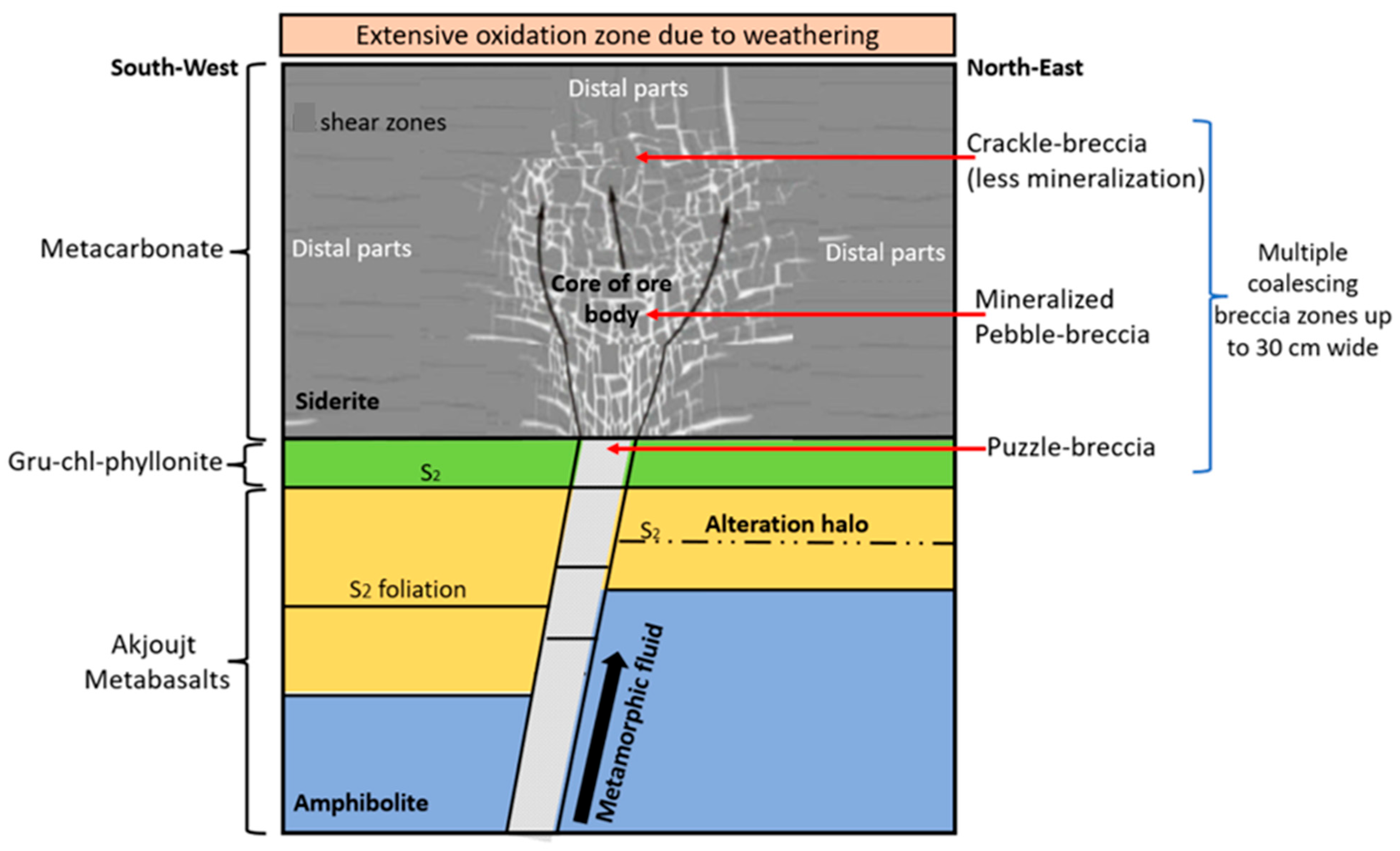
The Guelb Moghrein Mine is located close to the town known as Akjoujt, ~260 km northeast of Nouakchott (capital city) in the Inchiri Province of Mauritania [17]. The Guelb Moghrein Fe oxide-Cu-Au deposit is situated within the Mauritanide fold-and-thrust belt that accreted during the Variscan orogeny along the western edge of the West African craton [18]. Bound by thrust faults, a Ferro-Magnesian Carbonate unit (FMC) hosts the two outcropping and lensoid Oriental and Occidental deposits of the Guelb Moghrein [19]. The FMC is situated between meta-basalt and mafic schists along its contacts [17]. The coarse-grained, massive metacarbonate Oriental and Occidental bodies are host to the iron oxide–copper–gold mineralisation [19]. These metacarbonate bodies primarily consist of siderite with the accompanying ore and alteration minerals chalcopyrite, pyrrhotite, pentlandite, gold, graphite magnetite and Fe-Mg clinoamphibole [20]. Surrounding the mineralised metacarbonate bodies is an extensive zone of oxidation that formed during weathering and is characterised by hematite, goethite and siderite with minor graphite and anthophyllite [21]. The epigenetic Fe oxide-Cu-Au mineralisation is controlled by a range of shear and fault zones [22]. The metacarbonate is truncated by discrete shear zones characterising brittle deformation along which the Fe oxide-Cu-Au-Co mineralisation is concentrated [22]. The stage of mineralisation is regarded as coeval with this shearing, as the resulting breccia in the metacarbonate is always host to some Fe oxide-Cu-Au-Co mineralisation [21]. Multiple, merging breccia zones up to 30 cm in width and consisting of siderite, magnetite, Fe-Mg clinoamphibole, graphite and sulphide assemblages constitute the shear zones in the mineralised metacarbonate (Figure 2) [21].
The lithology and associated textures are characterised by their horizontal location transversing along the distal least-altered and mineralised wall rocks through to the zones of alteration and eventually the massive ore zones at the core of the mineralised metacarbonate body (Figure 2). Progressing with horizontal distance towards the central ore zones is a systematic increase in brecciation (fragment size and clast–matrix ratio [21]).

3. Materials and Methods

3.1. Sample Location and Collection

Fibrous mineral-containing rock fragments representing different lithotypes, textures and modes of NOA occurrence and mineralisation were collected from the Guelb Moghrein Mine (19°44′45″ latitude and 14°25′40″ longitude) (Figure S1). The Guelb Moghrein mine comprises a single open pit, 141 m above sea level, from which the ore body is removed and stockpiled [17]. The ore mined from the pit can be classified as structurally-modified iron oxide–copper–gold (IOCG) mineralisation, and it is hosted in a unit of coarse-grained ferro-magnesian carbonate. The deposit is marked by a siliceous gossan that outcrops as two hills [23]. The samples were collected from the open mine pit based on their visually apparent fibrous mineral content and varying lithotypes. The rock fragment lithologies are easily assigned because in-pit geology exposures allow for a greater understanding of close-space grade and geological continuity [17].

3.2. Sample Characterisation

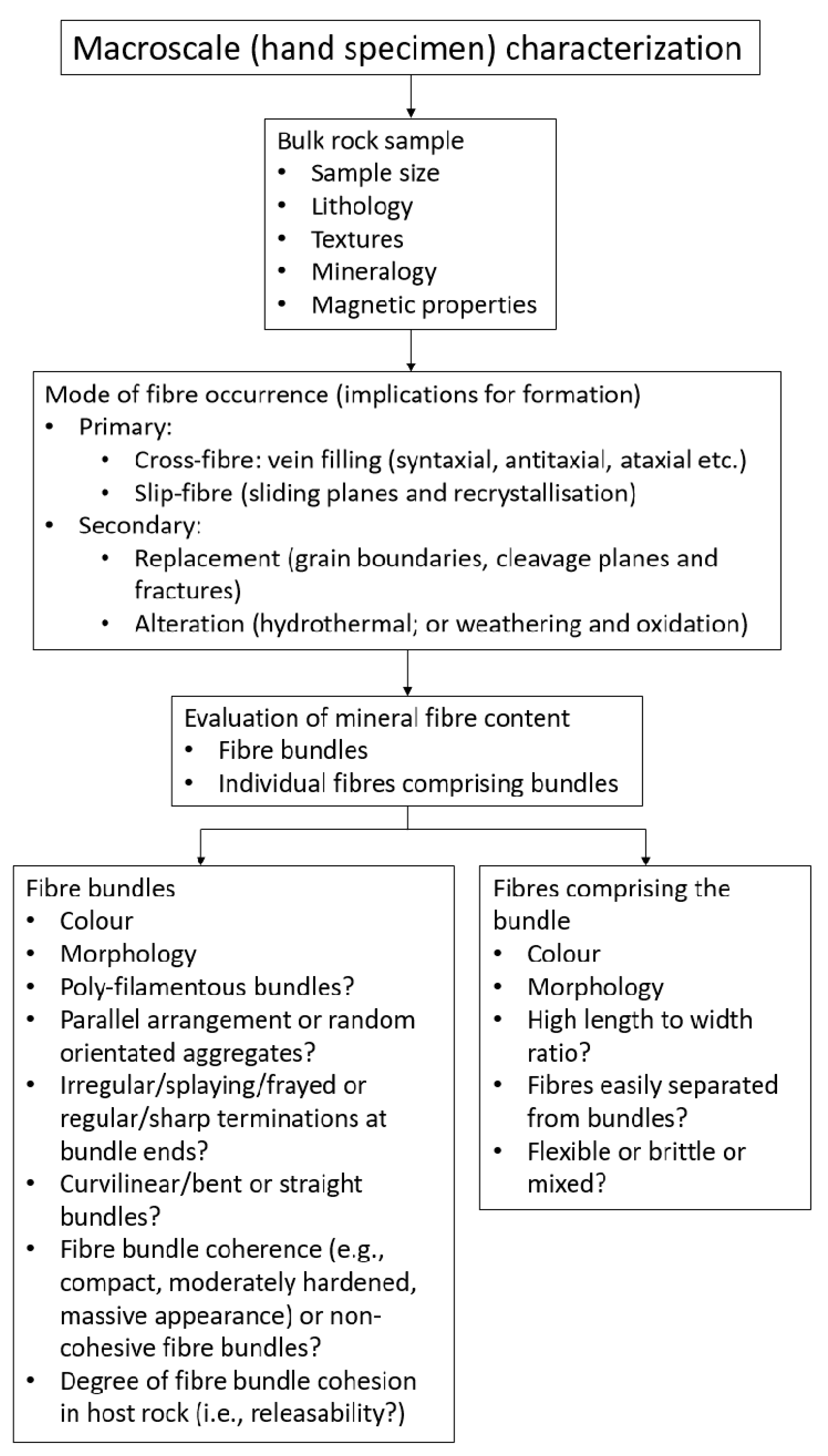
The samples were examined using a geological–structural approach to characterise the rock fabric and structural heterogeneities related to fibrous mineral occurrence and formation (Figure 3).
A geological–mineralogical description of the samples was recorded, and they were assigned to their respective lithological and corresponding structural classes. A total of 11 hand sample specimens were collected. The fibrous minerals identified in the rock samples were classified based on colour, morphology, habit and properties (Figure S2). Additionally, each sample was documented photographically.

3.3. Fibre Subsample Extraction; Distinguishing Fibrous and Pseudo-Fibrous Minerals; and Acid Test on Fibres (Determination of Carbonate Presence)

Fibres in each sample were identified and a small amount removed from the rock specimen using sterile stainless-steel forceps. The removed fibrous material was placed in a mortar and pestle and crushed to determine whether it was genuinely fibrous or pseudo-fibrous. This was accomplished by observing whether the crushed material (i) formed separable fibres or needles, (ii) matted together to form a ball, (iii) formed cleavage fragments or (iv) formed a powder, under a binocular microscope. Carbonate impurities or fibrous minerals were determined by exposing the extracted fibres to a weak solution of hydrochloric acid (10%).

3.4. Mineralogical and Geochemical Investigation

From the entire suite of studied samples, sample 1 was selected for mineralogical and geochemical analysis based on the peculiar characteristics of its fibrous mineral content. The mixed fibrous morphological types identified in sample 1 include (i) flexible to ridged fibrous crystals (asbestiform), (ii) brittle acicular crystals and (iii) coarsely acicular crystals with a more massive appearance. All occur within a large poly-filamentous bundle of parallel-arranged crystals within the metacarbonate host rock surface surrounding a siderite matrix. The long crystal lengths (up to 10 cm) and their restriction to the surface plane surrounding the pebble-like siderite matrix implies that the fibres are a slip-fibre type that formed by recrystallisation in highly sheared zones [24]. However, unlike those of the same lithology and slip-fibre origin demonstrated in samples 2 to 6, the fibrous bundles in sample 1 exhibited a strong reaction to the acid test and produced many morphological-type by-products (i.e., fibres, cleavage fragments and powder) following mechanical abrasion via crushing.

3.4.1. Polarised Light Microscopy (PLM)

The intact subsampled fibres from sample 1 were placed on a slide, covered and examined using Polarising Light Microscopy (Olympus BX53M). The thin sections were made parallel to the c-axis of the fibres to determine the textural and compositional relationships within and between individual fibres comprising parallel-arranged fibre bundles. The optical properties under plane polarising light (PPL) and cross-polarising light (XPL) were determined.

3.4.2. X-Ray Diffraction (XRD)

The fibrous subsamples from sample 1 were disaggregated and crushed in a mortar and pestle and submitted for XRD analysis to assess the bulk mineralogical composition. Diffractograms were obtained using a Malvern Panalytical Aeris (Malvern Panalytical, Worcestershire, UK) diffractometer with PIXcel detector (Malvern Panalytical, Worcestershire, UK) and fixed slits with Fe filtered Co-Kα radiation. The phases were identified using X’Pert Highscore plus software. The relative phase amounts (weight %) were estimated using the Rietveld method (quantitative analysis). The relative intensities (Equation (1)) and d-spacing (Equation (2)) were calculated from the diffractogram data.
Relative intensity (%) = I/I1 × 100
where I is the intensity of the peak and I1 is the intensity of the highest peak.
= 2d sinθ
where n = 1, λ (CoKα) = 1.78892 and d = nλ/2*sin(θ) (d-spacing value).

3.4.3. X-Ray Fluorescence (XRF)

Major and trace element concentrations of the four asbestos mineral fibre samples were determined using X-ray fluorescence (PANalytical Axios Max (Malvern Panalytical, Worcestershire, UK) and Panalytical Philips PW2404 X-ray spectrometer, Malvern Panalytical, Worcestershire, UK) at the Earth lab, Bernard Price Building, University of the Witwatersrand, South Africa. Major elements were determined using the Norrish Fusion 1 technique using in-house correction procedures. Calibration standards were from International Reference Materials USGS series (USA) and NIM series (South Africa). Precision was taken as 1% for elements in abundance greater than 5% by weight, and 5% for elements in abundance less than 5%. Pressed pellets were prepared for trace element analysis.

3.4.4. BET-N2 Specific Surface Area

The specific surface area of the asbestos rock samples was determined by the BET method using a Micromeritics TriStar 3000 V6.05 (Micromeritics Instrument Corporation, Norcross, GA, USA) at the University of the Witwatersrand, Johannesburg, South Africa and a surface area analyser with N2 as absorbing gas at the School of Chemistry, University of the Witwatersrand, South Africa. A mass of ~0.2 g of each sample was placed in BET sample tubes and degassed for 4 h. The samples were then loaded into the BET instrument and N2 absorption isotherms were obtained, and the specific surface area determined.

4. Results

4.1. Sample Characterisation

Sizes, lithological and textural assignment, mineralogy and magnetic properties for all samples as identified macroscopically are given in the Supplementary Material (Table S1).
Rock sample 1 (Figure S2) is composed of coarse-grained (~2 cm), light and dark coloured, compact siderite crystals that exhibit perfect cleavage. Small magnetite crystals are disseminated throughout the siderite crystal mass imparting weak magnetism to the sample. The fibrous material is restricted to the surface of the carbonate rock and has a white to clove-brown colour. The fibres occur in parallel fibre bundles showing a preferred orientation with the longest fibre length being 10 cm and having a high aspect ratio. The poly-filamentous fibre bundles show curvature and have splayed and/or irregular ends. The fibres constitute the parallel fibrous aggregates and show mixed morphologies: From fine, flexible asbestiform habits with individual fibres being readily separated from bundles to coarser, more brittle acicular and lamellar habits that are less easily resolved into individual fibrils and more tightly welded together. The mixed and heterogeneous morphology, habit and tensile strength across parts of this sample suggest some alteration. Crushing of the fibrous material subsamples resulted in a combination of fibres, cleavage fragments and powder material.
In sample 2 (Figure S3), the extremely long, fine, white to clove- and dark-brown fibrous amphibole surround a matrix of pebble-like siderite breccia. The length of these fibres and the slip-fibre occurrence indicates that their formation is a result of recrystallization along shear planes. The subsampled fibrous material is clearly asbestiform, being flexible and retaining its original aspect ratio and forming matted fibrous balls when crushed. Additionally, these fibres did not react to exposure to acid, indicating that they are not fibrous siderite or other carbonate minerals.
The four fibre bundles represented by samples 3–6 (Figure S4) show both fibrous and acicular morphology, having mixed flexible and brittle fibrous portions. The fibres are white to clove brown in colour and are arranged in parallel bundles. Sample 4 has splayed fibre bundle terminations and sample 5 shows matted fibre on the surface indicating that, although the fibres in these four bundles appear quite straight and ridged, they do possess a degree of flexibility.
The rock samples 7 to 11 are dark grey in colour and are composed mainly of siderite and magnetite with white, fibrous amphiboles and sulphide intergrowths, which appear to have a replacement paragenetic association of siderite. The texture in these samples is a crackle breccia, where incipient brecciation of the siderite is identified by less mineralisation and a weakly fractured appearance. These samples show structures that include variably orientated and healed microfractures filled with both sulphides and fibrous amphiboles. These veins resulted during in situ fragmentation of the primary siderite and subsequent syntaxial vein filling [21]. The very weak response of the fibrous material in these samples (besides sample 10 and 12) to acid implies that their bulk composition is most likely that typical of silicates. Additionally, the mechanical crushing test resulted in the formation of numerous individual finer fibres that retained their original aspect ratio in fibre bundles and can therefore be interpreted as asbestiform particles. Sample 7 (Figures S5 and S6) shows the textures and rock fabrics associated with the growth and occurrence of fibrous amphiboles. Sample 8 (Figures S7 and S8), sample 9 (Figure S9), sample 10 (Figure S10) and sample 11 (Figure S11) show fibrous amphibole occurrences because of vein filling and replacement growth.

4.2. Polarised Light Microscopy (PLM)

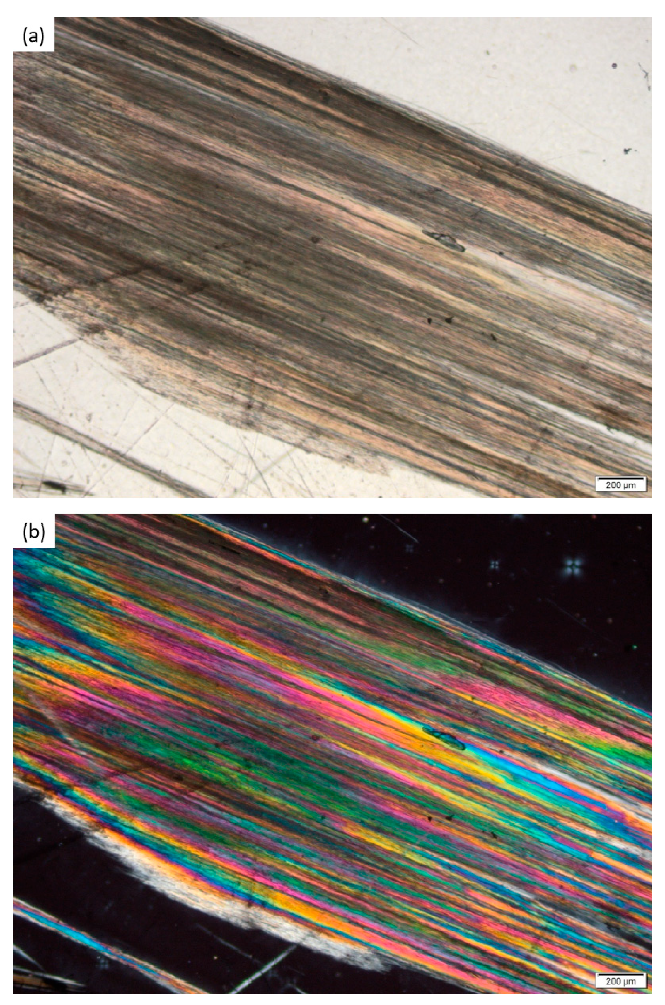
The photomicrographs of sample 1 are given in Figure 4 and Figure 5, showing a range of pleochroic and birefringence colours within and between fibres along the c-axis, indicating compositional heterogeneity. The PLM images show straight fibres forming parallel- arranged bundles. The fibres show both asbestiform (Figure 4) and acicular habits (Figure 5). The colours observed in PPL include a mixture of pale-yellow and green, blue, and light and dark shades of brown. Vibrant pink, blue, yellow, orange, green and white birefringent colours are observed under XPL, and the sample has a parallel extinction angle.

4.3. X-Ray Diffraction (XRD), X-Ray Fluorescence (XRF) and BET-N2-Pecific Surface Area

The XRD (λ (CoKα) = 1.78892) and Rietveld refinement data of the minerals detected in the fibrous material of sample 1 are shown in Figure 6, and the corresponding numerical position and intensity data given in Table S2. The relative intensity and d-spacing determined for the anthophyllite–gedrite phase (Table S3) is consistent with that of the known principle lattice spacings of anthophyllite [25].
X-ray fluorescence (XRF) was used to obtain the bulk geochemical major and trace elements of the fibrous material extracted from sample 1 (Table 1. The BET-N2-specific surface area of the fibrous material subsampled from sample 1 is 3.26 m2/g.

5. Discussion

NOA recognition and assessment actions are crucial for equipping industrial and regulatory agencies with practical knowledge to implement procedures for controlling exposure to NOA and other fibrous minerals during mining, construction and other geo-disturbing activities. Worldwide, many worker populations or communities surrounding NOA-bearing disturbed and/or exploited geological sites are potentially exposed to asbestos, fibrous minerals and/or EMPs (e.g., [28,29,30,31,32,33,34,35,36,37]), and awareness is growing. Incidences of malignant mesothelioma and lung cancer have, in recent years, excessively been reported in the USA, Corsica, Greece, New Caledonia, China, Italy and Turkey because of the presence of NOA [38].
Fibrous amphiboles, including anthophyllite, actinolite and tremolite, constitute 2% of the Guelb Moghrein deposit, where they commonly occur attached to the carbonate minerals, along fractures or as discrete fibrous aggregates [20]. Samples 1 to 6 represent slip-fibres that form in localised planes of shearing, resulting in the siderite being recrystallised to amphibole fibre that is orientated in the direction of shear. Slip-fibre inference is based on the long-lengths of the fibres observed in samples 1 to 6, as well as their occurrence as slip-fibre coatings on the surfaces of metacarbonate host rocks. The nature of fibres observed in samples 2 to 6 is unlike that in sample 1, which appears to have been altered by weathering processes to some degree. Fracture-filling hydrothermal fibrous and replacement-type fibrous mineral occurrences, as shown in samples 7 to 11, represent an early hydrothermal event and late replacement stages, respectively. The fibrous mineral content of samples 7 to 11 are smaller and more irregularly distributed than that of samples 1 to 6. Additionally, much longer fibre lengths are observed in samples 1 to 6 (>6 cm), whereas those in samples 7 to 11 do not exceed ~2 cm.
Asbestiform habit was established for the fibrous minerals in all 11 samples. However, unlike samples 2 to 11, sample 1 showed mixed and interlocking elongated crystal habits within its fibrous mineral content, with asbestiform being subordinate relative to acicular. The asbestiform habit of the minute fibres in samples 7 to 11 are easily identified using a hand lens; they project from the fibrous bundle or aggregate mass. The macroscale habit of fibrous minerals often does not correspond to that observed at the microscale, aiding in the difficulty of distinguishing asbestos and non-asbestiform mineral fibres under regulatory and scientific guidelines [39,40]. However, simple crushing tests allow fibrous material from non- or pseudo-fibrous material to be distinguished based on flexibility and aspect ratio retention of the crushed material using a binocular microscope.
The development of secondary textures of fibrous amphiboles is observed in samples 7 to 11, where the fibres appear to have originated at the siderite crystal boundaries, cleavage planes and fracture. Those formed by siderite replacement along cleavage planes are generally no longer than 4 mm in length; their preferred direction of growth is related to the nature of the siderite surface. When fibre development due to siderite replacement is advanced, mats of both parallel and/or random amphibole fibres are found surrounding and covering the rock mineral substrate. The parallel mineral fibres displayed in samples 9 and 11 appear to produce a foliation covering the original rock minerals. Replacement during late-stage, distal hydrothermal alteration has produced mesh textures in samples 8 and 10, where tiny fibrous minerals of interpenetrating and orientated bundles are arranged along the siderite cleavage cracks. The samples studied here illustrate that, within a single deposit type, several modes of fibrous mineral occurrences, textures and mineral assemblages can occur.
The fibrous Fe-Mg clino-amphibole (i.e., cummingtonite, grunerite, tremolite and actinolite) minerals are formed during equilibrium reactions, and directly replace siderite [21]. Hydrothermal metamorphism causes the siderite of the metacarbonate to metamorphically react with silica-bearing hydrothermal fluids, leading to the paragenetic association of quartz-free magnetite and fibrous amphibole minerals [41].
The unusual, intermixed fibre morphologies and compositions identified during the crushing and acid tests of sample 1 were confirmed by bulk mineralogical and geochemical analysis. The fibrous mineral content of sample 1 is unique in that it embodies all the attributes of asbestos occurrences in complex mineralogical environments showing a wide range of morphologies and intergrown mineral varieties. Bulk mineralogical and geochemical analysis was performed only on sample 1, as its fibrous phases appear unlike those observed in samples 2 to 11. Research pertaining to unusual but potentially harmful fibrous minerals, not using the established parameters for defining asbestos in the regulatory sense, are greatly lacking due to the challenges involved in identifying and characterising non-conventional fibrous minerals in the natural environment. Optically, fibre colours observed in sample 1 are heterogeneous on the micrometre scale, exhibiting colour banding within and between individual fibres (Figure 4 and Figure 5). This variation in colour suggests that the fibres are chemically heterogeneous, providing evidence to either multiple generations of mineral fibre growth or the partial replacement of original fibres [42]. The variety of pleochroic colours observed under PPL (Figure 4a and Figure 5a) suggests that transition elements are incorporated within the mineral fibre structure [42]. The irregular colour zoning forms distinct colour patches along the c-axis (Figure 4b and Figure 5b), which are compatible with formation during dissolution and reprecipitation [42]. Despite their minute size, the fibres show complex and strong colour zoning in PLM, representing compositional heterogeneity and most likely reflecting features typical of complex mineral fibre dissolution and reprecipitation reactions [43]. This compositional heterogeneity observed in PLM is confirmed in XRD, XRF and BET-specific surface area measurements that are unlike those of UICC anthophyllite asbestos. The quantitative phase analysis of XRD data, using the Rietveld method, shows that fibrous material of sample 1 is composed of three intergrown mineral phases being siderite (65.7%), anthophyllite–gedrite (25.9%) and talc (8.3%). This mineral, and therefore chemical, heterogeneity accounts for the anomalous and heterogeneous pleochroic and birefringent colours observed within and between the crystals under PLM. Although talc (8.3%), as observed in sample 1, is known to occur as in intergrowth within anthophyllite at the submicrometric scale [31], to the best of our knowledge, this is the first documented case of fibrous anthophyllite coexisting with fibrous siderite. Talc, whether in the fibrous or non-fibrous form, has been shown to result in the induction of lung tumours in rats following chronic particle inhalation [44]. There is no research or knowledge on the carcinogenic and pathogenic effects of asbestos-associated fibrous siderite mineral phases. Although these associated phases are thought to be harmless, little information on their potential toxic effects is known [45], although asbestos-associated mineral phases should not be neglected when considering the combined factors encompassing the toxicity of NOA. Thus, the characterisation of natural assemblages should include all phases occurring with asbestos fibres [46].
Bulk rock geochemistry of the fibrous material subsampled from sample 1 shows that Fe2O3 (34.1 wt%), SiO2 (19.7 wt%) and MgO (17.7 wt%) are the main components of the fibrous content. The iron, magnesium, calcium and manganese values are consistent with those of the siderite previously reported for this deposit by [21]. The significantly higher SiO2 content measured here is a result of the anthophyllite–gedrite and talc mineral phases as identified by XRD. The elevated Ni and Cu values are also consistent with those given by [21]. However, the Sr concentration is significantly greater and may also be attributed to the additional mineral phase. Sakellaris [21] attributed Cu values > 1500 ppm to the occurrence of local sulphide (chalcopyrite) mineralisation in the siderite rock. However, the lack of sulphide mineral phase detection by XRD indicates that the 7420 ppm Cu measured by XRF must be incorporated within the structure of one or more of the three mineral phases reported by XRD in this study. This is an interesting finding and should be considered for further examination.
The metal concentrations measured in the bulk, mixed mineral phase fibrous material of sample 1 that are above the threshold values (ppm) in soil [27] and human lungs [26] (Table 1 are copper (7491ppm), nickel (43.9 ppm), lead (8.42 ppm) and strontium (1.5 ppm), and are to be considered an ecological and human health risk. The fibrous minerals isolated in sample 1 are composed of an intergrowth of three mineral phases, namely siderite, anthophyllite–gedrite and talc, and therefore it is important to regard their contribution to the bulk major and trace elemental composition determined by XRF or XRD analysis. The trace element concentrations are highly variable compared to UICC standards due to the different mineral phase intergrowths and the independent geochemical mechanisms influencing their genesis [33,46]. The specific surface area of UICC anthophyllite asbestos is 4.4 m2/g [47], which is greater than measured here (3.26 m2/g). The BET surface area of asbestos and other fibrous minerals influences the potential bioavailability and toxicity of the minerals following inhalation, as the total surface area of these minerals controls the extent of mineral fibre and biological system interactions. The total surface area of the anthophyllite asbestos measured in sample 1 is lower than that of the UICC anthophyllite sample and therefore it can be deduced that this sample has a reduced bioavailability and toxicology. The heterogeneous and mixed mineral phase nature of NOA in natural environmental settings is excellently depicted in sample 1.

6. Implications for In Situ Identification and Health Risk Assessment

The samples and their fibrous mineral content represent different lithotypes within the deposit, and their various textural associations are attributed to distinct geological processes. This study highlights that the nucleation and growth of fibrous amphiboles occur in numerous environments via different mechanisms within a single type of deposit. The length of fibre growth also appears to be dependent upon the location or occurrence. Replacement at siderite cleavage planes and grain boundaries having short fibre lengths (maximum of 4 mm) and recrystallisation at shear planes or slip-fibre type resulted in the greatest lengths (11 cm shown in sample 2 and 3). Fibres formed as a product of fracture filling show intermediate lengths between replacement and recrystallisation mechanisms of formation. The study identifies that fibrous amphibole and/or fibrous mineral formation requires open space (i.e., cleavage cracks, fractures and/or shear planes). Mitigating the potential risks of NOA exposure from mining activities requires an extensive understanding of the geological setting and associated conditions favouring asbestos mineralisation. Knowledge pertaining to NOA geological environments allows predictive asbestos management tools to be developed that are critical for protecting public health.

7. Conclusions

In this study, metacarbonate rocks with visible fibrous minerals were collected from the Guelb Moghrein mining site. The fibrous minerals correspond to fracture fill, slip-fibre and replacement fibre modes. The asbestiform nature of the fibrous material content was easily discernible in hand samples or under hand lens. The variable compositions obtained for the fibrous mineral content of sample 1 are due to the mixed fibre content and mineral phases. The coexistence of fibrous siderite and anthophyllite asbestos was determined by XRF analysis in sample 1 from this deposit, which is a novel observation not ever before recorded in the scientific literature. NOA occurrences are highly dependent upon the geological conditions, and understanding these in various settings may allow for the development of predictive strategies necessary for the in situ identification and hazard assessment required for health-risk mitigation in the context of mining and other geologically disruptive activities. Asbestos and fibrous minerals have been shown to host various heavy metals that impart their toxicity once inhaled. Apoptosis and cellular damage are caused by the exposure of cells to Ni ions and the subsequent induction of tumours [48]. Lead is highly toxic and has extensively contaminated the environment and led to numerous health issues in numerous parts of the globe [49]. Brain damage, mental retardation, kidney damage and birth defects are some of the effects caused by chronic exposure to lead [50]. Future research should include incorporating optical reactions into PLM investigations and measuring the content of asbestos fibres in the air at the mine site.

Supplementary Materials

The following supporting information can be downloaded at https://www.mdpi.com/article/10.3390/min15090991/s1: Figure S1: Photographs of different parts of the Guelb Moghrein mine; Figure S2. Rock sample 1, showing least-altered siderite with white to clove-brown-coloured, ‘slip-fibre’ type poly-filamentous, parallel-arranged fibre bundles on the surfaces. Curvature of fibres and bundles, and splayed and/or frayed fibre bundle ends are observed; Figure S3. Rock sample 2, showing long pebble-like, low clast/matrix ratio siderite breccia surrounded by very long, fine fibrous amphibole; Figure S4. Samples 3 to 6, showing discrete, white to clove-brown parallel-arranged fibre bundles ranging in length from 11 to 6 cm. The bundles of fibres demonstrate quite rigid acicular habit. The bundle ends in sample 4 are splayed and have longitudinal splitting in flexible fibres. Matted fibre masses are observed on the surface of sample 5; Figure S5. Sample 7, showing (a) white fibres occurring as mats encompassing and surrounding different minerals. The flexible white fibrous bundles formed around mineral grain boundaries; within cleavage planes and fractures, they directly replace siderite. (b) Fibre bundles as fracture filling in siderite crystals. The fibrous amphibole grew as fracture filling material in siderite microfractures and shows clear crystallographic preferred orientation with respect to the fracture; Figure S6. Textures observed in fibrous amphiboles, sulphides and magnetite within the siderite host rock; Figure S7. Sample 8, showing (a) white- to tan-coloured vein filling asbestos in siderite organised in bundles; bundles have a curvilinear trend orientated parallel to vein wall. Fibre bundle ends show individual fibrils disseminated/splaying into the siderite matrix, and the boundary between the vein and the host rock is sharp. (b) Replacement of siderite along cleavage cracks by fibrous amphiboles; Figure S8. Fibrous amphibole mesh textures observed in sample 8 replacing siderite along cleavage cracks and grain boundaries, forming thin sub-parallel bands; Figure S9. Sample 9, showing large mats of fibrous mineral replacement of siderite overprinting original rock mineralogy and texture in both (a) and (b); Figure S10. Sample 10 (a and b) shows replacement of siderite by fibrous amphiboles along grain boundaries and cleavage cracks. The orientation of fibre growth conforms to that of the cleavage microcracks and boundaries of the siderite grains; Figure S11. Sample 11, showing large mats of replacement material having an elongated crystal habit covering original mineralogy and rock textures. Table S1. General macroscopic description and lithotype category of the different fibrous mineral-containing rock fragments; Table S2: Values of °2θ and intensity (I) recorded for the peaks of each phase from the X-ray diffraction record (λ (CoKα) = 1.78892); Table S3: Reduction of data from X-ray diffractogram (λ (CoKα) = 1.78892). The value of I/I1 (relative intensity %) is determined from Equation (1), where I1 is the intensity of the highest peak of the phase and d (Ǻ) is calculated from Equation (2) assuming n = 1.

Author Contributions

Conceptualization, J.S.S. and R.B.; methodology, J.S.S.; formal analysis, J.S.S.; investigation, J.S.S.; writing—original draft preparation, J.S.S.; writing—review and editing, J.S.S. and R.B.; visualization, J.S.S. and R.B.; supervision, R.B.; project administration, R.B.; funding acquisition, R.B. All authors have read and agreed to the published version of the manuscript.

Funding

The support of the Department of Science, Technology and Innovation through its funding agency, the National Research Foundation, and the Centre of Excellence for Integrated Mineral and Energy Resource Analysis (DSTI-NRF CIMERA) towards this research is hereby acknowledged. Opinions expressed and conclusions arrived at, are those of the author(s) and are not necessarily to be attributed to the CoE, DSTI or NRF.

Data Availability Statement

The original contributions presented in this study are included in the article/Supplementary Material. Further inquiries can be directed to the corresponding author.

Conflicts of Interest

Both authors declare no conflict of interest.

References

  1. Lahondère, D.; Cagnard, F.; Wille, G.; Duron, J. Naturally occurring asbestos in an alpine ophiolitic complex (northern Corsica, France). Environ. Earth Sci. 2019, 78, 540. [Google Scholar] [CrossRef]
  2. Wagner, J. Analysis of serpentine polymorphs in investigations of natural occurrences of asbestos. Environ. Sci. Process. Impacts 2015, 17, 985–996. [Google Scholar] [CrossRef]
  3. Meeker, G.; Bern, A.; Brownfield, I.; Lower, H.; Sutley, S.; Hoefen, T.; Vance, J. The Composition and Morphology of Amphiboles from the Rainy Creek Complex, Near Libby, Montana. Am. Mineral. 2003, 88, 1955–1969. [Google Scholar] [CrossRef]
  4. Punturo, R.; Ricchiuti, C.; Bloise, A. Assessment of Serpentine Group Minerals in Soils: A Case Study from the Village of San Severino Lucano (Basilicata, Southern Italy). Fibers 2019, 7, 18. [Google Scholar] [CrossRef]
  5. National Institute for Occupational Safety and Health (NIOSH). Asbestos Fibers and Other Elongate Mineral Particles: State of the Science and Roadmap for Research. In Current Intelligence Bulletin; NIOSH: Cincinnati, OH, USA, 2011; Volume 62. [Google Scholar]
  6. Lucci, F.; Della Ventura, G.; Conte, A.; Nazzari, M.; Scarlato, P. Naturally Occurring Asbestos (NOA) in Granitoid Rocks, A Case Study from Sardinia (Italy). Minerals 2018, 8, 442. [Google Scholar] [CrossRef]
  7. Harper, M. 10th Anniversary critical review: Naturally occurring asbestos. J. Environ. Monit. 2008, 10, 1394–1408. [Google Scholar] [CrossRef]
  8. Metcalf, R.V.; Buck, B.J. Genesis and health risk implication of an unusual occurrence of NaFe3+ amphibole. Geology 2015, 43, 63–66. [Google Scholar] [CrossRef]
  9. Vignaroli, G.; Ballirano, P.; Belardi, G.; Rossetti, F. Asbestos fibre identification vs. evaluation of asbestos hazard in ophiolitic rock mélanges, a case study from the Ligurian Alps (Italy). Environ. Earth Sci. 2014, 72, 3679–3698. [Google Scholar] [CrossRef]
  10. Lockey, J.E.; Parry, W.T. Health implications of naturally occurring mineral fibers. Fam. Community Health 1984, 7, 1–7. [Google Scholar] [CrossRef]
  11. Vignaroli, G.; Rossetti, F.; Belardi, G.; Billi, A. Linking rock fabric to fibrous mineralisation: A basic tool for the asbestos hazard. Nat. Hazards Earth Syst. Sci. 2011, 11, 1267–1280. [Google Scholar] [CrossRef]
  12. Schapira, J.S.; Bolhar, R.; Master, S.; Wilson, A.H. Mineralogical, Petrological and Geochemical Characterisation of Chrysotile, Amosite and Crocidolite Asbestos Mine Waste from Southern Africa in Context of Risk Assessment and Rehabilitation. Minerals 2023, 13, 1352. [Google Scholar] [CrossRef]
  13. Giacomini, F.; Boerio, V.; Polattini, S.; Tiepolo, M.; Tribuzio, R.; Zanetti, A. Evaluating asbestos fibre concentration in meta-ophiolites: A case study from the Voltri Massif and Sestri–Voltaggio Zone (Liguria; NW Italy). Environ. Earth Sci. 2010, 61, 1621–1639. [Google Scholar]
  14. Lescano, L.; Marfil, S.; Maiza, P.; Sfragulla, J.; Bonalumi, A. Amphibole in vermiculite mined in Argentina: Morphology; quantitative and chemical studies on the different phases of production and their environmental impact. Environ. Earth. Sci. 2013, 70, 1809–1821. [Google Scholar]
  15. Vignaroli, G.; Rossetti, F.; Billi, A.; Theye, T.; Belardi, G. Structurally controlled growth of fibrous amphibole in tectonised metagabbro: Constraints on asbestos concentration in non-serpentinised rocks. J. Geol. Soc. 2020, 177, 103–119. [Google Scholar] [CrossRef]
  16. Van Orden, D.; Allison, K.; Lee, R. Differentiating amphibole asbestos from non-asbestos in a complex mineral environment. Indoor Built Environ. 2008, 17, 58–68. [Google Scholar] [CrossRef]
  17. Gray, D.; Cameron, T.; Briggs, A. Guelb Moghrein Copper Gold Mine, Inchiri, Mauritania; Guelb Moghrein Copper Gold Mine Technical Report NI 43-101; 2016; p. 109. Available online: https://minedocs.com/12/GuelbMoghrein_Technical_032016.pdf (accessed on 25 August 2025).
  18. Martyn, J.E.; Strickland, C.D. Stratigraphy, structure, and mineralization of the Akjoujt area, Mauritania. J. Afr. Earth Sci. 2004, 38, 489–503. [Google Scholar]
  19. Kirschbaum, M.J.; Hitzman, M.W. Guelb Moghrein: An unusual carbonate-hosted iron oxide copper-gold deposit in Mauritania, Northwest Africa. Econ. Geol. 2016, 111, 763–770. [Google Scholar] [CrossRef]
  20. Strickland, C.D.; Martyn, J.E. The Guelb Moghrein Fe-oxide copper–gold–cobalt deposit and associated mineral occurrences, Mauritania: A geological introduction. In Hydrothermal Iron Oxide Copper–Gold & Related Deposits: A Global Perspective; Porter, T.M., Ed.; PGC Publishing: Adelaide, Australia, 2002; pp. 275–291. [Google Scholar]
  21. Sakellaris, G.A. Petrology, Geochemistry, Stable and Radiogenic Isotopy of the Guelb Moghrein Iron oxide-Copper-Gold-Cobalt Deposit, Mauritania. Ph.D. Thesis, RWTH Aachen University, Aachen, Germany, 2007; 444p. [Google Scholar]
  22. Kolb, J.; Sakellaris, G.A.; Meyer, F.M. Controls on hydrothermal Fe oxide–Cu–Au–Co mineralization at the Guelb Moghrein deposit, Akjoujt area, Mauritania. Mineral. Depos. 2006, 41, 68–81. [Google Scholar]
  23. Bargmann, C.J.; Dominy, S.C. Technical Report on the Mauritania Copper Mines: Guelb Moghrein Resource Estimation Project No. L00131; NI 43-101 Technical Report for Mauritania Copper Mines; Snowden Mining Industry Consultants Limited: Perth, Australia, 2008. [Google Scholar]
  24. Abbot, P. A Review of Asbestos Resources. Master’s Thesis, Rhodes University, Grahams-Town, South Africa, 1983. Available online: https://core.ac.uk/download/pdf/145045443.pdf (accessed on 25 August 2025).
  25. Karunaratne, P.C.T.; Fernando, G.W.A.R. Characterisation and radiation impact of corrugated asbestos roofing sheets in Sri Lanka. J. Geol. Soc. Sri Lanka 2015, 17, 31–40. [Google Scholar]
  26. Vanoeteren, C.; Cornelis, R.; Verbeeck, P. Evaluation of trace elements in human lung tissue III. Correspondence analysis. Sci. Total Environ. 1986, 54, 237–245. [Google Scholar]
  27. Toth, G.; Hermann, T.; Da Silva, M.R.; Montanarella, L. Heavy metals in agricultural soils of the European Union with implications for food safety. Environ. Int. 2016, 88, 299–309. [Google Scholar] [CrossRef]
  28. Buck, B.J.; Goossens, D.; Metcalf, R.V.; McLaurin, B.; Ren, M.; Freudenberger, F. Naturally occurring asbestos: Potential for human exposure, Southern Nevada, USA. Soil Sci. Soc. Am. J. 2013, 77, 2192–2204. [Google Scholar] [CrossRef]
  29. Punturo, R.; Bloise, A.; Critelli, T.; Catalano, M.; Fazio, E.; Apollaro, C. Environmental implications related to natural asbestos occurrences in the ophiolites of the Gimigliano-Mount Reventino Unit (Calabria, southern Italy). Int. J. Environ. Res. 2015, 9, 405–418. [Google Scholar]
  30. Bloise, A.; Belluso, E.; Critelli, T.; Catalano, M.; Apollaro, C.; Miriello, D.; Barrese, E. Amphibole asbestos and other fibrous minerals in the meta-basalt of the Gimigliano-Mount Reventino Unit (Calabria, south-Italy). Rend. Online Soc. Geol. Ital. 2012, 21, 847–848. [Google Scholar]
  31. Bloise, A.; Barca, D.; Gualtieri, A.F.; Pollastri, S.; Belluso, E. Trace elements in hazardous mineral fibres. Environ. Pollut. 2016, 216, 314–323. [Google Scholar] [CrossRef]
  32. Bloise, A.; Catalano, M.; Barrese, E.; Gualtieri, A.F.; Gandolfi, N.B.; Capella, S.; Belluso, E. TG/DSC study of the thermal behaviour of hazardous mineral fibres. J. Therm. Anal. Calorim. 2016, 123, 2225–2239. [Google Scholar] [CrossRef]
  33. Bloise, A.; Punturo, R.; Catalano, M.; Miriello, D.; Cirrincione, R. Naturally occurring asbestos (NOA) in rock and soil and relation with human activities: The monitoring example of selected sites in Calabria (southern Italy). Ital. J. Geosci. 2016, 135, 268–279. [Google Scholar] [CrossRef]
  34. Bloise, A.; Catalano, M.; Critelli, T.; Apollaro, C.; Miriello, D. Naturally occurring asbestos: Potential for human exposure, San Severino Lucano (Basilicata, southern Italy). Environ. Earth Sci. 2017, 76, 648. [Google Scholar] [CrossRef]
  35. Bloise, A.; Ricchiuti, C.; Giorno, E.; Fuoco, I.; Zumpano, P.; Miriello, D.; Apollaro, C.; Crispini, A.; De Rosa, R.; Punturo, R. Assessment of naturally occurring asbestos in the area of Episcopia (Lucania, Southern Italy). Fibers 2019, 7, 45. [Google Scholar] [CrossRef]
  36. Petriglieri, J.R.; Laporte-Magoni, C.; Gunkel-Grillon, P.; Tribaudino, M.; Bersani, D.; Sala, O.; Salvioli-Mariani, E. Mineral fibres and environmental monitoring: A comparison of different analytical strategies in New Caledonia. Geosci. Front. 2020, 11, 189–202. [Google Scholar] [CrossRef]
  37. Dichicco, M.C.; Paternoster, M.; Rizzo, G.; Sinisi, R. Mineralogical asbestos assessment in the Southern Apennines (Italy): A Review. Fibers 2019, 7, 24. [Google Scholar] [CrossRef]
  38. Case, B.W.; Marinaccio, A. Epidemiological approaches to health effects of mineral fibres: Development of knowledge and current practice. In Mineral Fibers: Crystal Chemistry, Chemical-Physical Properties, Biological Interaction and Toxicity; Gualtier, A.F., Ed.; European Mineralogical Union: London, UK, 2017; Volume 18, pp. 376–406. [Google Scholar]
  39. Punturo, R.; Cirrincione, R.; Pappalardo, G.; Mineo, S.; Fazio, E.; Bloise, A. Preliminary laboratory characterization of serpentinite rocks from Calabria (southern Italy) employed as stone material. J. Mediterr. Earth Sci. 2018, 10, 79–87. [Google Scholar]
  40. Militello, G.M.; Bloise, A.; Gaggero, L.; Lanzafame, G.; Punturo, R. Multi-Analytical Approach for Asbestos Minerals and Their Non-Asbestiform Analogues: Inferences from Host Rock Textural Constraints. Fibers 2019, 7, 42. [Google Scholar] [CrossRef]
  41. Ellis, D.J.; Hiroi, Y. Secondary siderite–oxide–sulphide and carbonate–andalusite assemblages in cordierite granulites from Sri Lanka: Post-granulite facies fluid evolution during uplift. Contrib. Mineral. Petrol. 1997, 127, 315–335. [Google Scholar] [CrossRef]
  42. Dutrow, B.L.; Henry, D.J. Fibrous tourmaline: A sensitive probe of fluid compositions and petrologic environments. Can. Mineral. 2016, 54, 311–335. [Google Scholar] [CrossRef]
  43. Dutrow, B.L.; Henry, D.J. Complexly zoned fibrous tourmaline, Cruzeiro mine, Minas Gerais, Brazil: A record of evolving magmatic and hydrothermal fluids. Can. Mineral. 2000, 38, 131–143. [Google Scholar] [CrossRef]
  44. Oberdörster, G. Toxicokinetics and effects of fibrous and nonfibrous particles. Inhal. Toxicol. 2002, 14, 29–56. [Google Scholar] [CrossRef]
  45. Vigliaturo, R.; Ventura, G.D.; Choi, J.K.; Marengo, A.; Lucci, F.; O’Shea, M.J.; Pérez-Rodríguez, I.; Gieré, R. Mineralogical Characterization and Dissolution Experiments in Gamble’s Solution of Tremolitic Amphibole from Passo di Caldenno (Sondrio, Italy). Minerals 2018, 8, 557. [Google Scholar] [CrossRef] [PubMed]
  46. Bloise, A.; Ricchiuti, C.; Punturo, R.; Pereira, D. Potentially toxic elements (PTEs) associated with asbestos chrysotile, tremolite and actinolite in the Calabria region (Italy). Chem. Geol. 2020, 558, 119896. [Google Scholar] [CrossRef]
  47. Gualtieri, A.F.; Pollastri, S.; Gandolfi, N.B.; Gualtier, M.L. In vitro acellular dissolution of mineral fibres: A comparative study. Sci. Rep. 2018, 8, 7071. [Google Scholar] [CrossRef]
  48. Lee, Y.J.; Lim, S.S.; Baek, B.J.; An, J.M.; Nam, H.S.; Woo, K.M.; Cho, M.K.; Kim, S.H.; Lee, S.H. Nickel (II)-induced nasal epithelial toxicity and oxidative mitochondrial damage. Environ. Toxicol. Pharmacol. 2016, 42, 76–84. [Google Scholar] [CrossRef] [PubMed]
  49. Jaishankar, M.; Tseten, T.; Anbalagan, N.; Mathew, B.B.; Beeregowda, K.N. Toxicity, mechanism, and health effects of some heavy metals. Interdiscip. Toxicol. 2014, 7, 60–72. [Google Scholar] [CrossRef] [PubMed]
  50. Martin, S.; Griswold, W. Human health effects of heavy metals. Environ. Ment. Sci. Technol. Briefs Citiz. 2009, 15, 1–6. [Google Scholar]
Figure 1. Graphical abstract of work investigated in the study.
Minerals 15 00991 g001
Figure 2. Schematic illustration of the Fe oxide-Cu-Au-Co mineralised metacarbonate deposit of the Guelb Moghrein (Gru–grunerite, chl-chlorite) (adapted from [21]).
Minerals 15 00991 g002
Figure 3. Flowchart for macroscale characterisation of the metacarbonate rocks.
Minerals 15 00991 g003
Figure 4. Sample 1: Photomicrographs of fibrous-acicular bundles in (a) plane polarising light and (b) cross-polarising light. The fibre bundles show heterogeneous birefringence colours, and the red oval highlights splayed ends of the asbestiform component.
Minerals 15 00991 g004
Figure 5. Sample 1: Photomicrographs of fibrous-acicular bundles in (a) plane polarising light and (b) cross-polarising light. Heterogeneous pleochroic and birefringence colours are observed along the length of the crystals. Notice the lenticular inclusion within the fibre bundle.
Minerals 15 00991 g005
Figure 6. X-Ray Diffraction and Rietveld refinement diffractogram of the fibrous material subsampled from sample 1.
Minerals 15 00991 g006
Table 1. Major (oxide wt%) and trace element (ppm) concentrations of fibrous material subsamples extracted from sample 1, values of elements in the human lungs (ppm) [26] and the threshold values (ppm) of metals in soils [27].
Oxides (wt%)Trace Elements (ppm)Elements in Human Lungs (ppm) (Vanoeteren et al., 1986) [26]Threshold Values (ppm) (Toth et al., 2016) [27]
SiO219.73Sc100NDNA
Al2O30.08VDL0.50100
Fe2O334.13CrDL0.50100
MnO1.43CoDL0.1020
MgO17.70Ni43.91.0050
CaO0.88Cu74195.00NA
Na2O0.00Zn1.330200
K2O0.02GaDLNANA
TiO20.01Rb0.6510NA
P2O50.12Sr1.51NA
Cr2O30.00Y7.01NDNA
NiO0.01Zr0.06NANA
LOI22.02Nb0.72NANA
Mo1.53NANA
BaDL>1.10NA
Pb8.420.5060
ThDLNDNA
UDL0.005NA
Total96.09 7585
DL: Detection limit; NA: Not available; ND: Not detected.
Disclaimer/Publisher’s Note: The statements, opinions and data contained in all publications are solely those of the individual author(s) and contributor(s) and not of MDPI and/or the editor(s). MDPI and/or the editor(s) disclaim responsibility for any injury to people or property resulting from any ideas, methods, instructions or products referred to in the content.