Review

Biomineralization Mediated by Iron-Oxidizing Microorganisms: Implication for the Immobilization and Transformation of Heavy Metals in AMD

1
State Key Laboratory of Geomicrobiology and Environmental Changes, China University of Geosciences, Wuhan 430074, China
2
Shanxi Center of Technology Innovation for Mining Groundwater Pollution Prevention and Remediation in Karst Area, School of Environmental Studies, China University of Geosciences, Wuhan 430074, China
*
Author to whom correspondence should be addressed.
Minerals 2025, 15(8), 868; https://doi.org/10.3390/min15080868
Submission received: 16 June 2025 / Revised: 7 August 2025 / Accepted: 15 August 2025 / Published: 17 August 2025

Abstract

Iron, an essential element for virtually all known organisms, serves not only as a micronutrient but also as an energy source for bacteria. Iron-oxidizing microorganisms mediate Fe(II) oxidation under diverse redox conditions, yielding amorphous iron (hydr)oxides or crystalline iron minerals. This globally significant biogeochemical process drives modern iron cycling across terrestrial and aquatic ecosystems. The resulting biomineralization not only produces secondary minerals but also effectively immobilizes heavy metals, offering a sustainable strategy for environmental remediation. This review systematically examines (1) the biogeochemical mechanisms and mineralogical signatures of Fe(II) oxidation by four distinct iron oxidizers: acidophilic aerobes (e.g., Acidithiobacillus), neutrophilic microaerophiles (e.g., Gallionella), nitrate-reducing anaerobes (e.g., Acidovorax), and anoxygenic phototrophs (e.g., Rhodobacter); (2) research advances in heavy metal immobilization by biogenic iron minerals: adsorption, coprecipitation, and structural incorporation; and (3) the impact of pH, temperature, organic matter, and coexisting ions on Fe(II) oxidation efficiency and iron mineral formation by iron-oxidizing bacteria. By characterizing iron-oxidizing bacterial species and their functional processes under varying pH and redox conditions, this study provides critical insights into microbial behaviors driving the evolution of acid mine drainage (AMD).

1. Introduction

Iron, the fourth most abundant crustal element, is essential for life and exists primarily as oxidized ferric iron (Fe(III)) or reduced ferrous iron (Fe(II)) in environmental systems [1,2]. While microbial iron oxidation was first described in the 19th century, early 20th-century studies still predominantly attributed iron oxidation to chemical oxidants such as O2 and NO2. However, subsequent isolation of nitrate-dependent iron-oxidizing bacteria (FeOB), anoxygenic phototrophic bacteria, and neutrophilic iron-metabolizing bacteria demonstrated that microbially mediated iron oxidation coexists with abiotic processes and plays a significant role in iron cycling [3,4,5]. Microbial iron transformations are deeply embedded in global biogeochemical cycles, serving as critical links between the lithosphere, hydrosphere, and biosphere [6]. Beyond regulating iron speciation and mineral formation, these processes significantly influence redox chemistry and the bioavailability of essential nutrients (e.g., C, N, P) and trace metals (e.g., Mn, Cr, As) through coupled biogeochemical reactions. Furthermore, they actively shape redox conditions across diverse environments, from ancient sedimentary deposits to modern aquatic systems [7,8,9].
With the rapid development of industrialization and urbanization, heavy metal pollution has become increasingly prevalent and has emerged as a global environmental issue. Heavy metal pollution is characterized by persistence, concealment, and the inability to be remediated through biodegradation. It is consistently present in smelting operations, metal-bearing minerals, coal gangue, and mine drainage [10], which has severely impacted the environment and human health [11,12,13]. Among the current commonly used heavy metal remediation methods, microbial remediation has gained increasing attention as it reduces issues such as secondary pollution and high costs associated with traditional physical and chemical approaches [14]. Microorganisms can remediate heavy metals by altering their physicochemical properties [15]. The mechanisms of bioremediation for heavy metal pollution primarily involve biosorption, bioaccumulation, bioleaching, bioprecipitation, and biomineralization [12]. In the following, this paper will focus exclusively on biomineralization.
Biomineralization, broadly defined as a mineral-forming process mediated by organisms, is generally categorized into two distinct pathways: biologically controlled mineralization (BCM) and biologically induced mineralization (BIM) [16,17,18]. The former involves direct biological regulation of mineral nucleation, growth, and final morphology, while the latter—BIM—refers to indirect mineral formation induced by microbial activities through the release of metabolic byproducts and surface interactions with ions in open environments [19]. By altering physicochemical conditions (e.g., pH, redox potential), microorganisms create localized supersaturation that influences the BIM process. In BIM, microbial cell surfaces often serve as nucleation sites for subsequent mineral growth [20]. Among the diverse microorganisms capable of biomineralization, bacteria are the most widely applied in practice. The main functional groups include carbonate-mineralizing bacteria (CMB) [21,22,23], phosphate-solubilizing bacteria (PSB) [24,25,26], sulfate-reducing bacteria (SRB) [27], manganese-oxidizing bacteria (MOB) [28,29], and FeOB.
FeOB mediate the oxidation of Fe(II) to Fe(III), leading to the formation of iron (oxyhydr) oxides that subsequently transform into iron mineral phases. Current research has classified FeOB into four distinct physiological groups based on their environmental adaptations and metabolic characteristics: (1) acidophilic aerobic Fe(II)-oxidizers, thriving in oxygenated acidic environments; (2) neutrophilic microaerophilic Fe(II)-oxidizers, inhabiting neutral pH environments with limited oxygen availability; (3) nitrate-reducing Fe(II)-oxidizers, coupling iron oxidation to nitrate reduction in neutral pH anaerobic systems; and (4) anaerobic phototrophic Fe(II)-oxidizers, conducting anoxygenic photosynthesis in neutral pH anaerobic conditions. Different iron-oxidizing microorganisms mediate the formation of distinct Fe(III) mineral species with varying physicochemical properties and morphological characteristics, including ferrihydrite, goethite (α-FeOOH), lepidocrocite (γ-FeOOH), and schwertmannite. This biomineralization process is intrinsically coupled with biogeochemical cycles of sulfur, nitrogen, carbon, and oxygen, while simultaneously exerting significant influences on heavy metal toxicity and mobility in environmental systems. Microorganisms can effectively immobilize heavy metal ions in the environment through direct or indirect induction of mineral formation, while simultaneously altering the physicochemical properties of heavy metals and their compounds. For instance, various iron precipitates can adsorb and/or coprecipitate soluble heavy metals, enabling combined removal with other contaminants such as nitrates and sulfates [30]. Characterized by minimal chemical requirements, cost-effectiveness, and environmental compatibility, biomineralization serves as an effective remediation strategy for heavy metal contamination. This process can utilize naturally in situ substrates (e.g., nitrate, sulfate), making it a sustainable solution for environmental restoration, such as AMD.
This review systematically examines four prevalent iron-oxidizing microorganisms in various natural environments, focusing on their mechanisms in mediating Fe(II) oxidation and subsequent biomineralization processes. We analyze key influencing factors and summarize heavy metal immobilization through adsorption and coprecipitation during/after mineral formation. The synthesis provides critical references for applying biomineralization technology in heavy metal contamination remediation.

2. Microbially Mediated Fe(II) Oxidation and Mineralization

2.1. Mechanisms of Microbial Fe(II) Oxidation

2.1.1. Acidophilic Aerobic Fe(II)-Oxidizers

Acidophilic aerobic FeOB typically thrive in acidic environments (pH 1.0–4.0), such as acidic leachates, AMD, deep-sea hydrothermal vents, and hot springs, which are rich in iron, sulfur, and other metal elements. Due to their unique survival environments and functional mechanisms, these bacteria are widely applied in AMD pollution control, hydrometallurgical bioleaching, and remediation of heavy metal-contaminated soils [31,32].
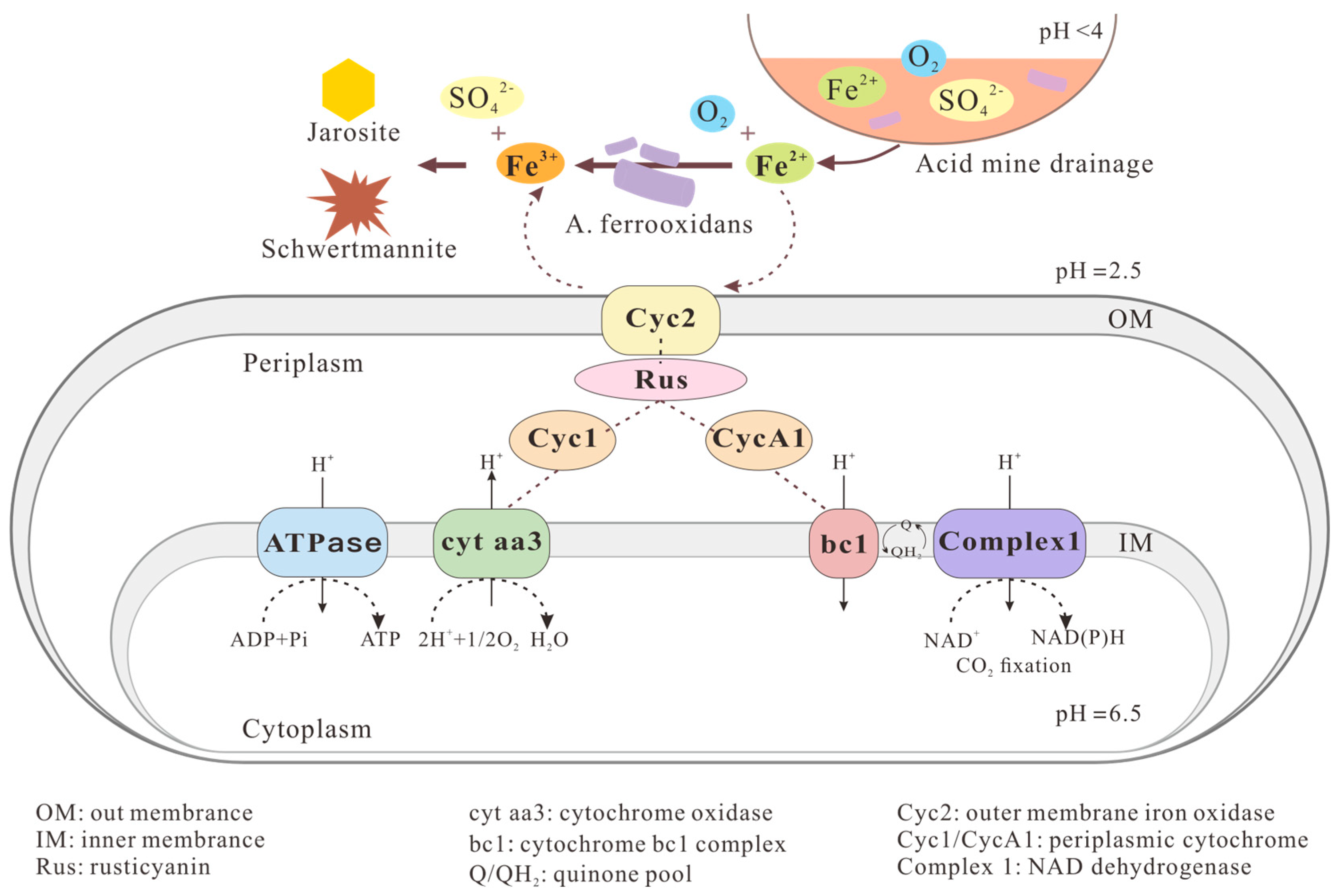
With the ever-increasing demand for mineral resources and the rapid development of the mining industry, a series of environmental issues have inevitably emerged, among which the environmental problems caused by AMD are particularly severe [33]. During the mining and processing of mineral resources, large quantities of tailings are generated. The metal sulfides present in these tailings undergo oxidation reactions in the presence of oxygen, water, and microorganisms, progressively producing large volumes of wastewater rich in sulfates and metal ions—known as acid mine drainage. AMD is typically acidic, with pH usually below 4 [34]. When discharged into the environment, these untreated effluents can enter water bodies and infiltrate soils and groundwater systems, posing severe threats to surrounding ecosystems and ultimately endangering human health. Under acidic conditions, the chemical oxidation of ferrous iron is unfavorable, while the presence of acidophilic FeOB can significantly accelerate this reaction, consequently influencing the water quality evolution of AMD. Acidophilic FeOB primarily utilize elemental sulfur or ferrous iron as electron donors; oxygen, sulfate, or nitrate as electron acceptors; and either organic or inorganic carbon as carbon sources for their metabolic activities and growth [35]. Acidithiobacillus ferrooxidans (A. ferrooxidans) is a predominant iron-oxidizing microorganism in AMD [36,37]. As a chemoautotroph, it obtains energy for growth and metabolism by aerobically oxidizing Fe2+ and S0 to Fe3+ and SO42−, with optimal growth at 30 °C and pH 2.0 [38,39]. The Fe(II) oxidation mechanism of A. ferrooxidans represents the most complete metabolic model among existing studies (Figure 1). The cytochrome c protein (Cyc2) embedded in its outer membrane can directly contact insoluble substrates such as pyrite, transferring electrons generated from Fe2+ oxidation by this bacterium to rusticyanin (Rus). Subsequently, some electrons are transported along the downhill potential gradient pathway. Finally, cytochrome c oxidase (Cox) on the inner membrane delivers the electrons—which pass through Cyc1 and cytochrome aa3 oxidase—to O2, where they combine with protons to form H2O, thereby providing energy for growth and metabolism [40,41,42]. The electron transfer pathway proceeds as follows: Fe(II)→Cyc2→Rus→Cyc1→Cyt aa3→O2. Approximately 5% of the electrons bifurcate at Rus and enter the uphill pathway to the quinone pool, where they are utilized by NADH dehydrogenase to generate NAD(P)H for cellular biosynthetic processes. The electron transfer pathway proceeds as follows: Fe(II)→Cyc2→Rus→CycA1→bc1→Complex I→NADH.
In addition to A. ferrooxidans, bacteria of the Leptospirillum genus are also widely present in AMD environments. Based on 16S rRNA gene phylogenetic classification, the Leptospirillum genus is divided into three groups: Leptospirillum ferrooxidans (Group I), Leptospirillum ferriphilum (Group II), and Leptospirillum ferrodiazotrophum (Group III) [43,44,45]. All these species exhibit remarkable acid tolerance and heavy metal resistance, capable of obtaining energy solely through Fe2+ oxidation while utilizing CO2 as their carbon source for chemolithoautotrophic growth. In contrast to A. ferrooxidans, Leptospirillum species are moderately thermophilic with an optimal growth temperature around 40 °C, and exhibit greater tolerance to extremely low pH conditions, thriving best at approximately pH 1.5 [46]. Furthermore, Leptospirillum demonstrates significantly higher iron-oxidizing activity, though it lacks the ability to oxidize sulfur or reduced sulfur compounds [47,48].
Figure 1. Acidophilic aerobic FeOB-mediated Fe(II) oxidation and mineralization. Modified from Quatrini et al. [49] and Zhang et al. [50].
Minerals 15 00868 g001

2.1.2. Neutrophilic Microaerophilic Fe(II)-Oxidizers

Neutrophilic microaerophilic FeOB utilize O2 as an electron acceptor and CO2 as their carbon source under near-neutral pH conditions, oxidizing Fe(II) to produce Fe(III) (oxyhydr)oxides [51]. Under oxygen-rich and neutral pH conditions, Fe(II) can undergo rapid abiotic oxidation, while excessively low oxygen levels may inhibit microbial activity [52]; therefore, neutrophilic microaerophilic FeOB typically colonize the oxic–anoxic transition zones [53], such as lake sediments, marine hydrothermal vents, wetlands, rhizosphere soils, and groundwater [54]. In these suboxic zones (O2 concentration < 50 μmol·L−1, optimal range 5–20 μmol·L−1) [52], neutrophilic microaerophilic FeOB can effectively compete with the chemical oxidation of Fe(II) by O2 [55,56].
4 F e 2 + + 10 H 2 O + O 2 4 Fe OH 3 + 8 H +
Current studies indicate that microaerophilic Fe(II)-oxidizing bacteria employ two distinct pathways for Fe(II) oxidation: the iron oxidase system (MtoAB) and the cytochrome c protein (Cyc2)-mediated pathway (Figure 2) [57,58]. MtoAB is a decaheme cytochrome-porin complex homologous to the Fe(III) reductase MtrAB from the dissimilatory iron-reducing bacterium Shewanella oneidensis, which can directly mediate solid-phase Fe(II) oxidation [59]. The lithoautotrophic bacterium Sideroxydans lithotrophicus ES-1 employs the Mto pathway to oxidize structural Fe(II) in montmorillonite, with the resulting Fe(III) remaining bound within the clay mineral structure [60]. Cyc2, a homolog of the Fe(II) oxidase identified in A. ferrooxidans, is widely distributed among neutrophilic microaerophilic FeOB [56,61]. This cytochrome primarily oxidizes soluble Fe(II) and demonstrates significantly higher expression levels than MtoAB during Fe(II) oxidation, indicating its dominant role in this process. Microaerophilic Fe(II)-oxidizing bacteria transfer electrons derived from Fe(II) oxidation via Cyc2 to periplasmic cytochromes. Under high-O2 conditions, these electrons are ultimately channeled through the terminal oxidase cyt-aa3 to reduce molecular oxygen. Under low-O2 conditions, microaerophilic Fe(II)-oxidizing bacteria employ the cyt-cbb3 oxidase to reduce O2, representing the so-called “downhill” pathway where oxygen serves as the terminal electron acceptor. Simultaneously, electrons transferred via periplasmic cytochromes are reverse-transported to Complex I (NADH dehydrogenase) through the ACIII-bc1 complex, reducing NAD+ to NADH in what is termed the “uphill” pathway. More importantly, the NADH generated through the uphill pathway can participate in the Calvin cycle to fix CO2, thereby influencing biomass yield [56,57,62].

2.1.3. Nitrate-Reducing Fe(II)-Oxidizers

Nitrate-reducing FeOB thrive under neutral pH and anaerobic conditions, with a wide distribution across various environments including groundwater, river/lake sediments, wetlands, and paddy soils. The microbially mediated process of nitrate reduction coupled with Fe(II) oxidation (NRFO) constitutes a critical component of iron/nitrogen biogeochemical cycling [64]. This process plays a significant role in generating Fe(II) and mixed-valent iron minerals in subsurface environments.
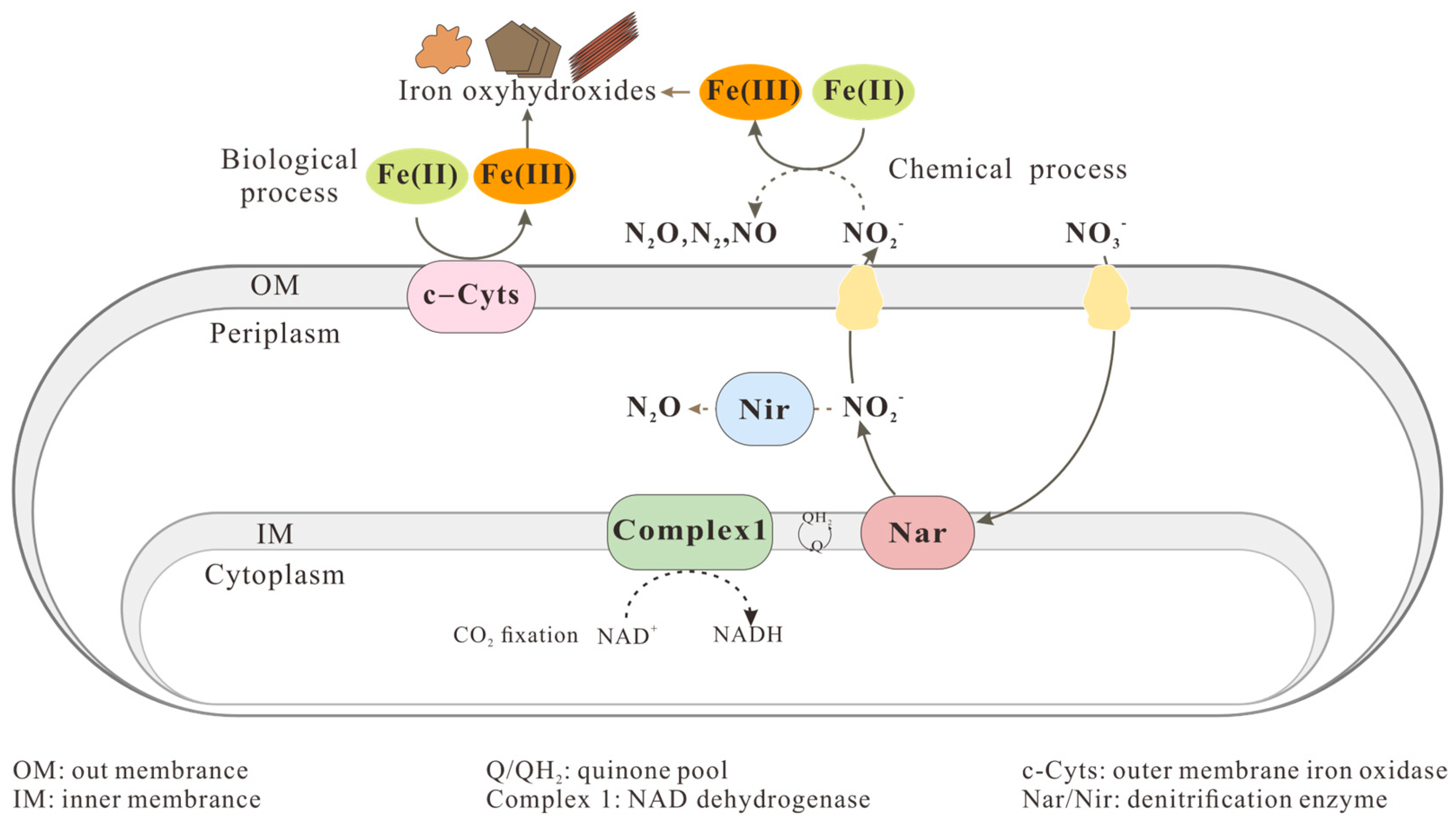
Nitrate has become an increasingly abundant potential electron acceptor for Fe(II) oxidation in groundwater [65]. Driven by FeOB, the coupled process of nitrate reduction and Fe(II) oxidation leads to the formation of iron minerals and N2 (Equation (2)) [66,67]. However, in the absence of catalysts, NO3 cannot directly oxidize Fe(II) [68]. Current research indicates that nitrate-reducing FeOB can be categorized into three metabolic types: autotrophic, mixotrophic, and heterotrophic. Most nitrate-reducing FeOB are mixotrophic, with only a few exceptional strains. These microorganisms possess complex ferrous iron oxidation electron transfer mechanisms, demonstrating dual metabolic capabilities: they can not only utilize organic compounds (such as acetate) to provide electrons for nitrate reduction to sustain cellular growth, but can also use Fe(II) as an electron donor [69]. It is generally recognized that both biological and chemical mechanisms coexist during the nitrate-dependent Fe(II) oxidation process (Figure 3). The biological Fe(II) oxidation mechanism involves microbial enzymatic pathways where iron-oxidizing enzymes (e.g., cytochrome c in Pseudogulbenkiania strain 2002) serve as electron acceptors to directly catalyze the oxidation of Fe(II) to Fe(III). However, the specific enzymes responsible for this process have not yet been fully characterized [70]. The chemical oxidation mechanism of Fe(II) involves NO2—an intermediate generated during microbial nitrate reduction—which indirectly drives Fe(II) oxidation through abiotic processes [71,72,73]. Specifically, nitrate-reducing FeOB oxidize organic substrates to generate electrons, which are then utilized by the membrane-associated nitrate reductase (Nar) to reduce NO3 to the primary product NO2 which is then further catalyzed by the periplasmic nitrite reductase (Nir) to NO or N2O, thereby facilitating rapid chemodenitrification between dissolved/solid-phase Fe(II) and biogenic NO2, ultimately generating Fe(III) (Equation (3)) [66,67,68]. Both biological and chemical mechanisms concurrently operate in microbially mediated NRFO processes, though their specific pathways and relative contributions remain unclear [74]. Chen et al. [64] demonstrated through dual N-O isotope analysis and kinetic modeling that the chemical pathway predominates in the NRFO process mediated by Acidovorax sp. strain BoFeN1; in Pseudogulbenkiania sp. strain 2002, competition occurs between chemical and biological oxidation pathways of Fe(II), with the biological oxidation rate likely exceeding that of chemical oxidation [68]. In summary, significant debates persist regarding microbially mediated NRFO processes, warranting further systematic investigation.
10 Fe II + 2 NO 3 + 12 H + 10 Fe III + N 2 + 6 H 2 O
4 F e 2 + + 2 N O 2 + 5 H 2 O 4 FeOOH +   N 2 O g + 6 H +

2.1.4. Anaerobic Phototrophic Fe(II)-Oxidizers

Anaerobic phototrophic FeOB can utilize Fe(II) as an electron donor and CO2/bicarbonate as both electron acceptor and carbon source for biomass production, with light energy driving this process under anoxic conditions [75]. This process, known as photoferrotrophy [76], commonly occurs in the photic zones of freshwater and marine sediments [77,78,79]. Fe(II)-oxidizing phototrophic bacteria are restricted to a narrow pH range (typically 6.5–7.0) and can only oxidize dissolved Fe2+, rendering them incapable of utilizing sparingly soluble iron minerals. To date, all known phototrophic iron-oxidizing bacteria belong to distinct groups of anaerobic photosynthetic bacteria, classified as green sulfur bacteria [80,81], purple sulfur bacteria [82], and purple non-sulfur bacteria [83].
HCO 3   + 4 F e 2 + + 10 H 2 O hv 4 Fe OH 3 + CH 2 O + 7 H +
Anaerobic photosynthesis utilizing Fe(II) as an electron donor is considered one of Earth’s most ancient metabolic processes, which evolutionarily predates oxygenic photosynthesis [84]. It has been proposed that direct phototrophic utilization of Fe(II) through anaerobic photoferrotrophy served as a key mechanism generating extensive iron oxide deposition in Archean banded iron formations (BIFs) within oxygen-deficient marine environments [85,86,87]. Consequently, studying photoferrotrophy provides critical insights into the biogeochemical cycling of iron on ancient Earth.
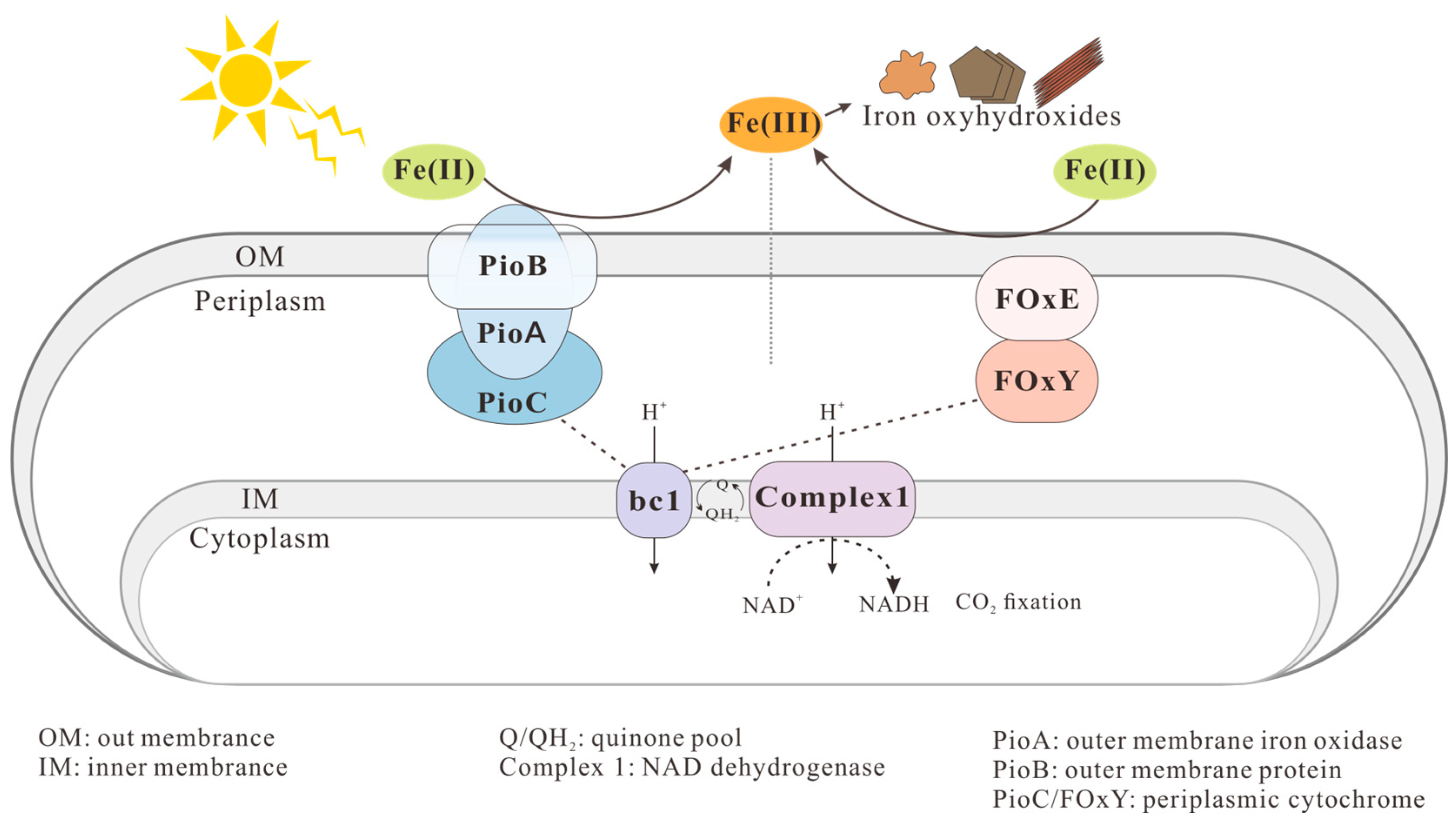
Currently isolated anaerobic phototrophic FeOB exhibit significant metabolic flexibility. These microorganisms can utilize multiple substrates, employing H2 or H2S as alternative electron donors to Fe(II) for autotrophic CO2 fixation, while also capable of utilizing various organic substrates such as acetate and lactate for heterotrophic growth [83,88,89]. Previous studies have primarily focused on two model strains: Rhodopseudomonas palustris TIE-1 and Rhodobacter ferrooxidans SW2, which serve as key systems for understanding phototrophic iron oxidation mechanisms. In strain TIE-1, Fe(II) oxidation is mediated by the pio operon encoding three key comprises: (1) PioA, a periplasmic decaheme c-type cytochrome; (2) PioB, an outer membrane β-barrel protein; and (3) PioC, a periplasmic high-potential iron-sulfur protein (HiPIP). Based on the well characterized electron functions of cytochromes and HiPIPs, a mechanistic model is proposed wherein PioA and PioC collectively mediate transmembrane electron transfer from Fe(II) to intracellular carriers, while PioB facilitates the transmembrane transport of iron species (Fe2+/Fe3+) [90,91,92]. Strain SW2 mediates Fe(II) oxidation through the Fox operon, which encodes (1) FoxE, a periplasmic decaheme c-type cytochrome; (2) FoxY, a quinone reactive protein; and (3) FoxZ, an inner membrane transporter [93,94,95]. Due to the metabolic versatility of anaerobic phototrophic iron-oxidizing microorganisms, their Fe(II) oxidation electron transport pathways remain incompletely characterized and cannot be generalized by a single mechanism (Figure 4).
In certain natural environments, the coexistence of diverse FeOB within overlapping ecological niches may lead to competitive interactions among these microorganisms. In oxygen gradient zones (oxic–anoxic interfaces), microaerophilic FeOB may compete with anaerobic microorganisms for ecological niches and resources. Laufer et al. [97] documented the coexistence of neutrophilic microaerophilic, anaerobic phototrophic, and anaerobic nitrate-reducing FeOB in two coastal marine sediments. Their Fe(II) oxidation activities were strongly constrained by geochemical gradients, particularly the vertical stratification of O2, light availability, N O 3 , and Fe(II) within the sediment column. Melton et al. [79] observed the coexistence of anaerobic phototrophic and nitrate-reducing FeOB in shallow freshwater lake sediments, with temporal niche separation driven by diel cycles. In this system, nitrate-reducing Fe(II) oxidizers dominated Fe(II) oxidation during dark periods, while phototrophs were primarily responsible for daytime Fe(II) oxidation.

2.2. Iron Mineral Formation and Characteristics

2.2.1. Formation of Fe(III) Minerals by Acidophilic Aerobic Fe(II)-Oxidizers

Acidithiobacillus ferrooxidans can utilize dissolved iron and sulfate ions in AMD to induce the formation of iron hydroxysulfate minerals (commonly referred to as iron–alum compounds) such as schwertmannite (Fe8O8(OH)8−2x(SO4)x, 1 ≤ x ≤ 1.75) and jarosite (MFe3(SO4)2(OH)6, M = K+, Na+, NH4+, H3O+). This biomineralization process occurs through the templating effect of extracellular macromolecules including polysaccharides, lipids, and proteins, ultimately precipitating and removing iron and sulfate from the wastewater system [98,99,100]. In the A. ferrooxidans-mediated ferrous iron oxidation and iron-sulfate mineral formation process, the biological oxidation of Fe2+ is an acid-consuming reaction (Equation (5)), while the hydrolysis and mineralization of Fe3+ represents an acid-generating reaction (Equation (6)) [101,102]. Consequently, this typically results in a characteristic pH fluctuation pattern featuring an initial increase followed by a subsequent decrease.
4 Fe 2 + + O 2 + 4 H + 4 F e 2 + + 2 H 2 O   microbial   involvement
8 Fe 3 + + 14 H 2 O +   SO 4 2 Fe 8 O 8 OH 6 SO 4 schwertmannite + 22 H +
Schwertmannite is a poorly crystalline, ochre-yellow secondary iron oxyhydroxy-sulfate mineral with a characteristic “sea-urchin-like” morphology, high specific surface area, and inherent instability in natural environments [103]. The main function groups of schwertmannite are -OH, H-O-H, S O 4 2 , and FeO6 coordination octahedral [104]. The bacterium A. ferrooxidans contains tryptophan, which can significantly alter the interfacial potential, thereby facilitating electron transfer from Fe2+ to schwertmannite and promoting the formation of needle-like goethite on the surface of its oval-shaped aggregates [105,106]. Meanwhile, at pH < 3, jarosite tends to form readily (Equation (7)). Additionally, monovalent cations in solution can also drive the transformation of schwertmannite into jarosite.
M + + 3 F e 3 + + 2 S O 4 2 + 6 H 2 O MF e 3 S O 4 2 OH 6   jarosite + 6 H +
In the equation, M = K+, Na+, NH4+, H3O+, etc. (monovalent cations).
At ambient conditions, the spontaneous hydrolysis of Fe3+ and its combination with S O 4 2 to form schwertmannite is thermodynamically unfavorable ( r G m = 6.63 kJ·mol−1). However, when Fe3+ is biologically generated by A. ferrooxidans, schwertmannite formation becomes favorable ( f G m = −34.12 kJ·mol−1). Simultaneously, jarosite formation from biologically oxidized Fe2+ shows enhanced thermodynamic favorability ( f G m decreasing from −22.20 to −67.45 kJ·mol−1). These ΔG° reductions demonstrate A. ferrooxidans’ crucial role in promoting iron mineral precipitation [107].

2.2.2. Formation of Fe(III) Minerals by Neutrophilic Microaerophilic Fe(II)-Oxidizers

In neutrophilic microaerophilic FeOB, Cyc2 and MtoAB function cooperatively to mediate extracellular electron transfer reactions in bacterial systems [108]. This process ultimately oxidizes Fe(II) to form amorphous iron oxides, which subsequently assemble into distinctive extracellular structures—either spiral stalk-like or elongated sheath-like morphologies, or alternatively precipitate as dispersed mineral phases surrounding bacterial cells [109,110], preventing the encrustation of either intracellular or extracellular surfaces during microbial Fe(II) oxidation.
Microaerophilic FeOB such as Gallionella, Leptothrix, and Sideroxydans species secrete extracellular polymeric substances (EPSs) that template the formation of either twisted stalks (Gallionella-type) [3,111] or tubular sheaths (Leptothrix-type). This mechanism provides a structural template for controlled iron oxide deposition while preventing cell death caused by disrupted nutrient circulation. The stalk structures consist of organic fibers composed primarily of polysaccharides and lipids, which incorporate nanoparticles of Fe(III) oxyhydroxides [112]; these iron oxides typically consist of ferrihydrite or lepidocrocite phases [113]. The tubular sheaths likely consist of an elastic fibrous framework. These characteristic stalk and sheath structures serve as distinctive morphological markers, enabling the identification of neutrophilic microaerophilic FeOB. Moreover, EPSs acidify the local microenvironment, triggering iron oxide precipitation away from cells [63].

2.2.3. Formation of Fe(III) Minerals by Nitrate-Reducing Fe(II)-Oxidizers

Nitrate-reducing FeOB mediate Fe(II) oxidation, yielding both poorly crystalline iron hydroxides (e.g., green rust and ferrihydrite) and more crystalline phases such as goethite, lepidocrocite, and magnetite [68,114]. During Fe(II) oxidation by strain BoFeN1, green rust forms as a transient intermediate that subsequently catalyzes extracellular Fe(II) oxidation [115]. Sun et al. [116] demonstrated through microcosm experiments containing natural aquifer sediments and groundwater that microbial nitrate-dependent Fe(II) oxidation can lead to magnetite formation. Kiskira et al. [117] utilized Pseudogulbenkiania strain 2002 to produce hematite and maghemite. Sunhwa Park et al. [118] demonstrated that Paracoccus denitrificans mediates nitrite-driven Fe(II) oxidation both intracellularly and extracellularly, leading to the formation of lepidocrocite. As Fe(III) minerals continue to accumulate, the negatively charged cell surface may adsorb iron minerals with higher point-of-zero charge (PZC), forming a dense encrustation layer. Simultaneously, minerals trapped in the periplasmic space could also lead to encrustation due to their inability to be expelled or dissolved. Microscopic observations revealed rapid precipitation of Fe(III) minerals within the periplasm, on the cell surface, and in the vicinity of BoFeN1 cells [119]. This encrustation phenomenon reduces cellular metabolic efficiency, inhibits microbial metabolic activity, and ultimately leads to cell death and lysis. Although nitrate-reducing FeOB have not evolved efficient encrustation-prevention mechanisms like neutrophilic microaerophilic iron oxidizers or phototrophic iron oxidizers, some microorganisms can mitigate encrustation effects by secreting EPSs. Klueglein et al. [120] demonstrated that EPSs can inhibit the formation of large mineral crystals, thereby reducing their encrustation potential on cells. However, whether this phenomenon is universally prevalent, and whether nitrate-reducing FeOB employ additional encrustation-avoidance strategies in natural environments, requires further investigation.

2.2.4. Formation of Fe(III) Minerals by Phototrophic Fe(II)-Oxidizers

Phototrophic FeOB initially produce poorly crystalline iron (oxyhydr)oxide precipitates (e.g., ferrihydrite) [77,78,89,121]. These metastable phases gradually transform into more crystalline iron minerals such as goethite, hematite, and lepidocrocite over time [122,123]. Gupta et al. [96] isolated the marine anaerobic phototrophic bacterium Rhodovulum sulfidophilum AB26 from an estuary, demonstrating its capability to utilize non-chelated Fe(II) as the sole electron donor for phototrophic growth. This process generates divalent, mixed-valent, and trivalent iron mineral products including siderite (FeCO3), vivianite [(Fe3(PO4)2•8(H2O)], carbonate green rust [GR( CO 3 2 )], goethite, and akageneite. Previous experiments revealed that while the produced Fe(III) minerals maintain close association with anaerobic phototrophic FeOB, they do not form dense cellular encrustations as observed in anaerobic nitrate-reducing iron oxidizers [96,121,124]. Instead, these minerals precipitate at a certain distance from the cell surface, thereby minimizing adverse effects on cellular growth. This phenomenon may be explained by fellow mechanisms: (1) the localized acidic microenvironments generated during Fe(II) oxidation near cell surfaces may inhibit immediate Fe(III) mineral precipitation, thereby preventing cellular encrustation [125]; (2) bacteria EPSs can bind Fe(III) and spatially direct mineral precipitation away from cell surfaces, forming native cell–EPS–mineral aggregates [75,126]; and (3) complexation-mediated dissolution maintains Fe(III) in soluble form [127]. Furthermore, Gauger et al. [128] demonstrated that ferrihydrite particles formed by strains TIE-1 and SW2 provide additional UV-radiation protection to cells when associated with their surfaces.
Collectively, diverse iron-oxidizing microbes mediate Fe(II) oxidation, generating multiple iron mineral species (Table 1).

3. Influencing Factors of Fe(II) Bio-Oxidation and Mineralization

Current research on the factors influencing microbially mediated iron oxidation and mineralization primarily focuses on laboratory-based experiments, with most studies targeting model microbial strains whose underlying mechanisms are relatively well understood. In the following sections, we will summarize these studies, focusing on key influencing factors, including pH, temperature, organic matter, and coexisting ions.

3.1. Impact of pH and Temperature

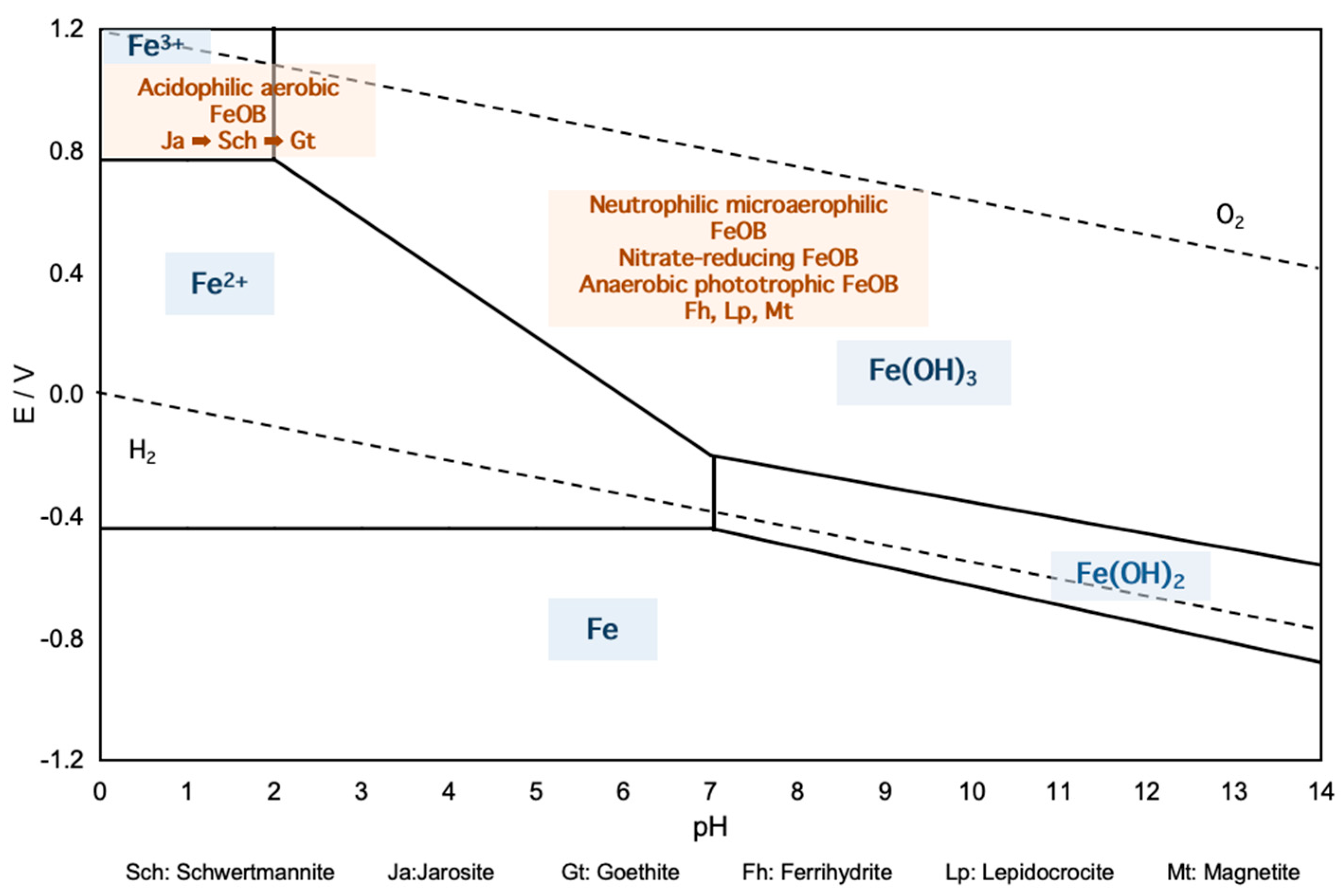
pH and temperature, as key environmental factors, strongly impact microbial/enzyme-driven mineralization, microbial community composition, and metabolic activity. As is well-known, pH can influence the chemical oxidation of Fe(II) (Figure 5). Meanwhile, different pH conditions lead to distinct Fe(II) oxidation efficiencies by microbial activity and generate varying Fe(III) mineral products. The Fe(II) oxidation rate and mineral products mediated by the iron-oxidizing phototrophic bacterium Rhodopseudomonas palustris Strain TIE-1 are pH-dependent. Optimal Fe(II) oxidation occurs at pH 6.5–6.9. At lower pH (<7.0), poorly crystalline iron (hydr)oxides and goethite dominate, whereas magnetite (Fe3O4) becomes the predominant phase at higher pH (>7.2 ± 0.2) [83]. Solution pH critically influences the concentration of hydroxyl complexes and the transformation of monomers into polymers, thereby further governing mineral formation [105]. During the A. ferrooxidans-mediated mineralization process, the final mineral products vary significantly with pH. Studies demonstrate that schwertmannite formation is most favorable at pH 3.0. In the presence of cations, jarosite precipitates preferentially at pH 2.0–2.5 through Fe(III) and sulfate precipitation. In systems with pH > 3.5, goethite becomes the dominant phase, with higher pH values promoting the transformation of schwertmannite to goethite [134,135,136].
Most bacterial strains exhibit an optimal temperature range for maximal metabolic activity. The iron-oxidizing bacterium A. ferrooxidans demonstrates optimal growth between 25–30 °C, while the microaerophilic iron-oxidizer Gallionella ferruginea growth is best at 20–25 °C. Song et al. [137] experimentally confirmed that low temperatures significantly inhibit A. ferrooxidans oxidation activity, with Fe2+ oxidation rates reaching only 11.81% at 10 °C compared to complete oxidation at 28 °C after 5 days of cultivation. Comparative analysis by Nicole Dopffel et al. [138] revealed that biological Fe(II) oxidation by nitrate-reducing FeOB (strain BoFeN1) dominates over abiotic processes at environmentally relevant temperatures (30 °C), where abiotic oxidation contributes minimally. Temperature further regulates secondary mineral speciation, with A. ferrooxidans preferentially forming schwertmannite at 10 °C and jarosite at 28 °C.

3.2. Effects of Organic Matter

Dissolved organic matter (DOM) is ubiquitously present in natural water bodies, sediments, and suspended particulate matter, primarily comprising humic substances, low-molecular-weight organic acids, amino acids, and polysaccharides. DOM contains functional groups (e.g., hydroxyl, carboxyl) that can complex or coordinate with heavy metal ions. It also serves as a carbon source for heterotrophic microorganisms to sustain cellular metabolic activity, thereby influencing Fe(II) oxidation and mineral formation. Organic matter at certain concentrations may promote Fe(II) oxidation by FeOB. Low concentrations of fulvic acid (FA) enhance electron transfer capacity through ion-bridge formation between its reactive groups (e.g., quinone and phenolic hydroxyl) and solution cations, thereby increasing both the oxidation efficiency and rate of Fe(II) by A. ferrooxidans [139,140,141]. Different organic compounds exert varying effects on Fe(II) oxidation. Experimental studies by Peng et al. [142] demonstrated that when iron exists as Fe(II)-DOM complexes (with citrate, EDTA, PPHA, or SRFA), Rhodopseudomonas palustris TIE-1 exhibits accelerated Fe(II) oxidation rates. In contrast, Zhou et al. [143] demonstrated that citrate showed no observable effect on biological Fe(II) oxidation when studying the microaerophilic bacterium Sideroxydans lithotrophicus ES-1 isolated from groundwater. Conversely, elevated DOM concentrations may inhibit microbial activity. Peng et al. [144] revealed that Fe(II)-OM complexes suppress both Fe(II) oxidation and nitrate reduction in the nitrate-reducing iron oxidizer BoFeN1. This inhibition likely occurs because Fe(II)-OM complexation alters Fe(II) charge state or ionic radius, hindering Fe(II) translocation into the periplasm. Without soluble Fe(II) in the periplasmic space, nitrite accumulation ceases, preventing rapid Fe(II) oxidation. Similarly, Li et al. [145] demonstrated that low-molecular-weight organic acids (formate, acetate, propionate, and lactate) disrupt the outer membrane and cell wall of A. ferrooxidans, interfering with critical metabolic pathways and ultimately inhibiting microbial Fe(II) oxidation. On the other hand, DOM may influence the size of biogenic minerals. Hädrich et al. [146] demonstrated that the addition of peat humic acid extract (HA) to ES-1 liquid cultures enhanced the Fe(II) oxidation rate and promoted the formation of smaller nanoscale iron (hydr)oxides, which is consistent with the findings of Peng et al. [142]. In summary, organic matter significantly affects Fe(II) bio-oxidation and mineral formation.

3.3. Characteristics of Coexisting Ions

In addition to iron ions, other ions are also present in natural environments and reaction systems, which can similarly influence Fe(II) bio-oxidation and mineral formation. Ions within certain concentration ranges can enhance the growth activity of iron-oxidizing microorganisms, their Fe2+ oxidation capacity, or Fe3+ hydrolysis, but exceeding critical concentrations leads to inhibitory effects. For instance, metal ions such as K+, Ca2+, Na+, and Mg2+—as well as their concentrations—significantly influence microbial activity and the types of minerals formed [147,148]. Ca2+ concentration of 50–100 mg/L accelerates the Fe(II) oxidation rate by A. ferrooxidans and simultaneously acts as a crystal seed in biogenic secondary iron minerals, promoting Fe(III) mineral synthesis and enhancing the total iron precipitation efficiency [148]. When the Na+ concentration remains within the tolerance range of A. ferrooxidans, it does not affect Fe2+ oxidation or total Fe precipitation efficiency. At Fe/Na molar ratios of 1.0 and 2.0, the secondary iron minerals formed consist exclusively of pure schwertmannite. However, when the Fe/Na molar ratio decreases to 0.5—despite its relatively weak alunite-forming capability—it still facilitates the formation of natrojarosite in the system [149]. Mg2+ serves as an essential cofactor for certain microbial enzymes and plays a critical role in stabilizing cellular structures such as ribosomes and membranes. Additionally, it can promote microbial aggregation by forming cation bridges within EPSs. Liu et al. [147] observed that in systems containing 48 mg/L Mg2+, biogenic iron minerals encapsulated A. ferrooxidans cells and adhered tightly to the reactor bottom, consequently impairing subsequent Fe(II) oxidation and total iron precipitation efficiency.
Similarly, when anion concentrations remain within the tolerance range of A. ferrooxidans, they exhibit negligible effects on Fe(II) bio-oxidation while accelerating solution acidification (pH decrease). However, elevated anion concentrations significantly inhibit the oxidative capacity of A. ferrooxidans, thereby indirectly impairing the Fe(III) hydrolysis and mineralization processes. This ultimately reduces both the total iron precipitation efficiency and secondary mineral yield in the system. The anion tolerance of A. ferrooxidans follows the descending order PO43− > NO3− > Cl [150]. Xiong et al. [151] demonstrated that in 0.1 M FeCl2 solution, schwertmannite formed through A. ferrooxidans-mediated oxidation exhibited distinct color variations depending on the Cl/SO42− molar ratio: reddish-brown at ratios of 1 and 3, versus yellowish-orange at ratios of 6 and 10, with the latter conditions additionally yielding akaganéite. These observations confirm Cl’s significant role in modulating schwertmannite formation and phase transformation.

4. Heavy Metal Immobilization by Biogenic Iron Minerals

Under various natural conditions, diverse iron-oxidizing microorganisms can oxidize Fe(II) to Fe (III) minerals. These biogenic minerals are characterized by high porosity, large specific surface area, and highly reactive surfaces [152], consequently exhibiting strong heavy metal binding capacity [30]. They can interact with heavy metal oxyanions through ligand exchange. The following briefly introduces the immobilization mechanism of typical heavy metals in various biogenic iron minerals.

4.1. Adsorption and Coprecipitation of Heavy Metals

The iron oxyhydroxysulfate secondary mineral schwertmannite, formed through Fe(II) oxidation by the acidophilic iron-oxidizing bacterium A. ferrooxidans, is a ubiquitous mineral in AMD environments. Characterized by unique tunnel structure, high specific surface area, low crystallinity, enriched hydroxyl groups, and sulfate ligands, schwertmannite could provide abundant ion-exchange sites, surface complexation opportunities, and lattice vacancies for metal removal. These characteristics make it an effective scavenger for heavy metals in sulfate-rich acid mine drainage environments [153]. Compared to abiogenic counterparts, biogenic schwertmannite exhibits a larger specific surface area, which significantly enhances its heavy metal immobilization capacity. Structurally, Fe(III) in its framework can be substituted by cations such as Pb2+, Cu2+, and Zn2+. The SO42− groups at structural sites preferentially exchange with heavy metal oxyanions (particularly AsO43− and CrO42−) that have comparable ionic radii and stronger binding affinity for Fe3+ coordination sites. The ≡Fe–OH/OH2 groups on schwertmannite surfaces can undergo ligand exchange with Sb(OH)6 and AsO43− to form stable surface complexes [154,155,156]. Previous studies have confirmed that schwertmannite retains its distinctive “sea-urchin” morphology after heavy metal adsorption. However, during coprecipitation under high heavy metal loading conditions (e.g., As/Fe molar ratio > 0.15), the surface acicular morphology may disappear or be accompanied by the formation of poorly crystalline ferric hydroxyarsenate (FeOHAs) phases [157,158]. Furthermore, arsenic (As) adsorption or coprecipitation can enhance the environmental stability of the mineral and retard its transformation into more stable goethite [159]. Experimental studies by Min GAN [98] demonstrated that in biological systems containing Cu, Pb, and Cd, the hydroxyl and sulfate groups of biogenic iron minerals were occupied by heavy metals. Furthermore, bidentate inner-sphere complexes formed between copper and singly coordinated surface sites, enabling hydrolysis and dimer formation of Cu surface species. These characteristics endow schwertmannite with significant application potential for heavy metal pollution remediation.
Under neutral pH conditions, certain anaerobic iron-oxidizing microorganisms—including phototrophic FeOB (e.g., Rhodobacter ferrooxidans strain SW2) and nitrate-dependent FeOB (e.g., Acidovorax sp. strain BoFeN1, Pseudogulbenkiania sp. strain 2002, and Citrobacter freundII strain PXL1)—can mediate Fe(II) oxidation to form iron (oxyhydr)oxides such as goethite (α-FeOOH), poorly crystalline ferrihydrite, and lepidocrocite (β-FeOOH). Studies have shown that these biogenic iron minerals exhibit significant arsenic immobilization effects. Both As(III) and As(V) can be fixed by iron minerals formed through anaerobic nitrate-dependent FeOB, with coprecipitation demonstrating higher arsenic fixation rates than adsorption [160]. This is likely because the coprecipitation process maximizes exposure of arsenic-removal active sites, enabling As(V) to make better contact with reactive sites for enhanced removal [156,161]. Li et al. [162] demonstrated that strain PXL1 cells themselves have minimal arsenic adsorption capacity, indicating that arsenic immobilization in solution primarily relies on iron oxides produced by microbial activity. During this process, arsenic is not incorporated into crystalline phases, but instead forms inner-sphere surface complexes on iron (oxyhydr)oxide surfaces. EXAFS analysis reveals that arsenic primarily immobilizes through distinct coordination modes with Fe(III) minerals, including binuclear bidentate corner-sharing (2C) complexes, mononuclear bidentate edge-sharing (2E) complexes, and mononuclear monodentate corner-sharing (1V) [114,163,164,165,166]. Additionally, certain FeOB possess the As(III) oxidase enzyme gene aioA, which oxidizes As(III) to the less toxic As(V) prior to its adsorption [72], thereby reducing arsenic bioavailability [167]. Furthermore, the iron (oxyhydr)oxides formed by Pseudogulbenkiania sp. strain 2002 (including hematite, akaganeite, and maghemite) can effectively immobilize heavy metals such as Cu, Zn, and Ni [117]. Notably, Zn2+ and Ni2+ not only promote the formation of maghemite but can also substitute for Fe3+ in the mineral lattice. Zheng et al. [168] demonstrated that in low-concentration Zn(II) systems, lepidocrocite (γ-FeOOH) formed via nitrate-dependent iron oxidation by Pseudomonas stutzeri strain LS-2 serves as an effective adsorbent for environmental zinc immobilization. At elevated Zn(II) concentrations (4 mmol/L), Zn(II) readily undergoes substitution for Fe at octahedral sites within the mineral structure, ultimately forming zinc ferrite (ZnFe2O4) spinel.

4.2. Formation of Mixed Fe(III)-As Minerals

Beyond adsorption and coprecipitation, microbially generated Fe(III) can directly bind heavy metals to form distinct Fe(III) mineral phases. Under anaerobic neutral conditions, the iron-oxidizing bacterium Ochrobactrum sp. EEELC W01 can utilize nitrate as an electron acceptor to oxidize Fe2+, thereby driving the formation of Fe-As minerals. During this process, arsenic becomes structurally incorporated into the iron mineral lattice, yielding two highly crystalline arsenic-bearing mineral phases: angelellite (Fe4As2O11) and loellingite (FeAs2) [165]. Under ambient pressure at 70–80 °C and pH 1–2, certain thermoacidophilic iron-oxidizing microorganisms (e.g., Acidianus sulfidivorans, Acidianus brierleyi) can oxidize Fe(II) to Fe(III) using oxygen as the electron acceptor, even in the absence of primary minerals or seed crystals. Concurrently, dissolved AsO43− reacts rapidly with Fe(III) to form scorodite (FeAsO4·2H2O) (Equation (9)). Furthermore, the thermos-acidophilic iron-oxidizing archaeon Acidianus brierleyi can independently oxidize and immobilize As(III) synchronously with Fe(II) oxidation. Under acidic high-temperature conditions, this process generates an amorphous ferric arsenate precursor (FeAsO4·(2+n)H2O), which subsequently undergoes gradual recrystallization into scorodite [169]. Scanning electron microscopy (SEM) observations by Silvia Vega-Hernandez et al. [170] revealed rod-shaped microorganisms tightly adhered to scorodite surfaces, suggesting microbial cells serve as interfaces for heterogeneous nucleation. This conclusion aligns with the findings of Gonzalez-Contreras et al. [171].
F e 2 + + 0.25 O 2 +   H + F e 3 + + 0.5 H 2 O
F e 3 +   +   As O 4 3   +   2 H 2   O     FeAs O 4   · 2 H 2 O s
Extracellular polymeric substances play a crucial role in microbial scorodite formation. At cell surfaces, EPSs create localized high-concentration microenvironments by accumulating Fe(III) and As(III), providing specific reactive zones for the coupled As(III) oxidation–Fe(III) reduction reaction. This process promotes initial scorodite nucleation while simultaneously protecting cells from extreme pH stress [169,172].
Scorodite is a well-crystallized iron-arsenate compound [173]. Among common arsenic-bearing minerals, scorodite exhibits the lowest solubility and dissolution rate, along with superior stability, high density, and low iron demand (Fe/As ≈ 1) [174,175,176]. These properties establish scorodite as a secure carrier medium for long-term arsenic sequestration [177]. Compared to chemically formed scorodite, biogenic scorodite exhibits characteristic spherical morphology with high crystallinity, low sulfate doping, and almond-like particles covering the crystal surfaces, demonstrating higher stability [178]. However, the biological formation of scorodite is dependent on strict conditions, such as acidic and high-temperature environments, and is highly sensitive to coexisting minerals like jarosite [172]. Optimizing the initial Fe/As ratio can prevent the formation of jarosite.
Due to the superior heavy metal immobilization capacity of biogenic iron minerals, FeOB are used to mediate Fe(II) oxidation and mineral formation for immobilizing environmental heavy metals.

5. Knowledge Gaps and Future Recommendations

Following the initial discovery of microbial iron oxidation in the 19th century, comprehensive studies have been conducted on microbially driven iron oxidation and its biogeochemical cycling. Research has confirmed the effective sequestration of heavy metals and other pollutants by biogenic iron oxides, indicating substantial potential for practical applications. However, the path forward still presents unresolved questions and challenges:
(1)
Current research has confirmed that anaerobic nitrate-reducing Fe(II)-oxidizing bacteria mediate the NRFO process through both biological and chemical mechanisms. However, the specific pathways and relative contributions of these biological/chemical mechanisms need to be further elucidated. Additionally, these microorganisms can alleviate crust formation by secreting EPSs, but whether this phenomenon is widespread and whether other anti-crusting mechanisms exist still require further investigation. Previous studies on anaerobic phototrophic Fe(II)-oxidizing microbes have primarily focused on strains TIE-1 and SW2, demonstrating their reliance on the Pio and Fox operons, respectively. Nevertheless, the electron transfer pathways in these microorganisms remain unclear. Future research should employ genomic, transcriptomic, and metabolomic approaches to explore Fe(II)-oxidation-related functional genes and proteins, thereby refining the Fe(II) oxidation pathways and electron transfer mechanisms.
(2)
Current research on microbially mediated Fe2+ oxidation and mineralization primarily focuses on heavy metal immobilization, particularly arsenic species [As(III)/As(V)], while largely neglecting its implications for natural water quality and soil evolution. Therefore, research on the biomineralization processes mediated by FeOB at the field scale are necessary.
(3)
In natural environments, FeOB typically coexist with heterotrophic bacteria and sulfur-cycling microorganisms. Moreover, different types of FeOB with varying metabolic requirements may co-occur in certain habitats. Current research has primarily focused on laboratory experiments using single-strain cultures, leaving the interspecies interactions within these microbial communities poorly understood. Future studies should investigate the electron transfer mechanisms and elemental cycling processes among mixed microbial consortia.
(4)
As mentioned above, adsorption is an effective method for heavy metal removal, and thus the selection of appropriate adsorbents is crucial. Nanomaterials are widely used in heavy metal adsorption due to their stable mechanical structure, high adsorption capacity, and cost-effectiveness [179]. However, traditional methods for synthesizing nanomaterials may release volatile compounds, leading to secondary pollution. Therefore, biogenic minerals offer an environmentally friendly alternative for nanomaterial synthesis. Further optimization of biogenic minerals is still needed. Under the premise of not harming bacterial activity, mild, efficient, and sustainable modification strategies should be adopted to regulate mineral morphology, structure, and surface properties, thereby enhancing the adsorption capacity and stability of biogenic minerals.

6. Implications

Acid mine drainage (AMD) refers to highly acidic and severely polluted leachate generated in underground workings of closed/abandoned mines, as well as from waste rocks and tailings of mineral processing and coal washing operations [180]. In China, where coal resources are predominantly extracted through underground mining, the voids left behind by abandoned old mines are termed mined-out areas. Groundwater, atmospheric precipitation, and surface water infiltrate into these areas through water-conducting pathways, where prolonged complex reactions lead to the formation of AMD [181]. Improper mining practices and the closure of numerous coal mines without adequate remediation have resulted in the widespread occurrence of AMD in China’s coal mining areas, severely impacting regional groundwater and surface water quality and posing catastrophic risks to water resources and ecological environments [182].
As previously mentioned, acidophilic FeOB are widely present in AMD [183], and can oxidize Fe(II)to Fe(III), generating secondary iron hydroxysulfate minerals such as schwertmannite and jarosite. Moreover, multiple studies have demonstrated that acidophilic FeOB participate in the oxidation of pyrite (FeS2), playing a crucial role in the formation of acidic goaf water [184].
7 Fe S 2 + 2 H 2 O + 7 O 2 2 Fe SO 4 + 2 H 2 SO 4
When AMD is discharged from mining areas into surface water bodies, its chemical and biogeochemical characteristics undergo significant changes due to environmental conditions. Initially, dilution through mixing with natural water and neutralization by carbonate rocks gradually raise the pH to near-neutral levels. During this process, the microbial community shifts from acidophilic FeOB to neutrophilic FeOB and phototrophic FeOB, which influence the migration and transformation of iron and heavy metals through distinct metabolic pathways. FeOB play a pivotal role throughout the AMD lifecycle, not only driving the formation and maintenance of acidic conditions but also contributing to the natural attenuation of pollutants in neutral environments. Therefore, investigating and discussing FeOB in different environments can deepen our understanding of the formation and evolution patterns of AMD.

7. Conclusions

Iron-oxidizing microorganisms play a crucial role in iron biogeochemical cycling and heavy metal immobilization through biomineralization. Current research has classified Fe(II)-oxidizing bacteria into four major functional groups: (1) acidophilic aerobic, (2) neutrophilic microaerophilic, (3) anaerobic nitrate-reducing, and (4) anoxygenic phototrophic FeOB, each employing distinct metabolic pathways to mediate Fe(II) oxidation and mineral formation. Environmental factors such as pH, temperature, and coexisting ions significantly influence these processes by regulating microbial activity and mineral properties. Biogenic iron minerals, characterized by high surface reactivity and porosity, effectively immobilize heavy metals via adsorption, coprecipitation, or structural incorporation into mineral lattices (e.g., scorodite formation). This microbially driven approach offers an eco-friendly and sustainable strategy for heavy metal remediation, demonstrating considerable potential for environmental applications.

Author Contributions

S.L.: conceptualization, writing—original draft preparation; C.L.: writing—review and editing, funding acquisition, project administration; X.G.: writing—review and editing; M.Z.: visualization; H.L.: investigation; X.W.: investigation. All authors have read and agreed to the published version of the manuscript.

Funding

This research was funded by the National Key R&D Program of China (Grant No. 2023YFC3710002), National Natural Science Foundation of China (No. 42272287), and Open fund project of Shanxi Center of Technology Innovation for Mining Groundwater Pollution Prevention and Remediation in Karst Area (No. 2025-02).

Data Availability Statement

No data was used for the research described in this article.

Acknowledgments

The authors would like to thank the editors and reviewers for their constructive comments and suggestions.

Conflicts of Interest

The authors declare no conflicts of interest.

Abbreviations

The following abbreviations are used in this manuscript:
BIMBiologically induced mineralization
FeOBIron-oxidizing bacteria
AMDAcid mine drainage
A. ferrooxidansAcidithiobacillus ferrooxidans
NRFONitrate reduction coupled with Fe(II) oxidation
EPSsExtracellular polymeric substances
DOMDissolved organic matter

References

  1. Weber, K.A.; Achenbach, L.A.; Coates, J.D. Microorganisms Pumping Iron: Anaerobic Microbial Iron Oxidation and Reduction. Nat. Rev. Microbiol. 2006, 4, 752–764. [Google Scholar] [CrossRef] [PubMed]
  2. Emerson, D.; Fleming, E.J.; McBeth, J.M. Iron-Oxidizing Bacteria: An Environmental and Genomic Perspective. Annu. Rev. Microbiol. 2010, 64, 561–583. [Google Scholar] [CrossRef] [PubMed]
  3. Melton, E.D.; Swanner, E.D.; Behrens, S.; Schmidt, C.; Kappler, A. The Interplay of Microbially Mediated and Abiotic Reactions in the Biogeochemical Fe Cycle. Nat. Rev. Microbiol. 2014, 12, 797–808. [Google Scholar] [CrossRef] [PubMed]
  4. Posth, N.R.; Canfield, D.E.; Kappler, A. Biogenic Fe(III) Minerals: From Formation to Diagenesis and Preservation in the Rock Record. Earth-Sci. Rev. 2014, 135, 103–121. [Google Scholar] [CrossRef]
  5. Tagliabue, A.; Bowie, A.R.; Boyd, P.W.; Buck, K.N.; Johnson, K.S.; Saito, M.A. The Integral Role of Iron in Ocean Biogeochemistry. Nature 2017, 543, 51–59. [Google Scholar] [CrossRef]
  6. Ehrlich, H. Geomicrobiology: Its Significance for Geology. Earth-Sci. Rev. 1998, 45, 45–60. [Google Scholar] [CrossRef]
  7. Ehrlich, H.L. Microbes and Metals. Appl. Microbiol. Biotechnol. 1997, 48, 687–692. [Google Scholar] [CrossRef]
  8. Ehrlich, H.L. How Microbes Influence Mineral Growth and Dissolution. Chem. Geol. 1996, 132, 5–9. [Google Scholar] [CrossRef]
  9. Ehrlich, H.L. How Microbes Mobilize Metals in Ores: A Review of Current Understandings and Proposals for Further Research. Min. Metall. Explor. 2002, 19, 220–224. [Google Scholar] [CrossRef]
  10. Li, H.; Ji, H.; Cui, X.; Che, X.; Zhang, Q.; Zhong, J.; Jin, R.; Wang, L.; Luo, Y. Kinetics, Thermodynamics, and Equilibrium of As(III), Cd(II), Cu(II) and Pb(II) Adsorption Using Porous Chitosan Bead-Supported MnFe2O4 Nanoparticles. Int. J. Min. Sci. Technol. 2021, 31, 1107–1115. [Google Scholar] [CrossRef]
  11. Wang, C.-C.; Zhang, Q.-C.; Yan, C.-A.; Tang, G.-Y.; Zhang, M.-Y.; Ma, L.Q.; Gu, R.-H.; Xiang, P. Heavy Metal(Loid)s in Agriculture Soils, Rice, and Wheat across China: Status Assessment and Spatiotemporal Analysis. Sci. Total Environ. 2023, 882, 163361. [Google Scholar] [CrossRef]
  12. Song, P.; Xu, D.; Yue, J.; Ma, Y.; Dong, S.; Feng, J. Recent Advances in Soil Remediation Technology for Heavy Metal Contaminated Sites: A Critical Review. Sci. Total Environ. 2022, 838, 156417. [Google Scholar] [CrossRef] [PubMed]
  13. Selvi, A.; Rajasekar, A.; Theerthagiri, J.; Ananthaselvam, A.; Sathishkumar, K.; Madhavan, J.; Rahman, P.K.S.M. Integrated Remediation Processes Toward Heavy Metal Removal/Recovery From Various Environments-A Review. Front. Environ. Sci. 2019, 7, 66. [Google Scholar] [CrossRef]
  14. Shi, J.; Wu, X.; Zhao, X.; Zhou, J.; Liu, S.; Li, B.; Zhang, J.; Li, W.; Zeng, X.; Wang, X.; et al. Remediation of Heavy Metal-Contaminated Estuarine Sediments by Strengthening Microbial in-Situ Mineralization. Appl. Geochem. 2024, 169, 106051. [Google Scholar] [CrossRef]
  15. Rajendran, S.; Priya, T.A.K.; Khoo, K.S.; Hoang, T.K.A.; Ng, H.-S.; Munawaroh, H.S.H.; Karaman, C.; Orooji, Y.; Show, P.L. A Critical Review on Various Remediation Approaches for Heavy Metal Contaminants Removal from Contaminated Soils. Chemosphere 2022, 287, 132369. [Google Scholar] [CrossRef]
  16. Mann, S. Mineralization in Biological Systems. In Inorganic Elements in Biochemistry; Connett, P.H., Folłmann, H., Lammers, M., Mann, S., Odom, J.D., Wetterhahn, K.E., Eds.; Springer: Berlin/Heidelberg, Germany, 1983; pp. 125–174. [Google Scholar]
  17. Bazylinski, D.; Frankel, R. Biologically Controlled Mineralization in Prokaryotes. In Biomineralization; Dove, P., DeYoreo, J., Weiner, S., Eds.; Shorte Course on Biomineralization: Napa Valley, CA, USA, 2003; Volume 54, pp. 217–247. ISBN 1529-6466. [Google Scholar]
  18. Lowenstam, H.A. Minerals Formed by Organisms. Science 1981, 211, 1126–1131. [Google Scholar] [CrossRef]
  19. Weiner, S.; Dove, P. An Overview of Biomineralization Processes and the Problem of the Vital Effect. In Biomineralization; Dove, P., DeYoreo, J., Weiner, S., Eds.; Shorte Course on Biomineralization: Napa Valley, CA, USA, 2003; Volume 54, pp. 1–29. ISBN 1529-6466. [Google Scholar]
  20. Ehrlich, H.; Bailey, E.; Wysokowski, M.; Jesionowski, T. Forced Biomineralization: A Review. Biomimetics 2021, 6, 46. [Google Scholar] [CrossRef]
  21. Bian, Z.; Dong, W.; Li, X.; Song, Y.; Huang, H.; Hong, K.; Hu, K. Enrichment of Terbium(III) under Synergistic Effect of Biosorption and Biomineralization by Bacillus Sp. DW015 Sporosarcina pasteurii. Microbiol. Spectr. 2024, 12, e00760-24. [Google Scholar] [CrossRef]
  22. Wang, X.; Wang, Z.; Su, J.; Li, X.; Wen, G.; Li, X. Simultaneous Removal of Calcium, Phosphorus, and Bisphenol A from Industrial Wastewater by Stutzerimonas Sp. ZW5 via Microbially Induced Calcium Precipitation (MICP): Kinetics, Mechanism, and Stress Response. J. Hazard. Mater. 2024, 473, 134700. [Google Scholar] [CrossRef]
  23. Zhang, L.; Zhang, J.; Zhou, R.; Si, Y. β-Tricalcium Phosphate Enhanced Biomineralization of Cd2+ and Pb2+ by Sporosarcina ureilytica HJ1 and Sporosarcina pasteurii HJ2. J. Hazard. Mater. 2024, 474, 134624. [Google Scholar] [CrossRef]
  24. Zhao, X.; Do, H.; Zhou, Y.; Li, Z.; Zhang, X.; Zhao, S.; Li, M.; Wu, D. Rahnella Sp. LRP3 Induces Phosphate Precipitation of Cu (II) and Its Role in Copper-Contaminated Soil Remediation. J. Hazard. Mater. 2019, 368, 133–140. [Google Scholar] [CrossRef]
  25. Yu, X.; Jiang, J. Phosphate Microbial Mineralization Consolidation of Waste Incineration Fly Ash and Removal of Lead Ions. Ecotoxicol. Environ. Saf. 2020, 191, 110224. [Google Scholar] [CrossRef]
  26. Yang, H.; Wang, J.; Zhang, Z. Bacillus megaterium Y5 Fixed Mechanism on Lead. J. Jilin Agric. Univ. 2024, 1–9. [Google Scholar] [CrossRef]
  27. Yang, F.; Zhai, W.; Li, Z.; Huang, Y.; Manzoor, M.; Yang, B.; Hou, Y.; Lei, L.; Tang, X. Immobilization of Lead and Cadmium in Agricultural Soil by Bioelectrochemical Reduction of Sulfate in Underground Water. Chem. Eng. J. 2021, 422, 130010. [Google Scholar] [CrossRef]
  28. Wang, Y.; Tsang, Y.F.; Wang, H.; Sun, Y.; Song, Y.; Pan, X.; Luo, S. Effective Stabilization of Arsenic in Contaminated Soils with Biogenic Manganese Oxide (BMO) Materials. Environ. Pollut. 2020, 258, 113481. [Google Scholar] [CrossRef] [PubMed]
  29. Nelson, Y.; Lion, L.; Shuler, M.; Ghiorse, W. Effect of Oxide Formation Mechanisms on Lead Adsorption by Biogenic Manganese (Hydr)Oxides, Iron (Hydr)Oxides, and Their Mixtures. Environ. Sci. Technol. 2002, 36, 421–425. [Google Scholar] [CrossRef] [PubMed]
  30. Ahoranta, S.H.; Kokko, M.E.; Papirio, S.; Özkaya, B.; Puhakka, J.A. Arsenic Removal from Acidic Solutions with Biogenic Ferric Precipitates. J. Hazard. Mater. 2016, 306, 124–132. [Google Scholar] [CrossRef] [PubMed]
  31. Sand, W.; Gehrke, T. Extracellular Polymeric Substances Mediate Bioleaching/Biocorrosion via Interfacial Processes Involving Iron(III) Ions and Acidophilic Bacteria. Res. Microbiol. 2006, 157, 49–56. [Google Scholar] [CrossRef]
  32. Vera, M.A.; Rohwerder, T.; Bellenberg, S.; Sand, W.; Denis, Y.; Bonnefoy, V. Characterization of Biofilm Formation by the Bioleaching Acidophilic Bacterium Acidithiobacillus ferrooxidans by a Microarray Transcriptome Analysis. Adv. Mater. Res. 2009, 71–73, 175–178. [Google Scholar] [CrossRef]
  33. Rodríguez-Galán, M.; Baena-Moreno, F.; Vázquez, S.; Arroyo-Torralvo, F.; Vilches, L.; Zhang, Z. Remediation of Acid Mine Drainage. Environ. Chem. Lett. 2019, 17, 1529–1538. [Google Scholar] [CrossRef]
  34. Yuan, J.; Ding, Z.; Bi, Y.; Li, J.; Wen, S.; Bai, S. Resource Utilization of Acid Mine Drainage (AMD): A Review. Water 2022, 14, 2385. [Google Scholar] [CrossRef]
  35. Johnson, D.B.; Hallberg, K.B. The Microbiology of Acidic Mine Waters. Res. Microbiol. 2003, 154, 466–473. [Google Scholar] [CrossRef] [PubMed]
  36. Johnson, D.B. Importance of Microbial Ecology in the Development of New Mineral Technologies. Hydrometallurgy 2001, 59, 147–157. [Google Scholar] [CrossRef]
  37. Johnson, D.B. Biodiversity and Ecology of Acidophilic Microorganisms. FEMS Microbiol. Ecol. 1998, 27, 307–317. [Google Scholar] [CrossRef]
  38. Rawlings, D.E.; Kusano, T. Molecular Genetics of Thiobacillus ferrooxidans. Microbiol. Rev. 1994, 58, 39–55. [Google Scholar] [CrossRef]
  39. Wang, Y.; Liu, X.; Li, Y.; Zhang, Y.; Wang, X. Progress in Structure Regulation and Environmental Applications of Biologically Synthesized Schwertmannite. Mod. Chem. Ind. 2025, 45, 19–23. [Google Scholar] [CrossRef]
  40. Bonnefoy, V.; Holmes, D.S. Genomic Insights into Microbial Iron Oxidation and Iron Uptake Strategies in Extremely Acidic Environments. Environ. Microbiol. 2012, 14, 1597–1611. [Google Scholar] [CrossRef]
  41. Zhan, Y.; Yang, M.; Zhang, S.; Zhao, D.; Duan, J.; Wang, W.; Yan, L. Iron and Sulfur Oxidation Pathways of Acidithiobacillus ferrooxidans. World J. Microbiol. Biotechnol. 2019, 35, 60. [Google Scholar] [CrossRef]
  42. Wang, R. Basic Study on the Electron Transport Chain of Ferrous Pxidation in Acidithiobacillus ferrooxidans. Master’s Thesis, Shandong University, Jinan, China, 2022. [Google Scholar]
  43. Zhang, C.; Zhang, S.; Liu, T.; Zhao, Z.; Yan, L. Condition Optimization and Identification of Secondary Minerals Produced by Leptospirillum Ferrodiazotrophum. Microbiol. China 2024, 51, 3954–3969. [Google Scholar] [CrossRef]
  44. Tyson, G.W.; Lo, I.; Baker, B.J.; Allen, E.E.; Hugenholtz, P.; Banfield, J.F. Genome-Directed Isolation of the Key Nitrogen Fixer Leptospirillum ferrodiazotrophum Sp. Nov. from an Acidophilic Microbial Community. Appl. Environ. Microbiol. 2005, 71, 6319–6324. [Google Scholar] [CrossRef]
  45. Goltsman, D.S.A.; Denef, V.J.; Singer, S.W.; VerBerkmoes, N.C.; Lefsrud, M.; Mueller, R.S.; Dick, G.J.; Sun, C.L.; Wheeler, K.E.; Zemla, A.; et al. Community Genomic and Proteomic Analyses of Chemoautotrophic Iron-Oxidizing “Leptospirillum Rubarum” (Group II) and “Leptospirillum Ferrodiazotrophum” (Group III) Bacteria in Acid Mine Drainage Biofilms. Appl. Environ. Microbiol. 2009, 75, 4599–4615. [Google Scholar] [CrossRef] [PubMed]
  46. Zhou, Y.; Wang, J.; Liu, Y.; Yang, B.; Wu, B.; Li, H.; Liu, H.; Yu, S.; Liu, S.; Liao, R. Research Progress and Application in Biomineralization of Acidophilic Iron-Oxidizing Bacteria. J. Biol. 2024, 41, 69–76. [Google Scholar]
  47. Sand, W.; Rohde, K.; Sobotke, B.; Zenneck, C. Evaluation of Leptospirillum ferrooxidans for Leaching. Appl. Environ. Microbiol. 1992, 58, 85–92. [Google Scholar] [CrossRef] [PubMed]
  48. Liu, Y.; Lin, J.; Yan, W. Bioming Microbes—Recent Research Progress of Bacteria Belonging to the Genus Leptospirillum. Microbiol. China 2006, 02, 151–155. [Google Scholar] [CrossRef]
  49. Quatrini, R.; Appia-Ayme, C.; Denis, Y.; Jedlicki, E.; Holmes, D.S.; Bonnefoy, V. Extending the Models for Iron and Sulfur Oxidation in the Extreme Acidophile Acidithiobacillus ferrooxidans. BMC Genom. 2009, 10, 394. [Google Scholar] [CrossRef]
  50. Zhang, S.; Yan, L.; Xing, W.; Chen, P.; Zhang, Y.; Wang, W. Acidithiobacillus ferrooxidans and Its Potential Application. Extremophiles 2018, 22, 563–579. [Google Scholar] [CrossRef]
  51. Li, X.; Mou, S.; Chen, Y.; Liu, T.; Dong, J.; Li, F. Microaerobic Fe(II) Oxidation Coupled to Carbon Assimilation Processes Driven by Microbes from Paddy Soil. Sci. China Earth Sci. 2019, 49, 1948–1959. [Google Scholar] [CrossRef]
  52. Maisch, M.; Lueder, U.; Laufer, K.; Scholze, C.; Kappler, A.; Schmidt, C. Contribution of Microaerophilic Iron(II)-Oxidizers to Iron(III) Mineral Formation. Environ. Sci. Technol. 2019, 53, 8197–8204. [Google Scholar] [CrossRef]
  53. Emerson, D.; Moyer, C. Isolation and Characterization of Novel Iron-Oxidizing Bacteria That Grow at Circumneutral pH. Appl. Environ. Microbiol. 1997, 63, 4784–4792. [Google Scholar] [CrossRef]
  54. Hedrich, S.; Schlömann, M.; Johnson, D.B. The Iron-Oxidizing Proteobacteria. Microbiology 2011, 157, 1551–1564. [Google Scholar] [CrossRef]
  55. Emerson, D. Microbial Oxidation of Fe(II) and Mn(II) at Circumneutral pH. In Environmental Microbe-Metal Interactions; Wiley: Hoboken, NJ, USA, 2000; pp. 31–52. ISBN 978-1-68367-249-4. [Google Scholar]
  56. Zhou, N.; Keffer, J.L.; Polson, S.W.; Chan, C.S. Unraveling Fe(II)-Oxidizing Mechanisms in a Facultative Fe(II) Oxidizer, Sideroxydans lithotrophicus Strain ES-1, via Culturing, Transcriptomics, and Reverse Transcription-Quantitative PCR. Appl. Environ. Microbiol. 2022, 88, e0159521. [Google Scholar] [CrossRef]
  57. Li, J.; Tong, H.; Chen, M.; Liu, C.; Jiang, Q.; Yi, X. Formation of Fe(III) Minerals by Microaerophilic Fe(II)-Oxidizing Bacteria and Its Effect on Immobilization of Heavy Metals: A Review. Ecol. Environ. Sci. 2024, 33, 310–320. [Google Scholar] [CrossRef]
  58. He, S.; Barco, R.A.; Emerson, D.; Roden, E.E. Comparative Genomic Analysis of Neutrophilic Iron(II) Oxidizer Genomes for Candidate Genes in Extracellular Electron Transfer. Front. Microbiol. 2017, 8, 1584. [Google Scholar] [CrossRef]
  59. Liu, J.; Wang, Z.; Belchik, S.M.; Edwards, M.J.; Liu, C.; Kennedy, D.W.; Merkley, E.D.; Lipton, M.S.; Butt, J.N.; Richardson, D.J.; et al. Identification and Characterization of MtoA: A Decaheme c-Type Cytochrome of the Neutrophilic Fe(II)-Oxidizing Bacterium Sideroxydans lithotrophicus ES-1. Front. Microbiol. 2012, 3, 37. [Google Scholar] [CrossRef]
  60. Zhou, N.; Kupper, R.J.; Catalano, J.G.; Thompson, A.; Chan, C.S. Biological Oxidation of Fe(II)-Bearing Smectite by Microaerophilic Iron Oxidizer Sideroxydans lithotrophicus Using Dual Mto and Cyc2 Iron Oxidation Pathways. Environ. Sci. Technol. 2022, 56, 17443–17453. [Google Scholar] [CrossRef]
  61. McAllister, S.M.; Polson, S.W.; Butterfield, D.A.; Glazer, B.T.; Sylvan, J.B.; Chan, C.S. Validating the Cyc2 Neutrophilic Iron Oxidation Pathway Using Meta-Omics of Zetaproteobacteria Iron Mats at Marine Hydrothermal Vents. mSystems 2020, 5, e00553-19. [Google Scholar] [CrossRef]
  62. Chen, Y.; Li, X.; Liu, T.; Li, F.; Sun, W.; Young, L.Y.; Huang, W. Metagenomic Analysis of Fe(II)-Oxidizing Bacteria for Fe(III) Mineral Formation and Carbon Assimilation under Microoxic Conditions in Paddy Soil. Sci. Total Environ. 2022, 851, 158068. [Google Scholar] [CrossRef]
  63. Lin, C.; Gong, J. Recent Progress in Research on Neutrophilic, Microaerophilic Iron(II)-Oxidizing Bacteria. Acta Ecol. Sin. 2012, 32, 5889–5899. [Google Scholar] [CrossRef]
  64. Chen, D.; Cheng, K.; Liu, T.; Chen, G.; Kappler, A.; Li, X.; Zeng, R.J.; Yang, Y.; Yue, F.; Hu, S.; et al. Novel Insight into Microbially Mediated Nitrate-Reducing Fe(II) Oxidation by Acidovorax Sp. Strain BoFeN1 Using Dual N–O Isotope Fractionation. Environ. Sci. Technol. 2023, 57, 12546–12555. [Google Scholar] [CrossRef]
  65. Smith, R.L.; Kent, D.B.; Repert, D.A.; Böhlke, J.K. Anoxic Nitrate Reduction Coupled with Iron Oxidation and Attenuation of Dissolved Arsenic and Phosphate in a Sand and Gravel Aquifer. Geochim. Cosmochim. Acta 2017, 196, 102–120. [Google Scholar] [CrossRef]
  66. Zhao, L.; Dong, H.; Kukkadapu, R.; Agrawal, A.; Liu, D.; Zhang, J.; Edelmann, R.E. Biological Oxidation of Fe(II) in Reduced Nontronite Coupled with Nitrate Reduction by Pseudogulbenkiania Sp. Strain 2002. Geochim. Cosmochim. Acta 2013, 119, 231–247. [Google Scholar] [CrossRef]
  67. Straub, K.L.; Benz, M.; Schink, B.; Widdel, F. Anaerobic, Nitrate-Dependent Microbial Oxidation of Ferrous Iron. Appl. Environ. Microbiol. 1996, 62, 1458–1460. [Google Scholar] [CrossRef] [PubMed]
  68. Chen, D.; Liu, T.; Li, X.; Li, F.; Luo, X.; Wu, Y.; Wang, Y. Biological and Chemical Processes of Microbially Mediated Nitrate-Reducing Fe(II) Oxidation by Pseudogulbenkiania Sp Strain 2002. Chem. Geol. 2018, 476, 59–69. [Google Scholar] [CrossRef]
  69. Liu, T.; Cheng, K.; Chen, D.; Wang, Y.; Yin, Y.; Li, F. Formation of Fe (III) -Minerals by Microbially Mediated Coupling of Nitrate Reduction and Fe (II) Oxidation: A Review. Ecol. Environ. Sci. 2019, 28, 620–628. [Google Scholar] [CrossRef]
  70. Peng, R.; Shen, J.; Huang, Y.; Liu, H. Isolation, Characterization and Function of a Nitrate-Dependent Iron-Oxidizing Bacterium Aquabacterium olei HJ-4. Saf. Environ. Eng. 2022, 29, 204–211. [Google Scholar] [CrossRef]
  71. Kampschreur, M.J.; Kleerebezem, R.; de Vet, W.W.J.M.; van Loosdrecht, M.C.M. Reduced Iron Induced Nitric Oxide and Nitrous Oxide Emission. Water Res. 2011, 45, 5945–5952. [Google Scholar] [CrossRef]
  72. Zhang, J.; Chai, C.-W.; ThomasArrigo, L.K.; Zhao, S.-C.; Kretzschmar, R.; Zhao, F.-J. Nitrite Accumulation Is Required for Microbial Anaerobic Iron Oxidation, but Not for Arsenite Oxidation, in Two Heterotrophic Denitrifiers. Environ. Sci. Technol. 2020, 54, 4036–4045. [Google Scholar] [CrossRef]
  73. Zhang, J.; Zhao, S.; Xu, Y.; Zhou, W.; Huang, K.; Tang, Z.; Zhao, F.-J. Nitrate Stimulates Anaerobic Microbial Arsenite Oxidation in Paddy Soils. Environ. Sci. Technol. 2017, 51, 4377–4386. [Google Scholar] [CrossRef]
  74. Feng, Q.; Li, C.; Hu, N.; Li, S.; He, Y.; Li, Y.; Guo, T.; Hu, W.; Dong, Y. Research Progress in the Microbially Catalyzed Reactions Associated with Coupled Nitrogen and Iron Transformation in Lake Ecosystems. Saf. Environ. Eng. 2024, 31, 271–281. [Google Scholar] [CrossRef]
  75. Widdel, F.; Schnell, S.; Heising, S.; Ehrenreich, A.; Assmus, B.; Schink, B. Ferrous Iron Oxidation by Anoxygenic Phototrophic Bacteria. Nature 1993, 362, 834–836. [Google Scholar] [CrossRef]
  76. Huang, L.; Li, L.; Jiang, H. Formation and Iron Oxidation Mechanisms of BIFs:Research Progress Review and Outlook. Earth Sci. Front. 2023, 30, 333–346. [Google Scholar] [CrossRef]
  77. Straub, K.; Rainey, F.; Widdel, F. Rhodovulum iodosum Sp. Nov, and Rhodovulum robiginosum Sp. Nov., Two New Marine Phototrophic Ferrous-Iron-Oxidizing Purple Bacteria. Int. J. Syst. Bacteriol. 1999, 49, 729–735. [Google Scholar] [CrossRef] [PubMed]
  78. Swanner, E.D.; Wu, W.; Schoenberg, R.; Byrne, J.; Michel, F.M.; Pan, Y.; Kappler, A. Fractionation of Fe Isotopes during Fe(II) Oxidation by a Marine Photoferrotroph Is Controlled by the Formation of Organic Fe-Complexes and Colloidal Fe Fractions. Geochim. Cosmochim. Acta 2015, 165, 44–61. [Google Scholar] [CrossRef]
  79. Melton, E.D.; Schmidt, C.; Kappler, A. Microbial Iron(II) Oxidation in Littoral Freshwater Lake Sediment: The Potential for Competition between Phototrophic vs. Nitrate-Reducing Iron(II)-Oxidizers. Front. Microbio. 2012, 3, 197. [Google Scholar] [CrossRef]
  80. Crowe, S.A.; Jones, C.; Katsev, S.; Magen, C.; O’Neill, A.H.; Sturm, A.; Canfield, D.E.; Haffner, G.D.; Mucci, A.; Sundby, B.; et al. Photoferrotrophs Thrive in an Archean Ocean Analogue. Proc. Natl. Acad. Sci. USA 2008, 105, 15938–15943. [Google Scholar] [CrossRef]
  81. Heising, S.; Richter, L.; Ludwig, W.; Schink, B. Chlorobium ferrooxidans Sp. Nov., a Phototrophic Green Sulfur Bacterium That Oxidizes Ferrous Iron in Coculture with a “Geospirillum” Sp. Strain. Arch. Microbiol. 1999, 172, 116–124. [Google Scholar] [CrossRef]
  82. Croal, L.R.; Johnson, C.M.; Beard, B.L.; Newman, D.K. Iron Isotope Fractionation by Fe(II)-Oxidizing Photoautotrophic Bacteria. Geochim. Cosmochim. Acta 2004, 68, 1227–1242. [Google Scholar] [CrossRef]
  83. Jiao, Y.; Kappler, A.; Croal, L.R.; Newman, D.K. Isolation and Characterization of a Genetically Tractable Photoautotrophic Fe(II)-Oxidizing Bacterium, Rhodopseudomonas palustris Strain TIE-1. Appl. Environ. Microbiol. 2005, 71, 4487–4496. [Google Scholar] [CrossRef]
  84. Xiong, J. Photosynthesis: What Color Was Its Origin? Genome Biol. 2007, 7, 245. [Google Scholar] [CrossRef]
  85. Bryce, C.; Franz-Wachtel, M.; Nalpas, N.C.; Miot, J.; Benzerara, K.; Byrne, J.M.; Kleindienst, S.; Macek, B.; Kappler, A. Proteome Response of a Metabolically Flexible Anoxygenic Phototroph to Fe(II) Oxidation. Appl. Environ. Microbiol. 2018, 84, e01166-18. [Google Scholar] [CrossRef]
  86. Kappler, A.; Pasquero, C.; Konhauser, K.O.; Newman, D.K. Deposition of Banded Iron Formations by Anoxygenic Phototrophic Fe(II)-Oxidizing Bacteria. Geology 2005, 33, 865. [Google Scholar] [CrossRef]
  87. Posth, N.R.; Konhauser, K.O.; Kappler, A. Microbiological Processes in Banded Iron Formation Deposition. Sedimentology 2013, 60, 1733–1754. [Google Scholar] [CrossRef]
  88. Melton, E.D.; Schmidt, C.; Behrens, S.; Schink, B.; Kappler, A. Metabolic Flexibility and Substrate Preference by the Fe(II)-Oxidizing Purple Non-Sulphur Bacterium Rhodopseudomonas Palustris Strain TIE-1. Geomicrobiol. J. 2014, 31, 835–843. [Google Scholar] [CrossRef]
  89. Laufer, K.; Niemeyer, A.; Nikeleit, V.; Halama, M.; Byrne, J.M.; Kappler, A. Physiological Characterization of a Halotolerant Anoxygenic Phototrophic Fe(II)-Oxidizing Green-Sulfur Bacterium Isolated from a Marine Sediment. FEMS Microbiol. Ecol. 2017, 93, fix054. [Google Scholar] [CrossRef]
  90. Bose, A.; Gardel, E.J.; Vidoudez, C.; Parra, E.A.; Girguis, P.R. Electron Uptake by Iron-Oxidizing Phototrophic Bacteria. Nat. Commun. 2014, 5, 3391. [Google Scholar] [CrossRef]
  91. Jiao, Y.; Newman, D.K. The Pio Operon Is Essential for Phototrophic Fe(II) Oxidation in Rhodopseudomonas palustris TIE-1. J Bacteriol. 2007, 189, 1765–1773. [Google Scholar] [CrossRef]
  92. Bird, L.J.; Bonnefoy, V.; Newman, D.K. Bioenergetic Challenges of Microbial Iron Metabolisms. Trends Microbiol. 2011, 19, 330–340. [Google Scholar] [CrossRef]
  93. Saraiva, I.H.; Newman, D.K.; Louro, R.O. Functional Characterization of the FoxE Iron Oxidoreductase from the Photoferrotroph Rhodobacter ferrooxidans SW2. J. Biol. Chem. 2012, 287, 25541–25548. [Google Scholar] [CrossRef]
  94. Bryce, C.; Blackwell, N.; Schmidt, C.; Otte, J.; Huang, Y.; Kleindienst, S.; Tomaszewski, E.; Schad, M.; Warter, V.; Peng, C.; et al. Microbial Anaerobic Fe(II) Oxidation–Ecology, Mechanisms and Environmental Implications. Environ. Microbiol. 2018, 20, 3462–3483. [Google Scholar] [CrossRef]
  95. Croal, L.R.; Jiao, Y.; Newman, D.K. The Fox Operon from Rhodobacter Strain SW2 Promotes Phototrophic Fe(II) Oxidation in Rhodobacter capsulatus SB1003. J. Bacteriol. 2007, 189, 1774–1782. [Google Scholar] [CrossRef]
  96. Gupta, D.; Guzman, M.S.; Rengasamy, K.; Stoica, A.; Singh, R.; Ranaivoarisoa, T.O.; Davenport, E.J.; Bai, W.; McGinley, B.; Meacham, J.M.; et al. Photoferrotrophy and Phototrophic Extracellular Electron Uptake Is Common in the Marine Anoxygenic Phototroph Rhodovulum sulfidophilum. ISME J. 2021, 15, 3384–3398. [Google Scholar] [CrossRef]
  97. Laufer, K.; Nordhoff, M.; Røy, H.; Schmidt, C.; Behrens, S.; Jørgensen, B.B.; Kappler, A. Coexistence of Microaerophilic, Nitrate-Reducing, and Phototrophic Fe(II) Oxidizers and Fe(III) Reducers in Coastal Marine Sediment. Appl. Environ. Microbiol. 2016, 82, 1433–1447. [Google Scholar] [CrossRef] [PubMed]
  98. Gan, M.; Li, M.-M.; Zeng, J.; Liu, X.-X.; Zhu, J.-Y.; Hu, Y.-H.; Qiu, G.-Z. Acidithiobacillus ferrooxidans Enhanced Heavy Metals Immobilization Efficiency in Acidic Aqueous System through Bio-Mediated Coprecipitation. Trans. Nonferrous Met. Soc. China 2017, 27, 1156–1164. [Google Scholar] [CrossRef]
  99. Xie, Y.; Zhou, L. Thermodynamics and Kinetics of Adsorption of Arsenite in Acid Mine Drainage by Biogenic Secondary Iron Minerals. Acta Pedol. Sin. 2012, 49, 481–490. [Google Scholar]
  100. Zhong, P.; Wu, Z.; Ji, C.; Xiao, Q. Research Progress on Source Control Technologies of Biological Mineralization for Acid Mine Drainage. Hydrometall. China 2022, 41, 289–294. [Google Scholar] [CrossRef]
  101. Bigham, J.M.; Schwertmann, U.; Traina, S.J.; Winland, R.L.; Wolf, M. Schwertmannite and the Chemical Modeling of Iron in Acid Sulfate Waters. Geochim. Cosmochim. Acta 1996, 60, 2111–2121. [Google Scholar] [CrossRef]
  102. Hedrich, S.; Johnson, D.B. A Modular Continuous Flow Reactor System for the Selective Bio-Oxidation of Iron and Precipitation of Schwertmannite from Mine-Impacted Waters. Bioresour. Technol. 2012, 106, 44–49. [Google Scholar] [CrossRef]
  103. Regenspurg, S.; Brand, A.; Peiffer, S. Formation and Stability of Schwertmannite in Acidic Mining Lakes. Geochim. Cosmochim. Acta 2004, 68, 1185–1197. [Google Scholar] [CrossRef]
  104. Zhu, J.; Chen, F.; Gan, M. Controllable Biosynthesis of Nanoscale Schwertmannite and the Application in Heavy Metal Effective Removal. Appl. Surf. Sci. 2020, 529, 147012. [Google Scholar] [CrossRef]
  105. Feng, K.; Wang, X.; Zhou, B.; Xu, M.; Liang, J.; Zhou, L. Hydroxyl, Fe2+, and Acidithiobacillus ferrooxidans Jointly Determined the Crystal Growth and Morphology of Schwertmannite in a Sulfate-Rich Acidic Environment. ACS Omega 2021, 6, 3194–3201. [Google Scholar] [CrossRef]
  106. Feng, K.; Wang, X.; Ding, B.; Xu, M.; Liang, J.; Zhou, L. Acidithiobacillus ferrooxidans Mediates Morphology Evolution of Schwertmannite in the Presence of Fe2+. Chem. Geol. 2022, 598, 120828. [Google Scholar] [CrossRef]
  107. Song, Y.; Yang, L.; Wang, H.; Sun, X.; Bai, S.; Wang, N.; Liang, J.; Zhou, L. The Coupling Reaction of Fe2+ Bio-Oxidation and Resulting Fe3+ Hydrolysis Drastically Improve the Formation of Iron Hydroxysulfate Minerals in AMD. Environ. Technol. 2021, 42, 2325–2334. [Google Scholar] [CrossRef]
  108. Sun, M.; Su, L.; Li, J. Diversity, Biomineralization, and Metabolic Characteristics of Neutrophilic Microaerophilic Iron-Oxidizing Bacteria Inseafloor Hydrothermal Environment. Acta Microbiol. Sin. 2022, 62, 2119–2135. [Google Scholar] [CrossRef]
  109. Chan, C.S.; Emerson, D.; Iii, G.W.L. The Role of Microaerophilic Fe-oxidizing Micro-organisms in Producing Banded Iron Formations. Geobiology 2016, 14, 509–528. [Google Scholar] [CrossRef] [PubMed]
  110. Chen, Y.; Li, F.; Li, X. Diversity and Biomineralization of Microaerophilic Iron-Oxidizing Bacteria in Paddy Soil. Ecol. Environ. Sci. 2016, 25, 547–554. [Google Scholar] [CrossRef]
  111. McAllister, S.M.; Moore, R.M.; Gartman, A.; Luther, G.W.; Emerson, D.; Chan, C.S. The Fe(II)-Oxidizing Zetaproteobacteria: Historical, Ecological and Genomic Perspectives. FEMS Microbiol. Ecol. 2019, 95, fiz015. [Google Scholar] [CrossRef]
  112. Chan, C.S.; Fakra, S.C.; Emerson, D.; Fleming, E.J.; Edwards, K.J. Lithotrophic Iron-Oxidizing Bacteria Produce Organic Stalks to Control Mineral Growth: Implications for Biosignature Formation. ISME J. 2011, 5, 717–727. [Google Scholar] [CrossRef]
  113. Tong, H.; Liu, C.; Hao, L.; Swanner, E.D.; Chen, M.; Li, F.; Xia, Y.; Liu, Y.; Liu, Y. Biological Fe(II) and As(III) Oxidation Immobilizes Arsenic in Micro-Oxic Environments. Geochim. Cosmochim. Acta 2019, 265, 96–108. [Google Scholar] [CrossRef]
  114. Xiu, W.; Guo, H.; Shen, J.; Liu, S.; Ding, S.; Hou, W.; Ma, J.; Dong, H. Stimulation of Fe(II) Oxidation, Biogenic Lepidocrocite Formation, and Arsenic Immobilization by Pseudogulbenkiania Sp. Strain 2002. Environ. Sci. Technol. 2016, 50, 6449–6458. [Google Scholar] [CrossRef]
  115. Pantke, C.; Obst, M.; Benzerara, K.; Morin, G.; Ona-Nguema, G.; Dippon, U.; Kappler, A. Green Rust Formation during Fe(II) Oxidation by the Nitrate-Reducing Acidovorax Sp. Strain BoFeN1. Environ. Sci. Technol. 2012, 46, 1439–1446. [Google Scholar] [CrossRef]
  116. Sun, J.; Chillrud, S.N.; Mailloux, B.J.; Stute, M.; Singh, R.; Dong, H.; Lepre, C.J.; Bostick, B.C. Enhanced and Stabilized Arsenic Retention in Microcosms through the Microbial Oxidation of Ferrous Iron by Nitrate. Chemosphere 2016, 144, 1106–1115. [Google Scholar] [CrossRef]
  117. Kiskira, K.; Papirio, S.; Mascolo, M.C.; Fourdrin, C.; Pechaud, Y.; Van Hullebusch, E.D.; Esposito, G. Mineral Characterization of the Biogenic Fe(III)(Hydr)Oxides Produced during Fe(II)-Driven Denitrification with Cu, Ni and Zn. Sci. Total Environ. 2019, 687, 401–412. [Google Scholar] [CrossRef]
  118. Park, S.; Lee, J.-H.; Shin, T.J.; Hur, H.-G.; Kim, M.G. Adsorption and Incorporation of Arsenic to Biogenic Lepidocrocite Formed in the Presence of Ferrous Iron during Denitrification by Paracoccus denitrificans. Environ. Sci. Technol. 2018, 52, 9983–9991. [Google Scholar] [CrossRef]
  119. Miot, J.; Benzerara, K.; Morin, G.; Kappler, A.; Bernard, S.; Obst, M.; Férard, C.; Skouri-Panet, F.; Guigner, J.-M.; Posth, N.; et al. Iron Biomineralization by Anaerobic Neutrophilic Iron-Oxidizing Bacteria. Geochim. Cosmochim. Acta 2009, 73, 696–711. [Google Scholar] [CrossRef]
  120. Klueglein, N.; Zeitvogel, F.; Stierhof, Y.-D.; Floetenmeyer, M.; Konhauser, K.O.; Kappler, A.; Obst, M. Potential Role of Nitrite for Abiotic Fe(II) Oxidation and Cell Encrustation during Nitrate Reduction by Denitrifying Bacteria. Appl. Environ. Microbiol. 2014, 80, 1051–1061. [Google Scholar] [CrossRef] [PubMed]
  121. Han, X.; He, Y.; Li, J.; Kappler, A.; Pan, Y. Iron Isotope Fractionation in Anoxygenic Phototrophic Fe(II) Oxidation by Rhodobacter ferrooxidans SW2. Geochim. Cosmochim. Acta 2022, 332, 355–368. [Google Scholar] [CrossRef]
  122. Wu, W.; Swanner, E.D.; Hao, L.; Zeitvogel, F.; Obst, M.; Pan, Y.; Kappler, A. Characterization of the Physiology and Cell–Mineral Interactions of the Marine Anoxygenic Phototrophic Fe(II) Oxidizer Rhodovulum iodosum–Implications for Precambrian Fe(II) Oxidation. FEMS Microbiol. Ecol. 2014, 88, 503–515. [Google Scholar] [CrossRef]
  123. Swanner, E.D.; Bayer, T.; Wu, W.; Hao, L.; Obst, M.; Sundman, A.; Byrne, J.M.; Michel, F.M.; Kleinhanns, I.C.; Kappler, A.; et al. Iron Isotope Fractionation during Fe(II) Oxidation Mediated by the Oxygen-Producing Marine Cyanobacterium Synechococcus PCC 7002. Environ. Sci. Technol. 2017, 51, 4897–4906. [Google Scholar] [CrossRef]
  124. Gauger, T.; Byrne, J.M.; Konhauser, K.O.; Obst, M.; Crowe, S.; Kappler, A. Influence of Organics and Silica on Fe(II) Oxidation Rates and Cell–Mineral Aggregate Formation by the Green-Sulfur Fe(II)-Oxidizing Bacterium Chlorobium ferrooxidans KoFox–Implications for Fe(II) Oxidation in Ancient Oceans. Earth Planet. Sci. Lett. 2016, 443, 81–89. [Google Scholar] [CrossRef]
  125. Hegler, F.; Schmidt, C.; Schwarz, H.; Kappler, A. Does a Low-pH Microenvironment around Phototrophic FeII Oxidizing Bacteria Prevent Cell Encrustation by FeIII Minerals? FEMS Microbiol. Ecol. 2010, 74, 592–600. [Google Scholar] [CrossRef]
  126. Miot, J.; Benzerara, K.; Obst, M.; Kappler, A.; Hegler, F.; Schädler, S.; Bouchez, C.; Guyot, F.; Morin, G. Extracellular Iron Biomineralization by Photoautotrophic Iron-Oxidizing Bacteria. Appl. Environ. Microbiol. 2009, 75, 5586–5591. [Google Scholar] [CrossRef] [PubMed]
  127. Schädler, S.; Burkhardt, C.; Hegler, F.; Straub, K.L.; Miot, J.; Benzerara, K.; Kappler, A. Formation of Cell-Iron-Mineral Aggregates by Phototrophic and Nitrate-Reducing Anaerobic Fe(II)-Oxidizing Bacteria. Geomicrobiol. J. 2009, 26, 93–103. [Google Scholar] [CrossRef]
  128. Gauger, T.; Konhauser, K.; Kappler, A. Protection of Phototrophic Iron(II)-Oxidizing Bacteria from UV Irradiation by Biogenic Iron(III) Minerals: Implications for Early Archean Banded Iron Formation. Geology 2015, 43, 1067–1070. [Google Scholar] [CrossRef]
  129. Jin, D.; Wang, X.; Liu, L.; Liang, J.; Zhou, L. A Novel Approach for Treating Acid Mine Drainage through Forming Schwertmannite Driven by a Mixed Culture of Acidiphilium multivorum and Acidithiobacillus ferrooxidans Prior to Lime Neutralization. J. Hazard. Mater. 2020, 400, 123108. [Google Scholar] [CrossRef]
  130. Zhou, Y.; Huang, W.; Nie, Z.; Liu, H.; Liu, Y.; Wang, C.; Xia, J.; Shu, W. Fe/S Oxidation-Coupled Arsenic Speciation Transformation Mediated by AMD Enrichment Culture under Different pH Conditions. J. Environ. Sci. 2024, 137, 681–700. [Google Scholar] [CrossRef]
  131. Wang, H.; Bigham, J.M.; Tuovinen, O.H. Formation of Schwertmannite and Its Transformation to Jarosite in the Presence of Acidophilic Iron-Oxidizing Microorganisms. Mater. Sci. Eng. C 2006, 26, 588–592. [Google Scholar] [CrossRef]
  132. Kappler, A.; Newman, D.K. Formation of Fe(III)-Minerals by Fe(II)-Oxidizing Photoautotrophic Bacteria. Geochim. Cosmochim. Acta 2004, 68, 1217–1226. [Google Scholar] [CrossRef]
  133. Chaudhuri Swades, K.; Lack Joseph, G.; Coates John, D. Biogenic Magnetite Formation through Anaerobic Biooxidation of Fe(II). Appl. Environ. Microbiol. 2001, 67, 2844–2848. [Google Scholar] [CrossRef]
  134. Nurmi, P.; Özkaya, B.; Sasaki, K.; Kaksonen, A.H.; Riekkola-Vanhanen, M.; Tuovinen, O.H.; Puhakka, J.A. Biooxidation and Precipitation for Iron and Sulfate Removal from Heap Bioleaching Effluent Streams. Hydrometallurgy 2010, 101, 7–14. [Google Scholar] [CrossRef]
  135. Burgos, W.D.; Borch, T.; Troyer, L.D.; Luan, F.; Larson, L.N.; Brown, J.F.; Lambson, J.; Shimizu, M. Schwertmannite and Fe Oxides Formed by Biological Low-pH Fe(II) Oxidation versus Abiotic Neutralization: Impact on Trace Metal Sequestration. Geochim. Cosmochim. Acta 2012, 76, 29–44. [Google Scholar] [CrossRef]
  136. Fu, Y.; Zhang, R.; Wang, N.; Wu, P.; Zhang, Y.; An, L.; Zhang, Y. Effects of Initial pH and Carbonate Rock Dosage on Bio-Oxidation and Secondary Iron Mineral Synthesis. Toxics 2023, 11, 224. [Google Scholar] [CrossRef]
  137. Song, Y.; Zhao, B.; Huo, M.; Cui, C.; Zhou, L. Effect of Temperature on Activity of Acidithiobacillus ferrooxidan and Formation of Biogenic Secondary Iron Minerals. Environ. Sci. 2013, 34, 3264–3271. [Google Scholar] [CrossRef]
  138. Dopffel, N.; Jamieson, J.; Bryce, C.; Joshi, P.; Mansor, M.; Siade, A.; Prommer, H.; Kappler, A. Temperature Dependence of Nitrate-Reducing Fe(II) Oxidation by Acidovorax Strain BoFeN1–Evaluating the Role of Enzymatic vs. Abiotic Fe(II) Oxidation by Nitrite. FEMS Microbiol. Ecol. 2022, 97, fiab155. [Google Scholar] [CrossRef] [PubMed]
  139. Huang, H.; Geng, K.; Wang, C.; Wu, X.; Wei, C. Impact of Fulvic Acid and Acidithiobacillus ferrooxidan Inoculum Amount on the Formation of Secondary Iron Minerals. Int. J. Environ. Res. Public Health 2023, 20, 4736. [Google Scholar] [CrossRef]
  140. Bao, Y.; Lai, J.; Wang, Y.; Fang, Z.; Su, Y.; Alessi, D.S.; Bolan, N.S.; Wu, X.; Zhang, Y.; Jiang, X.; et al. Effect of Fulvic Acid Co-Precipitation on Biosynthesis of Fe(III) Hydroxysulfate and Its Adsorption of Lead. Environ. Pollut. 2022, 295, 118669. [Google Scholar] [CrossRef]
  141. Huang, H.; Ji, Y.; Wang, C.; Geng, K.; Wu, X.; Wei, C. Effect of Rotation Speed and Fulvic Acid Concentration on Biogenic Secondary High-Iron Mineral Synthesis. Water 2024, 16, 2092. [Google Scholar] [CrossRef]
  142. Peng, C.; Bryce, C.; Sundman, A.; Borch, T.; Kappler, A. Organic Matter Complexation Promotes Fe(II) Oxidation by the Photoautotrophic Fe(II)-Oxidizer Rhodopseudomonas palustris TIE-1. ACS Earth Space Chem. 2019, 3, 531–536. [Google Scholar] [CrossRef]
  143. Zhou, N.; Luther, G.W.; Chan, C.S. Ligand Effects on Biotic and Abiotic Fe(II) Oxidation by the Microaerophile Sideroxydans lithotrophicus. Environ. Sci. Technol. 2021, 55, 9362–9371. [Google Scholar] [CrossRef]
  144. Peng, C.; Sundman, A.; Bryce, C.; Catrouillet, C.; Borch, T.; Kappler, A. Oxidation of Fe(II)−Organic Matter Complexes in the Presence of the Mixotrophic Nitrate-Reducing Fe(II)-Oxidizing Bacterium Acidovorax Sp. BoFeN1. Environ. Sci. Technol. 2018, 52, 5753–5763. [Google Scholar] [CrossRef]
  145. Li, W.; Feng, Q.; Li, Z.; Jin, T.; Zhang, Y.; Southam, G. Inhibition of Iron Oxidation in Acidithiobacillus ferrooxidans by Low-Molecular-Weight Organic Acids: Evaluation of Performance and Elucidation of Mechanisms. Sci. Total Environ. 2024, 927, 171919. [Google Scholar] [CrossRef]
  146. Hädrich, A.; Taillefert, M.; Akob, D.M.; Cooper, R.E.; Litzba, U.; Wagner, F.E.; Nietzsche, S.; Ciobota, V.; Rösch, P.; Popp, J.; et al. Microbial Fe(II) oxidation by Sideroxydans lithotrophicus ES-1 in the presence of Schlöppnerbrunnen fen-derived humic acids. FEMS Microbiol. Ecol. 2019, 95, fiz034. [Google Scholar] [CrossRef]
  147. Liu, F.; Gao, S.; Wang, M.; Bu, Y.; Cui, C.; Zhou, L. Effect of Magnesium Ions on the Formation of Secondary Iron Minerals Facilitated by Acidithiobacillus ferrooxidans. China Environ. Sci. 2014, 34, 713–719. [Google Scholar]
  148. Liu, F.; Gao, S.; Cui, C.; Liang, J.; Zhou, L. Effect of Calcium Ions on Secondary Iron Minerals Formation in Sulfate-Rich Acidic Environment. China Environ. Sci. 2015, 35, 1142–1148. [Google Scholar]
  149. Song, Y.; Wang, H.; Liang, J.; Zhou, L.; Cao, Y.; Zhou, J. Factors Affecting the Formation of Secondary Iron Minerals Mediated by Acidithiobacillus ferrooxidans. Acta Sci. Circumstantiae 2018, 38, 1024–1030. [Google Scholar] [CrossRef]
  150. Song, Y.; Chen, T.; Wang, H.; Yang, J.; Cao, Y.; Zhou, L. Effect of Anions on the Oxidation Activity of Acidithiobacillus ferrooxidans and the Formation of Secondary Iron Minerals. China Environ. Sci. 2018, 38, 574–580. [Google Scholar] [CrossRef]
  151. Xiong, H.; Liao, Y.; Zhou, L. Influence of Chloride and Sulfate on Formation of Akaganéite and Schwertmannite through Ferrous Biooxidation by Acidithiobacillus ferrooxidans Cells. Environ. Sci. Technol. 2008, 42, 8681–8686. [Google Scholar] [CrossRef] [PubMed]
  152. Muehe, E.M.; Scheer, L.; Daus, B.; Kappler, A. Fate of Arsenic during Microbial Reduction of Biogenic versus Abiogenic As–Fe(III)–Mineral Coprecipitates. Environ. Sci. Technol. 2013, 47, 8297–8307. [Google Scholar] [CrossRef] [PubMed]
  153. Maillot, F.; Morin, G.; Juillot, F.; Bruneel, O.; Casiot, C.; Ona-Nguema, G.; Wang, Y.; Lebrun, S.; Aubry, E.; Vlaic, G.; et al. Structure and Reactivity of As(III)- and As(V)-Rich Schwertmannites and Amorphous Ferric Arsenate Sulfate from the Carnoulès Acid Mine Drainage, France: Comparison with Biotic and Abiotic Model Compounds and Implications for As Remediation. Geochim. Cosmochim. Acta 2013, 104, 310–329. [Google Scholar] [CrossRef]
  154. Liao, Y.; Liang, J.; Zhou, L. Adsorptive Removal of As(III) by Biogenic Schwertmannite from Simulated As-Contaminated Groundwater. Chemosphere 2011, 83, 295–301. [Google Scholar] [CrossRef]
  155. Jiang, F.; Xue, C.; Zeng, L.; Zheng, Y.; Wang, Y.; Jin, X.; Yi, X.; Dang, Z. Effects of Fe(II) Bio-Oxidation Rate and Alkali Control on Schwertmannite Microstructure and Adsorption of Oxyanions: Characteristics, Performance and Mechanism. Sci. Total Environ. 2024, 930, 172844. [Google Scholar] [CrossRef]
  156. Jia, Y.; Demopoulos, G.P. Adsorption of Arsenate onto Ferrihydrite from Aqueous Solution: Influence of Media (Sulfate vs. Nitrate), Added Gypsum, and pH Alteration. Environ. Sci. Technol. 2005, 39, 9523–9527. [Google Scholar] [CrossRef]
  157. Carlson, L.; Bigham, J.M.; Schwertmann, U.; Kyek, A.; Wagner, F. Scavenging of As from Acid Mine Drainage by Schwertmannite and Ferrihydrite:  A Comparison with Synthetic Analogues. Environ. Sci. Technol. 2002, 36, 1712–1719. [Google Scholar] [CrossRef]
  158. Park, J.H.; Han, Y.-S.; Ahn, J.S. Comparison of Arsenic Co-Precipitation and Adsorption by Iron Minerals and the Mechanism of Arsenic Natural Attenuation in a Mine Stream. Water Res. 2016, 106, 295–303. [Google Scholar] [CrossRef]
  159. Burton, E.D.; Johnston, S.G.; Watling, K.; Bush, R.T.; Keene, A.F.; Sullivan, L.A. Arsenic Effects and Behavior in Association with the Fe(II)-Catalyzed Transformation of Schwertmannite. Environ. Sci. Technol. 2010, 44, 2016–2021. [Google Scholar] [CrossRef] [PubMed]
  160. Fuller, C.C.; Davis, J.A.; Waychunas, G.A. Surface Chemistry of Ferrihydrite: Part 2. Kinetics of Arsenate Adsorption and Coprecipitation. Geochim. Cosmochim. Acta 1993, 57, 2271–2282. [Google Scholar] [CrossRef]
  161. Xiu, W.; Guo, H.; Liu, Q.; Liu, Z.; Zou, Y.; Zhang, B. Arsenic Removal and Transformation by Pseudomonas Sp. Strain GE-1-Induced Ferrihydrite: Co-Precipitation Versus Adsorption. Water Air Soil Pollut. 2015, 226, 167. [Google Scholar] [CrossRef]
  162. Li, B.; Pan, X.; Zhang, D.; Lee, D.-J.; Al-Misned, F.A.; Mortuza, M.G. Anaerobic Nitrate Reduction with Oxidation of Fe(II) by Citrobacter freundii Strain PXL1–a Potential Candidate for Simultaneous Removal of As and Nitrate from Groundwater. Ecol. Eng. 2015, 77, 196–201. [Google Scholar] [CrossRef]
  163. Hohmann, C.; Winkler, E.; Morin, G.; Kappler, A. Anaerobic Fe(II)-Oxidizing Bacteria Show As Resistance and Immobilize As during Fe(III) Mineral Precipitation. Environ. Sci. Technol. 2010, 44, 94–101. [Google Scholar] [CrossRef]
  164. Omoregie, E.O.; Couture, R.-M.; Van Cappellen, P.; Corkhill, C.L.; Charnock, J.M.; Polya, D.A.; Vaughan, D.; Vanbroekhoven, K.; Lloyd, J.R. Arsenic Bioremediation by Biogenic Iron Oxides and Sulfides. Appl. Environ. Microbiol. 2013, 79, 4325–4335. [Google Scholar] [CrossRef]
  165. Luo, X.; Jiang, X.; Xue, S.; Tang, X.; Zhou, C.; Wu, C.; Qian, Z.; Wu, K. Arsenic Biomineralization by Iron Oxidizing Strain (Ochrobactrum Sp.) Isolated from a Paddy Soil in Hunan, China. Land Degrad. Dev. 2021, 32, 2082–2093. [Google Scholar] [CrossRef]
  166. Hohmann, C.; Morin, G.; Ona-Nguema, G.; Guigner, J.-M.; Brown, G.E.; Kappler, A. Molecular-Level Modes of As Binding to Fe(III) (Oxyhydr)Oxides Precipitated by the Anaerobic Nitrate-Reducing Fe(II)-Oxidizing Acidovorax Sp. Strain BoFeN1. Geochim. Cosmochim. Acta 2011, 75, 4699–4712. [Google Scholar] [CrossRef]
  167. Luo, X.; Wu, C.; Lin, Y.; Li, W.; Deng, M.; Tan, J.; Xue, S. Soil Heavy Metal Pollution from Pb/Zn Smelting Regions in China and the Remediation Potential of Biomineralization. J. Environ. Sci. 2023, 125, 662–677. [Google Scholar] [CrossRef] [PubMed]
  168. Zheng, C.; Chen, M.; Tong, H.; Meng, F.; Lv, Y.; Liu, C. Effect of Zn on Fe(II) Oxidation and Nitrate Reduction by a Denitrifying Bacterium, Pseudomonas stutzeri LS-2. Acta Microbiol. Sin. 2021, 61, 1463–1473. [Google Scholar] [CrossRef]
  169. Okibe, N.; Koga, M.; Morishita, S.; Tanaka, M.; Heguri, S.; Asano, S.; Sasaki, K.; Hirajima, T. Microbial Formation of Crystalline Scorodite for Treatment of As(III)-Bearing Copper Refinery Process Solution Using Acidianus Brierleyi. Hydrometallurgy 2014, 143, 34–41. [Google Scholar] [CrossRef]
  170. Vega-Hernandez, S.; Weijma, J.; Buisman, C.J.N. Immobilization of Arsenic as Scorodite by a Thermoacidophilic Mixed Culture via As(III)-Catalyzed Oxidation with Activated Carbon. J. Hazard. Mater. 2019, 368, 221–227. [Google Scholar] [CrossRef]
  171. Gonzalez-Contreras, P.; Weijma, J.; Weijden, R.V.D.; Buisman, C.J.N. Biogenic Scorodite Crystallization by Acidianus sulfidivorans for Arsenic Removal. Environ. Sci. Technol. 2010, 44, 675–680. [Google Scholar] [CrossRef]
  172. Okibe, N.; Koga, M.; Sasaki, K.; Hirajima, T.; Heguri, S.; Asano, S. Simultaneous Oxidation and Immobilization of Arsenite from Refinery Waste Water by Thermoacidophilic Iron-Oxidizing Archaeon, Acidianus brierleyi. Miner. Eng. 2013, 48, 126–134. [Google Scholar] [CrossRef]
  173. Yang, Y.; Xie, Z.; Wang, J.; Chen, M. Thiosulfate Driving Bio-Reduction Mechanisms of Scorodite in Groundwater Environment. Chemosphere 2023, 311, 136956. [Google Scholar] [CrossRef]
  174. Drahota, P.; Filippi, M. Secondary Arsenic Minerals in the Environment: A Review. Environ. Int. 2009, 35, 1243–1255. [Google Scholar] [CrossRef]
  175. Tanaka, M.; Sasaki, K.; Okibe, N. Behavior of Sulfate Ions during Biogenic Scorodite Crystallization from Dilute As(III)-Bearing Acidic Waters. Hydrometallurgy 2018, 180, 144–152. [Google Scholar] [CrossRef]
  176. Paktunc, D.; Bruggeman, K. Solubility of Nanocrystalline Scorodite and Amorphous Ferric Arsenate: Implications for Stabilization of Arsenic in Mine Wastes. Appl. Geochem. 2010, 25, 674–683. [Google Scholar] [CrossRef]
  177. Vega-Hernandez, S.; Weijma, J.; Buisman, C.J.N. Particle Size Control of Biogenic Scorodite during the GAC-Catalysed As(III) Oxidation for Efficient Arsenic Removal in Acid Wastewaters. Water Resour. Ind. 2020, 23, 100128. [Google Scholar] [CrossRef]
  178. Tanaka, M.; Okibe, N. Factors to Enable Crystallization of Environmentally Stable Bioscorodite from Dilute As(III)-Contaminated Waters. Minerals 2018, 8, 23. [Google Scholar] [CrossRef]
  179. Zeng, G.; Feng, S.; Jia, M.; Yan, C.; Liu, H.; Xu, H. Performance Enhancement of Biogenic Hydroxyapatite for Cd Adsorption: Role of Inorganic Mineral and Extracellular Polymeric Substances. Environ. Res. 2025, 282, 122043. [Google Scholar] [CrossRef] [PubMed]
  180. Tabelin, C.B.; Corpuz, R.D.; Igarashi, T.; Villacorte-Tabelin, M.; Alorro, R.D.; Yoo, K.; Raval, S.; Ito, M.; Hiroyoshi, N. Acid Mine Drainage Formation and Arsenic Mobility under Strongly Acidic Conditions: Importance of Soluble Phases, Iron Oxyhydroxides/Oxides and Nature of Oxidation Layer on Pyrite. J. Hazard. Mater. 2020, 399, 122844. [Google Scholar] [CrossRef]
  181. Tomiyama, S.; Igarashi, T.; Tabelin, C.B.; Tangviroon, P.; Ii, H. Acid Mine Drainage Sources and Hydrogeochemistry at the Yatani Mine, Yamagata, Japan: A Geochemical and Isotopic Study. J. Contam. Hydrol. 2019, 225, 103502. [Google Scholar] [CrossRef]
  182. Akcil, A.; Koldas, S. Acid Mine Drainage (AMD): Causes, Treatment and Case Studies. J. Clean. Prod. 2006, 14, 1139–1145. [Google Scholar] [CrossRef]
  183. Méndez-García, C.; Peláez, A.; Mesa, V.; Sánchez, J.; Golyshina, O.; Ferrer, M. Microbial Diversity and Metabolic Networks in Acid Mine Drainage Habitats. Front. Microbiol. 2015, 6, 475. [Google Scholar] [CrossRef]
  184. Jiang, C.; Gao, X.; Hou, B.; Zhang, S.; Zhang, J.; Li, C.; Wang, W. Occurrence and Environmental Impact of Coal Mine Goaf Water in Karst Areas in China. J. Clean. Prod. 2020, 275, 123813. [Google Scholar] [CrossRef]
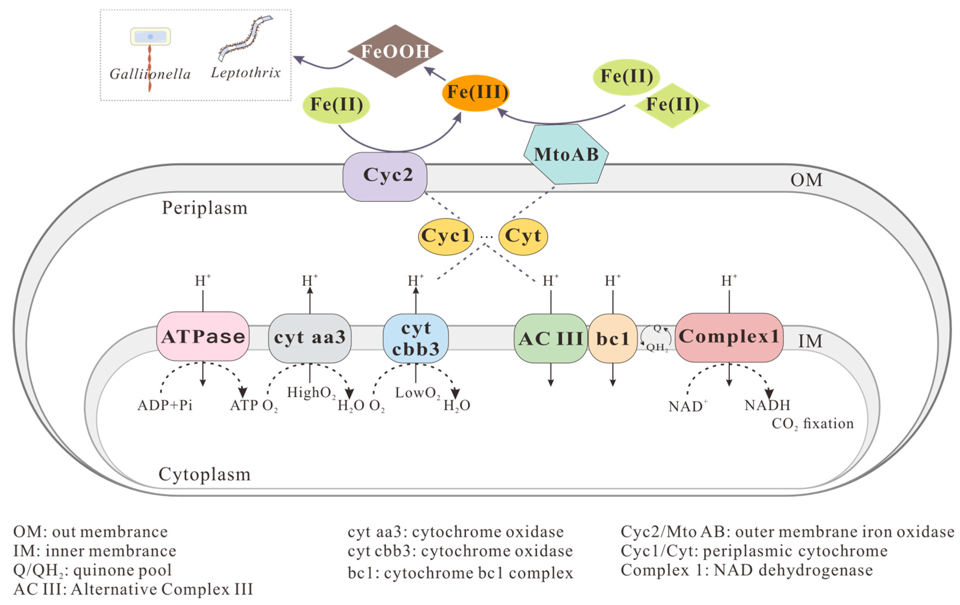
Figure 2. Neutrophilic microaerophilic FeOB-mediated Fe(II) oxidation and mineralization. Modified from Li et al. [57], McAllister et al. [61], and Lin et al. [63].
Minerals 15 00868 g002
Figure 3. Nitrate-reducing FeOB mediate Fe(II) oxidation and mineralization through chemo-/bio-coupled pathways. Modified from Liu et al. [69]; Chen et al. [64].
Minerals 15 00868 g003
Figure 4. Anaerobic phototrophic FeOB-mediated Fe(II) oxidation and mineralization. Modified from Gupta et al. [96]; Bird et al. [92]; Bryce et al. [94].
Minerals 15 00868 g004
Figure 5. Fe-H2O E-pH pourbaix diagram. For simplicity, Fe(OH)3 is used as approximation for the Fe(III) precipitates that are formed.
Minerals 15 00868 g005
Table 1. The representative iron minerals formed by different iron-oxidizing microorganisms.
Typical MineralsFormulaReferences
SchwertmanniteFe8O8(OH)8−2x(SO4)x, 1 ≤ x ≤ 1.75[101,102,129]
JarositeMFe3(SO4)2(OH)6, M = K+, Na+, NH4+, H3O+[130,131]
FerrihydriteSimplified:Fe(OH)3[113,132]
Lepidocrociteγ-FeOOH[118,132]
Goethiteα-FeOOH[105,106,122]
MagnetiteFe3O4[116,117,133]
Disclaimer/Publisher’s Note: The statements, opinions and data contained in all publications are solely those of the individual author(s) and contributor(s) and not of MDPI and/or the editor(s). MDPI and/or the editor(s) disclaim responsibility for any injury to people or property resulting from any ideas, methods, instructions or products referred to in the content.