Article

Reduction-Driven Mobilization of Structural Fe in Clay Minerals with High Fe Content

by
Anke Neumann
1,2,*,
Luiza Notini
3,
W. A. P. Jeewantha Premaratne
4,
Drew E. Latta
5 and
Michelle M. Scherer
5,6
1
School of Engineering, Newcastle University, Newcastle upon Tyne NE1 7RU, UK
2
PSI Center for Nuclear Engineering and Sciences, 5232 Villigen PSI, Switzerland
3
Civil, Construction, and Environmental Engineering, University of Delaware, Newark, DE 19716, USA
4
Department of Chemistry, University of Kelaniya, Kelaniya 11600, Sri Lanka
5
Civil and Environmental Engineering, The University of Iowa, Iowa City, IA 52242, USA
6
Civil, Environmental, and Geospatial Engineering, Michigan Technological University, Houghton, MI 49931, USA
*
Author to whom correspondence should be addressed.
Minerals 2025, 15(7), 713; https://doi.org/10.3390/min15070713
Submission received: 10 April 2025 / Revised: 20 June 2025 / Accepted: 27 June 2025 / Published: 4 July 2025
(This article belongs to the Special Issue Redox Reactivity of Iron Minerals in the Geosphere, 2nd Edition)

Abstract

Clay minerals contain significant amounts of Fe in their alumosilicate framework, and this structural Fe can be reduced and re-oxidized, constituting a potentially renewable source of reduction equivalents in sedimentary environments. However, dissolution and/or clay mineral transformations during microbial Fe reduction contradict this concept. Here, we investigate how Fe reduction and re-oxidation affect the propensity of Fe to be released from the clay mineral structure and use selective sequential extractions in combination with Mössbauer spectroscopy. Negligible amounts of Fe were released in the sequential extraction of high Fe content clay minerals NAu-1 and NAu-2. Once aqueous Fe(II) was added as a reductant, the extraction procedure recovered the initially added Fe amount and up to 30% of the Fe from the clay mineral structure as both Fe(II) and Fe(III). Similar extents of Fe mobilization were found for clay minerals partly reduced (7%–20%) with dithionite, suggesting that mobilization was reduction-induced and independent of the source of reduction equivalents (Fe(II), dithionite). Although higher Fe reduction extents mobilized more structural Fe, i.e., >90% in fully reduced clay minerals, re-oxidation largely reverted the reduction-induced Fe mobilization in clay minerals. Our finding of reduction-driven Fe mobilization provides a plausible explanation for conflicting reports on Fe release from clay minerals and how extensive Fe atom exchange between aqueous and clay mineral Fe occurs.

1. Introduction

Clay minerals are ubiquitous in soils and sediments and are the namesake and main constituents of claystone [1,2]. Most clay minerals, a term we use synonymously for phyllosilicates, contain Fe in their structure, ranging from trace concentrations to up to 30 wt% in nontronites [3]. This Fe can be bound in both tetrahedral and octahedral coordination, isomorphically substituting for either silicon or aluminum, respectively, in the clay mineral structure [4]. Similar to other Fe-bearing minerals, Fe(III) in the structure of clay minerals can also be utilized by microorganisms as a terminal electron acceptor for respiration [5,6,7,8], which consequently leads to the production of Fe(II)-bearing clay minerals. Clay mineral-bound Fe(II), in turn, has been found to be a relevant reductant for a range of organic and inorganic contaminants, including chlorinated compounds, nitroaromatics, Hg(II), Cr(VI), Tc(VIII), and Se(IV) [9,10,11,12,13,14,15], and to contribute to the (bio)geochemical cycling of elements such as N and S [16,17,18].
Importantly, the alumosilicate framework of clay minerals renders the structurally bound Fe less susceptible to reductive dissolution [19] compared to other Fe(III)-bearing minerals such as oxides or oxyhydroxides. Therefore, clay mineral Fe has been proposed to function as an important renewable source of reduction equivalents in the environment [20], reversibly undergoing Fe reduction and re-oxidation, much like a biogeobattery [21]. This concept has been supported by experimental studies on microbial reduction [17,19,22,23] and studies employing chemical reductants such as dithionite [23,24,25,26,27]. Changes to clay mineral structure and redox reactivity upon Fe reduction/re-oxidation have been reported and were largely reversible in low Fe content clay minerals, such as montmorillonite [26,27], and for low Fe reduction extents in high Fe content clay minerals like nontronite [23,24,25,26,28,29], irrespective of whether microbial activity or chemical reductants were applied. Other studies have, however, reported a reductive dissolution of Fe from the clay mineral structure [30,31,32,33,34,35,36] and/or mineral transformations, including fast smectite-to-illite transformation [32,37], mainly when microorganisms were used to facilitate clay mineral Fe reduction. The chemical reduction of clay mineral Fe, on the other hand, released Fe only rarely and to limited extents [38,39,40,41]. This raises the fascinating questions of how sustainable the redox cycling of clay mineral Fe can be, and to what extent the release of Fe from the clay mineral structure is driven by Fe reduction compared to solution factors such as microbial exudates or the presence of natural organic compounds [30,37].
Additionally, we, and others, have demonstrated that clay mineral Fe can also be reduced when in contact with aqueous Fe(II) [29,42,43,44,45], a relevant and abundant reductant in subsurface environments. Reduction extents have been limited to a maximum of 20% of the total Fe in high Fe content clay minerals [29,42], while more than 80% Fe(II)/Fe(total) have been found in low Fe content clay minerals [44,45]. In addition to yielding similar reduction extents as those obtained with microbial Fe reduction [46], electron transfer from aqueous Fe(II) and microorganisms is also thought to occur via the same pathway, i.e., predominantly via the clay mineral edge surfaces [43,47]. We therefore suggest that reduction with aqueous Fe(II) could serve as a surrogate for microbial Fe reduction, with the advantage of eliminating additional interfering factors such as microbial growth media components or exudates. To date, no study has assessed whether clay mineral Fe reduction with aqueous Fe(II) also leads to the net release of structural Fe or whether this reduction pathway could result in largely reversible reduction and re-oxidation cycles. Some indication that the reaction of Fe-bearing clay minerals with aqueous Fe(II) may result in the somewhat increased propensity of Fe to be released from the structure comes from the observation of up to 20% atom exchange between the aqueous and structural Fe pools [48] and requires further scrutiny.
Here, we investigate how and to what extent Fe reduction mobilizes Fe from the clay mineral structure, and we compare reduction by aqueous Fe(II) as a model for microbial reduction with reduction by dithionite as the typical chemical used in laboratory experiments. To estimate the mobilized Fe in the clay minerals, we developed a sequential extraction procedure based on our previous work [43] and applied the procedure to non-reduced, partly reduced, completely reduced, and reduced re-oxidized samples of Fe-rich clay minerals NAu-1 (∼21 wt% Fe) and NAu-2 (∼23 wt% Fe). We complemented this wet-chemical analysis with the use of isotope-selective Mössbauer spectroscopy combined with aqueous Fe(II) enriched in the Mössbauer-active 57Fe isotope to characterize the different solid-bound Fe pools before and after extraction.

2. Materials and Methods

2.1. Clay Mineral Preparation

Clay minerals NAu-1 ( Na 0.53 ( Al 0.15 Mg 0.02 Fe 1.84 )( Si 3.49 Al 0.51 ) O 10 (OH)2 [49], 20.5–22.4 wt% Fe) and NAu-2 ( Na 0.36 ( Al 0.17 Mg 0.03 Fe 1.77 )( Si 3.78 Al 0.08 Fe 0.15 ) O 10 (OH)2 [50], 22.5–24.0 wt% Fe) were obtained from the Source Clays Repository of The Clay Mineral Society (www.clays.org) and prepared as described previously [43]. In short, the clay minerals were ground, size-fractionated (<0.5 µm) by centrifugation, Na + -homoionized, freeze-dried, and passed through a sieve (100 mesh/150 µm) before use. The absence of relevant admixed phases was confirmed spectroscopically (infra-red (IR): kaolinite, silica, quartz; Mössbauer: Fe(III) oxides and oxyhydroxides, Fe(II)-containing phases). The size-fractionated, homoionized clay minerals resulting from this procedure are referred to as native or untreated clay minerals.

2.2. Clay Mineral Reduction and Re-Oxidation

Anaerobic conditions were ensured by carrying out all experiments inside an anaerobic chamber ( N 2 /H 2 : 93/7, O 2 < 0.1 ppm), purging all solutions with N 2 for at least 2 h prior to transfer into the anaerobic chamber, and degassing all powder reagents and glass/plasticware inside the anaerobic chamber for a minimum of 12 h prior to use.
For reduction with aqueous Fe(II), Fe(II) stock solutions (∼150 mM Fe(II)) were prepared by dissolving metallic Fe in 1 M HCl at ∼60 °C overnight, followed by filtration and dilution with deoxygenated DI water. Reduction experiments with aqueous Fe(II) were carried out in analogy to our previous experiments demonstrating electron transfer from sorbed Fe(II) to clay mineral Fe(III) [42,43,44]. Instead of using Mössbauer-invisible 56Fe(II), here, we used aqueous Fe(II), either in its natural abundance or highly enriched in the Mössbauer-visible 57Fe isotope (>92% 57Fe/∑iFe, Isoflex, San Francisco, CA, USA), to study the fate of the added aqueous Fe(II). Reduction reactors contained 15 mL of 25 mM MES (2-(N-morpholino)ethanesulfonic acid, p K a 6.06 [51]), buffer adjusted to pH 6.00 ± 0.05, or HEPES (N-2-hydroxyethylpiperazine-N’-2-ethane-sulfonic acid, p K a 7.55 [52]), buffer adjusted to pH 7.50 ± 0.05, 50 mM NaCl as ionic strength buffer, and 2 mM aqueous Fe(II). Clay minerals (30.0 ± 0.2 mg) were added to start the reduction, and the reaction was stopped after 1–24 h equilibration in the dark using centrifugation (13,000 rpm, 15 min). The supernatant was filtered (0.2 µm, nylon), acidified, and analyzed for Fe(II) and Fe(total) using the 1,10-phenanthroline method [53]. The solid phase was subjected to the sequential extraction described below or analyzed with Mössbauer spectroscopy.
For comparison, clay mineral Fe was reduced to similar extents as resulting from reaction with 2 mM aqueous Fe(II) at pH 7.5 (NAu-1: 9% [43], NAu-2: 15% [42]) and to the maximum extent achievable (‘complete dithionite-reduced’), using a modified dithionite–citrate–bicarbonate method [26] that minimizes clay mineral dissolution [39]. The reduced clay minerals were washed with deionized water, Na + -homoionized, and stored as suspensions in 50 mM NaCl to prevent both re-oxidation during freeze-drying [54] and dissolution during storage. A portion of each reduced clay mineral suspension was re-oxidized outside the anaerobic chamber using H 2 O 2 (30%, 2:1 stoichiometric excess, reaction over night) [55], washed, homoionized, deoxygenated with N 2 , and transferred into the anaerobic chamber prior to further use. Hydrogen peroxide was chosen as the oxidant to ensure complete re-oxidation of the clay mineral Fe(II) within a reasonable timeframe. As an in situ intermediate of oxidation, H 2 O 2 is expected to have the same effect on the clay mineral structure as oxidation with O 2 . All dithionite-treated clay minerals (reduced and reduced re-oxidized) were analyzed for their Fe(II) and Fe(total) content, using both Mössbauer spectroscopy and UV-vis spectrometry after complete digestion with HF [26,56].

2.3. Sequential Extraction

To account for all Fe pools in our experiments, we extended our repeatedly tested sequential extraction procedure [43,44] from targeting only sorbed species to also include different operationally defined solid phases. We previously demonstrated that extraction with 1 M CaCl 2 solution (4 h, pH ∼ 7) selectively and quantitatively liberates basal plane sorbed Fe, and subsequent treatment with 1 M NaH 2 PO 4 (20 h, pH 5) extracted edge OH-group bound Fe. Here, we included a subsequent extraction step with 0.5 M HCl (20 h) targeting Fe (oxyhydr)oxide phases [57,58] that were observed in suspensions of Fe(II)-reacted clay minerals [42,44,59]. In the last step, structurally bound Fe in the clay minerals was quantitatively liberated during digestion with HF [60], with simultaneous stabilization of any residual clay mineral Fe(II) by complexation with 1,10-phenanthroline [26,56]. Sequential extractions were carried out in triplicate, using a 10 mL extraction solution in each step, mixing end-over-end for the appropriate time in the dark, and were stopped by centrifugation (13,000 rpm, 15 min). The supernatant was filtered (0.2 µm, nylon), acidified with concentrated HCl to pH < 1, and analyzed for Fe(II) and Fe(total) using the 1,10-phenanthroline method [53]. After each extraction step, any residual extractant was removed in a 30 min wash step with deionized water.
An additional set of 8 reactors was subjected to the sequential extraction procedure, and one of the duplicates was sacrificed after each extraction step. The supernatant was treated and analyzed as described above, and the solid was collected for subsequent Mössbauer analysis.
To explore the effect of pH on Fe mobilization, aliquots of partly and complete dithionite-reduced NAu-1 were resuspended in ionic strength buffered (50 mM NaCl) solution maintained at pH 4 (25 mM PIPPS: piperazine-1,4-bis(propanesulfonic acid)), reacted with 2 mM Mössbauer-visible 57Fe(II), and subsequently extracted with 1 M CaCl 2 solution (4 h, pH ∼ 7). Samples of both aqueous and solid phase were retrieved from sacrificial reactors after each step and analyzed for aqueous Fe(II) and Fe (total) and with Mössbauer spectroscopy.

2.4. Mössbauer Spectroscopy

Solid samples were sealed between two pieces of Kapton tape to avoid sample oxidation during transfer to the Mössbauer spectrometer. Mössbauer spectra were collected in transmission mode with a spectrometer supplied by Web Research, Inc. (Edina, MN, USA) and equipped with a closed-cycle cryostat (CCS-850 System, Janis Research Co., Wilmington, MA, USA), or with a spectrometer supplied by SEE Co. (Edina, MN, USA) and equipped with closed-cycle cryostat (SHI-850 System, Janis Research Co., Wilmington, MA, USA). Data acquisition was carried out at 13 K, with the exception of completely dithionite-reduced NAu-1 samples, which were measured at T ≥ 50 K to avoid ordering at lower temperatures [61]. Measured spectra were fit using Voigt-based fitting [62] as implemented in the software Recoil (Ottawa, ON, Canada). The Mössbauer parameter center shift (CS) is reported relative to 30 µm α -Fe(0) foil, which was measured as the calibration standard at room temperature. In our analysis, we assume that the recoilless fraction, or f-factor, of all Fe sites in our samples have equal values, allowing for the assignment of relative abundances of these species based on their spectral area [63]. To achieve good and realistic fits of the Mössbauer spectra, we used the reduced χ 2 value returned by the fitting software Recoil as an indicator, alongside the residual and the plausibility of the hyperfine parameter values obtained [63].

3. Results and Discussion

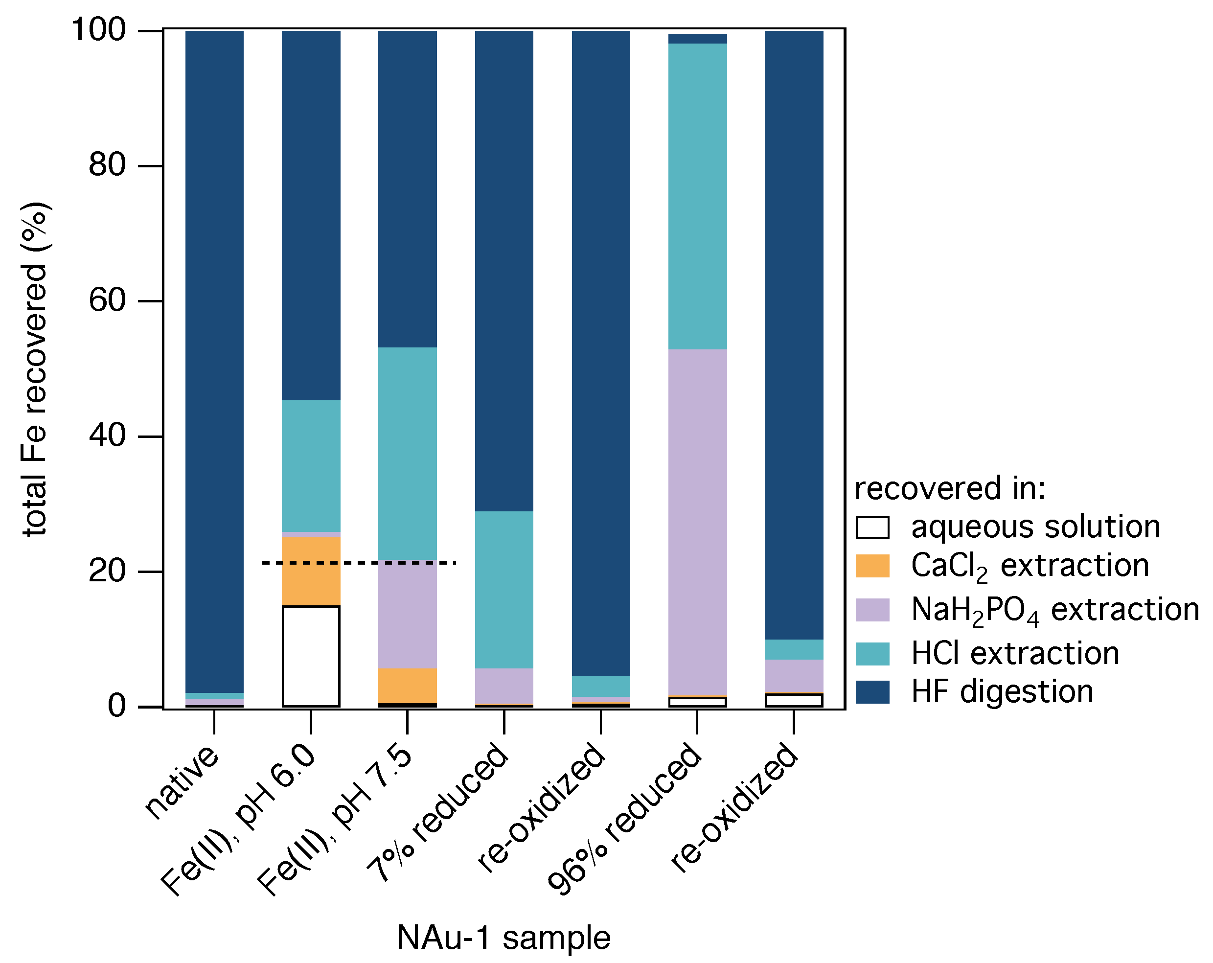
3.1. Quantification of Fe Pools in Fe(II)-Reacted Clay Minerals

To determine how sustainable the redox cycling of Fe-bearing clay minerals with aqueous Fe(II) as a reductant is, Fe in clay mineral and added pools and their speciation need to be quantified. To this end, we developed a sequential extraction procedure to selectively target sorbed Fe(II) species and solid phase Fe(III) pools. In previous work, we demonstrated that extraction with CaCl 2 selectively removes basal plane sorbed Fe(II), and subsequent treatment with NaH 2 PO 4 targets edge OH-group bound Fe(II) [43]. Here, we included a subsequent extraction step with HCl, which targets Fe (oxyhydr)oxide phases [57,58]. This extraction step was designed to selectively dissolve the solid Fe(III) oxidation product that forms during the electron transfer reaction between sorbed Fe(II) and structural Fe in clay minerals [42,44,45,59,64,65]. Structurally bound Fe in the clay minerals can be liberated quantitatively only in a (final) digestion step with HF [60].
To verify that our extraction procedure does not produce false positive results, i.e., liberate structural Fe from clay minerals in any step other than the HF digestion [40], we subjected size-fractionated, Na + -homoionized and non-reduced clay minerals NAu-1 and NAu-2 to the sequential extraction procedure. All extractions steps targeting non-structural Fe pools yielded negligible amounts of Fe, whereas 98%–99% of the recovered Fe was found as Fe(III) in the HF extract (Figure 1 and Figure S1; Table 1 and Table S1). The absence of significant Fe amounts in the HCl extract confirms that 0.5 M HCl alone is not capable of releasing structural Fe(III) from fully oxidized clay minerals [60,66]. Our results also confirm that structural Fe in clay minerals can only be dissolved in HF [60]. In combination with our previous method validation, we conclude that our extended sequential extraction procedure can effectively distinguish and quantify Fe that is sorbed (basal/edge), present in the oxidation product, and in the clay mineral.
Both clay minerals showed similar trends across all experiments, and we use NAu-1 as an example here and discuss the effect of subtle structural differences between NAu-1 and NAu-2 in Section S1. After reacting Fe-bearing clay mineral NAu-1 with aqueous Fe(II), 32%–46% of the aqueous Fe(II) sorbed at pH 6.0, and at pH 7.5 98% of Fe(II) was removed from the aqueous phase (Table 1). This significant sorption of Fe(II) to clay minerals is in agreement with previous assessments of Fe(II) uptake to NAu-1 and NAu-2 [29,34,42,43,59] and the well-studied pH-dependent sorption edge of smectites [67,68,69]. Low and pH-independent Fe(II) sorption at pH values between 4.0 and 6.0 is indicative of Fe(II) sorption to basal planes in an ion exchange reaction [68], whereas a sharp increase in sorption to pH-dependent edge OH-groups occurs at higher pH values (Figure S2).
For clay mineral NAu-1 reacted at pH 6.0, extraction with CaCl 2 liberated 5%–10% of total recovered Fe, which was exclusively Fe(II), and only negligible Fe amounts (1% of total recovered Fe) were recovered in the subsequent extraction with NaH 2 PO 4 (Table 1, Figure 1). In contrast, Fe recovery from the same clay mineral reacted at pH 7.5 was low in the CaCl 2 extraction step (1%–5% of total recovered Fe) and substantial in the subsequent NaH 2 PO 4 extraction (16%–22% of total recovered Fe; ≥87% Fe(II)/Fe(total)). These results confirm that Fe(II) sorption at a low pH value occurs predominantly at clay mineral basal planes, while sorption is dominated by edge OH groups at higher pH values [43]. The combined amounts of Fe recovered in the aqueous phase, CaCl 2 , and NaH 2 PO 4 extraction amount to 19%–27% of the total recovered Fe and equal the initially added aqueous Fe(II) (19%–21% of total Fe; Table 1). Our results for Fe(II) sorption and extraction are in agreement with our previous findings that (i) Fe(II) sorption to clay minerals occurs predominantly at basal planes at pH 6.0 and at edge OH-groups at pH 7.5, and (ii) all sorbed Fe(II) can be recovered in a CaCl 2 NaH 2 PO 4 extraction procedure [43].
In the subsequent HCl extraction, significant amounts of Fe were released (19%–31% of total recovered Fe), mostly as Fe(III) (87%–97%), consistent with the dissolution of a solid Fe(III) oxidation product. In the preceding extraction steps, we did, however, recover all initially added aqueous Fe(II) (Table 1, dashed line in Figure 1), which was the source of this Fe(III) oxidation product [42,44,45,59,64,65]. This finding leads to the intriguing question of which Fe pool is indeed represented by the HCl-extracted Fe(III): the Fe(III) oxidation product or the clay mineral structural Fe(III). From our sequential extraction of untreated clay mineral (Figure 1), it seems unlikely that extraction with 0.5 M HCl could liberate clay mineral Fe, which is also in agreement with negligible metal release during acid-leaching of clay minerals [66]. Our results and mass balance suggest, however, that a significant amount of clay mineral structural Fe(III) in NAu-1 (24%–37%) became accessible to this mild acidic extraction (0.5 M HCl) upon reaction of NAu-1 with aqueous Fe(II).
Further digestion of the clay mineral with HF recovered the remaining structural Fe as predominantly Fe(III) (93%–99%; Table 1), with an overall Fe(II) and total Fe recovery of 88%–109% and 62%–85%, respectively. The absence of significant Fe(II) amounts in the HF extraction is consistent with the recovery of the initially added aqueous Fe(II) in the preceding extraction steps, mainly in the CaCl 2 and NaH 2 PO 4 extractions. However, the absence of structural Fe(II) appears to contradict our spectroscopic evidence of interfacial electron transfer from sorbed Fe(II) to structural Fe(III), which resulted in the formation of significant amounts of structural Fe(II) [29,42,43,44,45] that should be released quantitatively only during HF digestion [60]. Different mechanisms could reconcile our apparently contradicting experimental results, including the following: (i) interfacial electron transfer was reversed in one of the extraction steps preceding HF digestion; (ii) formed structural Fe(II) was released from the clay mineral lattice during one or more of the extraction steps; and (iii) formed structural Fe(II) was spontaneously released during reduction, re-adsorbed to the clay mineral surface, and was finally extracted in one of the extraction steps preceding HF digestion, as suggested previously [34,40]. The wet chemical data alone do not provide unambiguous evidence for neither the source of extracted and mobilized clay mineral Fe pools nor the mobilization mechanism. Thus, additional direct observation of mineral changes during the extraction procedure is necessary.
Table 1. Amounts of Fe(II) and total Fe present in the extraction experiments with clay mineral NAu-1, and total Fe and relative Fe(II) recovered in the aqueous phase, CaCl 2 extraction, NaH 2 PO 4 extraction, HCl extraction, and after HF digestion of the remaining clay mineral. Data for clay mineral NAu-2 is provided in Table S1.
Aqueous Fe(II) CaCl 2 Extracted Fe NaH 2 PO 4 Extracted FeHCl Extracted FeHF Extracted FeTotal FeFe(II)
NAu-1 SampleInitialof
Total a
Finalof Recovered bSorbed cFe(II)/ Fe(tot)Fe(tot)of Recovered bFe(II)/ Fe(tot)Fe(tot)of Recovered bFe(II)/ Fe(tot)Fe(tot)of Recovered bFe(II)/ Fe(tot)Fe(tot)of Recovered bIntial dRecov- eredof Initialof initial
μmol%μmol%μmol%μmol%%μmol%%μmol%%μmol%μmolμmol%%
untreated
 NAu-10.04(0.02)<11000.05(0.02)<1150.61(0.04)1110.55(0.16)1258.1(6.4)98129.4(0.6)59.4(6.6)46
 NAu-1 e,f1000.01(0.01)<150.77(0.14)1290.14(0.07)<1 112.3(5.7)
Fe(II)-reacted, pH 6.0
 NAu-132.3(0.2)2117.5(0.3)1514.8(0.5)9611.8(0.2)10140.90(0.01)1722.7(0.3)19763.8(4.2)55152.0(0.7)116.7(4.9)77109
 NAu-1 e29.9(3.1)2020.5(0.4)219.61(3.50)1007.40(0.14)8248.26(0.08)8320.7(0.2)21142.343148.4(1.3)99.2(0.7)67106
Fe(II)-reacted, pH 7.5
 NAu-132.5(0.5)210.65(0.00)131.9(0.5)935.42(0.09)58717.2(0.5)161333.5(0.8)31750.0(7.6)47152.5(0.7)106.7(9.0)7088
 NAu-1 e27.9(0.1)192.48(0.06)225.5(0.2)1002.30(0.11)29618.6(0.5)16927.2(1.6)24164.156147.7(0.1)114.5(2.3)7888
 NAu-1 g32.6(0.8)210.88(0.01)131.7(0.4)1001.28(0.04)19629.3(0.2)221130.8(0.3)23169.953155.0(0.8)132.1(0.6)85101
partly dithionite-reduced h
 NAu-10.02(0.02)<1830.12(0.08)<1484.15(2.42)5918.1(0.5)23155.5(2.6)71103.9(1.3)77.9(5.6)7560
 NAu-1 e0.00(0.00)0930.28(0.01)2799.74(0.56)59194.32(0.15)2642.241472.7(0.3)16.59(0.73)2365
partly dithionite-reduced, re-oxidized i
 NAu-10.01(0.01)<11000.03(0.03)<170.61(0.49)141.79(0.74)3157.7(0.8)96102.0(7.2)60.1(2.1)59
completely dithionite-reduced j
 NAu-13.32(1.27)11001.27(0.03)<1100124.8(4.3)5195109.7(16.3)4573.55(0.46)1238.2(2.4)242.6(22.4)102104
 NAu-1 e0.28(0.05)01000.69(0.02)0100123.7(0.5)877214.9(4.3)1042.662158.8142.2(4.9)9092
completely dithionite-reduced, re-oxidized k
 NAu-1 j0.02(0.00)2n.d. l0<12.51(0.02)631.40(0.16)3136.628867.6(0.4)41.45(0.55)61
a Calculated as % of initially present total Fe. b Calculated as % of recovered total Fe. c Calculated as difference of initial and final aqueous Fe(II). d Initial total Fe in the system was calculated from initial aqueous Fe(II) and Fe in the clay mineral. The latter was calculated from the total Fe content determined after HF digestion, according to [11,54,56,70]. e Two sample reactors were sacrificed after each extraction step, leading to less reliable results for the HCl and HF extraction steps, for which only two and one replicates were carried out, respectively. f This extraction procedure was carried out to obtain samples for Mössbauer spectroscopy and without final HF digestion. The relative amounts of total Fe recovered in each extraction step (‘of recovered’) are expressed relative to the initial total Fe in the reactors. g Extraction data for 57Fe(II)-reacted NAu-1 after 187 days of reaction (more than 6 months), whereas all other reactions of Fe(II) with NAu-1 were stopped within 1 hour to 3 days. h Reduction with sodium dithionite according to [39,54] to similar extent as was achieved after reaction with aqueous Fe(II) (extraction samples: 6.8(0.2)%; Mössbauer samples: 18.9(0.1)%). i Re-oxidized samples were produced from dithionite-reduced NAu-1 suspensions by reaction with H 2 O 2 according to [55]; final Fe(II) content was 1.1(0.1)%. j Reduction with sodium dithionite according to [39,54] to maximum extent (extraction and Mössbauer samples: 96.4(0.6)%). k The re-oxidized clay mineral sample was produced from dithionite-reduced clay mineral suspension by reaction with H 2 O 2 according to [55]; initial Fe(II) content: 86.7(0.4)%; final Fe(II) content: 0.5(0.6)%. l not detected.

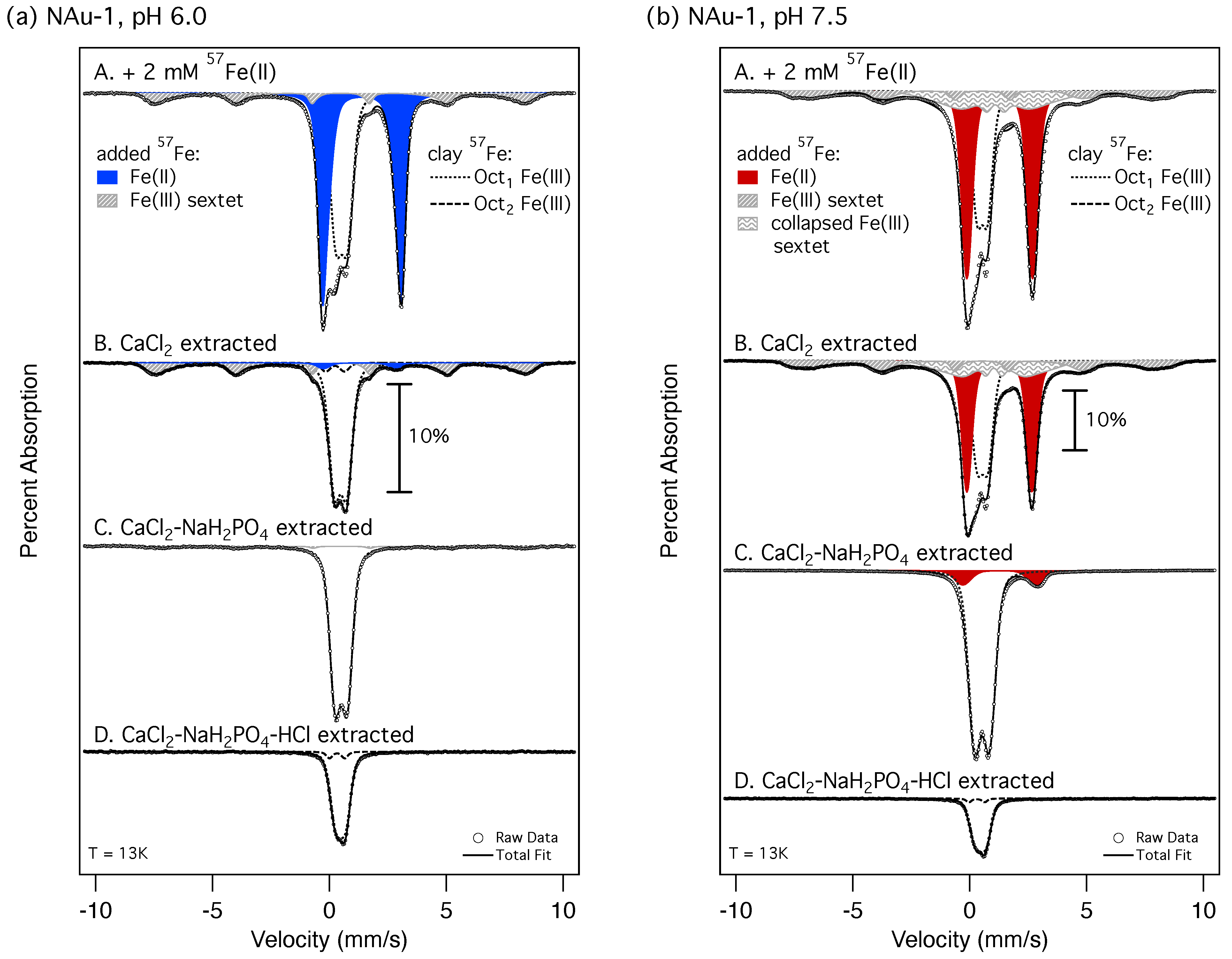
3.2. Source of Extractable Fe Pools in Fe(II)-Reduced Clay Minerals

To directly observe which mineral-bound Fe species were liberated in each extraction step, we used 57Fe-Mössbauer spectroscopy in combination with aqueous Fe(II) enriched in the Mössbauer-active 57Fe isotope ( 92 % 57Fe/∑iFe). This way, the Mössbauer signal of added Fe, or non-structural Fe pools, was increased relative to clay mineral Fe, which is present in its natural composition (57Fe/∑iFe: 2.2%). Consequently, spectral areas in the Mössbauer spectra do not reflect the actual abundance of each Fe pool; while Fe taken up from the aqueous phase represents 8% and 18%–21% of the total Fe at pH 6 and 7.5 (Table 1), the contribution of sorbed 57Fe in the Mössbauer spectra amounts to 77% and over 90%, respectively (Table S2). Therefore, the results from Mössbauer spectral analysis can be compared only qualitatively with the wet chemical extraction data.
After the reaction of clay mineral NAu-1 with aqueous 57Fe(II) at pH 6.0 and pH 7.5, we observed the presence of a large Fe(II) doublet and an Fe(III) sextet, in addition to the clay mineral Fe(III) doublet (Figure 2). The Mössbauer parameters of the Fe(II) doublet in NAu-1 reacted at pH 7.5 (red doublet in Figure 2b) are consistent with structural/edge OH-group sorbed Fe(II) (CS: 1.27 mm/s; QS: 2.83 mm/s; Table S3). The larger values for the center shift (CS: 1.39 mm/s) and quadrupole splitting (QS: 3.30 mm/s) of the Fe(II) doublet in NAu-1 reacted at pH 6.0 (blue doublet in Figure 2a) are characteristic for basal plane sorbed Fe(II) [43,44]. The presence of Fe(II) doublets with large spectral areas (50% and 46% at pH 6.0 and pH 7.5, respectively) is consistent with significant sorption of 57Fe(II) to clay mineral NAu-1 and only partial participation of sorbed Fe(II) in the interfacial electron transfer reaction, as recently found for NAu-1 [29].
The oxidation product of this interfacial electron transfer reaction manifests as a well-ordered Fe(III) sextet in both spectra. The sextet’s Mössbauer parameters are highly similar at both pH values (Table S3) and consistent with the formation of lepidocrocite [71], which was also found after reacting NAu-2 with aqueous Fe(II) at pH 7.5 [42]. A range of Fe (oxyhydr)oxides has been found for different reaction conditions and different Fe-bearing clay minerals [42,44,59,64,65], and an unambiguous phase assignment based on Mössbauer parameters alone is very difficult. Hence, we cannot rule out that an Fe(III) mineral with similar values for CS, QS, and hyperfine magnetic field (Bhf) formed, and we provide a more detailed discussion in Section S2. Moreover, the spectrum of NAu-1 reacted at pH 7.5 exhibits an additional collapsed Fe sextet, which is characterized by an unusually high CS value for Fe(III) (1.09 mm/s) and a very low hyperfine magnetic field with a large distribution (13.7 ± 6.1 T; Table S3). Collapsed sextets with similar Mössbauer parameters have been interpreted as originating from mixed-valent Fe(II)-Fe(III) phases [72,73] or Fe(III) phases of low crystallinity and/or of very small particle size [74,75]. Irrespective of the specific identity of new solid phase(s) formed as a result of interfacial electron transfer, these phases contain substantial amounts of Fe(III) and are clearly distinguishable in the Mössbauer spectra from Fe(II) sorbed to both basal surfaces and edge OH-groups. Selective removal of any of these different species by the sequential extraction steps can therefore be directly verified in the Mössbauer spectra.
Extraction with CaCl 2 resulted in very small changes in the Mössbauer spectrum of NAu-1 reacted at pH 7.5 (Figure 2b-B). This finding agrees well with the recovery of minor amounts of Fe in this extraction step (1%–5%; Table 1) and confirms the absence of Fe(II) sorbed to basal surfaces. In contrast, this extraction step removed almost all of the Fe(II) doublet area in the spectrum of NAu-1 reacted at pH 6.0 (Figure 2a-B), corroborating that this Fe(II) was indeed sorbed via ion exchange at the clay mineral basal surfaces. The remaining small Fe(II) doublet (3% of the spectral area) can be attributed to either residual basal plane sorbed 57Fe(II) or to clay mineral Fe(II) formed during the interfacial electron transfer. In our previous work with 56Fe(II), which allowed us to exclusively monitor the changes to the naturally abundant 57Fe present in the clay mineral structure [29,42,43,44,45], we found similar hyperfine parameters for structural Fe(II) in NAu-1 reacted at pH 6 and basal plane sorbed Fe(II) [43]. This similarity makes it impossible to differentiate between these two species in the Mössbauer spectrum. Importantly, the Fe(III) sextet(s), which directly result from the interfacial electron transfer from sorbed Fe(II), remained largely unchanged by the CaCl 2 extraction.
Subsequent extraction with NaH 2 PO 4 , designed to quantitatively remove OH-group bound Fe(II), did indeed remove the majority of the Fe(II) doublet area in the spectrum of NAu-1 reacted at pH 7.5 (Figure 2b-C). Although the remaining Fe(II) doublet area (10%; Table S3) is consistent with the extent of clay mineral Fe reduction in our previous work (8%–9%), structural clay mineral Fe(II) was absent in 56Fe(II)-reacted NAu-1 after extraction with NaH 2 PO 4 [43]. This suggests that the remaining Fe(II) doublet here is due to residual sorbed 57Fe(II), magnified in the Mössbauer spectrum due to its high enrichment. Surprisingly, the Fe(III) sextet(s) were also completely removed in both 57Fe(II)-reacted samples after extraction with NaH 2 PO 4 , which was not intended to dissolve solid Fe(III)-containing phases. Nevertheless, the Fe(II)/Fe(total) ratios quantified in the extracts (Table 1) match well with the Fe(II)/Fe(total) ratios derived from the spectral areas of Fe(II) doublets and Fe(III) sextets removed (pH 6: 14%–24% vs. 13%; pH 7.5: 87%–96% vs. 45%–72%). For NAu-1 reacted at pH 7.5, values are closer when higher Fe(II) contents of the collapsed sextet are assumed (Table S3). Our Mössbauer spectral analyses therefore confirm the finding from the wet-chemical extractions that all initially added Fe(II) is recovered after CaCl 2 NaH 2 PO 4 extraction. Importantly, the spectra provide direct evidence that the initially added Fe is recovered both as Fe(II), as intended, and additionally in the form of the Fe(III) oxidation product. We suspect that the slightly acidic NaH 2 PO 4 -extraction solution dissolved these freshly formed Fe(III) phases. Extraction of 6-month-aged samples (Figure S3, Table 1) suggests that these Fe(III) phase(s) remain susceptible to dissolution even after extended periods of time that would enable recrystallization [76,77,78] and ripening to more crystalline minerals [72,73,79,80].
Because all added 57Fe(II) had been recovered in the aqueous phase and CaCl 2 NaH 2 PO 4 extraction steps, the Fe recovered in further extraction with HCl must have been released from the clay mineral structure. Although Mössbauer spectroscopy cannot provide absolute amounts of Fe in a sample, we can semi-quantitatively interpret changes in signal intensity within each set of extraction samples: all samples initially contained the same 57Fe amount and were packed in the same way for Mössbauer analysis, and the resulting absorption data were plotted relative to the background rather than normalized to the highest peak. Hence, we suggest that the sharp drop in overall signal intensity after extraction with HCl (Figure 2-D) can reasonably be interpreted as a net loss of Fe, which was higher at pH 7.5 than 6 (two-thirds vs. one half). Both observations are consistent with the extraction data, which showed 19%–21% Fe recovered for NAu-1 reacted at pH 6.0 compared to 23%–31% Fe recovered for NAu-1 reacted at pH 7.5 (Table 1). The Mössbauer spectra comprised exclusively octahedral Fe(III) components after extraction with HCl and were already dominated by these components before this extraction step (Figure 2-C). This observation further supports that HCl liberated clay mineral Fe(III) from the Fe(II)-reacted NAu-1 rather than Fe(III) in the oxidation product as intended. Again, 6-month aged samples behaved highly similarly (Figure S3), and the HCl-extracted Fe was predominantly Fe(III) in all samples (≥87%; Table 1), indicating that the fraction of HCl-extractable Fe(III) persisted over time. Intriguingly, HCl extraction of native (un-reduced) NAu-1 neither released significant amounts of structural Fe from the Fe(III)-containing clay mineral (Table 1) nor led to significant changes in the Mössbauer spectrum (Figure S4a). Our combined evidence from sequential extractions and Mössbauer spectroscopy suggest that reaction with aqueous Fe(II) led to 19%–21% (pH 6.0) and 23%–31% (pH 7.5) of structural Fe(III) in clay mineral NAu-1 becoming susceptible to extraction, or mobilized. It is, however, unclear whether this observed structural Fe mobilization is specific to the interaction of clay minerals with aqueous Fe(II) or whether the process of structural Fe reduction is causing the observed mobilization.

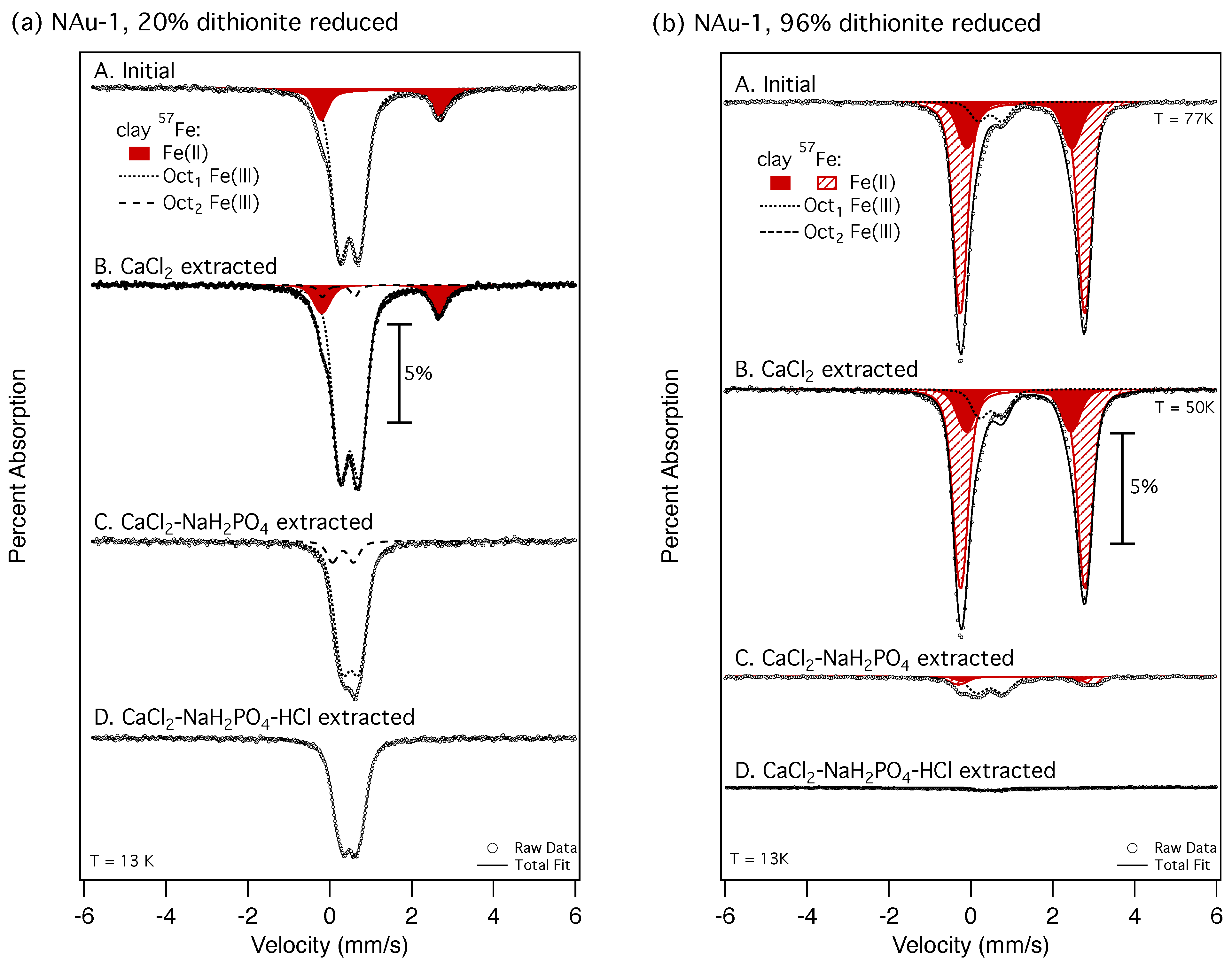
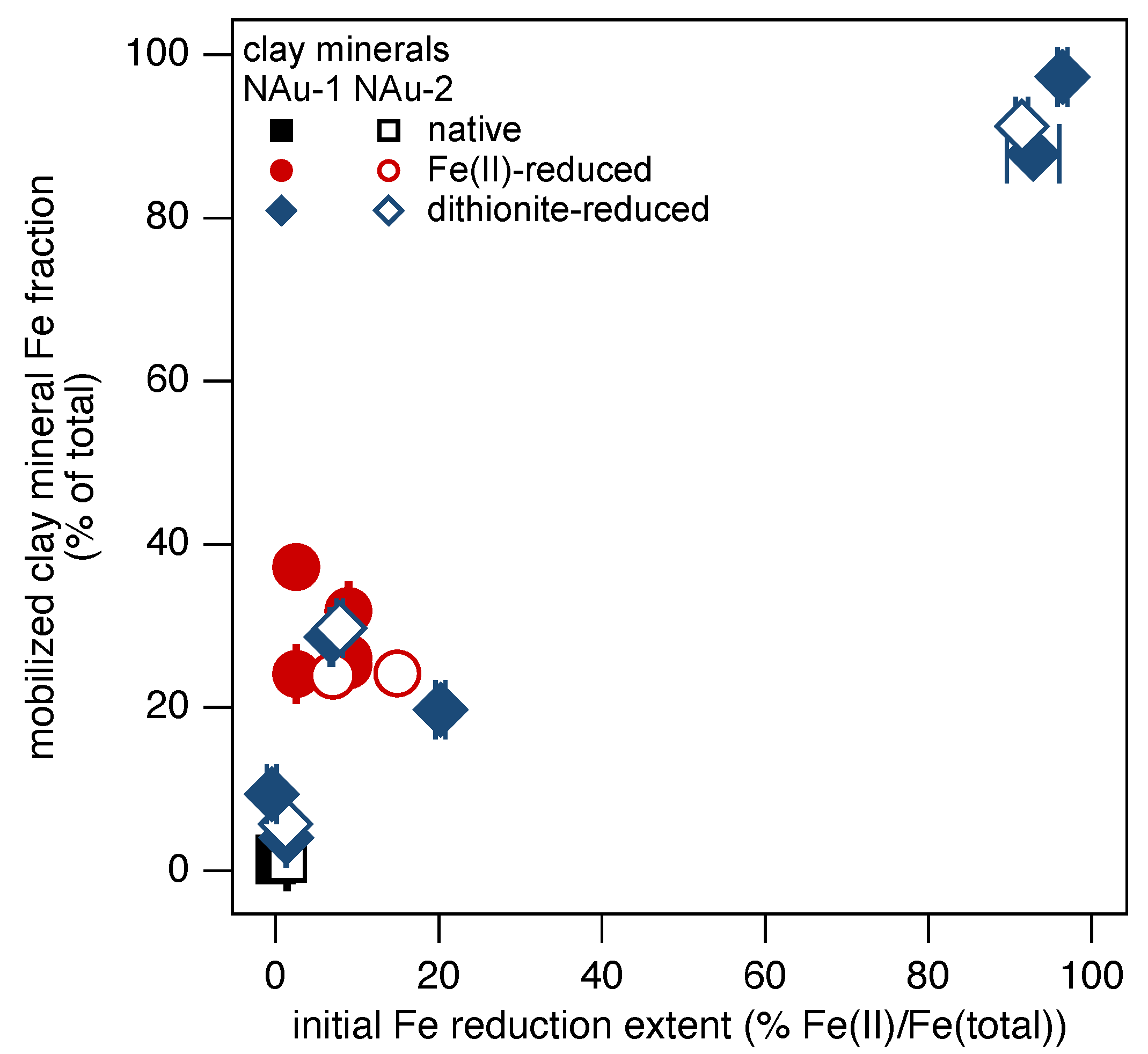
3.3. Iron Mobilization in Dithionite-Reduced Clay Minerals

To explore the effect of structural Fe reduction on Fe mobilization in clay minerals in the absence of added aqueous Fe(II), we reduced NAu-1 and NAu-2 partly (7%–8%) and fully (91%–96%) with the chemical dithionite. We subjected these chemically reduced minerals to the same sequential extractions as their Fe(II)-reacted counterparts (Table 1 and Table S1) and complemented the wet chemical analyses with Mössbauer spectroscopy. The reduction extents obtained photometrically after HF digestion and from Mössbauer spectral analysis agree well (Table S4). The Mössbauer spectra of partly and fully reduced NAu-1 exhibit one and two Fe(II) doublets, respectively (Figure 3), that exhibit hyperfine parameters within the expected range for structural Fe(II) in clay minerals (Table S3). Two doublets may be required for fitting the Mössbauer spectra of Fe-rich smectites with high reductions extents [29], which can be rationalized with the presence of several structural Fe(II) binding environments [23,24,25,26,81,82]. As trends were again highly similar for both clay minerals, we present the results for NAu-1 in detail and include the data for NAu-2 in Figure S1 and Table S1.
As found for native, or un-reduced, clay minerals, extraction with CaCl 2 also led to negligible changes in the Mössbauer spectra of dithionite-reduced NAu-1 (Figure 3-B), which is reflected in minor recovery of Fe(II) in the extracts (Table 1). These data confirm the absence of Fe(II) sorbed at basal surfaces in these chemically reduced clay minerals. In contrast, subsequent extraction with the NaH 2 PO 4 of partly reduced NAu-1 liberated 5% of the total recovered Fe (Table 1) as both Fe(II) and Fe(III). We suspect that the higher initial reduction extent in the samples used for Mössbauer analysis compared to the one used to quantify release in the extractions (19% vs. 7%; Table S4) caused the release of more total Fe with a higher Fe(II) ratio (79% Fe(II)/Fe(total) vs. 48% Fe(II)/Fe(total); Table 1). The Mössbauer spectrum of partly reduced NAu-1 corroborates the quantitative extraction data and shows complete removal of the Fe(II) doublet and alteration of the Fe(III) doublet shape (Figure 3a-B). Our results also suggest that the chemical reduction of clay mineral Fe, just like reduction with aqueous Fe(II), made a substantial fraction of clay mineral Fe more susceptible to release in mildly acidic conditions such as the NaH 2 PO 4 extraction solution.
Neither the wet chemical extraction data nor the hyperfine parameters of the Fe(II) doublet in partly reduced NAu-1 allow us to determine whether the Fe(II) originated from the clay mineral octahedral sheet or was bound to edge OH-groups. Several studies found that reduction with dithionite released structural Fe, in some cases up to 20%, which may re-adsorb to edge OH-groups [15,40,83]. In contrast, other studies found no such release during reduction with the citrate-bicarbonate-dithionite method [10,38,39]. It is possible that reduction with dithionite mobilized and released structural Fe(II), which subsequently sorbed to clay mineral surfaces and transferred electrons back to structural Fe(III), leading to a mixture of sorbed Fe(II) and Fe(III) at edge OH-groups, yet without the formation of an Fe(III) oxidation product that is usually observed as a result of reduction with aqueous Fe(II) (Figure 2). Considering additional evidence from fully reduced and reduced and re-oxidized clay minerals, a more plausible explanation is that both the Fe(II) and Fe(III) liberated during extraction with H 2 PO 4 resided within the structure of the clay mineral.
First, more than 50% of the Fe(II) in fully reduced clay mineral NAu-1 (51%–87%; Table 1) were removed during extraction with NaH 2 PO 4 , which also resulted in a 90% decrease in the overall Mössbauer signal intensity in fully reduced NAu-1 (Figure 3b-C). It is impossible that these extensive amounts of clay mineral Fe were released during reduction with dithionite without completely destroying the clay mineral structure, which previous work has ruled out based on reduced and re-oxidized samples [23,24,25,26]. Second, and consistent with these reports, re-oxidation of partly and fully reduced clay minerals restored all Fe(II) to Fe(III) (Table S4), albeit with slightly altered binding environments when high Fe reduction extents had been applied. However, no ancillary Fe(III) phases were observed (Figure S4b-A), which would be expected to form if significant amounts of surface-bound Fe(II) had been present during re-oxidation. We propose that structural Fe became mobilized, i.e., more susceptible to release into the aqueous phase under appropriate conditions such as acidic pH and/or the presence of complexing agents like HPO 4 2 upon formation of structural Fe(II) through reduction with either dithionite or aqueous Fe(II). We attribute this to the higher solubility of Fe(II) compared to Fe(III) [67], and the additional substantial mobilization of structural Fe(III) to its spatial co-location with the Fe(II) formed during reduction. As electron hopping is fast between adjacent Fe atoms in Fe-rich clay minerals [84], effective reduction, and hence mobilization, may not be localized to one specific Fe atom [42] and also affect surrounding Fe atoms.
This mobilization mechanism can also rationalize the additional release of 23%–26% Fe from partly reduced NAu-1 upon subsequent extraction with HCl (Table 1). Importantly, the extracted Fe was predominantly Fe(III) (81%–91%; Table 1), confirming that the initially extraction-resistant Fe(III) in the clay mineral (Figure S4a) also became mobilized by the reduction process, and remained so even after the majority of Fe(II) had been removed. Again, the effect of this reduction-induced Fe(III) mobilization was more pronounced for the fully reduced NAu-1 compared to the partly reduced sample, as after extraction with HCl the Mössbauer spectral signal of fully reduced NAu-1 was reduced to levels close to noise (Figure 3b-D). The remaining amounts of Fe were released in the final HF digestion step and were very low for fully reduced NAu-1 (1%–2% of total recovered Fe; Table 1). In contrast, around 70% of the clay mineral Fe was found in the HF digests from partly reduced NAu-1, indicating that this portion of Fe(III) remained in its original structural bonding environment.
Adding the amounts of Fe released in all extracts other than the HF digestion allows us to calculate the overall structural Fe mobilization in dithionite-reduced clay minerals. The same procedure can be applied to Fe(II)-reduced clay minerals when this value is corrected for the initially added aqueous Fe(II). Interestingly, the amounts of structural Fe mobilization in partly dithionite-reduced clay minerals and in Fe(II)-reduced clay minerals are similar (Figure 4), suggesting that the initial extent of structural Fe reduction, rather than the presence of aqueous Fe(II), determines the extent of structural Fe mobilization. However, the absolute extent of structural Fe reduction was between 3% (Fe(II)-reduced NAu-1, pH 6.0 [43]) and 20% (dithionite-reduced NAu-1; Table S4), whereas 20%–37% of structural Fe were mobilized (Table 1). In fully reduced clay minerals, more than 90% of structural Fe were released during the sequential extraction and were thus mobilized. Although tempting, a simple linear relationship is unlikely to describe the observed reduction-driven Fe mobilization in clay minerals because the cause is most likely linked to structural changes in the clay mineral lattice during structural Fe reduction, which are known to be non-linear [29,38,82]. Moreover, our data (Figure 4) has a clear bias towards low (≤20%) and very high reduction extents (>90%), and intermediate reduction extents should be explored in future research.
We therefore conclude that clay mineral Fe reduction results in apparent mobilization of up to one third of structural Fe in partly reduced clay minerals, independent of the source of redox equivalents (Fe(II), dithionite). More extensive structural Fe mobilization of more than 90% was found in fully reduced clay minerals, suggesting that structural Fe(II) in clay minerals is more prone to release from the clay mineral structure during changes in solution chemistry. If our finding is indeed of general validity, the increased amounts of HCl-extracted Fe from microbially reduced clay minerals might be due to the same mechanism of increased accessibility of structural Fe to extraction [30,31,33,85]. However, as microbial studies commonly use exclusively HCl extractions (compared to sequential extractions) with slightly differing conditions, the results from our work and microbial clay mineral reduction cannot be compared directly.

3.4. Reversibility of Iron Mobilization in Clay Minerals

Mobilization of more than 90% of clay mineral Fe during structural Fe reduction raises the question of whether this extensive mobilization is reversible and/or to what extent. Previous spectroscopic evidence suggests that low Fe reduction extents lead to reversible clay mineral structural changes, whereas high reduction extents yield some irreversible alterations [23,24,25,26,27,28,55]. Our Mössbauer spectrum of fully reduced, re-oxidized NAu-1 (Figure S4b-A) confirms different Fe(III) binding compared to the native, or un-reduced, sample (Figure S4a-A), with larger QS values for both octahedral Fe components (0.67 and 1.37 mm/s; Table S3) that are consistent with previous reports [23,27,55]. Similar Mössbauer parameters have been observed for native NAu-2 (CS: 0.52–0.53 mm/s, QS: 1.23–1.27 mm/s [42,86]) and re-oxidized SWa-1 (CS: 0.44 mm/s, QS: 0.75 mm/s [28,55]), and large QS values are generally linked to distorted octahedral binding environments [87,88].
Despite these significant structural differences caused by Fe reduction and re-oxidation, sequential extraction of fully dithionite-reduced, re-oxidized NAu-1 recovered less than 10% of total Fe in the CaCl 2 , NaH 2 PO 4 , and HCl extraction steps (Figure 1). Similarly, 94%–96% of the solid phase Fe were released only in the HF digestion process when partly dithionite-reduced, re-oxidized NAu-1 and NAu-2 were subjected to the sequential extraction procedure (Table 1 and Table S1). Our data demonstrates that re-oxidation returned the structural Fe that was accessible to extraction with NaH 2 PO 4 and HCl after reduction with dithionite to the same inaccessible state as in the non-reduced materials. We conclude that the effect of reduction on structural Fe mobilization is largely reversible and appears to be mostly unconnected to the initial Fe reduction extent (4%–10% residually mobilized Fe for all reduction extents and both clay minerals) and therefore also unaffected by the clay mineral structural changes during Fe reduction.
We note, however, that overall Fe recoveries were less complete for native and re-oxidized samples (46%–64%) compared to both dithionite-reduced and Fe(II)-reduced clay minerals (76%–102%; Table 1 and Table S1). We suspect that this might be due to most of the Fe in the native and re-oxidized samples remaining in the clay mineral structure, which is liberated only in the final extraction step. The overall extraction procedure comprises four consecutive extraction steps and additional DI water washes, which are all potential sources of mass loss, particularly via colloids that clay minerals are known to form [69,89]. Adding high ionic strength solutions such as 1 M CaCl 2 and NaH 2 PO 4 extraction solutions effectively prevents colloid formation [69,89] and hence enables high solid recoveries. In contrast, the HCl extraction solution lacks cations for clay flocculation [69,89], and consequently, a loss of solids can occur. We suggest that a loss of colloids was also responsible for the intensity loss, and hence the apparent mass loss, observed in the Mössbauer spectrum of fully dithionite-reduced, re-oxidized NAu-1 after extraction with HCl (Figure 3b-D). As samples are prepared via filtration for measurements with the Mössbauer spectrometer, colloids might be lost, but they would be present in the full suspension used for HF digestion. Indeed, photometric analysis of the extracts accounted for 3% of the total Fe recovered in the HCl extraction, where the intensity in the Mössbauer spectrum was reduced to almost background levels, but 88% were recovered in the HF digestion (Table 1).
Even though re-oxidation can largely revert the reduction-induced Fe mobilization in clay minerals, changes in solution chemistry may irreversibly release the mobilized Fe from clay minerals. To illustrate this point, we suspended the partly and fully dithionite-reduced samples of clay mineral NAu-1 in solutions buffered at pH 4. In both spectra, we observed a significant decrease in the Mössbauer spectral area of Fe(II), which was more pronounced in the partly reduced NAu-1 sample (20% to 5% and 78% to 75%, Figure S5). This spectral area loss corresponds to a net release of clay mineral Fe(II) that was also accessible in our extraction experiments (Table 1), which we termed mobilized. Interestingly, the hyperfine parameters of the remaining Fe(II) doublet in the partly reduced NAu-1 at pH 4 are more similar to those indicative of Fe(II) sorbed to clay mineral basal surfaces (Figure S5a-B; Table S5) [43]. This could suggest a release of structural Fe(II) and its re-adsorption to basal surfaces. Sorption of additional 57Fe(II) at this low pH value increased the area of the Fe(II) doublet, which retained identical Mössbauer parameters (Figure S5a-C). Subsequent extraction with CaCl 2 removed the added 57Fe(II) while retaining a Fe(II) doublet of similar spectral area as before 57Fe(II)-addition (Figure S5a-D). In an identical experiment with fully reduced NAu-1 at pH 4, the addition of 57Fe(II) at this acidic pH yielded a second Fe(II) doublet with hyperfine parameters indicative of basal plane sorbed Fe(II), which was also completely removed with CaCl 2 extraction (Figure S5b). In both cases, the remaining Fe(II) doublet exhibited the same hyperfine parameters and spectral area as before the addition of 57Fe(II), suggesting that both Fe(II) doublets corresponded to structurally bound Fe(II). In summary, even though a substantial portion of reduction-mobilized clay mineral Fe was released at low pH values, some (low initial reduction extent) or the majority (high reduction extent) of clay mineral Fe(II) remained structurally bound and can undergo reversible reduction and re-oxidation (Table 1 and Table S1, Figure S4).

4. Conclusions

Our results show that clay mineral Fe reduction mobilizes a substantial amount of the formed Fe(II) and Fe(III). We define mobilization to mean that the Fe remains bound in the structure of the clay mineral yet has a greatly increased susceptibility to being released upon changes in solution conditions such as pH and/or the presence of complexing agents. This reduction-driven mobilization somewhat resembles the reductive dissolution process of Fe (oxyhydr)oxides but is more complicated due to the mobilization of both Fe(II) and Fe(III), presumably present in spatial proximity to each other and hence connected via fast electron hopping [84]. Our demonstration that mobilized Fe is released from the clay mineral structure at low pH value further suggests that other solution conditions conducive to Fe complexation, such as the presence of organic material, may result in similar or even higher Fe(II) loss from clay minerals [90,91]. We also hypothesize that microbial dissolution of clay mineral Fe [30,31,32,33,34,35,36] may actually be linked to its reduction-induced mobilization and subsequent release via complexation with organic material exuded by the microbes.
Other work has reported Fe recovery from clay minerals when exposing dithionite-reduced samples to citrate-bicarbonate or acetic acid-buffered citrate [38,40,92], and our results provide a mechanistic explanation. Moreover, we also demonstrate that mildly acidic conditions (pH 5) are sufficient to release some structural Fe from reduced clay minerals and that this amount strongly depends on the initial reduction extent and the specific extraction procedure applied. We note that the sequential CaCl 2 NaH 2 PO 4 -extraction, which we developed to assess interfacial electron transfer reactions between Fe(II) sorbed to different clay mineral surfaces and clay mineral Fe [43], has also been applied to microbially reduced clay minerals to differentiate ‘exterior’ ( CaCl 2 NaH 2 PO 4 -extracted Fe) and ‘interior’ (HCl-extracted Fe) Fe [85,93]. In particular, our results from fully reduced clay minerals demonstrate that Fe liberated during extraction with NaH 2 PO 4 cannot stem exclusively from, or close to, clay mineral surfaces, and we strongly caution against the designation of NaH 2 PO 4 -extracted Fe in reduced clay minerals as ‘exterior’.
Finally, we propose that our finding of reduction-induced Fe mobilization could provide a plausible mechanism for how extensive Fe atom exchange between aqueous and clay mineral Fe occurs [48]. Of the three potential mechanisms, i.e., Fe diffusion through mineral pores, Fe atom diffusion within the crystal lattice, and electron conduction connecting spatially separated oxidative growth and the reductive dissolution of the Fe mineral, we ruled out the first and found the last to be most plausible given our dataset at the time. However, considering the higher propensity of Fe(II) and some Fe(III) in reduced clay mineral to be released from the structure, the diffusion of these mobilized Fe atoms within the crystal lattice may indeed be another plausible mechanism. Consistent with this idea, the final extent of atom exchange we found previously is higher when the average extent of Fe mobilization is higher in the respective clay minerals (Figure S6). This relationship may yet simply be caused by a co-dependence of Fe mobilization extent and Fe reduction extent, which also affected the extent of atom exchange observed, and thus needs to be tested in future research.

Supplementary Materials

The following supporting information can be downloaded at https://www.mdpi.com/article/10.3390/min15070713/s1: wet chemical extraction data on clay mineral NAu-2; Fe(II) pH-adsorption edges for both NAu-1 and NAu-2; a calculation on the contribution of added 57Fe to the Mössbauer signal; Mössbauer spectra of samples before and after each extraction step of NAu-1 reacted with 57Fe(II) for six months, native NAu-1, fully dithionite-reduced and re-oxidized NAu-1, and partly and fully dithionite-reduced NAu-1 resuspended at pH 4; Mössbauer hyperfine parameters of all spectra presented in this study; a comparison of the relative Fe(II) content of samples used in the study as determined after HF digestion vs measured with Mössbauer spectroscopy; and a comparison of Fe mobilization extent and the extent of Fe atom exchange observed in NAu-1 and NAu-2 after reaction with aqueous Fe(II).

Author Contributions

Conceptualization, A.N. and M.M.S.; methodology, A.N.; formal analysis, A.N., L.N. and W.A.P.J.P.; investigation, A.N., L.N., W.A.P.J.P. and D.E.L.; resources, A.N. and M.M.S.; data curation, A.N., L.N. and M.M.S.; writing—original draft preparation, A.N.; writing—review and editing, A.N., L.N., D.E.L. and M.M.S.; visualization, A.N.; supervision, A.N. and M.M.S.; project administration, A.N.; funding acquisition, A.N. and M.M.S. All authors have read and agreed to the published version of the manuscript.

Funding

This research was funded by the Swiss National Science Foundation (grant number PBEZP2_137292) and the German Research Foundation (grant number NE 1715/1-1).

Data Availability Statement

The original contributions presented in this study are included in the article/Supplementary Materials. Further inquiries can be directed to the corresponding author.

Acknowledgments

We acknowledge help in the laboratory by Tyler L. Olson, and Katherine A. Rothwell.

Conflicts of Interest

The authors declare no conflicts of interest. The funders had no role in the design of the study; in the collection, analyses, or interpretation of data; in the writing of the manuscript; or in the decision to publish the results.

References

  1. Amonette, J.E. Iron redox chemistry of clays and oxides: Environmental applications. In Electrochemical Properties of Clays; CMS Workshop Lectures; Fitch, A., Ed.; The Clay Minerals Society: Aurora, CO, USA, 2002; Volume 10, pp. 89–146. [Google Scholar]
  2. Murad, E.; Fischer, W.R. The Geobiochemical Cycle of Iron. In Iron in Soils and Clay Minerals; Stucki, J.W., Goodman, B.A., Schwertmann, U., Eds.; Springer: Dordrecht, The Netherlands, 1988; pp. 1–18. [Google Scholar] [CrossRef]
  3. Stucki, J.W. Structural Iron in Smectites. In Iron in Soils and Clay Minerals; Stucki, J.W., Goodman, B.A., Schwertmann, U., Eds.; Springer: Dordrecht, The Netherlands, 1988; pp. 625–675. [Google Scholar] [CrossRef]
  4. Stucki, J.W. Properties and Behavior of Iron in Clay Minerals. In Handbook of Clay Science; Bergaya, F., Theng, B., Lagaly, G., Eds.; Elsevier: Amsterdam, The Netherlands, 2006; Volume 1, pp. 423–477. [Google Scholar]
  5. Stucki, J.W.; Komadel, P.; Wilkinson, H.T. Microbial reduction of structural Iron(III) in smectites. Soil Sci. Soc. Am. J. 1987, 51, 1663–1665. [Google Scholar] [CrossRef]
  6. Dong, H.; Jaisi, D.P.; Kim, J.; Zhang, G. Microbe-clay mineral interactions. Am. Mineral. 2009, 94, 1505–1519. [Google Scholar] [CrossRef]
  7. Kostka, J.E.; Dalton, D.D.; Skelton, H.; Dollhopf, S.; Stucki, J.W. Growth of iron(III)-reducing bacteria on clay minerals as the sole electron acceptor and comparison of growth yields on a variety of oxidized iron forms. Appl. Environ. Microbiol. 2002, 68, 6256–6262. [Google Scholar] [CrossRef]
  8. Kukkadapu, R.K.; Zachara, J.M.; Fredrickson, J.K.; Kennedy, D.W. Biotransformation of two-line silica-ferrihydrite by a dissimilatory Fe(III)-reducing bacterium: Formation of carbonate green rust in the presence of phosphate. Geochim. Cosmochim. Acta 2004, 68, 2799–2814. [Google Scholar] [CrossRef]
  9. Neumann, A.; Hofstetter, T.B.; Skarpeli-Liati, M.; Schwarzenbach, R.P. Reduction of Polychlorinated Ethanes and Carbon Tetrachloride by Structural Fe(II) in Smectites. Environ. Sci. Technol. 2009, 43, 4082–4089. [Google Scholar] [CrossRef]
  10. Hofstetter, T.B.; Neumann, A.; Schwarzenbach, R.P. Reduction of nitroaromatic compounds by Fe(II) species associated with iron-rich smectites. Environ. Sci. Technol. 2006, 40, 235–242. [Google Scholar] [CrossRef] [PubMed]
  11. Neumann, A.; Hofstetter, T.B.; Lussi, M.; Cirpka, O.A.; Petit, S.; Schwarzenbach, R.P. Assessing the redox reactivity of structural iron in smectites using nitroaromatic compounds as kinetic probes. Environ. Sci. Technol. 2008, 42, 8381–8387. [Google Scholar] [CrossRef]
  12. O’Loughlin, E.J.; Boyanov, M.I.; Kemner, K.M.; Thalhammer, K.O. Reduction of Hg(II) by Fe(II)-Bearing Smectite Clay Minerals. Minerals 2020, 10, 1079. [Google Scholar] [CrossRef]
  13. Brigatti, M.F.; Franchini, G.; Lugli, C.; Medici, L.; Poppi, L.; Turci, E. Interaction between aqueous chromium solutions and layer silicates. Appl. Geochem. 2000, 15, 1307–1316. [Google Scholar] [CrossRef]
  14. Peretyazhko, T.; Zachara, J.M.; Heald, S.M.; Jeon, B.H.; Kukkadapu, R.K.; Liu, C.; Moore, D.; Resch, C.T. Heterogeneous reduction of Tc(VII) by Fe(II) at the solid-water interface. Geochim. Cosmochim. Acta 2008, 72, 1521–1539. [Google Scholar] [CrossRef]
  15. Qian, Y.; Scheinost, A.C.; Grangeon, S.; Hoving, A.; Churakov, S.V.; Marques Fernandes, M. Influence of structural Fe content in clay minerals on selenite redox reactions: Kinetics and structural transformations. Geochim. Cosmochim. Acta 2024, 377, 19–33. [Google Scholar] [CrossRef]
  16. Ernstsen, V. Reduction of nitrate by Fe2+ in clay minerals. Clays Clay Miner. 1996, 44, 599–608. [Google Scholar] [CrossRef]
  17. Zhao, L.; Dong, H.; Kukkadapu, R.K.; Zeng, Q.; Edelmann, R.E.; Pentrák, M.; Agrawal, A. Biological Redox Cycling of Iron in Nontronite and Its Potential Application in Nitrate Removal. Environ. Sci. Technol. 2015, 49, 5493–5501. [Google Scholar] [CrossRef] [PubMed]
  18. Hadi, J.; Greneche, J.M.; Wersin, P.; Koho, P.; Pastina, B. Determination of Sulfide Consumption by Fe-bearing Components of Bentonites. Clays Clay Miner. 2023, 71, 577–599. [Google Scholar] [CrossRef]
  19. Stucki, J.W. A review of the effects of iron redox cycles on smectite properties. Comptes Rendus Geosci. 2011, 343, 199–209. [Google Scholar] [CrossRef]
  20. Ernstsen, V.; Gates, W.P.; Stucki, J.W. Microbial reduction of structural iron in clays—A renewable source of reduction capacity. J. Environ. Qual. 1998, 27, 761–766. [Google Scholar] [CrossRef]
  21. Peiffer, S.; Kappler, A.; Haderlein, S.B.; Schmidt, C.; Byrne, J.M.; Kleindienst, S.; Vogt, C.; Richnow, H.H.; Obst, M.; Angenent, L.T.; et al. A biogeochemical–hydrological framework for the role of redox-active compounds in aquatic systems. Nat. Geosci. 2021, 14, 264–272. [Google Scholar] [CrossRef]
  22. Lee, K.; Kostka, J.E.; Stucki, J.W. Comparisons of structural Fe reduction in smectites by bacteria and dithionite: An infrared spectroscopic study. Clays Clay Miner. 2006, 54, 195–208. [Google Scholar] [CrossRef]
  23. Ribeiro, F.R.; Fabris, J.D.; Kostka, J.E.; Komadel, P.; Stucki, J.W. Comparisons of structural iron reduction in smectites by bacteria and dithionite: II. A variable-temperature Mossbauer spectroscopic study of Garfield nontronite. Pure Appl. Chem. 2009, 81, 1499–1509. [Google Scholar] [CrossRef]
  24. Fialips, C.I.; Huo, D.; Yan, L.B.; Wu, J.; Stucki, J.W. Infrared study of reduced and reduced-reoxidized ferruginous smectite. Clays Clay Miner. 2002, 50, 455–469. [Google Scholar] [CrossRef]
  25. Fialips, C.I.; Huo, D.F.; Yan, L.B.; Wu, J.; Stucki, J.W. Effect of Fe oxidation state on the IR spectra of Garfield nontronite. Am. Mineral. 2002, 87, 630–641. [Google Scholar] [CrossRef]
  26. Neumann, A.; Petit, S.; Hofstetter, T.B. Evaluation of redox-active iron sites in smectites using middle and near infrared spectroscopy. Geochim. Cosmochim. Acta 2011, 75, 2336–2355. [Google Scholar] [CrossRef]
  27. Gorski, C.A.; Klüpfel, L.E.; Voegelin, A.; Sander, M.; Hofstetter, T.B. Redox Properties of Structural Fe in Clay Minerals: 3. Relationships between Smectite Redox and Structural Properties. Environ. Sci. Technol. 2013, 47, 13477–13485. [Google Scholar] [CrossRef]
  28. Komadel, P.; Madejova, J.; Stucki, J.W. Reduction and Reoxidation of Nontronite-Questions of Reversibility. Clays Clay Miner. 1995, 43, 105–110. [Google Scholar] [CrossRef]
  29. Rothwell, K.A.; Pentrak, M.P.; Pentrak, L.A.; Stucki, J.W.; Neumann, A. Reduction Pathway-Dependent Formation of Reactive Fe(II) Sites in Clay Minerals. Environ. Sci. Technol. 2023, 57, 10231–10241. [Google Scholar] [CrossRef]
  30. Kostka, J.E.; Haefele, E.; Viehweger, R.; Stucki, J. Respiration and dissolution of iron(III)-containing clay minerals by bacteria. Environ. Sci. Technol. 1999, 33, 3127–3133. [Google Scholar] [CrossRef]
  31. Dong, H.L.; Kostka, J.E.; Kim, J. Microscopic evidence for microbial dissolution of smectite. Clays Clay Miner. 2003, 51, 502–512. [Google Scholar] [CrossRef]
  32. Kim, J.; Dong, H.L.; Seabaugh, J.; Newell, S.W.; Eberl, D.D. Role of microbes in the smectite-to-illite reaction. Science 2004, 303, 830–832. [Google Scholar] [CrossRef]
  33. Jaisi, D.P.; Kukkadapu, R.K.; Eberl, D.D.; Dong, H.L. Control of Fe(III) site occupancy on the rate and extent of microbial reduction of Fe(III) in nontronite. Geochim. Cosmochim. Acta 2005, 69, 5429–5440. [Google Scholar] [CrossRef]
  34. Jaisi, D.P.; Dong, H.L.; Morton, J.P. Partitioning of Fe(II) in reduced nontronite (NAu-2) to reactive sites: Reactivity in terms of Tc(VII) reduction. Clays Clay Miner. 2008, 56, 175–189. [Google Scholar] [CrossRef]
  35. Yang, J.; Kukkadapu, R.K.; Dong, H.; Shelobolina, E.S.; Zhang, J.; Kim, J. Effects of redox cycling of iron in nontronite on reduction of technetium. Chem. Geol. 2012, 291, 206–216. [Google Scholar] [CrossRef]
  36. Bishop, M.E.; Dong, H.; Glasser, P.; Briggs, B.R.; Pentrak, M.; Stucki, J.W. Microbially mediated iron redox cycling of subsurface sediments from Hanford Site, Washington State, USA. Chem. Geol. 2020, 546, 119643. [Google Scholar] [CrossRef]
  37. Zhang, G.; Kim, J.; Dong, H.; Sommer, A.J. Microbial effects in promoting the smectite to illite reaction: Role of organic matter intercalated in the interlayer. Am. Mineral. 2007, 92, 1401–1410. [Google Scholar] [CrossRef]
  38. Stucki, J.W.; Golden, D.C.; Roth, C.B. Effects of reduction and reoxidation of structural iron on the surface charge and dissolution of dioctahedral smectites. Clays Clay Miner. 1984, 32, 350–356. [Google Scholar] [CrossRef]
  39. Stucki, J.W.; Golden, D.C.; Roth, C.B. Preparation and handling of dithionite-reduced smectite suspensions. Clays Clay Miner. 1984, 32, 191–197. [Google Scholar] [CrossRef]
  40. Hepburn, L.E.; Butler, I.B.; Boyce, A.; Schröder, C. The use of operationally-defined sequential Fe extraction methods for mineralogical applications: A cautionary tale from Mössbauer spectroscopy. Chem. Geol. 2020, 543, 119584. [Google Scholar] [CrossRef]
  41. Qian, Y.; Scheinost, A.C.; Grangeon, S.; Greneche, J.M.; Hoving, A.; Bourhis, E.; Maubec, N.; Churakov, S.V.; Fernandes, M.M. Oxidation State and Structure of Fe in Nontronite: From Oxidizing to Reducing Conditions. ACS Earth Space Chem. 2023, 7, 1868–1881. [Google Scholar] [CrossRef]
  42. Schaefer, M.V.; Gorski, C.A.; Scherer, M.M. Spectroscopic Evidence for Interfacial Fe(II)-Fe(III) Electron Transfer in a Clay Mineral. Environ. Sci. Technol. 2011, 45, 540–545. [Google Scholar] [CrossRef]
  43. Neumann, A.; Olson, T.L.; Scherer, M.M. Spectroscopic evidence for Fe(II)-Fe(III) electron transfer at clay mineral edge and basal sites. Environ. Sci. Technol. 2013, 47, 6969–6977. [Google Scholar] [CrossRef]
  44. Latta, D.E.; Neumann, A.; Premaratne, W.A.P.J.; Scherer, M.M. Fe(II)-Fe(III) Electron Transfer in a Clay Mineral with Low Fe Content. ACS Earth Space Chem. 2017, 1, 197–208. [Google Scholar] [CrossRef]
  45. Entwistle, J.; Latta, D.E.; Scherer, M.M.; Neumann, A. Abiotic Degradation of Chlorinated Solvents by Clay Minerals and Fe(II): Evidence for Reactive Mineral Intermediates. Environ. Sci. Technol. 2019, 53, 14308–14318. [Google Scholar] [CrossRef] [PubMed]
  46. Luan, F.; Gorski, C.A.; Burgos, W.D. Thermodynamic Controls on the Microbial Reduction of Iron-Bearing Nontronite and Uranium. Environ. Sci. Technol. 2014, 48, 2750–2758. [Google Scholar] [CrossRef] [PubMed]
  47. Komadel, P.; Madejova, J.; Stucki, J.W. Structural Fe(III) reduction in smectites. Appl. Clay Sci. 2006, 34, 88–94. [Google Scholar] [CrossRef]
  48. Neumann, A.; Wu, L.; Li, W.; Beard, B.L.; Johnson, C.M.; Rosso, K.M.; Frierdich, A.J.; Scherer, M.M. Atom Exchange between Aqueous Fe(II) and Structural Fe in Clay Minerals. Environ. Sci. Technol. 2015, 49, 2786–2795. [Google Scholar] [CrossRef]
  49. Keeling, J.L.; Raven, M.D.; Gates, W.P. Geology and characterization of two hydrothermal nontronites from weathered metamorphic rocks at the Uley Graphite Mine, South Australia. Clays Clay Miner. 2000, 48, 537–548. [Google Scholar] [CrossRef]
  50. Gates, W.P.; Slade, P.G.; Manceau, A.; Lanson, B. Site occupancies by iron in nontronites. Clays Clay Miner. 2002, 50, 223–239. [Google Scholar] [CrossRef]
  51. Kandegedara, A.; Rorabacher, D.B. Noncomplexing tertiary amines as "better" buffers covering the range of pH 3-11. Temperature dependence of their acid dissociation constants. Anal. Chem. 1999, 71, 3140–3144. [Google Scholar] [CrossRef]
  52. Good, N.E.; Winget, G.D.; Winter, W.; Connolly, T.N.; Izawa, S.; Singh, R.M.M. Hydrogen Ion buffers for biological research. Biochemistry 1966, 5, 467. [Google Scholar] [CrossRef]
  53. Schilt, A.A. Applications of 1,10-Phenanthroline and Related Compounds, 1st ed.; Pergamon Press: Oxford, UK, 1969. [Google Scholar]
  54. Stucki, J.W.; Su, K.; Pentráková, L.; Pentrák, M. Methods for handling redox-sensitive smectite dispersions. Clay Miner. 2014, 49, 359–377. [Google Scholar] [CrossRef]
  55. Gorski, C.A.; Klüpfel, L.; Voegelin, A.; Sander, M.; Hofstetter, T.B. Redox Properties of Structural Fe in Clay Minerals. 2. Electrochemical and Spectroscopic Characterization of Electron Transfer Irreversibility in Ferruginous Smectite, SWa-1. Environ. Sci. Technol. 2012, 46, 9369–9377. [Google Scholar] [CrossRef]
  56. Amonette, J.E.; Templeton, J.C. Improvements to the quantitative assay of nonrefractory minerals for Fe(II) and total Fe using 1,10-phenanthroline. Clays Clay Miner. 1998, 46, 51–62. [Google Scholar] [CrossRef]
  57. Cornell, R.M.; Schwertmann, U. Dissolution. In The Iron Oxides: Structure, Properties, Reactions, Occurences and Uses; Book Section 12; Wiley: Weinheim, Germany, 2003; pp. 297–344. [Google Scholar]
  58. Hyacinthe, C.; Bonneville, S.; Van Cappellen, P. Reactive iron(III) in sediments: Chemical versus microbial extractions. Geochim. Cosmochim. Acta 2006, 70, 4166–4180. [Google Scholar] [CrossRef]
  59. Tsarev, S.; Waite, T.D.; Collins, R.N. Uranium Reduction by Fe(II) in the Presence of Montmorillonite and Nontronite. Environ. Sci. Technol. 2016, 50, 8223–8230. [Google Scholar] [CrossRef]
  60. Anastacio, A.S.; Harris, B.; Yoo, H.I.; Fabris, J.D.; Stucki, J.W. Limitations of the ferrozine method for quantitative assay of mineral systems for ferrous and total iron. Geochim. Cosmochim. Acta 2008, 72, 5001–5008. [Google Scholar] [CrossRef]
  61. Rancourt, D.G.; Christie, I.A.D.; Lamarche, G.; Swainson, I.; Flandrois, S. Magnetisms of synthetic and natural annite mica–ground state and nature of excitation in an exchange-wise 2-dimensional easy-plane ferromagnet with disorder. J. Magn. Magn. Mater. 1994, 138, 31–44. [Google Scholar] [CrossRef]
  62. Rancourt, D.G.; Ping, J.Y. Voigt-based method for arbritrary-shape static hyperfine parameter distributions on Mossbauer-spectroscopy. Nucl. Instrum. Methods Phys. Res. Sect. B Beam Interact. Mater. At. 1991, 58, 85–97. [Google Scholar] [CrossRef]
  63. Thompson, A.; Byrne, J.M.; Dreher, C.L.; Grigg, A.R.C.; Joshi, P.; Latta, D.E.; Neumann, A.; Notini, L.; ONeill, K.E.B.; Rothwell, K.A. Current Practices for Analyzing Soils and Sediments via Mössbauer Spectroscopy. J. Plant Nutr. Soil Sci. 2025, in press. [Google Scholar]
  64. Soltermann, D.; Marques Fernandes, M.; Baeyens, B.; Dähn, R.; Joshi, P.A.; Scheinost, A.C.; Gorski, C.A. Fe(II) Uptake on Natural Montmorillonites. I. Macroscopic and Spectroscopic Characterization. Environ. Sci. Technol. 2014, 48, 8688–8697. [Google Scholar] [CrossRef] [PubMed]
  65. Jones, A.M.; Murphy, C.A.; Waite, T.D.; Collins, R.N. Fe(II) Interactions with Smectites: Temporal Changes in Redox Reactivity and the Formation of Green Rust. Environ. Sci. Technol. 2017, 51, 12573–12582. [Google Scholar] [CrossRef]
  66. Dousova, B.; Fuitova, L.; Kolousek, D.; Lhotka, M.; Grygar, T.M.; Spurna, P. Stability of iron in clays under different leaching conditions. Clays Clay Miner. 2014, 62, 145–152. [Google Scholar] [CrossRef]
  67. Stumm, W.; Morgan, J.J. Aquatic Chemistry: Chemical Equilibria and Rates in Natural Waters; John Wiley & Sons, Inc.: Hoboken, NJ, USA, 1996. [Google Scholar]
  68. Schultz, C.; Grundl, T. pH Dependence of ferrous sorption onto two smectite clays. Chemosphere 2004, 57, 1301–1306. [Google Scholar] [CrossRef] [PubMed]
  69. Sposito, G. The Chemistry of Soils; Oxford University Press: Oxford, UK, 2016. [Google Scholar] [CrossRef]
  70. Stucki, J.W. The quantitative assay of minerals for Fe2+ and Fe3+ using 1,10-phenanthroline: 2. A photochemical method. Soil Sci. Soc. Am. J. 1981, 45, 638–641. [Google Scholar] [CrossRef]
  71. Murad, E.; Cashion, J. Mössbauer Spectroscopy of Environmental Materials and Their Industrial Utilization; Kluwer Academic Publishers: Dordrecht, The Netherlands, 2004. [Google Scholar] [CrossRef]
  72. Notini, L.; Latta, D.E.; Neumann, A.; Pearce, C.I.; Sassi, M.; N’Diaye, A.T.; Rosso, K.M.; Scherer, M.M. The Role of Defects in Fe(II)-Goethite Electron Transfer. Environ. Sci. Technol. 2018, 52, 2751–2759. [Google Scholar] [CrossRef]
  73. Notini, L.; Latta, D.E.; Neumann, A.; Pearce, C.I.; Sassi, M.; N’Diaye, A.T.; Rosso, K.M.; Scherer, M.M. A Closer Look at Fe(II) Passivation of Goethite. ACS Earth Space Chem. 2019, 3, 2717–2725. [Google Scholar] [CrossRef]
  74. Thompson, A.; Rancourt, D.G.; Chadwick, O.A.; Chorover, J. Iron solid-phase differentiation along a redox gradient in basaltic soils. Geochim. Cosmochim. Acta 2011, 75, 119–133. [Google Scholar] [CrossRef]
  75. Pasakarnis, T.; McCormick, M.L.; Parkin, G.F.; Thompson, A.; Scherer, M.M. Feaq(II)-Fe-oxide(III) oxide electron transfer and Fe exchange: Effect of organic carbon. Environ. Chem. 2015, 12, 52–63. [Google Scholar] [CrossRef]
  76. Handler, R.M.; Frierdich, A.J.; Johnson, C.M.; Rosso, K.M.; Beard, B.L.; Wang, C.; Latta, D.E.; Neumann, A.; Pasakarnis, T.; Premaratne, W.A.P.J.; et al. Fe(II)-Catalyzed Recrystallization of Goethite Revisited. Environ. Sci. Technol. 2014, 48, 11302–11311. [Google Scholar] [CrossRef] [PubMed]
  77. Latta, D.E.; Gorski, C.A.; Scherer, M.M. Influence of Fe2+-catalysed iron oxide recrystallization on metal cycling. Biochem. Soc. Trans. 2012, 40, 1191–1197. [Google Scholar] [CrossRef]
  78. Gorski, C.A.; Scherer, M.M. Fe2+ sorption at the Fe oxide-water interface: A revised conceptual framework. In Aquatic Redox Chemistry; ACS Symposium Series; American Chemical Society: Washington, DC, USA, 2011; Volume 1071, pp. 315–343. [Google Scholar] [CrossRef]
  79. Cornell, R.M.; Schwertmann, U. The Iron Oxides: Structure, Properties, Reactions, Occurrences and Uses; Wiley-VCH: Weinheim, Germany, 2003. [Google Scholar] [CrossRef]
  80. Joshi, P.; Fantle, M.S.; Larese-Casanova, P.; Gorski, C.A. Susceptibility of Goethite to Fe2+-Catalyzed Recrystallization over Time. Environ. Sci. Technol. 2017, 51, 11681–11691. [Google Scholar] [CrossRef]
  81. Manceau, A.; Drits, V.A.; Lanson, B.; Chateigner, D.; Wu, J.; Huo, D.; Gates, W.P.; Stucki, J.W. Oxidation-reduction mechanism of iron in dioctahedral smectites: II. Crystal chemistry of reduced Garfield nontronite. Am. Mineral. 2000, 85, 153–172. [Google Scholar] [CrossRef]
  82. Drits, V.A.; Manceau, A. A model for the mechanism of Fe3+ to Fe2+ reduction in dioctahedral smectites. Clays Clay Miner. 2000, 48, 185–195. [Google Scholar] [CrossRef]
  83. Rozenson, I.; Hellerkallai, L. Reduction and oxidation of Fe3+ in dioctahedral smectites. 1. Reduction with hydrazine and dithionite. Clays Clay Miner. 1976, 24, 271–282. [Google Scholar] [CrossRef]
  84. Alexandrov, V.; Neumann, A.; Scherer, M.M.; Rosso, K.M. Electron Exchange and Conduction in Nontronite from First-Principles. J. Phys. Chem. C 2013, 117, 2032–2040. [Google Scholar] [CrossRef]
  85. Shi, B.; Liu, K.; Wu, L.; Li, W.; Smeaton, C.M.; Beard, B.L.; Johnson, C.M.; Roden, E.E.; Van Cappellen, P. Iron Isotope Fractionations Reveal a Finite Bioavailable Fe Pool for Structural Fe(III) Reduction in Nontronite. Environ. Sci. Technol. 2016, 50, 8661–8669. [Google Scholar] [CrossRef]
  86. Gorski, C.A.; Aeschbacher, M.; Soltermann, D.; Voegelin, A.; Baeyens, B.; Marques Fernandes, M.; Hofstetter, T.B.; Sander, M. Redox Properties of Structural Fe in Clay Minerals. 1. Electrochemical Quantification of Electron-Donating and -Accepting Capacities of Smectites. Environ. Sci. Technol. 2012, 46, 9360–9368. [Google Scholar] [CrossRef] [PubMed]
  87. Rancourt, D.G. Mossbauer spectroscopy in clay science. Hyperfine Interact. 1998, 117, 3–38. [Google Scholar] [CrossRef]
  88. Dyar, M.D.; Agresti, D.G.; Schaefer, M.W.; Grant, C.A.; Sklute, E.C. Mossbauer spectroscopy of earth and planetary materials. Annu. Rev. Earth Planet. Sci. 2006, 34, 83–125. [Google Scholar] [CrossRef]
  89. Lagaly, G. Colloid Clay Science. In Developments in Clay Science; Book Section 5; Bergaya, F., Theng, B.K.G., Lagaly, G., Eds.; Elsevier: Amsterdam, The Netherlands, 2006; Volume 1, pp. 141–245. [Google Scholar]
  90. Adamou, P.; Entwistle, J.; Graham, D.W.; Neumann, A. Mineral-Based Advanced Oxidation Processes for Enhancing the Removal of Antibiotic Resistance Genes from Domestic Wastewater. ACS ES&T Water 2025, 5, 2310–2321. [Google Scholar] [CrossRef]
  91. Yang, S.; Liu, D.; Zheng, W.; Fan, Q.; Wang, H.; Zhao, L. Microbial reduction and alteration of Fe(III)-containing smectites in the presence of biochar-derived dissolved organic matter. Appl. Geochem. 2023, 152, 105661. [Google Scholar] [CrossRef]
  92. Latta, D.E.; Boyanov, M.I.; Kemner, K.M.; O’Loughlin, E.J.; Scherer, M.M. Abiotic reduction of uranium by Fe(II) in soil. Appl. Geochem. 2012, 27, 1512–1524. [Google Scholar] [CrossRef]
  93. Zhou, N.; Kupper, R.J.; Catalano, J.G.; Thompson, A.; Chan, C.S. Biological Oxidation of Fe(II)-Bearing Smectite by Microaerophilic Iron Oxidizer Sideroxydans lithotrophicus Using Dual Mto and Cyc2 Iron Oxidation Pathways. Environ. Sci. Technol. 2022, 56, 17443–17453. [Google Scholar] [CrossRef] [PubMed]
Figure 1. Fe recovered in the different extraction steps as a percentage of the total recovered Fe for NAu-1. The dashed line represents the initially added aqueous Fe(II) in Fe(II)-reaction experiments. See Table 1 for the Fe speciation, i.e., Fe(II)/Fe(total), in each extraction step.
Minerals 15 00713 g001
Figure 2. Mössbauer spectra of clay mineral NAu-1 reacted with aqueous 57Fe(II) at (a) pH 6.0 and (b) pH 7.5 before (A) and after each step of the sequential extraction procedure (B–D). At both pH values, added 57Fe was initially present as sorbed Fe(II) and an Fe(III) oxidation product, overlaying the structural clay mineral components. (a) CaCl 2 extraction (B) substantially decreased the area of the Fe(II) doublet, extraction with NaH 2 PO 4 (C) removed the Fe(III) oxidation product, and HCl extraction (D) decreased the amount of the remaining 57Fe in NAu-1. (b) CaCl 2 extraction (B) decreased the Fe(II) doublet, extraction with NaH 2 PO 4 (C) removed the the remaining sorbed Fe(II) and the Fe(III) oxidation product, and HCl extraction (D) decreased the amount of the remaining 57Fe in NAu-1.
Minerals 15 00713 g002
Figure 3. Mössbauer spectra of clay mineral NAu-1 that was (a) partly and (b) fully reduced with dithionite before (A) and after each step of the sequential extraction procedure (B–D). The Fe(II) initially present in the structure of NAu-1 (red doublets) was resistant to extraction with CaCl 2 (B), whereas extraction with NaH 2 PO 4 (C) removed the majority of Fe(II) and, in partly reduced NAu-1 (a), a significant portion of structural Fe(III). Final extraction with HCl (D) decreased the amount of the remaining Fe in NAu-1.
Minerals 15 00713 g003
Figure 4. Extent of Fe mobilized during reduction with aqueous Fe(II) (red circles) and dithionite (blue diamonds) relative to the initial Fe reduction extent of clay minerals NAu-1 (filled markers) and NAu-2 (open markers). The extent of Fe mobilized was calculated as the sum of total Fe recovered in the aqueous phase, CaCl 2 extraction, NaH 2 PO 4 extraction, and HCl extraction for dithionite-reduced clay minerals; and for Fe(II)-reduced clay minerals, the initially added aqueous Fe(II) was deducted (data from Table 1 and Table S1). Values for native clay minerals (black squares) are included for reference.
Minerals 15 00713 g004
Disclaimer/Publisher’s Note: The statements, opinions and data contained in all publications are solely those of the individual author(s) and contributor(s) and not of MDPI and/or the editor(s). MDPI and/or the editor(s) disclaim responsibility for any injury to people or property resulting from any ideas, methods, instructions or products referred to in the content.

