Article

Mechanisms of Thermal Color Change in Brown Elbaite–Fluorelbaite Tourmaline: Insights from Trace Elements and Spectral Signatures

1
Jewelry Institute, Guangzhou Polytechnic University, Guangzhou 511483, China
2
Jewelry and Advanced Materials Research Center, Guangzhou Polytechnic University, Guangzhou 511483, China
*
Authors to whom correspondence should be addressed.
Minerals 2025, 15(10), 1032; https://doi.org/10.3390/min15101032
Submission received: 23 August 2025 / Revised: 19 September 2025 / Accepted: 21 September 2025 / Published: 29 September 2025

Abstract

This study investigates the mechanism behind the heat-induced color change (brown to yellowish green) in Mn- and Fe-rich elbaite tourmaline under reducing atmosphere at 500 °C. A combination of analytical techniques including gemological characterization, electron microprobe analysis (EMPA), laser ablation inductively coupled plasma mass spectrometry (LA-ICP-MS), Fourier-transform infrared spectroscopy (FTIR), Raman spectroscopy, and ultraviolet–visible (UV-Vis) spectroscopy was employed. Chemical analysis confirmed the samples as intermediate members of the elbaite–fluorelbaite series, with an average formula of X(Na0.660.26 Ca0.08) Σ1.00Y(Li1.29Al1.10Mn0.31 Fe2+0.15Ti0.01Zn0.01) Σ2.87 ZAl6T[Si6O18] (BO3)3V(OH)3.00W(OH0.51F0.49) Σ1.00, enriched in Mn (17,346–20,669 μg/g) and Fe (8396–10,750 μg/g). Heat treatment enhanced transparency and induced strong pleochroism (yellowish green parallel c-axis, brown perpendicular c-axis). UV-Vis spectroscopy identified the brown color origin in the parallel c-axis direction: absorption bands at 730 nm (Fe2+ dd transition, 5T2g5Eg), 540 nm (Fe2+→Fe3+ intervalence charge transfer, IVCT), and 415 nm (Fe2+→Ti4+ IVCT + possible Mn2+ contribution). Post-treatment, the 540 nm band vanished, creating a green transmission window and causing the color shift parallel the c-axis. The spectra perpendicular to the c-axis remained largely unchanged. The disappearance of the 540 nm band, attributed to the reduction of Fe3+ to Fe2+ eliminating the Fe2+–Fe3+ pair interaction required for IVCT, is the primary color change mechanism. The parallel c-axis section of the samples shows brown and yellow-green dichroism after heat treatment. A decrease in the IR intensity at 4170 cm−1 indicates a reduced Fe3+ concentration. The weakening or disappearance of the 4721 cm−1 absorption band of the infrared spectrum and the near-infrared 976 nm absorption band of the ultraviolet–visible spectrum provides diagnostic indicators for identifying heat treatment in similar brown elbaite–fluorelbaite.

1. Introduction

Tourmaline is a borosilicate mineral with the following general formula: XY3Z6[T6O18](BO3)3V3W. The X-site contains large cations such as Na+, Ca2+, K+, □(=vacancy); the octahedral Y-site hosts Mg2+, Fe2+, Fe3+, Mn2+, Al3+, Mn3+, Cr3+, Li+, Ti4+, etc.; the octahedral Z-site accommodates Al3+, Mg2+, Fe2+, Fe3+, Cr3+, and V3+; the T-site contains Si4+, Al3+, and B3+; the V-site includes O2− and OH; and the W-site includes F, OH, and O2− [1,2,3,4,5,6]. The varying charges and sizes of these cations enable their accommodation in the X and Y sites, contributing to tourmaline’s structural complexity.
Based on the occupancies of the X and Y sites, tourmalines are classified into end-members, including schorl (Y = Fe2+), dravite (Y = Mg), tsilaisite (Y = Mn), olenite (Y = Al), and elbaite (Y = Li, Al) [1,2]. Complete solid solutions commonly exist between Mg-Fe and Li-Fe end-members but not between Mg-Li end-members. The Z-site is generally occupied by Al in all tourmaline species, though it may occasionally host other trivalent cations. Tourmaline’s diverse colors arise from transition metal ions (Fe2+, Fe3+, Mn2+, Mn3+, Cu2+, Cr3+, V3+, Ti3+ and Ti4+, etc.) and their concentrations. Hydroxyl groups occupy two distinct structural sites: (OH1) at the W position and (OH3) at the V position. These OH groups (OH1, OH3) are coordinated by neighboring cations, so their stretching frequencies reflect local cation environments, making vibrational spectroscopy a powerful probe for compositional complexity near hydroxyl sites [7].
Heat treatment is a common gem enhancement technique to improve clarity, color, and thus value. Examples include beryl heat-treated to form aquamarine [8], amethyst heat-treated to form citrine [9], yellow titanite transformed to reddish-brown via heat treatment [10], green apatite converted to sky blue [11], and so on. Heat treatment is widely used due to its harmlessness, ability to produce stable colors, and low cost—and tourmaline is no exception. Previous studies have explored tourmaline heat treatment [12,13,14,15,16], such as most “Paraiba tourmaline” being heat-treated from purple Cu-bearing tourmaline [17,18]. Gem color changes during heat treatment typically result from valence state transitions of chromophoric elements (e.g., Fe3+⇆Fe2+, Mn3+⇆Mn2+) under oxidizing or reducing atmospheres.
This study presents the characteristics of brown elbaite before and after heat treatment, using electron microprobe analyses (EMPAs), laser ablation inductively coupled plasma mass spectrometry (LA-ICP-MS), infrared spectra (IR), Laser Raman, and ultraviolet–visible spectrophotometry (UV-Vis) analyses to discuss its properties and explore the color change mechanism.

2. Materials and Methods

2.1. Samples and Standard Gemological Methods

Samples were obtained from the jewelry market and numbered T2, T5, T23, and T29 (Figure 1), and the exact geological locality was not provided by the vendor. Surfaces were cut and polished for conventional gemological testing. Standard gemological characterization was performed at the Jewelry Testing Center of Guangzhou Polytechnic University to determine refractive index (RI), relative density, and fluorescence under long-wave (365 nm) and short-wave (254 nm) UV light. Optical properties and pleochroism were observed using a polariscope and dichroscope, respectively. Physical appearance was documented under fiber-optic lighting (ambient and transmitted) using a 60 W FABLE LED daylight bulb (5000–6000 K). Microscopic features were examined using a FABLE 4×–40× gemological microscope (Shenzhen, China).

2.2. Sample Preparation and Heat Treatment

Samples had table facets parallel to the c-axis and triangular cross-sections perpendicular to the c-axis, with an emerald cut. They were cut perpendicular to the c-axis, with one portion used for heat treatment and the other as a control (Figure 1a,b). A reducing atmosphere was created by adding graphite powder to the crucible and sealing the lid with clay. Heat treatment conditions are listed in Table 1.
Unheated and heat-treated samples were analyzed via IR, Raman, and UV-Vis spectroscopy. Unheated samples were additionally analyzed via EMPA and LA-ICP-MS for compositional analysis. IR, Raman, and UV-Vis analyses were conducted at the Jewelry and Advanced Materials Research Center of Guangzhou Polytechnic University; EMPA and LA-ICP-MS were performed at Guangzhou Tuoyan Analytical Technology Co., Ltd., Guangzhou, China.

2.3. EMPA and LA-ICP-MS

Major elements were analyzed with a JEOL JXA-iSP-100 Electron Probe Microanalyzer (JEOL Ltd., Tokyo, Japan) equipped with five wavelength-dispersive spectrometers (15 kV voltage, 20 nA current, 10 μm beam spot). Data were corrected using the ZAF method, with standards including British MAC mineral/metal standards and Chinese national standard GSB. Trace elements of samples were determined using a New Wave Research 193 nm ArF Excimer laser-ablation system (Elemental Scientific Inc., Omaha, NE, USA) coupled to an iCAP RQ (ICPMS, Thermo Fisher Scientific, Waltham, MA, USA). The ICPMS was tuned using NIST 610 standard glass to yield low oxide production rates. 0.7 L/min He carrier gas was fed into the cup, and the aerosol was subsequently mixed with 0.89 L/min Ar make-up gas. The laser fluence was 3.5 J/cm2, with a repetition rate of 6 Hz, a 30 μm spot size and analysis time of 45 s, followed by a 40 s background measurement.

2.4. Fourier-Transform Infrared (FTIR) Spectroscopy

FTIR analysis used a Bruker Tensor 27 Fourier-transform infrared spectrometer (Bremen, Germany, 32 scans, 4 cm−1 resolution). Reflection spectra (1600–400 cm−1; MIR: mid-Infrared) and transmission spectra (9000–4000 cm−1; NIR: near-Infrared) were collected. Reflection spectra (parallel and perpendicular to the c-axis; Figure 1c) and transmission spectra (perpendicular to the c-axis) of unheated and heat-treated samples used MIR and NIR lamp fittings, respectively. The crystal’s c-axis was parallel to the optical axis (OA) of the indicatrix.

2.5. Laser Raman (LR) Spectroscopy

Raman spectroscopy was performed using a Renishaw InVia Reflex confocal microscope (Renishaw plc, Gloucestershire, UK) with a high-sensitivity Centrus CCD detector (1040 × 256 pixels). Spectra were acquired using a 532 nm laser focused through a 50× long-working-distance (LWD) objective, with excitation power set to 100% instrument output (sample power uncalibrated). The system used 1800 lines/mm grating and 65 μm entrance slit for high spectral resolution. Scattered light was collected over 100–4000 cm−1 with 10 accumulations (2 s exposure each) to optimize signal-to-noise while minimizing laser-induced damage.

2.6. Ultraviolet–Visible (UV-Vis) Spectroscopy

UV-Vis analysis used a Guangdong BiaoQi Gem3000 spectrophotometer (Guangzhou, China, detection range: 1000–200 nm; signal-to-noise ratio: 450:1; 220 V, 250 W).

3. Results

3.1. Gemological Characteristics

Gemological properties (appearance, color, refractive index, density, UV fluorescence, pleochroism) are summarized in Table 2. The gemological properties of the samples are consistent with those of tourmaline. In particular, it is clear that the dichroism of the unheated samples is masked by their strong absorption, and the green and brown dichroism can be clearly seen in the parallel c-axis direction after heat treatment.

3.2. Chemical Composition and Species

The results of major and trace elements in the samples are listed in Table 3 and Table 4, respectively (two EMPA points per sample). The main components included SiO2 (38.59–38.94 wt.%), Al2O3 (38.47–38.85 wt.%), Na2O (2.05–2.32 wt.%), and a small amount of MnO (2.14–2.62 wt.%), FeO (1.03–1.46 wt.%), and TiO2 (0.02–0.09 wt.%). B2O3 and Li2O were calculated by the LA-ICP-MS results. H2O was calculated, and cation counts were determined assuming 31 anions (O, OH, F). In the structural formula calculation, all iron was assigned as Fe2+ to the Y-site, as the Fe3+/ΣFe ratio could not be determined by EMPA. It should be noted that some Fe3+ may occupy the Z-site. The provided formula is therefore an estimate based on this conventional approach. The calculation was performed on averaged compositional data, justified by the homogeneous nature of the samples without visible zoning. Aluminum was first fully allocated to the Z-site, with excess assigned to the Y-site. The average molecular formula was X(Na0.660.26 Ca0.08) Σ1.00Y(Li1.29Al1.10Mn0.31 Fe2+0.15Ti0.01Zn0.01) Σ2.87 ZAl6T[Si6O18] (BO3)3V(OH)3.00W(OH0.51F0.49) Σ1.00. The lack of Y-site cations also means the presence of Fe3+. LA-ICP-MS analysis indicated negligible V, with Cr, Co, and Ni below detection limits. Mg, K, and Cu contents were very low; detected transition metals included Fe, Ti, and Mn (Table 4).
It should be noted that the fluorine content (F apfu = 0.49–0.51, atoms per formula unit) places these samples close to the boundary between elbaite and fluorelbaite as defined by the IMA (International Mineralogical Association) nomenclature [19]. Given that F exceeds o.5 apfu in some individual spot analyses (e.g., T2-1, T5-1, T23-2), the samples are best described as intermediate members of the elbaite–fluorelbaite solid solution series.

3.3. Heat-Treated Samples’ Features

After heat treatment, the color changed from brown to yellowish green, with a lighter hue, improved transparency, and occasional cracks. Heat-treated samples exhibited strong pleochroism: brown in sections perpendicular to the c-axis (ordinary ray) and yellowish green in sections parallel to the c-axis (extraordinary ray) (Figure 1).

3.4. Infrared Spectroscopy

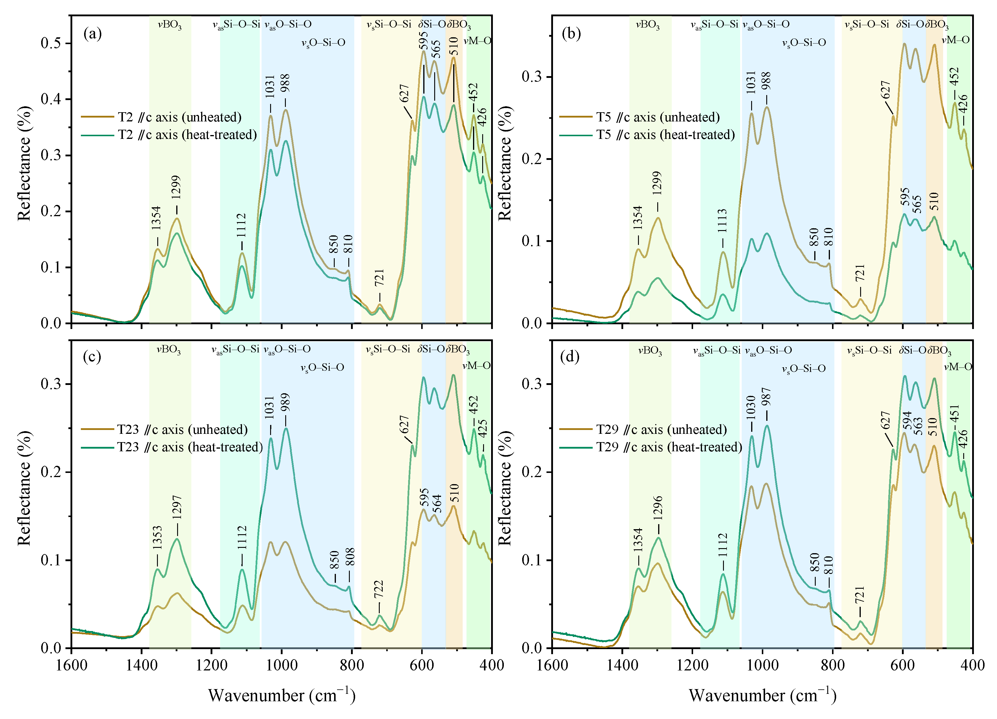
Spectra of unheated and heat-treated samples were compared (Figure 2, Figure 3 and Figure 4), with band assignments summarized in Table 5 and Table 6.

3.4.1. Reflective MIR Spectra (1600–400 cm−1)

Unheated samples showed two distinct spectral regions: 1400–800 cm−1 and 700–400 cm−1. In sections parallel to the c-axis (parallel to the optical axis), bands appeared at 1354, 1299, 1112, 1031, 988, 810, and 721 cm−1 and 627, 595, 565, 510, 452, and 426 cm−1 (Figure 2a–d). In sections perpendicular to the c-axis, bands included 1353, 1298, 1110, 1053, 1031, 992, 791, 753, and 716 cm−1, and 631, 596, 508, 453, and 429 cm−1 (Figure 3a–d). While band positions were similar, spectral morphologies differed: sections parallel to the c-axis had unique 810 and 565 cm−1 bands, while perpendicular sections had unique 791 and 753 cm−1 bands. The sharp, weak bands are observed in each spectrum, and these bands’ displacement show no change after being heat-treated. Sharp, weak bands showed no displacement post-heating, but reflectivity increased in both directions (Figure 2).
The mid-infrared spectra of tourmaline are dominated by fundamental vibrations of infrared-active structural units: YO6 octahedra, TO4 tetrahedra, BO3 triangles, hydroxyl groups, and water molecules.
The double bands at ~1353 cm−1 and 1255 cm−1 in schorl are attributed to antisymmetric BO3 stretching [22]. Some scholars classify bands near 1300 cm−1 as νas BO3 [20] or νs BO3 [21]. Thus, the 1354 and 1299 cm−1 bands here are assigned to BO3-related vibrations. The 510 cm−1 (parallel to c-axis) and 508 cm−1 (perpendicular to c-axis) bands are also BO3-related, attributed to deformation vibrations [21].
Infrared vibrational modes of [SiO4] tetrahedra ([TO4]) include νs Si–O–Si (symmetric stretching), νas Si–O–Si (asymmetric stretching), νs O–Si–O, νas O–Si–O, and δ Si–O (bending) [20,21]. Observed bands (Figure 3) are assigned as follows: 1112 cm−1 corresponds to the νas Si–O–Si mode; bands at 1031 cm−1 and 988 cm−1 are attributed to νas O–Si–O modes; features at 850 cm−1 and 810 cm−1 arise from νs O–Si–O modes; bands at 721 cm−1 and 627 cm−1 are assigned to the νs Si–O–Si mode; and absorptions at 595 cm−1 and 565 cm−1 originate from δ Si–O bending vibrations.
Absorptions at ~400 cm−1 relate to metal cations in YO6 octahedra (e.g., MgO6 at 470 cm−1, FeO6 at 400 cm−1). Bands at 449 cm−1 and 428 cm−1 in Fe-bearing elbaite are assigned to [MgO6] and [FeO6] [21], while 457 cm−1 and 431 cm−1 in elbaites correspond to ν Mg–O and ν Fe–O [20]. This study’s samples had weak bands at 452 cm−1 and 426 cm−1 (parallel to c-axis) and 453 cm−1 and 429 cm−1 (perpendicular to c-axis; Figure 2a), with minor band shifts across samples (Figure 2 and Figure 3). Given the samples’ Fe content (and low Mg), these bands may relate to Fe or Mn.

3.4.2. Transmission NIR Spectra (9000–4000 cm−1)

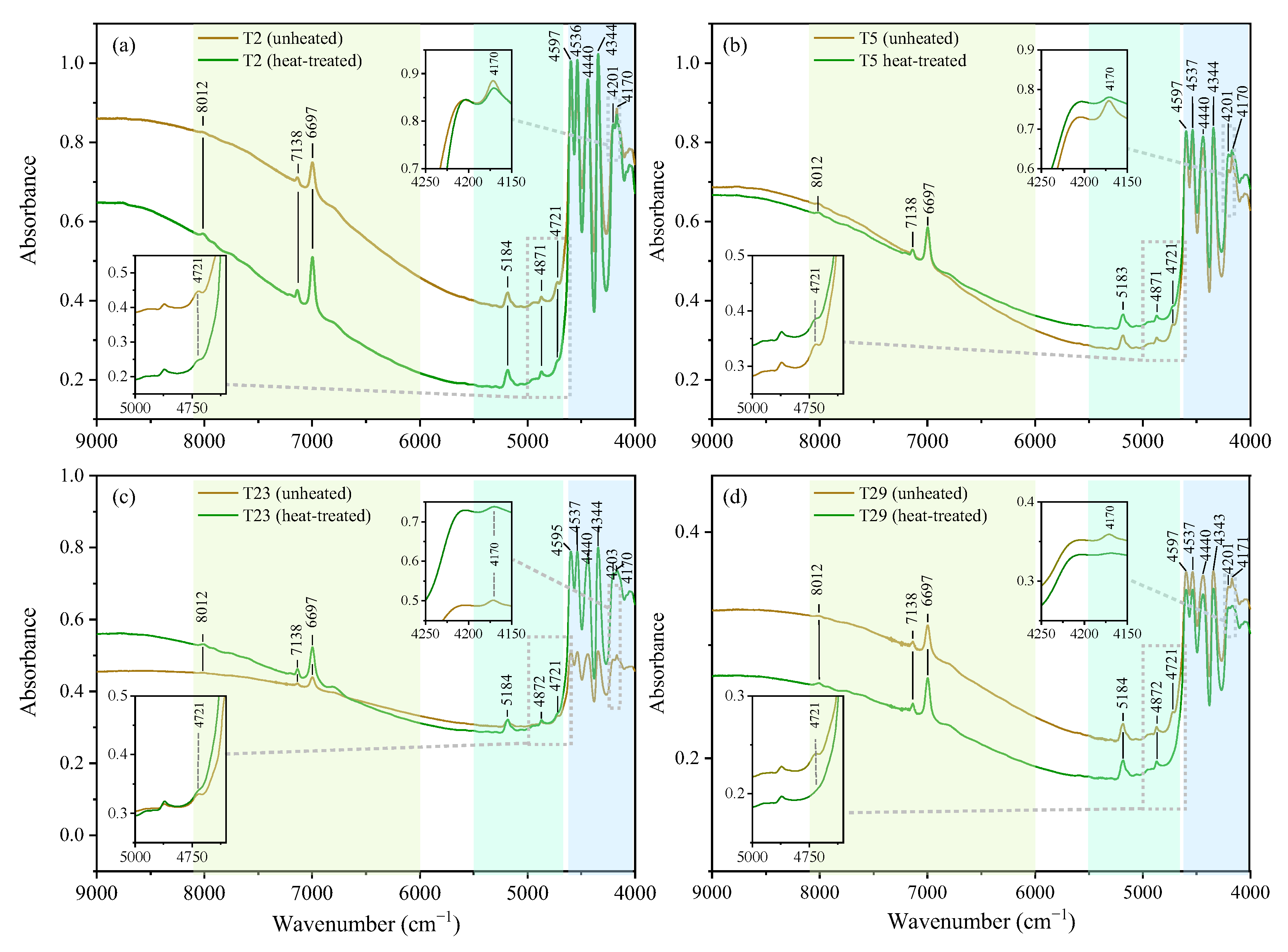
Bands in 8000–4000 cm−1 were divided into three groups: 8000–6000 cm–1, 5500–4700 cm–1, and 4600–4000 cm–1 (Figure 4). Unheated sample T2 showed third-group bands at 4597, 4536, 4440, 4344, 4201, and 4170 cm−1; first-group bands at 7138 and 6997 cm−1; and second-group bands at 5184, 4871, and 4721 cm−1 (Figure 4a). T5, T23, and T29 showed similar bands (Figure 4b–d).
Band positions were unchanged post-heating, but third-group absorption intensity increased in heat-treated samples (except T29; Figure 4). First- and second-group absorbance decreased in T2 and T29 but increased in T5 and T23. Notably, the 4721 cm−1 band weakened significantly (tending to disappear; Figure 4) and vanished entirely at >600 °C [23]. First-group bands (8012, 7138 and 6997 cm−1) enhanced post-treatment, with the 6997 cm−1 band showing the greatest increase. These phenomena may relate to compositional changes along the c-axis or varying thickness. Alternatively, intensity changes in 4600–4300 cm−1 post-heating/irradiation may reflect distances between metal ions and hydroxyl groups [13], as heat treatment alters cation valence states and ionic radii.
Bands in 8000–6000 cm–1 are attributed to the first overtones of hydroxyl stretching modes (3600–3460 cm–1) [4,24,25,26,27]. Specifically, 7000 and 6500 cm–1 correspond to the first overtones of OH3 groups at 3600 and 3460 cm–1 [4]. Others attribute 7138 and 6995 cm–1 to the first overtones of OH vibrations in complex tripartite rings [20,27], with strong 6995 cm−1 bands from pure hydroxyls and weak 7132 and 7185 cm−1 bands from water-bound hydroxyls [20]. Thus, the bands at 7138 and 6997 cm–1 are assigned to OH overtones, and 8012 cm–1 to Fe2+ 5T2g5Eg transitions [4].
Second-group bands (5500–4700 cm−1) included 5182, 4871, and 4721 cm−1 (Figure 4). The 4871 cm−1 band is the third overtone of νas Si–O–Si [20,27]. Bands in 5400–4900 cm−1 reflect combinations of water bending and stretching vibrations [27], with 5182 cm−1 indicating water content. The disappearance of 4721 cm−1 post-heating suggests that it relates to transition metal ions (e.g., Fe2+/3+) whose valence states changed during treatment.
Bands in 4800–4000 cm−1 arise from combinations of hydroxyl stretching and bending modes in the YM–OH unit (M = Al, Mg, Fe, Mn, etc.) [4]. Despite minor species-specific differences (due to Y-site cation variation), characteristic 4700–4100 cm−1 bands in tourmalines (4600, 4540, 4440, 4340, 4170 cm−1) are assigned to cationic hydroxyl unit vibrations (M–OH; M = Al, Mg, Fe, Mn, Li) [2,4,13,27]. YM–OH1 bands are influenced by Y-site cations; some attribute them to YAl–OH1 [24] or Al–OH combinations (~4600 cm−1 [4]). Bands in 4500–4300 cm−1 correspond to Fe-OH and Mg-OH, with 4540 cm−1 corresponding to the YFe-OH, 4440 cm−1 to the Y(Fe, Mg)-OH, and 4344 cm−1 to the YMg-OH. Two bands in the range of 4300–4100 cm−1 are assigned to the combination of stretching and bending modes of YM–OH3 (M = Al, Li, Fe, Mn) [24] or (Fe2+, Mn)-OH [20]. Given Y-site occupancy by Mn, Fe, Al, and Li, the bands at 4204 and 4171 cm−1 are assigned to Y (Mn, Fe, Al, Li)–OH3.
Table 6. Assignments of NIR bands (9000–4000 cm−1) in various tourmalines.
This StudyGreen Elbaite [20]Green
Elbaite [4]
Pink Tourmaline
[24]
Green Tourmaline [24]Brownish-Green [24]Assignment
UnheatedHeat-Treated
80128012 ↑ 8000 First overtones of hydroxyl stretching modes
7185
71387138 ↑71327140
69976997 ↑69957000
67836795
518351835180 The bending vibration of the water and the stretching vibration
487148714873 νas Si–O–Si
47214721 ↓
4597459745984597460446044596Combination of stretching and bending modes of M-OH unitsYM-OH1
4536453645444538454145454541YM-OH1
4440444044444433444444484444YM-OH1
4344434443444347434743474344ZM-OH3
420142014206 42114214YM-OH3
41704170↓41644171414641774175YM-OH3
Note: “↑”—increase in intensity; “↓”—decrease in intensity; vas—asymmetric stretching;YM and ZM —the corner markers Y and Z represent Y and Z sites, respectively, and M represents the metal cations.

3.5. Raman Spectroscopy

Laser Raman spectra were consistent across samples (Figure 5a–d). For T2, sections perpendicular to the c-axis showed no significant pre- vs. post-heating differences, with bands at 223, 242, 376, 408, 730, 756, 837, 1063, 1395, 3498, 3564, 3593, and 3653 cm−1(Figure 5a). Four main Raman band groups (centered at 220, 370, 730, and 1060 cm−1 in 1000–1200 cm−1) are assigned in Table 7. Band intensity and shifts were consistent, aligning with chemical composition and optical parameters.
Prior research links specific Raman shifts to tourmaline structural vibrations, with diagnostic bands for classification. The band at 223 cm−1 is assigned to δ(Si-O-Si) bending in SiO6 octahedra [7,26], a universal feature in all tourmalines. The band at 376 cm−1 corresponds to ν(Z-O) stretching in Al3+-dominated ZO6 octahedra and the sharp band with marked intensity at 376 cm−1 in both the spectra of the samples implies strong bonding of Al–O [4]. The band at 408 cm−1 arises from ν(O-Si-O) bending in SiO4 tetrahedra, characteristic of Fe-rich tourmaline and potentially linked to Fe3+ substitution at Y sites [28]. Bands at 636, 708, and 730 cm−1 are attributed to νs(B-O) stretching in BO3 triangles, indicative of Li-bearing tourmaline, while 756 cm−1 originates from ν(O-B-O) asymmetric stretching in BO3 groups. The region 837–1087 cm−1 results from ν(Si-O) stretching in SiO4 tetrahedra, with band shifts attributed to variations in Si-O bond lengths [26].
The bands ranging from 3400 to 3700 cm−1 are related to OH vibrations (Figure 5). In liddicoatite (calcic-lithium tourmaline), 3640 cm−1 corresponds to Y-site Al/Li occupancy; in Fe-substituted elbaite, this shifts to 3655 cm−1 [7]. In the paper, the observed band at 3652–3653 cm−1 aligns with elbaite (3655 cm−1), suggesting YAlYAlY(Li, Mn, Fe)-OH1 or YAlY(Li, Mn, Fe)Y(Li, Mn, Fe)-OH1configurations. For liddicoatite, 3597 and 3490 cm−1 reflect Y-site differences between Al and Li, assigned to ZAlZAlYAl-OH3 or ZAlZAlYLi-OH3; elbaite shows 3585 and 3500 cm−1 bands [7]. This study’s 3594 cm−1 band is intermediate between liddicoatite (3597 cm−1) and lithium tourmaline (3585 cm−1). The 3489 cm−1 band (parallel to c-axis) aligns with liddicoatite (3490 cm−1), while 3498 cm−1 (perpendicular to c-axis) matches elbaite (3500 cm−1), reflecting heterogeneous Y-site cation occupancy (Al, Fe, Li, Mn) in ZAlZAlY(Li, Al, Mn, Fe)-OH3. The 3560 cm−1 shoulder (diagnostic of X-site Na+; [7]) confirms classification as sodium-dominant elbaite.

3.6. UV-Vis Spectroscopy

UV-Vis absorption intensity showed polarization dependence: bands parallel to the c-axis were stronger and more distinct than those perpendicular to the c-axis (Figure 6). Unheated samples exhibited three visible-region bands (415, 540, and 730 nm), causing the brown color, plus two ultraviolet bands (379 and 395 nm) with no color impact. Heat-treated samples showed reduced overall absorption, with the 540 nm band absent. A green transmission window formed in the visible region, shifting color from brown to green (Figure 1).
Unheated brown samples (parallel to c-axis) had 730, 540, and 415 nm bands. Post-treatment, the 540 nm band vanished, while 730 and 415 nm bands persisted. Spectra perpendicular to the c-axis remained nearly unchanged, retaining the brown color. Thus, heat-treated samples showed strong pleochroism parallel to the c-axis.

4. Discussion

4.1. Tourmaline Species of Samples

Mostly brown tourmalines are primarily dravite and some are uvite, ferromanganese elbaite [12,29,30]. The composition of tourmaline measured in the paper is ferromanganese elbaite and enriched Mn- (MnO: 2.14–2.62 wt.%), Fe- (FeO: 1.03–1.46 wt.%), and Ti-bearing (TiO2: 0.02–0.09 wt.%), which lacks V, Cr, Cu, and Ni, and is likely colored by Fe, Mn, Ti, etc. [14]. The molecular formula calculation shows that the sum of Y-site cations is less than 3, and the number of silicon atoms is greater than 6, which means the existence of some trivalent iron. Fe3+ occupies the Z-site, resulting in the overall small Y-site, which also confirms the existence of Fe3+. Raman spectra show 3653 and 3585 cm−1 bands (elbaite markers) and a Li-related 730 cm−1 band [4,7]. The absence of the 1131 cm−1 band (exclusive to Fe-uvite and dravite; [7]) confirms the classification as Fe- and Mn-bearing elbaite–fluorelbaite, consistent with compositional analyses.

4.2. Spectral Response to 500 °C Heating and Structural Stability

Tourmaline’s complex composition leads to varied vibrational band displacements. IR spectra show subtle pre- vs. post-heating differences (Figure 2 and Figure 3), making distinction challenging. However, post-treatment, the 4721 cm−1 band vanished (Figure 4) and the near-infrared absorption at 976 nm weakened (Figure 6). Raman spectra perpendicular to the c-axis were unchanged post-heating, confirming structural integrity. Invariant OH-stretching bands (3498, 3593, and 3653 cm−1) indicate unaltered cation occupancy at crystallographic sites.

4.3. Color Origin and Color Change Mechanism Induced by Heat Treatment

Brown arises from absorption of blue-green and some red light, with yellow-orange light transmitted. These unheated samples have 415, 540, and 730 nm absorption bands, which are the main reasons for the brown color of the samples (Figure 6). After heat treatment, the 540 nm absorption band of the parallel c-axis section of the samples disappears, forming a transmission window near 540 nm, thus showing yellow-green. However, the absorption spectrum characteristics in the perpendicular c-axis section are basically unchanged, and the color is still brown. The sample shows yellow-green and brown dichroism (Figure 1b).
Based on previous studies and the characteristics of the samples in this paper, it is believed that the absorption bands at 730, 540, and 415 nm are caused by Fe2+ dd transition (5T2g5Eg) [21,31,32,33,34,35], Fe2+→Fe3+ intervalence charge transfer (IVCT) [32,36,37], and Fe2+→Ti4+ (IVCT) [32,35,36,37,38], respectively. And for the Mn2+ spin-forbidden transition 6A1g4A1g, 4TEg generated the 412/414 nm absorption band [38], superimposed onto the band 415 nm. Furthermore, the comprehensive review by Vigier et al. (2025) [39] emphasizes that Fe2+→Ti4+ (IVCT) is associated with a strong, broad absorption band in the visible range and is a common cause of brown coloration in minerals and glasses. This strongly supports our assignment of the 415 nm band to Fe2+→Ti4+ (IVCT), which is a significant contributor to the brown color of the untreated samples in this study, alongside the Fe2+→Fe3+ (IVCT) band at 540 nm.
Combined with previous studies, the blue tourmaline has 540 nm and 730 nm absorption bands [13,32,35,40,41]. The researchers found that the blue tourmaline generally does not contain Ti (<0.01% or below the detection limit). This means that the 540 nm absorption band has nothing to do with Ti. When the researchers heat-treated the blue tourmaline in the air, the absorption band at 540 nm was significantly enhanced (No.11 [40]; TB1-1, Faye et al., 1974 [32]), and the Fe2+/Fe3+ ratio changed from 24:1 to 2:1 (TB1-1). A reasonable explanation is that part of the Fe2+ was oxidized to the Fe3+ by heat treatment process, and the 540 nm absorption band was strengthened with the increase in Fe3+. Similarly, pink and yellow tourmaline are iron-free, or rather, below the detection limit [4,38,42]. The band of 520 nm in the pink tourmaline is related to the Mn, and the tail absorption 320 nm trending to ultraviolet region in the yellow tourmaline is due to Mn2+→Ti4+ intervalence charge transfer (IVCT) [43]. Therefore, the 520 and 320 nm bands are unrelated to Fe. Moreover, the iron-related absorption bands are missing in pink and yellow tourmalines, including the 540 nm band. In other words, the band at 540 nm is related to Fe.
The green tourmaline heat-treated in the air produced the 540 nm absorption band and the samples turned brown (No.3 [40]; TGr-2 [32]). The iron of the green tourmaline is dominated by Fe2+ [32,35]. This means that the Fe2+ cannot generate the absorption band alone. In general, Fe2+ can be oxidized to Fe3+ when the tourmaline is heat-treated in air (oxidizing atmosphere). In other words, the appearance of the 540 nm band is coupled with the appearance of Fe3+. Similarly, the green tourmaline produces a band that is centered at 550 nm after irradiation, which is caused by the Fe2+→Fe3+ electronic transition due to the oxidation of Fe2+ to Fe3+ by irradiation [13]. Additionally, the intensity of the infrared band at 4170 cm−1, attributed to Fe3+, significantly decreases after heat treatment (Figure 6), indicating a reduction in its concentration. Therefore, we believe that the 540 nm absorption band is caused by Fe2+→Fe3+ (IVCT).
The brown samples in this study turned green after heat treatment in the reducing atmosphere, and the absorption band at 540 nm disappeared. This is the reverse process of the above-mentioned irradiation or heat treatment in oxidizing atmosphere, that is, the reduction of Fe3+ to Fe2+ leads to the disappearance of the Fe2+→Fe3+ electronic transition that can produce the 540 nm band. In addition, the samples in the paper have high Mn contents (17,346.71–20,669.52 μg/g), and the absorption near 415 nm may have a 412/414 nm superposition caused by the Mn2+ dd (6A1g4A1g, 4TEg) spin-forbidden transition [38]. Heat treatment reduces part of Fe3+ to Fe2+ and Ti4+ to Ti3+, causing Fe2+→Ti4+ to decrease and the absorption at 415 nm to weaken. However, some of Mn3+ was reduced to Mn2+, and the Mn2+ that leads to an increase in the absorption band at 412/414 nm increases, so the absorption band intensity around 415 nm has changes insignificantly. The 520 nm absorption related to Mn3+ [13,25] also disappeared at the same time. The existence of the 520 nm absorption band may also be the reason for the asymmetrical absorption band of the 540 nm absorption band in the unheated samples.

5. Conclusions

The brown tourmaline belongs to Fe- and Mn-bearing elbaite–fluorelbaite, at 500 °C under neutral-reducing conditions, where its brown hue fades and transparency improves. The weakening or disappearance of IR bands at 4721 cm–1 serve as diagnostic indicators of heat treatment.
The UV-Vis spectra of the natural brown samples (parallel to c-axis) show 730, 540, and 415 nm bands, attributed to Fe2+ dd transitions (5T2g5Eg), Fe2+→Fe3+ IVCT, and Fe2+→Ti4+ IVCT, respectively. The 415 nm band may also include Mn2+ d–d transitions (6A1g4A1g, 4TEg) at 412/414 nm.
The reduction heat treatment leads to the transformation of Fe3+ to Fe2+, resulting in the disappearance of the 540 nm absorption band caused by Fe2+→Fe3+ IVCT in the parallel c-axis direction, forming a green light transmission window, which is the reason for the green color. The parallel c-axis section of the samples shows brown and yellow-green dichroism after heat treatment.

Author Contributions

Writing—original draft, K.L.; writing—review and editing, S.Y.; data analysis, K.L. and S.Y.; review comments modification—K.L. and S.Y. All authors have read and agreed to the published version of the manuscript.

Funding

This study is funded by the Guangdong Provincial Education Science Planning Project (2023GXJK846), Innovation team of Guangzhou Education Bureau Project—Gems and Jewelry Materials Innovation Team (202235328), and Guangzhou Polytechnic University Major Scientific and Technological Project (2021KJ03).

Data Availability Statement

Data are available from the authors upon reasonable request.

Conflicts of Interest

The authors declare no conflicts of interest.

References

  1. Hawthorne, F.C.; Henry, D.J. Classification of the Minerals of the Tourmaline Group. Eur. J. Mineral. 1999, 11, 201–216. [Google Scholar] [CrossRef]
  2. Prasad, P.S.R. Study of Structural Disorder in Natural Tourmalines by Infrared Spectroscopy. Gondwana Res. 2005, 8, 265–270. [Google Scholar] [CrossRef]
  3. Castaneda, C.; Eeckhout, S.G.; Da Costa, G.M.; Botelho, N.F.; De Grave, E. Effect of Heat Treatment on Tourmaline from Brazil. Phys. Chem. Min. 2006, 33, 207–216. [Google Scholar] [CrossRef]
  4. Reddy, B.J.; Frost, R.L.; Martens, W.N.; Wain, D.L.; Kloprogge, J.T. Spectroscopic Characterization of Mn-Rich Tourmalines. Vib. Spectrosc. 2007, 44, 42–49. [Google Scholar] [CrossRef]
  5. Henry, D.J.; Dutrow, B.L. Tourmaline Studies through Time: Contributions to Scientific Advancements. J. Geosci. 2018, 63, 77–98. [Google Scholar] [CrossRef]
  6. Pasetti, L.; Fornasini, L.; Mantovani, L.; Andò, S.; Raneri, S.; Palleschi, V.; Bersani, D. Study of Mg–Fe Content in Tourmalines from the Dravite–Schorl Series by Raman Spectroscopy. J. Raman Spectrosc. 2024, 55, 276–286. [Google Scholar] [CrossRef]
  7. Hoang, L.H.; Hien, N.T.M.; Chen, X.B.; Van Minh, N.; Yang, I. Raman Spectroscopic Study of Various Types of Tourmalines. J. Raman Spectrosc. 2011, 42, 1442–1446. [Google Scholar] [CrossRef]
  8. Yan, X.X.; Wang, P.L.; Yue, S.W. Spectroscopic Characteristics and Coloring Mechanism of Greenish-Yellow Beryl Under Heating Treatment. Spectrosc. Spectr. Anal. 2020, 40, 3795–3800, (In Chinese with English Abstract). [Google Scholar]
  9. Wu, W.Y.; Yue, S.W. Influence of Heat Treatment on Color of Amethyst and Its Spectral Characteristic. J. Gems Gemol. 2016, 18, 47–55, (In Chinese with English Abstract). [Google Scholar]
  10. Wang, P.L.; Yue, S.W.; Li, J.Y. Spectral Characteristics and Color Mechanism of Heat-Treated Gem-Quality Yellow Sphene. Spectrosc. Spectr. Anal. 2024, 44, 2545–2550, (In Chinese with English Abstract). [Google Scholar]
  11. Yue, S.W.; Yan, X.X.; Chen, S.M.; Luo, J. The Heat Treatment Process and Color Mechanism of Green Fluor-Hydroxyapatite. Spectrosc. Spectr. Anal. 2025, 45, 1961–1967, (In Chinese with English Abstract). [Google Scholar]
  12. Pezzotta, F.; Laurs, B.M. Tourmaline: The Kaleidoscopic Gemstone. Elements 2011, 7, 333–338. [Google Scholar] [CrossRef]
  13. Ahn, Y.K.; Seo, J.Y.; Park, J.W. Electronic and Vibrational Spectra of Tourmaline-The Impact of Electron Beam Irradiation and Heat Treatment. Vib. Spectrosc. 2013, 65, 165–175. [Google Scholar] [CrossRef]
  14. Maneewong, A.; Seong, B.S.; Shin, E.J.; Kim, J.S.; Kajornrith, V. Color Change of Tourmaline by Heat Treatment and Electron Beam Irradiation: UV-Visible, EPR, and Mid-IR Spectroscopic Analyses. J. Korean Phys. Soc. 2016, 68, 83–92. [Google Scholar]
  15. Thongnopkun, P.; Bamrungpol, J. Color Change of Green Tourmaline from Madagascar by Heat Treatment. Burapha Sci. J. 2017, 22, 299–308. [Google Scholar]
  16. Thongnopkun, P.; Naowabut, P. Effect of Heat Treatment on Madagascar Dravite Tourmaline: UV-Visible and Diffuse Reflectance Infrared Spectroscopic Characterization. J. Appl. Spectrosc. 2018, 85, 616–623. [Google Scholar] [CrossRef]
  17. Abduriyim, A.; Kitawaki, H.; Furuya, M.; Schwarz, D. “Paraíba”-Type Copper-Bearing Tourmaline from Brazil, Nigeria, and Mozambique: Chemical Fingerprinting By LA-ICP-MS. Gems Gemol. 2006, 42, 4–21. [Google Scholar]
  18. Laurs, B.M.; Zwaan, J.C.; Breeding, C.M.; Simmons, W.B.; Beaton, D.; Rijsdijk, K.F.; Befi, R.; Falster, A.U. Copper-Bearing (Paraíba-Type) Tourmaline from Mozambique. Gems Gemol. 2008, 44, 294–320. [Google Scholar] [CrossRef]
  19. Henry, D.J.; Novák, M.; Hawthorne, F.C.; Ertl, A.; Dutrow, B.L.; Uher, P.; Pezzotta, F. Nomenclature of the Tourmaline-Supergroup Minerals. Am. Mineral. 2011, 96, 895–913. [Google Scholar] [CrossRef]
  20. Chen, Y.F.; Xu, D.; Zhou, Z.Y.; Schwarz, D.; Zheng, J.H.; Zhang, L.M. Chemical Composition and Spectral Variation in Gem-Quality Blue Iron-Bearing Tourmaline from Brazil. Crystals 2024, 14, 877. [Google Scholar]
  21. Cui, L.Y.; Guo, Y.; Tang, J.; Yang, Y.S. Spectroscopy Characteristics and Color-Influencing Factors of Green Iron-Bearing Elbaite. Crystals 2023, 13, 1461. [Google Scholar]
  22. Makreski, P.; Jovanovski, G. Minerals from Macedonia: XXIII. Spectroscopic and Structural Characterization of Schorl and Beryl Cyclosilicates. Spectrochim. Acta A Mol. Biomol. Spectrosc. 2009, 73, 460–467. [Google Scholar] [CrossRef] [PubMed]
  23. Yue, S.W.; Yan, X.X.; Lin, J.Q.; Wang, P.L.; Liu, J.F. Spectroscopic Characteristics and Coloring Mechanism of Brown Tourmaline under Heating Treatment. Spectrosc. Spectr. Anal. 2021, 41, 2524–2529, (In Chinese with English Abstract). [Google Scholar]
  24. Liu, X.L.; Feng, X.Q.; Fan, J.L.; Guo, S.G. Optical Absorption Spectra of Tourmaline Crystals from Altay, China. Chin. Opt. Lett. 2011, 9, 083001–083014. [Google Scholar] [CrossRef]
  25. Phichaikamjornwut, B.; Pongkrapan, S.; Intarasiri, S.; Bootkul, D. Conclusive Comparison of Gamma Irradiation and Heat Treatment for Color Enhancement of Rubellite from Mozambique. Vib. Spectrosc. 2019, 103, 102926. [Google Scholar] [CrossRef]
  26. Bosi, F.; Celata, B.; Skogby, H.; Hålenius, U.; Tempesta, G.; Ciriotti, M.E.; Bittarello, E.; Marengo, A. Mn-Bearing Purplish-Red Tourmaline from the Anjanabonoina Pegmatite, Madagascar. Miner. Mag 2021, 85, 242–253. [Google Scholar] [CrossRef]
  27. Li, X.J.; Zu, E.D. Near-Infrared Spectrum Analysis of Cyclosilicates Gem Minerals. Bull. Chin. Ceram. Soc. 2016, 35, 1318–1321. [Google Scholar]
  28. Ertl, A.; Kolitsch, U.; Dyar, M.D.; Hughes, J.M.; Rossman, G.R.; Pieczka, A.; Henry, D.J.; Pezzotta, F.; Prowatke, S.; Lengauer, C.L. Limitations of Fe2+ and Mn2+ Site Occupancy in Tourmaline: Evidence from Fe2+- and Mn2+-Rich Tourmaline. Am. Mineral. 2012, 97, 1402–1416. [Google Scholar] [CrossRef]
  29. Robinson, G.W.; Chamberlain, S.C.; Walter, M. The History and Mineralogy of the Classic Brown Tourmaline Locality, Gouverneur, New York. Rocks Miner. 2016, 91, 520–529. [Google Scholar] [CrossRef]
  30. Fonseca-Zang, W.A.d.; Zang, J.W.; Hofmeister, W. The Ti-Influence on the Tourmaline Color. J. Braz. Chem. Soc. 2008, 19, 1186–1192. [Google Scholar] [CrossRef]
  31. Manning, P.G. An Optical Absorption Study of the Origin of Colour and Pleochroism in Pink and Brown Tourmalines. Can. Miner. 1969, 9, 678–690. [Google Scholar]
  32. Faye, G.H.; Manning, P.G.; Gosselin, J.R.; Tremblay, R.J. The Optical Absorption Spectra of Tourmaline; Importance of Charge-Transfer Processes. Can. Miner. 1974, 12, 370–380. [Google Scholar]
  33. Burns, R.G.; Simon, H.F. Cation Disorder in Tourmalines. Geol. Soc. Am. Ann. Meet. Dallas Abstr. 1973, 5, 563–564. [Google Scholar]
  34. de Camargo, M.B.; Isotani, S. Optical Absorption Spectroscopy of Natural and Irradiated Pink Tourmaline. Am. Mineral. 1988, 73, 172–180. [Google Scholar]
  35. da Silva, S.F.; Moura, M.A.; Queiroz, H.d.A.; Ardisson, J.D. Chemical and Spectroscopic Characterization of Tourmalines from the Mata Azul Pegmatitic Field, Central Brazil. J. Geosci. 2018, 63, 155–165. [Google Scholar] [CrossRef]
  36. Smith, G. Evidence for Absorption by Exchange-Coupled Fe2+-Fe3+ Pairs in the near Infra-Red Spectra of Minerals. Phys. Chem. Min. 1978, 3, 375–383. [Google Scholar] [CrossRef]
  37. Smith, G. A Reassessment of the Role of Iron in the 5,000-30,000 Cm-1 Region of the Electronic Absorption Spectra of Tourmaline. Phys. Chem. Min. 1978, 3, 343–373. [Google Scholar] [CrossRef]
  38. Rossman, G.R.; Mattson, S.M. Yellow, Mn-Rich Elbaite with Mn-Ti Intervalence Charge Transfer. Am. Mineral. 1986, 71, 599–602. [Google Scholar]
  39. Vigier, M.; Evans, H.; Rossman, G.R.; Jobic, S.; Fritsch, E. Fe-Ti vs. Fe-Fe Charge Transfers: A Comprehensive Review and Its Applications in Minerals and Glasses. Am. Mineral. 2025, 110. [Google Scholar] [CrossRef]
  40. Wilkins, R.W.T.; Farrell, E.F.; Naiman, C.S. The Crystal Field Spectra and Dichroism of Tourmaline. J. Phys. Chem. Solids 1969, 30, 43–56. [Google Scholar] [CrossRef]
  41. Kaewtip, M.; Limtrakun, P. Gemological and Chemical Characteristics of Green Tourmaline from Madagascar, Mozambique, and Tanzania. Walailak J Sci Technol 2016, 13, 985–992. [Google Scholar]
  42. Laurs, B.M.; Simmons, W.B.; Rossman, G.R.; Fritz, E.A.; Koivula, J.I.; Anckar, B.; Falster, A.U. Yellow Mn-Rich Tourmaline from the Canary Mining Area, Zambia. Gems Gemol. 2007, 43, 314–331. [Google Scholar] [CrossRef]
  43. Fritz, E.A.; Breeding, C.M.; Rossman, G.R.; Laurs, B.M.; Simmons, W.B.; Falster, A.U. Yellow-Green Clinohumite and Yellow Chondrodite from Tanzania. Gems Gemol. 2007, 43, 377–379. [Google Scholar] [CrossRef]
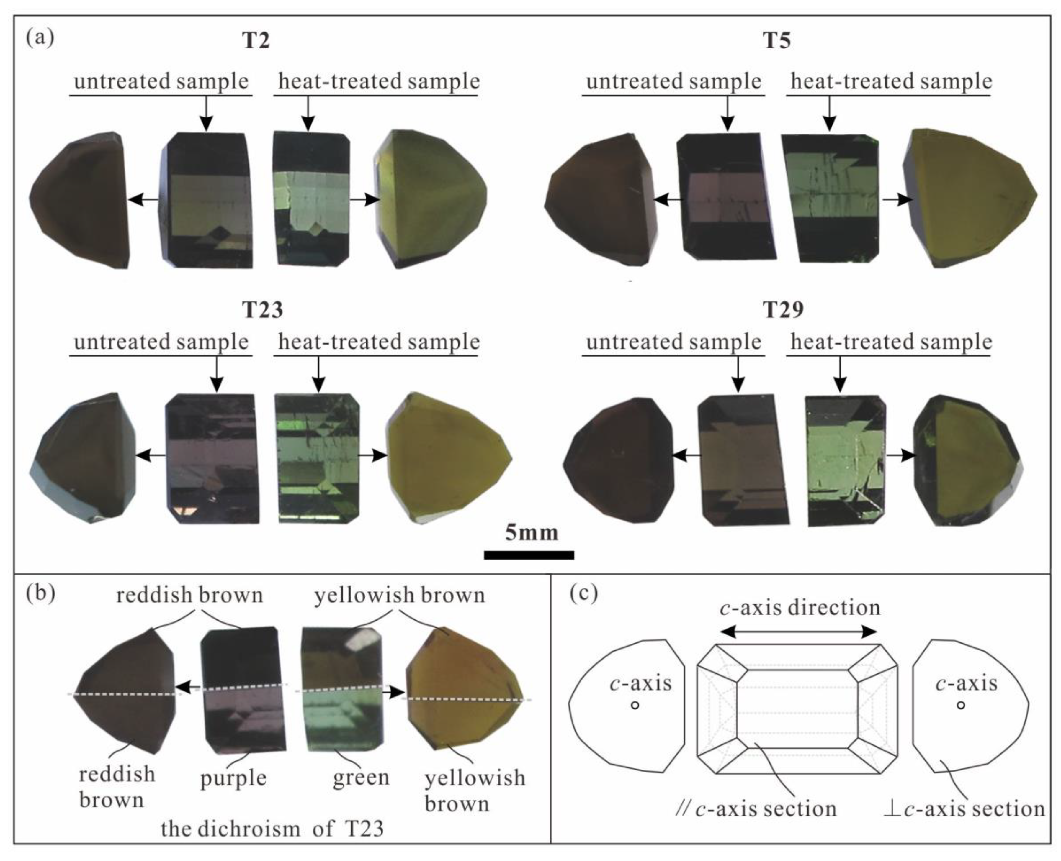
Figure 1. Characteristics of samples before and after heat treatment. (a) Unheated and heat-treated samples; (b) samples generally have obvious dichroism, and sample T23 is taken as example to show its dichroism characteristics: yellowish brown and green in section parallel to c-axis, and yellowish brown in section perpendicular to c-axis (gray dashed line = boundary in dichroscope). (c) Schematic of c-axis direction in samples.
Minerals 15 01032 g001
Figure 2. Characteristics of MIR spectra (1600–400 cm−1) of unheated and heat-treated tourmaline parallel c-axis sections (v—stretching vibrations; vs—symmetric stretching vibrations; vas—asymmetric stretching vibrations; δ—deformation vibration). (a) T2; (b) T5; (c) T23; (d) T29.
Minerals 15 01032 g002
Figure 3. MIR spectra (1600–400 cm−1) of unheated and heat-treated elbaite (sections perpendicular to c-axis) (v—stretching vibrations; vs—symmetric stretching vibrations; vas—asymmetric stretching vibrations; δ—deformation vibration). (a) T2; (b) T5; (c) T23; (d) T29.
Minerals 15 01032 g003
Figure 4. NIR spectra (9000–4000 cm−1) of tourmaline samples before and after heating. (a) T2; (b) T5; (c) T23; (d) T29.
Minerals 15 01032 g004
Figure 5. Comparison of Laser Raman spectra between unheated and heat-treated tourmaline samples. (a) T2; (b) T5; (c) T23; (d) T29.
Minerals 15 01032 g005
Figure 6. Comparison of UV-Vis spectra between unheated and heat-treated elbaite samples. (a) T2; (b) T5; (c) T23; (d) T29. The green rectangular area represents the green light transmission window.
Minerals 15 01032 g006
Table 1. Experimental protocol for heat treatment.
Sample No.Temperature (°C)Heating RateHolding Time (h)Atmosphere
°C/min
T250010.5reduction
T550030.5reduction
T2350041reduction
T2950020.5reduction
Table 2. The gemological characteristics of the brown elbaite.
Sample No.Weight (g)TransparencyRefractive IndexBirefringenceUV FluorescenceColorDichroism
NoNeLongShortUnheatedHeat-TreatedUnheatedHeat-Treated
T20.511transparent1.6401.6200.020inertinertyellowish brownyellowish greenreddish brown + yellowish brownyellowish brown + green
T50.470transparent1.6401.6200.020inertinertreddish brownyellowish greenBrown + light greenyellowish brown + green
T230.442transparent1.6401.6200.020inertinertreddish brownyellowish greenpurple + reddish brownyellowish brown + green
T290.407transparent1.6401.6200.020inertinertyellowish brownyellowish greenpink green + brownyellowish brown + green
Table 3. EMPA results for major elements and molecular formula calculation (wt.%).
Sample No.T2T5T23T29
12121212
SiO238.9438.7938.7638.8238.5938.6738.7938.63
TiO20.090.070.060.060.020.040.050.06
Al2O338.5738.7238.7938.8538.6038.5838.4738.69
FeO1.411.461.151.131.031.041.101.08
CaO0.400.420.460.430.530.500.500.50
MnO2.192.142.182.292.622.522.432.43
ZnO0.110.070.070.070.000.040.040.03
Na2O2.322.262.112.182.102.182.122.05
F1.040.961.020.940.101.030.100.94
H2O *3.363.403.373.413.373.363.373.40
B2O3 *11.6911.7111.7411.7411.7711.7711.6811.68
Li2O *2.042.042.072.072.072.072.072.07
Total102.18102.05101.79102.01101.72101.80101.63101.57
O=F0.440.410.430.400.420.430.420.39
Total *101.74101.64100.37101.61101.30101.37101.21101.17
T: Si6.066.046.046.046.026.036.066.03
B3.003.003.003.003.003.003.003.00
Z: Al6.006.006.006.006.006.006.006.00
Y: Al1.071.101.121.121.101.091.081.12
Ti0.010.010.010.010.000.000.010.01
Mn0.290.280.290.300.350.330.320.32
Fe2+0.180.190.150.150.130.140.140.14
Zn0.010.010.010.010.000.000.000.00
Li1.281.281.301.291.301.301.301.30
X: Ca0.070.070.080.070.090.080.080.08
Na0.700.680.640.660.640.660.640.62
V: OH3.003.003.003.003.003.003.003.00
W: OH0.490.530.500.540.510.490.510.54
F0.510.470.500.460.490.510.490.46
Note: The contents of B2O3 * and Li2O * were calculated by LA-ICP-MS results, the H2O * and Total * were calculated value; FeO = total Fe.
Table 4. LA-ICP-MS results for trace elements (μg/g).
Samples No.LiBNaMgKCaTiVCrMnFeCoNiCuZn
T29530.0136,297.6118,037.895.21165.333187.231111.7717,346.7110,750.6810.87736.06
T59571.5136,381.2817,977.814.47162.353480.91942.310.4017,871.539001.6810.02569.89
T239572.6936,533.5017,886.373.70167.763948.25582.430.3620,669.528396.468.27276.87
T299636.8736,172.7017,710.793.81162.994100.36748.2819,608.028612.7610.25331.12
Note: “—” below detection limits.
Table 5. Assignments of IR bands (1600–400 cm−1) in various tourmalines.
This StudyElbaite S5 [20]Elbaite Tg-1 [21]Schorl [22]Dravite [22]Assignment
c-Axis Sectionc-Axis Section
1354(m)1353(w)1355135713531345vs BO3 or vas BO3
1299(s)1298(s)1309129712551312
1165v Si–O–Al
1112(m)1110(s)1106111210851112vas Si–O–Si
1053(w) 1056 vas Si–O–Si
1031(s)1031(s)1031103110391065vas O–Si–O
988(s)992(s)9969889851040vas O–Si–O
850(w) vs O–Si–O
810(w) 800802vs O–Si–O
791(s)794790780787vs Si–O–Si
753(w)755752758727vs Si–O–Si
721(w)716(s)717716713720vs Si–O–Si
651650v M–O (M = Fe, Mg, Al)
627(s)631(s)629636631 vs Si–O–Si
595(s)596(m)593595608 δ Si–O
565(s) 535565590δ Si–O
510(s)508(s)515503510520δ BO3
483475δ Si–O
452(m)453(w)458449449 v M–O
426(m)429(w)431428422430v M–O
Note: m—middle; s—strong; w—weak. v—stretching vibrations; vs—symmetric stretching vibrations; vas—asymmetric stretching vibrations; δ—deformation vibrations; “‖” means parallel; “⊥” means perpendicular.
Table 7. Assignments of Raman bands in various tourmalines.
This StudyElbaite [4,7]Liddicoatite [7]Uvite [7]Assignment
c-Axis Section
223(s)222 YO6 vibrations
242(w)244 O–YAl–O bond bending
376(s)373383 ZAl–O bond stretching
408(w)407 Onon–Si–Onon bond bending
512(w)508 Oxygen vibrations in Si–O rings
637 Si–Obr bond rocking
727 “Breathing” of Obr in SiO rings
730(vs)731734728B–O bond stretching and O–B–O bond bending
756(sh)760 Si–O bond stretching and Si–O–Si bond bending
837(w)840839 Si–Onon bond stretching
1063(s)105910681070Si–Onon bond stretching
1131δMgOH
1395(w)140014001400B–O stretching mode
3460 ZAlZAlYAl-OH3
3498(m)34903500 Two Z-site cations and one Y-site cation link OH3:
ZAlZAlYAl-OH3 or ZAlZAlYLi-OH3 in liddicoatite; ZAlZAlYMg-OH3 in uvite
3560(sh)3560 3570
3593(m)35853597
3653(w)365536403728Three Y-site cations link OH1:
YAlYAlYLi-OH1 orYAlYLiYLi-OH1 in liddicoatite; YMgYMgYMg-OH1 in uvite
Note: m—middle; s—strong; vs—very strong; w—weak; sh—shoulder; Obr, bridging oxygens in Si–O rings, which link two SiO4 tetrahedra; Onon, non-bridging oxygens in Si–O rings; YAl and ZAl—the corner markers Y and Z represent Y and Z sites, respectively; “⊥” means perpendicular.
Disclaimer/Publisher’s Note: The statements, opinions and data contained in all publications are solely those of the individual author(s) and contributor(s) and not of MDPI and/or the editor(s). MDPI and/or the editor(s) disclaim responsibility for any injury to people or property resulting from any ideas, methods, instructions or products referred to in the content.