Article

Characterizing Microbial and CO2-Induced Carbonate Minerals: Implications for Soil Stabilization in Sandy Environments

by
Hamed Abdeh Keykha
1,
Alireza Zangani
1,
Hadi Mohamadzadeh Romiani
1,
Afshin Asadi
2,*,
Satoru Kawasaki
3 and
Niloofar Radmanesh
4
1
Department of Civil Engineering, Buein Zahra Technical University, Buein Zahra, Qazvin 3451866391, Iran
2
EnvoGéotechnique Ltd., Auckland 1010, New Zealand
3
Division of Sustainable Resources Engineering, Faculty of Engineering, Hokkaido University, Kita 13, Nishi 8, Kita-ku, Sapporo 060-8628, Japan
4
Department of Biology, College of Basic Science, Karaj Branch Islamic Azad University, Karaj 3149968111, Iran
*
Author to whom correspondence should be addressed.
Minerals 2023, 13(7), 976; https://doi.org/10.3390/min13070976
Submission received: 2 June 2023 / Revised: 20 July 2023 / Accepted: 21 July 2023 / Published: 23 July 2023
(This article belongs to the Section Biomineralization and Biominerals)

Abstract

:
This study aimed to investigate the structure and shape of carbonate crystals induced through microbial activity and carbon dioxide reactions in the sand. The strength of sandy soil treated with carbonate minerals was subsequently determined using unconfined compression strength (UCS) tests. Sporoscarcina pasteurii bacteria were used to produce an aqueous solution of free carbonate ions (CO32−) under laboratory circumstances called microbial-induced carbonate precipitation (MICP). In CO2-induced carbonate precipitation (CICP), carbon dioxide was added to a sodium hydroxide solution to form free carbonate ions (CO32−). Different carbonate mineral compositions were then provided by adding Fe2+, Mg2+, and Ca2+ ions to carbonate ions (CO32−). In the MICP and CICP procedures, the results of scanning electron microscopy (SEM) and X-ray powder diffraction (XRD) revealed a distinct morphology of any type of carbonate minerals. Vaterite (CaCO3), siderite (FeCO3), nesquehonite (MgCO3(H2O)3), and dolomite (CaMg(CO3)2 were produced in MICP. Calcite (CaCO3), siderite (FeCO3), nesquehonite (MgCO3(H2O)3), and high-Mg calcite (Ca-Mg(CO3)) were produced in CICP. According to UCS data, siderite and high-Mg calcite/dolomite had more effectiveness in increasing soil strength (63–72 kPa). The soils treated with nesquehonite had the lowest strength value (25–29 kPa). Mineral-treated soils in CICP showed a slightly higher UCS strength than MICP, which could be attributable to greater particle size and interlocking. This research focused on studying the mineralogical properties of precipitated carbonate minerals by CICP and MICP methods to suggest a promising environmental method for soil reinforcement.

1. Introduction

Sustainable precipitation of carbonate minerals may provide green means of mitigating some geotechnical challenges associated with soils. Microbial-induced carbonate precipitation (MICP) and CO2-induced carbonate precipitation (CICP) can induce interparticle cementation and mineral precipitation in soil pore space to address geotechnical problems such as soil erosion, slope instability, soil reinforcement, and immobilization of heavy metals [1,2,3,4,5]. The potential of applying the MICP technology to remediate and immobilize heavy metals such as Pb in water bodies and soil sites through PbCO3 precipitation was investigated in previous studies [5]. It was also reported that some bacteria species are able to immobilize Cd and Zn in contaminated aqueous solutions through calcium carbonate precipitation [6]. The previous research confirmed that the surface soil treated with carbonate minerals exhibited the lowest rate of wind erosion. The study confirms that the utilization of CO2-induced carbonate minerals has the potential for practical application in soil improvement and dust suppression [7]. It was also reported that the MICP technique has great potential to improve soil strength and slope stability [8].
Biologically induced mineralization is the chemical modification of an environment that comes about through the precipitation of minerals by microbial activity which may be extracellular precipitation. The carbonate precipitation as a result of extracellular mineralization is a well-known phenomenon in some classes of living organisms [9]. In some difficult natural environments (in terms of pH and temperature), bacteria can cause mineral precipitation in microenvironments by (1) changing surrounding environmental conditions and (2) acting as nucleation sites [10]. The negative surface charge of bacterial cells absorbs divalent cations (e.g., Ca2+, Mg2+) at neutral pH, making them suitable nucleation sites for calcite deposition [11,12]. MICP is a biogeochemical process that precipitates calcium carbonate as extracellular mineralization. MICP treatment is a CaCO3 precipitation biotechnology that uses ureolytic bacteria to hydrolyze urea to carbonate by producing urease enzyme release [13,14,15]. However, MICP in soils is challenging because it is conducted by living microorganisms. MICP can produce different polymorphs of CaCO3 such as aragonite, calcite, and vaterite, as well as CaCO3 monohydrate, CaCO3 hexahydrate, and amorphous CaCO3 [16,17]. The type of strain of bacteria can affect crystal morphology because different bacterial species precipitate different types and shapes of carbonate crystals from the same medium [18,19,20]. Depending on the polymorph of CaCO3 crystals (i.e., calcite, vaterite, or aragonite), the strength of bio-cemented soil can be affected [20,21]. Less information is available concerning the usage of other carbonate minerals for soil improvement.
The precipitation of carbonate minerals by CO2 is considered a promising option for carbon capture and storage (CCS) since (i) CO2 can be accumulated permanently and (ii) industrial disposals (i.e., cement and lime kiln dust, steel, stainless-steel slags, etc.) can be recycled into carbonate materials [22,23,24]. The precipitated mineral carbonates have a versatile application in industrial uses depending on their purity, polymorphism, shape, size, distribution, color, brightness, density, and many other physicochemical properties. Therefore, such a transformation of CO2 into value-added solid carbonates through ex situ mineral carbonation can partially offset the total cost of the carbon capture and storage process, thus making the mineral carbonation process more viable [24]. In addition, mineral carbonation also has unique advantages because it allows the utilization of industrial wastes (with hazardous effects), and given this, providing an appropriate method for disposal or recycling is a significant environmental issue. CO2 may be stored and converted to carbonate minerals for a long time, such as CaCO3 and MgCO3, because they are one of the most efficient carbon storage mediums due to their great chemical stability and are, therefore, of great interest for long-term carbon storage [25]. According to the Paris Agreement target at COP21 to reduce the risks and impacts of climate change, the global mean temperature increase must be held below 2 °C. To achieve this ambitious goal, high GHG-emitting and energy-intensive industries must be decarbonized. Reducing the amounts of CO2, acting as carbon sinks, and absorbing industrially produced CO2 to produce carbonate minerals with cost-effective processes that require low energy input. Finally, to meet the aforementioned goal, carbon capture and storage (CCS) technologies are considered to offer the greatest potential for CO2 mitigation from industrial sites.
According to a recent study, CICP improves soil characteristics by precipitating carbonate minerals between soil particles in an environmentally friendly method [3]. It entails capturing carbon dioxide (carbon sink) and producing carbonate minerals as a result (e.g., siderite, magnesite, and calcite). The carbonate minerals are environmentally safe and have the potential to physically and chemically bind soil particles. The developed CICP and their application in soil improvement is a promising result for new efforts because carbon dioxide is a contributing factor to the major global warming projected in future decades.
In 2015, the United Nations adopted the 17 Sustainable Development Goals (SDGs) to address global challenges and achieve a better and more sustainable future for all by 2030 [26]. By exploring the applications and implications of MICP and CICP in soil treatment, this study helps contribute to SDGs related to water quality, soil stability, climate action, and responsible consumption and production. MICP not only provides a sustainable and environmentally friendly technology, but it also has the potential to fulfill SDG 6 (Clean Water and Sanitation) by producing carbonate ions and contributing to water quality improvement. Furthermore, the biomineralization process in MICP can help achieve SDG 15 (Life on Land) by improving soil stability and minimizing soil erosion, thereby assisting in the maintenance of terrestrial ecosystems. CICP, on the other hand, offers the potential for carbon capture and storage (CCS), which directly aligns with SDG 13 (Climate Action). By capturing CO2 and converting it into stable carbonate minerals such as siderite, magnesite, and calcite, CICP can assist in reducing greenhouse gas emissions and mitigating climate change impacts. Furthermore, CICP presents an opportunity to repurpose industrial waste materials, addressing SDG 12 (Responsible Consumption and Production) by promoting the recycling of waste and the efficient use of resources. Understanding the structure and morphology of carbonate crystals formed through these processes can pave the way for sustainable and eco-friendly geotechnical solutions, aligning with global efforts to achieve the SDGs. This study aims to evaluate the structure and morphology of precipitated minerals using two different methods (i.e., microbial activity and CO2 reactions), the use of the minerals to improve soil properties and analysis of the minerals produced and of the soil properties.

2. Materials and Methods

2.1. Microbial Induced-Ammonium Free Carbonate Production

The Sporsarcina pasteurii (PTCC 1645), a urease-producing bacterium, was used in this study. S. pasteurii was grown in NH4-YE, a medium made up of 20.0 g yeast extract in 100 mL distilled water, 10.0 g (NH4)2SO4 in 100 mL distilled water, and 800 mL Tris-buffer 0.13 mol·L−1 (pH = 9.0). The pH was measured before autoclavation. After that, the materials were autoclaved separately (121 °C for 15 min), cooled, and then blended in an Erlenmeyer flask. The bacteria medium was cultured at a pH of 9.0, which is ideal for the growth of S. pasteurii. The bacteria were thereafter moved to the medium broth in an incubator (30 °C) and shaken at 200 rpm under aerobic conditions for 3 days. The bacterium was extracted from an overnight culture by centrifugation (Sartoriun AG, sigma 3–18 K, Göttingen, Germany) at 8000× g for 10 min with an optical density of 1.6 at 600 nm (about 108 cells·mL−1) (Labomed UVD 2950).
To prepare a free carbonate solution, 100 mL urea (1 M) was added to 100 mL of the bacterial culture. The bacteria were exposed to the urea and produced carbonate ions (CO32−) according to Equation (1).
CO ( NH 2 ) 2 + 2 H 2 O   u r e a s e   e n z y m e   CO 3 2 + 2 NH 4 + .
To separate the carbonate-producing bacteria, after 2 days, the solution was centrifuged at 10,000× g for 8 min. The remaining solution (supernatant) was collected with a pipette and passed through a Millipore HPLC Syringe Filter (0.25 µm). The concentration of carbonate ion in the cementation solution was measured by Thomas Combination Carbonate Ion-Selective Electrode (ISE). The standard solution was prepared with dried NH4Cl at a concentration of 2000 mg NH3-N/L. The phenol–hypochlorite method was used as a basis for comparison. This colorimetric method was very simple and rapid and successfully used for the determination of ammonia and nitrogen in the tested solution [7].
In the next step, natural zeolite (sodium–potassium form, Pars Chemical industry, Tehran, Iran) was used to reduce to reduce the concentration of ammonium ions according to the method developed by Keykha et al. [2]. In this study, different masses of zeolite powder were added to carbonate solution and remained for 3 days. The solution was then passed through filter papers and the ammonium concentration was measured using an Orion ion-selective ammonia probe (model no. 9512HPBNWP).

2.2. CO2 Induced-Carbonate Production

To produce free carbonate ions (CO32−) in this study, a 2.5 kg industrial cylinder of CO2 gas and sodium hydroxide (NaOH) were used.
Free carbonate ions (CO32−) were produced through laboratory-scale carbon capture technology [27]. Alkaline metal oxides and alkaline earth metal oxides, which easily react with CO2 to form carbonates, are candidates for solid CO2 absorbents. To prepare free carbonate ions (CO32−), the CO2 gas was injected into a solution of sodium hydroxide (2 M) at a 5 mL·min−1 rate for a period of 72 h [3,7]. In this process, when CO2 reacts with water, in the presence of sodium hydroxide (NaOH), it forms Na2CO3 according to Equations (2)–(4).
CO2 + NaOH + 2H2O → NaHCO3 (aq),
NaHCO3 (aq) + NaOH (aq) → Na2CO3,
Na2CO3 → 2Na+ (aq) + CO32− (aq).
The concentration of carbonate ions was eventually measured by Thomas Combination Carbonate Ion-Selective Electrode (ISE) at the end of the process. Before measurement, samples and standards should be aqueous and at the same temperature. In an analytical procedure, buffer solutions must be added to samples and standards before measurement. After addition of the buffer solution, all samples and standards should fall within the pH 4.8 to 5.2 ranges so that all bicarbonate and carbonate are converted to carbon dioxide and possible interferences are minimized.

2.3. Carbonate Minerals Production

A schematic diagram of the carbonate mineral precipitation techniques is shown in Figure 1. The chemical characteristics of the used materials (i.e., FeSO4, MgSO4, and CaCl2) are listed in Table 1.
In this study, the carbonate minerals were precipitated by adding Fe2+, Mg2+, and Ca2+ ions to free carbonate ions (CO32−) produced by microbial-induced carbonate precipitation (MICP) and CO2-induced carbonate precipitation (CICP). FeCO3 was precipitated by adding ferrous sulfate solution to free carbonate ions (CO32−) produced by microbial-induced carbonate or CO2-induced carbonate precipitation (Equation (5)). MgCO3 was precipitated by adding magnesium sulfate solution to free carbonate ions (CO32−) (Equation (6)). CaCO3 was precipitated by adding calcium chloride solution to free carbonate ions (CO32−) (Equation (7)).
FeSO4 (aq) + CO32− (aq) → FeCO3,
MgSO4 (aq) + CO32− (aq) → MgCO3,
CaCl2 (aq) + CO32− (aq) → CaCO3,
MgSO4(aq) + CaCl2 (aq) + CO32− (aq) → CaCO3 + CaMg (CO3)2.
In addition, a blend of magnesium sulfate and calcium chloride with 1:1 ratio was added to free carbonate ions (CO32−) to precipitate CaMg(CO3)2 (Equation (8)). These equations represent hypotheses about mineral precipitation. The carbonate solutions were centrifuged at 8000× g for 10 min and eventually filtered by filter paper to obtain a condensed carbonate mineral mass with moisture contents in the range of 30%–40%. The produced carbonate minerals from two different methods (Microbial-induced mineral carbonates and CO2-induced mineral carbonates) were used for SEM analysis and soil engineering properties in the next steps.

2.4. SEM, EDS and XRD Analysis

Microstructural analyses of selected samples were conducted using scanning electron microscopy (SEM) (TESCAN MIRA2, magnification: 50–200,000), energy-dispersive X-ray spectroscopy (EDS) (MIRA3TESCAN-XMU), and X-ray diffraction analysis (XRD) (Philips PW1730). The size, shape, and arrangement of the mineral crystals were examined using scanning electron microscopy (SEM) on the various mineral products from MICP and CICP. The dry powder specimens were gold-coated to make them conductive. For these trials, accelerating voltages were chosen between 2 and 10 kV. EDS analysis was performed on specimens at the same time with the same instrument. XRD was performed to identify the crystalline phases in the cement-stabilized matrix. XRD curves were obtained using Cu Kα radiation (λ = 1.5406 Å) with a voltage of 30 kV and a current setting of 30 mA.

2.5. Green Soil Improvement Using Carbonate Minerals

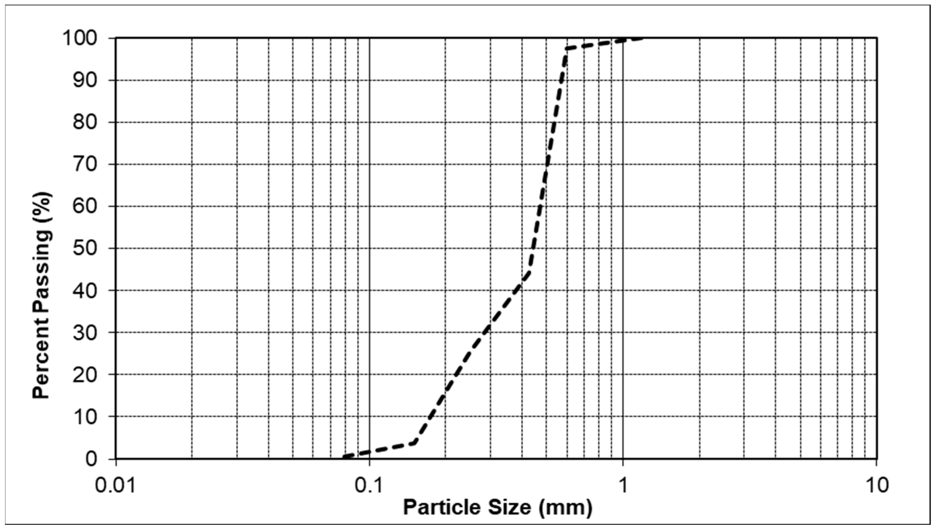
In this experiment, clean Firoozkuh silica sand with a uniformity coefficient of 2.74, a specific gravity of 2.65, and maximum and minimum void ratios of 0.90 and 0.57, respectively, was used (Figure 2).
The soil specimens were prepared by mixing condensed carbonate minerals. To obtain condensed carbonate minerals, the suspensions were centrifuged at 8000× g for 10 min and then filtered by filter paper [7]. The suspension of carbonate minerals was used as soil-stabilizing agents in the unconfined compression tests.

Unconfined Compressive Strength Testing

The unconfined compression strength (UCS) test was used for obtaining the approximate strength of the soil specimens. To perform the UCS test, the pre-weighed amount of sand was mixed with a suspension of carbonate minerals and placed in the two-piece split mold by slightly tamping to obtain a homogenous specimen of 50 mm diameter and 100 mm length by the dry density of about 19.2 kN·m−3. Before the UCS tests, the specimens were allowed to cure at room temperature for 7 days. The samples were tested with a 0.04 MPa/s loading rate at a constant strain rate of 1.5 mm·min−1. The UCS tests were repeated three times for each type of mineral and production method. The accuracy analysis was performed for parallel tests. The UCS was taken as the peak stress with the corresponding axial strain at failure in the stress–strain curve (ASTM D2166/D2166M-16, 2016) [28].
The carbonate mineral percentage of the samples was determined by using an acid-washing technique [2]. The soil samples were soaked into the 100 mL acid solution (HCl 5 M) for 1 h. The dissolved carbonates and acid wash solution were filtered through a filter paper (2 in) three times, allowing the dissolved salts to be rinsed from the soil while the soil was retained. Before and after the acid washing technique, the oven-dried mass of the soil samples across the specimen was measured by digital scales. The mass of carbonate minerals was calculated as the difference between the two observed masses.

3. Results and Discussion

3.1. Induced Carbonate Production (CO32−)

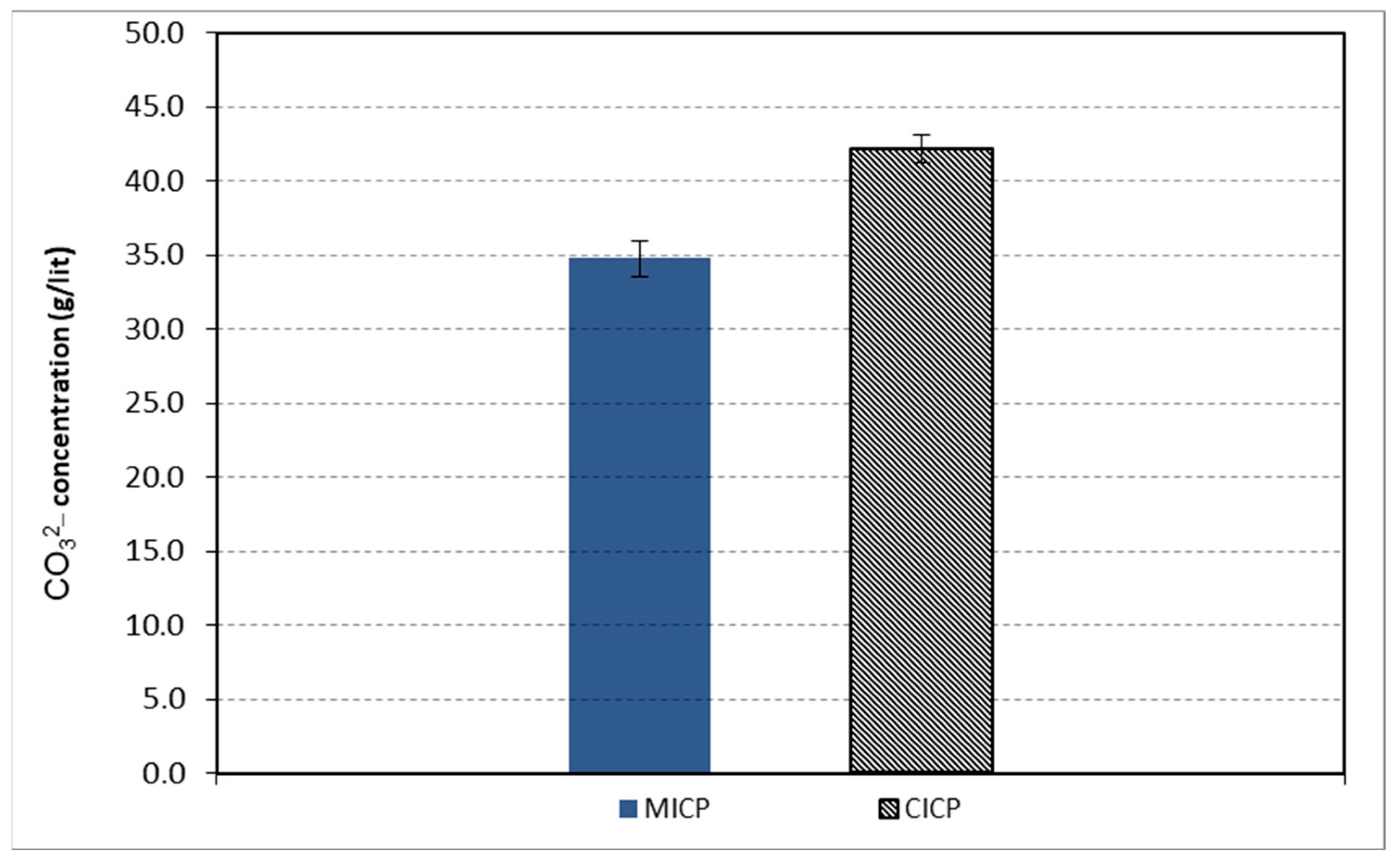
The microbial-induced and CO2-induced carbonate ions (CO32−) were compared in Figure 3. Average values of 42.1 and 34.8 g·L−1 were obtained for CICP and MICP methods, respectively.
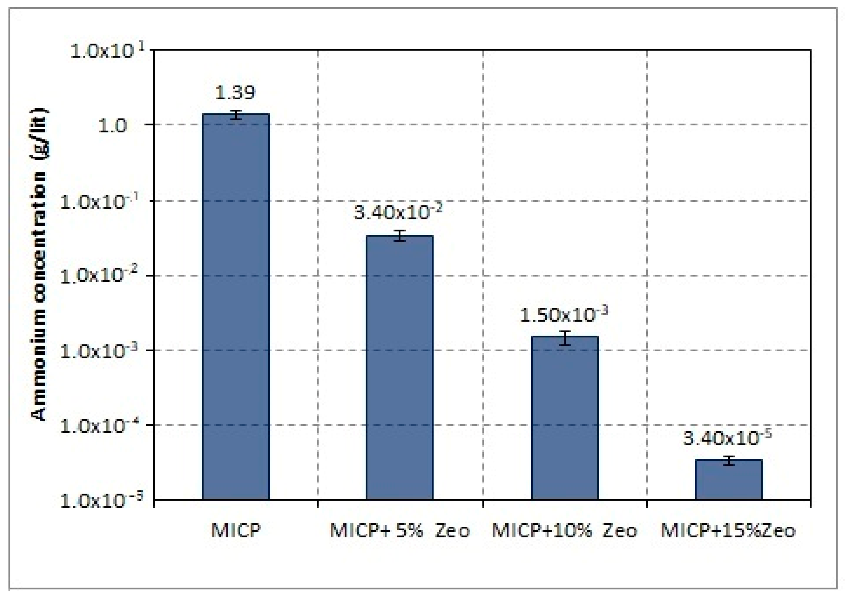
The effect of zeolite treatment on ammonium concentration for MICP to generate an ammonium-free carbonate solution is shown in Figure 4. The ammonium content of the solution was initially around 1.39 g·L−1, as seen in the figure. After 15 percent zeolite treatment, the ammonium concentration dropped to an average of 3.4 × 10⁻² g·L−1. The concentration meets the requirement specified by some drinking-water standards (e.g., lower than 5 × 10⁻⁴ g·L−1, Australian Drinking Water Guidelines and Guidelines for Drinking-water Quality Management for New Zealand, 2016).

3.2. Carbonate Mineral Production

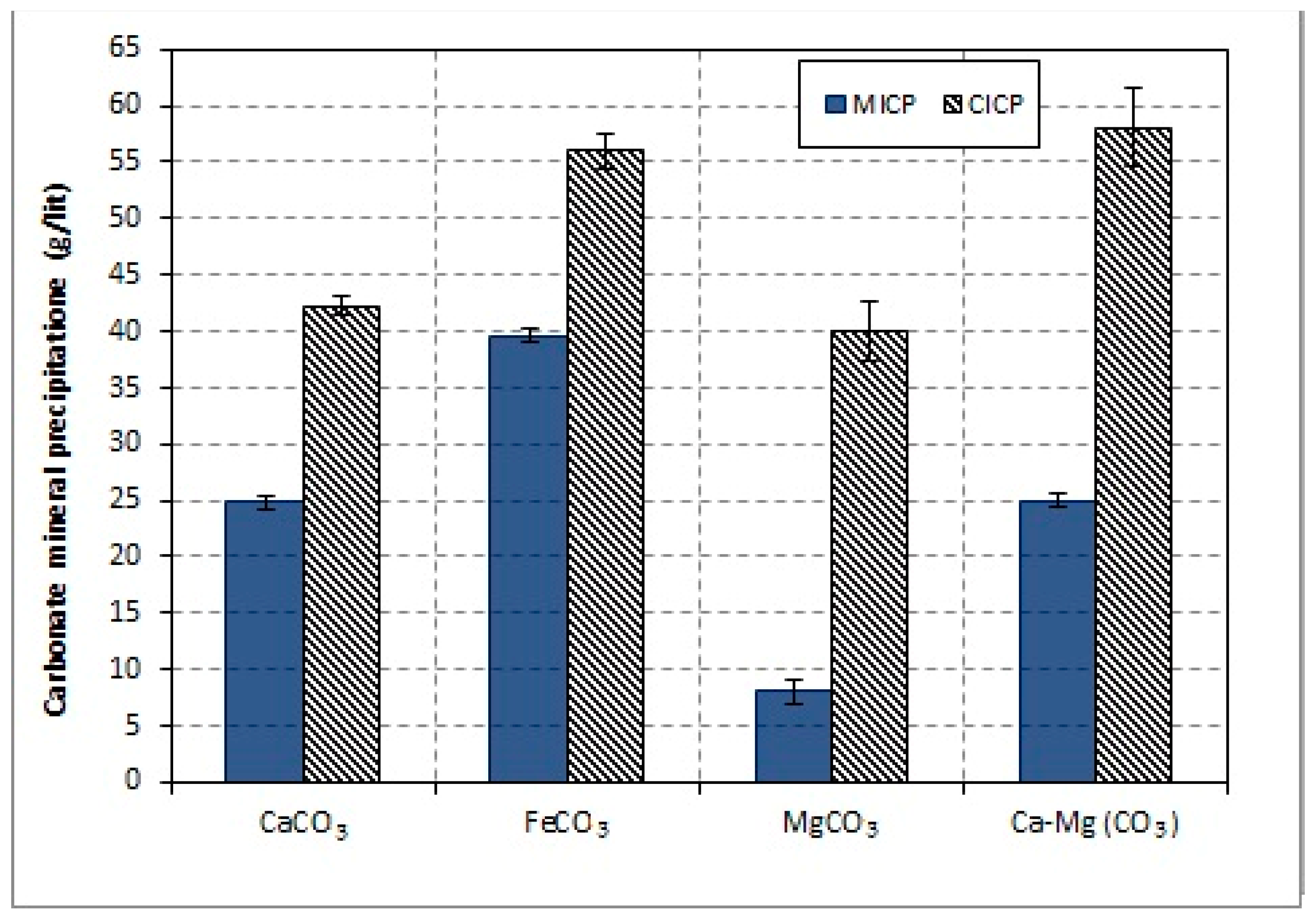
Carbonate minerals precipitated by MICP and CICP techniques are shown in Figure 5. The results demonstrate that the carbonate minerals precipitated by CICP had higher values than those precipitated by MICP. The largest carbonate minerals content was FeCO3 (55 g·L−1) and Ca-Mg (CO3) (58 g·L−1) precipitated by the CICP. The precipitation of CaCO3 and Ca-Mg (CO3) in MICP were identical (25 g·L−1). In MICP, the highest level of carbonate precipitation was found in FeCO3 (40 g·L−1), whereas the lowest level was found in MgCO3 (8 g·L−1). As can be seen from the results in Figure 5, MICP deposited fewer carbonate minerals than CICP. It is generally accepted that this microbial activity can be influenced by several physicochemical and biological parameters, and it is correlated to both metabolic activities and cell surface structures [29,30]. Multiple studies revealed that fresh bacterial cells are an essential step to keep the fast rate of precipitation, as desirable in certain applications [31]. CICP is a method based on the fixation of CO2 in the form of inorganic carbonates, also known as ‘mineral carbonation’. Since carbonate is the lowest energy state of carbon, these carbonation reactions are exothermic, and the formation of carbonates is favored at low temperatures and low pressures [32].

3.3. Carbonate Mineral Characterization

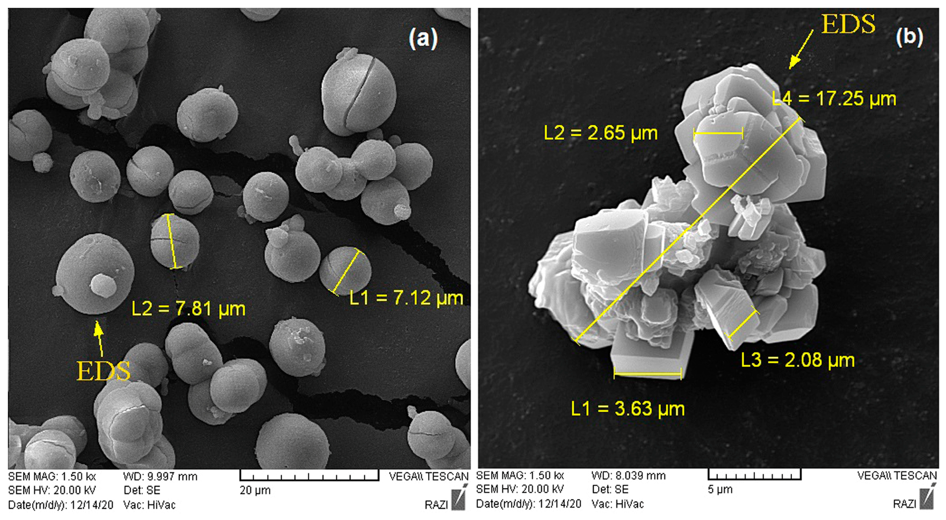
Figure 6 illustrates the crystal morphology of CaCO3 which was produced by MICP and CICP methods. As can be seen, the CaCO3 crystals have been grown spherical with diameters of 2–7 µm in MICP (Figure 6a), while crystals were in rhombohedral shape and smooth with diameters of 2–6 µm in CICP (Figure 6b). Figure 7 shows EDS and XRD analysis of CaCO3 deposition. The EDS layered image presents the distribution of C, O, and Ca elements in the test area that proved the importance of carbonate phases and the CaCO3 compound in the sample. The result of XRD justified vaterite minerals (Figure 7a) and calcite (Figure 7b) in MICP and CICP, respectively. Previous studies presented that the morphology of CaCO3 is dependent on additives such as CO2 and Ca2+, which are precipitated in three anhydrous polymorphic modifications (calcite, aragonite, and vaterite) and mostly calcite [33]. MICP is largely dependent on metabolic processes by ureolytic bacteria that influence crystal growth [34,35,36,37]. It has also been reported that the amino acid (Glu and Asp) in the urease enzyme favored the formation of vaterite [38].
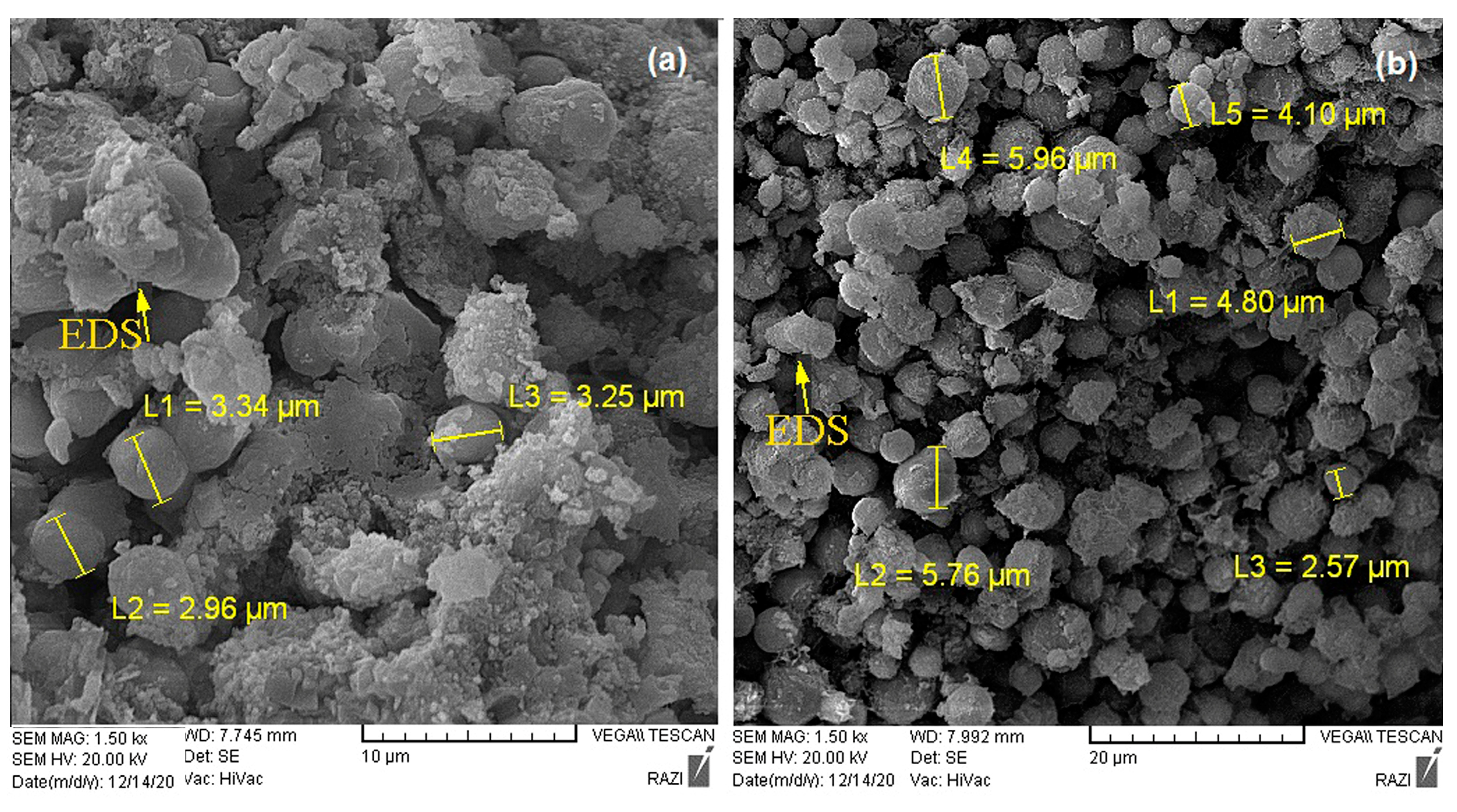
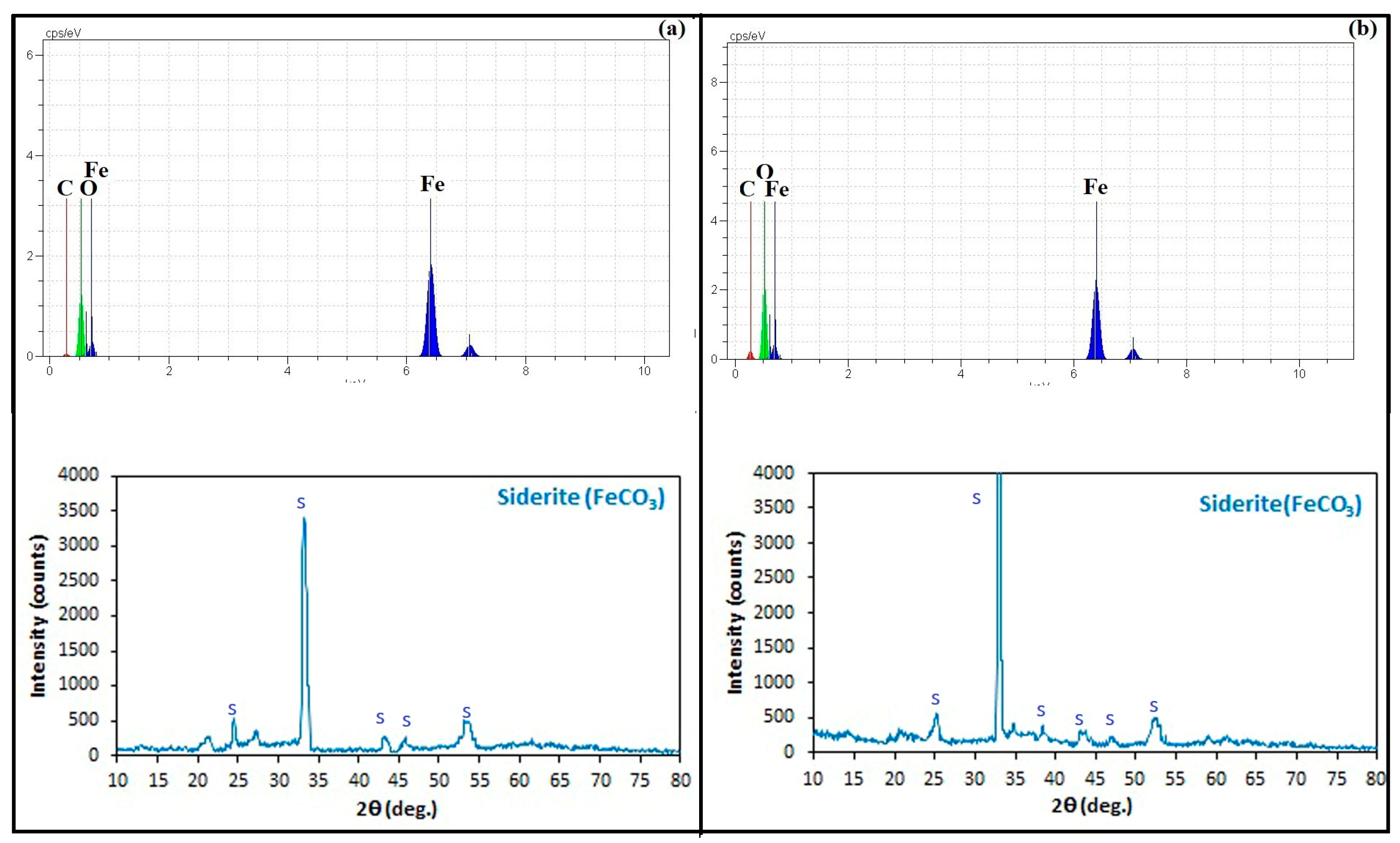
Figure 8 shows the crystal morphology of FeCO3, which was produced by MICP and CICP methods. There was a similar polycrystalline shape (spherical) for both of them. The particle size of FeCO3 in MICP was with diameters of 0.5–3 µm while the particle size was bigger (2–6 µm) in CICP. Figure 9 shows the EDS analysis of FeCO3 samples. The EDS spectrum evidently denoted the occurrence of C, O, and Fe atoms in the sample, which proved the importance of carbonate phases and FeCO3 compounds in the sample. The XRD analysis was conducted on the precipitated FeCO3; however, it did not reveal the presence of pure siderite minerals. The result was justified by the presence of siderite minerals with other compounds such as quartz, calcite, gypsum, and magnesite in this study. Several factors might have hindered the correct detection of pure siderite minerals in the XRD analysis. One possible reason is the presence of impurities or other minerals in the sample, which could have overlapped with the characteristic peaks of siderite, making it challenging to distinguish. Additionally, the particle size of the sample might have been too small, resulting in weak diffraction signals.
Less information is available concerning the mineralization or morphogenesis of iron carbonates (siderite). This is probably because natural siderite is often associated with other coexisting elements (e.g., Mn, Mg, and Ca) and contains a small amount of hematite (Fe2O3) due to partial oxidation in natural air [39,40]. The biological aspects of iron biomineralization have been investigated by studying iron-bearing biominerals, owing to their significance in identifying microbe-sediment-water interactions as well as mineral and biogenic origins [41,42].
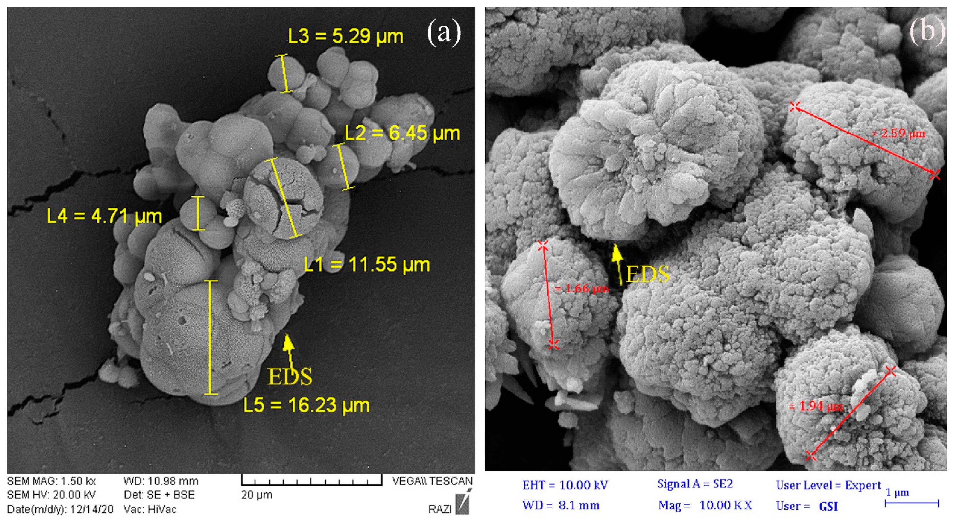
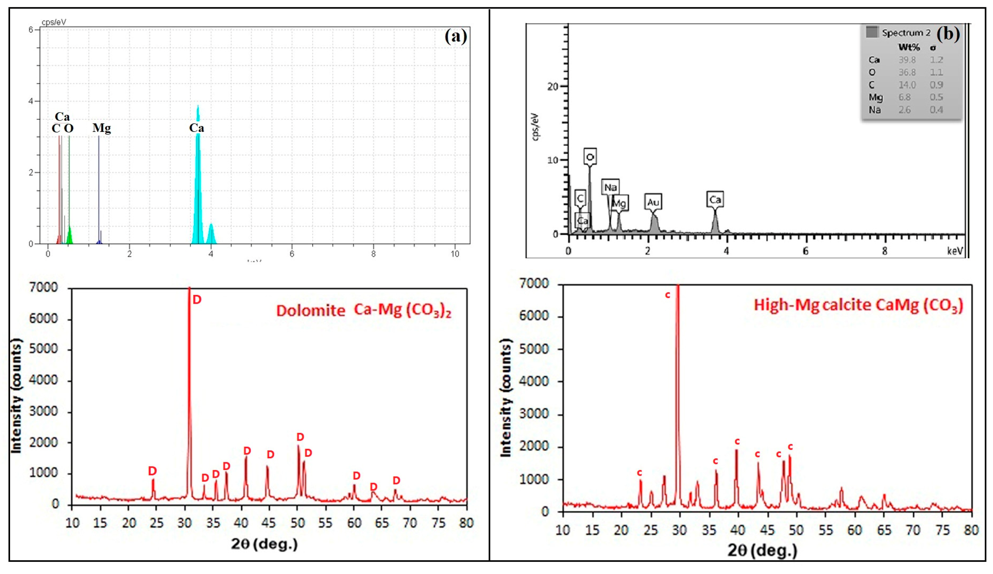
Figure 10 shows the crystal morphology of Ca-Mg(CO3), which is produced by a mixture of magnesium and calcium ions with free carbonate ions (CO32−). As can be seen from the figure, there was a botryoidal crystal shape of Ca-Mg(CO3) in the MICP and CICP methods. There was just one main difference between the shape and size of the crystals in both methods. Ca-Mg (CO3) was precipitated with diameters of 5–16 µm and 2–5 µm in MICP and CICP, respectively. Minerals were deposited at a larger size in MICP than in CICP, as can be seen clearly. Figure 11 shows EDS and XRD analyses of Ca-Mg (CO3) samples. The EDS spectra present the distribution of C, O, Mg, and Ca elements in the test area. It is proven that the importance of carbonate phases and Ca-Mg (CO3) compounds in the sample. XRD analysis of samples showed Ca-Mg (CO3) deposition as dolomite in MICP (Figure 11a) and high-Mg calcite in CICP (Figure 11b), respectively.
Previous laboratory research revealed that aerobic bacteria may have a role in the creation of high-magnesium calcite and dolomite, in the microenvironment around the cell [43,44]. Numerous field and laboratory studies claim that microbes catalyze nucleation and growth of dolomite and high-magnesium calcite by altering the chemistry of their environments through metabolic activity at temperatures common in low-temperature geologic environments (25–60 °C) [45,46]. González-Muñoz et al. [47] found that Mg probably plays a key role in the development of the morphologies of the precipitates since these morphologies had never been observed in the absence of Mg. The precipitated CaCO3 in seawater changes from calcite to aragonite, which is experimentally determined as a function of temperature and additives. Precipitation of high-Mg calcite and dolomite is largely dependent on the Mg:Ca ratio over a relatively small temperature range in an aqueous solution [48]. In conclusion, the morphology, type, and size of calcium magnesium carbonate minerals are influenced by the chemical and biochemical environments in CICP and MICP.
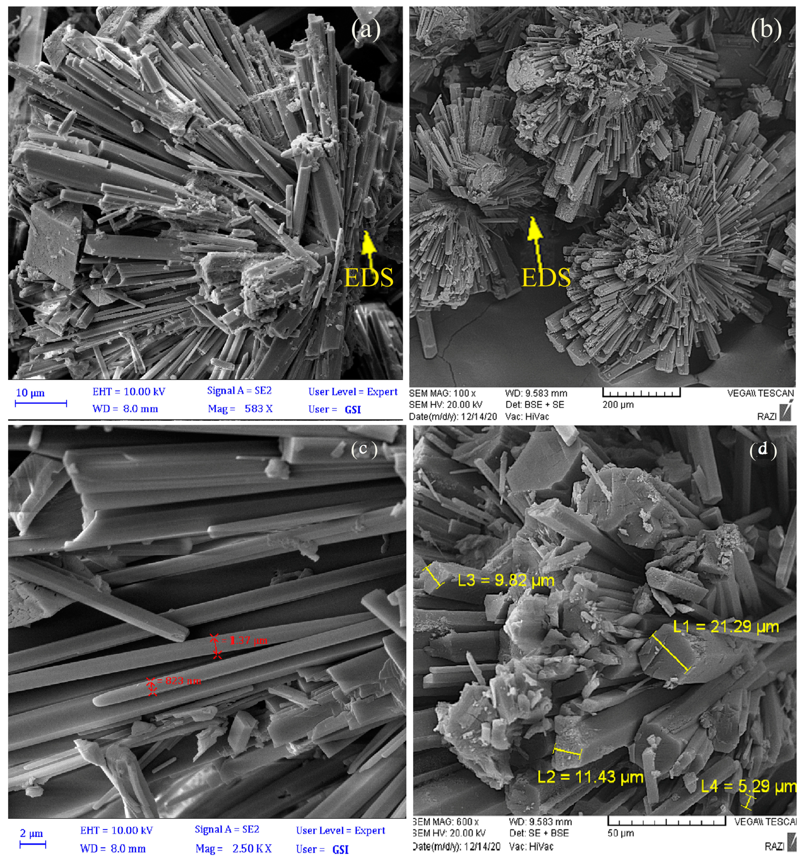
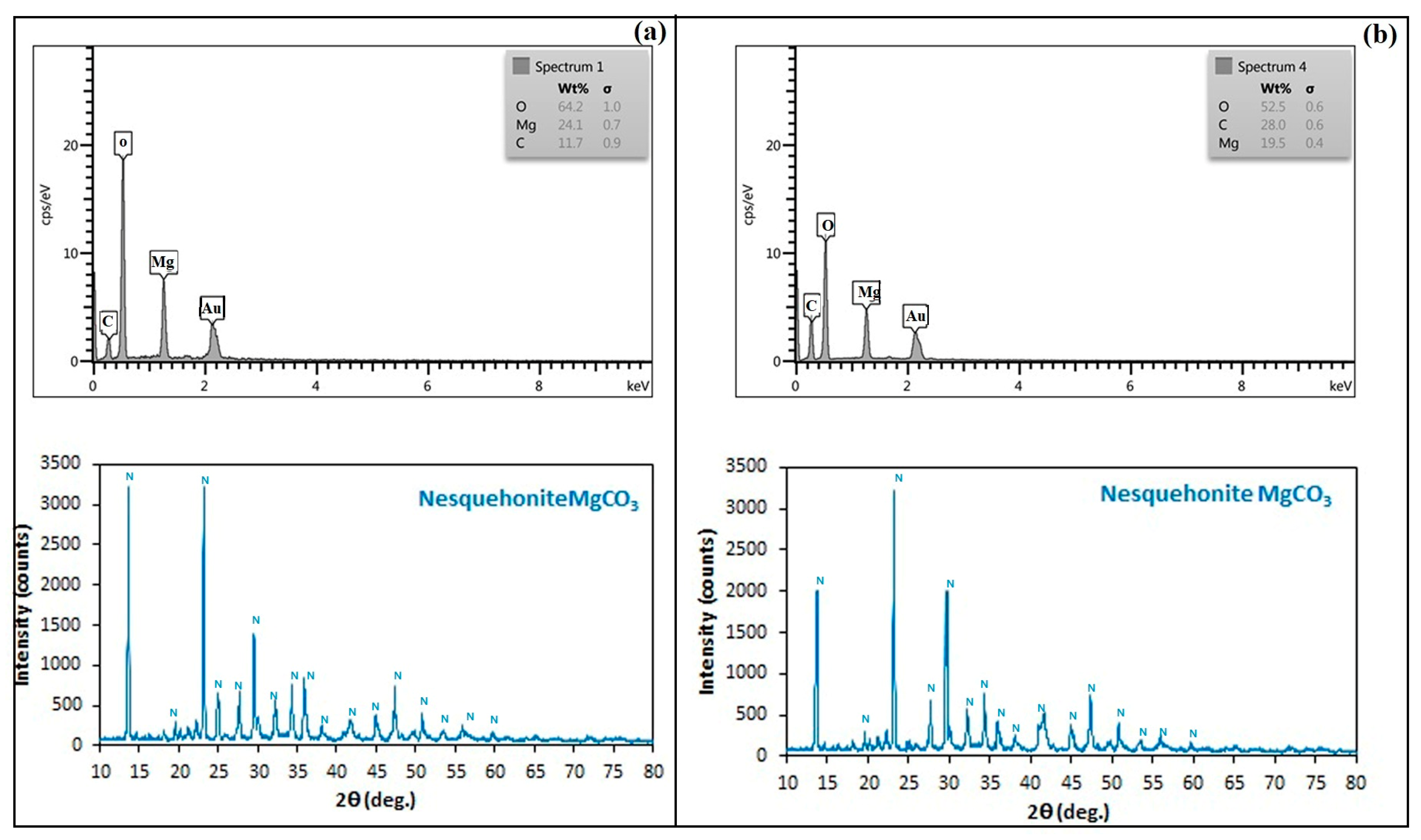
Figure 12 illustrates the crystal morphology of MgCO3, which was produced by MICP and CICP methods. As can be seen, the needle-shaped crystals of MgCO3 were arranged radially by MICP and CICP. There was a main difference between the sizes of the crystals in both methods. The thickness of needle-shaped crystals was 1–10 µm in MICP, whereas thicker crystals of about 5–20 µm precipitated in CICP. It was clearly shown that the potential of CICP to precipitate MgCO3 crystals is greater than that of MICP. Figure 13 shows EDS and XRD analyses of MgCO3 samples. The EDS spectrum evidently denoted the occurrence of C, O, and Mg atoms in the sample, which proved the importance of carbonate phases and the MgCO3 compound in the sample. The result of XRD shows the mineralization of nesquehonite for both methods (Figure 13). Previous studies presented that magnesium carbonate (as hydrate) can display four different morphologies during the reaction process, such as needle, sheet, rose, and nest-like, with different carbonation temperatures [49,50]. Chen et al. [50] presented that at lower temperatures, magnesium carbonate hydrate was prone to display needle-like morphology and, with increasing temperature, became sheet-like with tended crystallites, rose-like particles, and a nest-like structure. A previous study showed the same mineral (nesquehonite) with similar morphologies in MICP [51] and CICP [52].
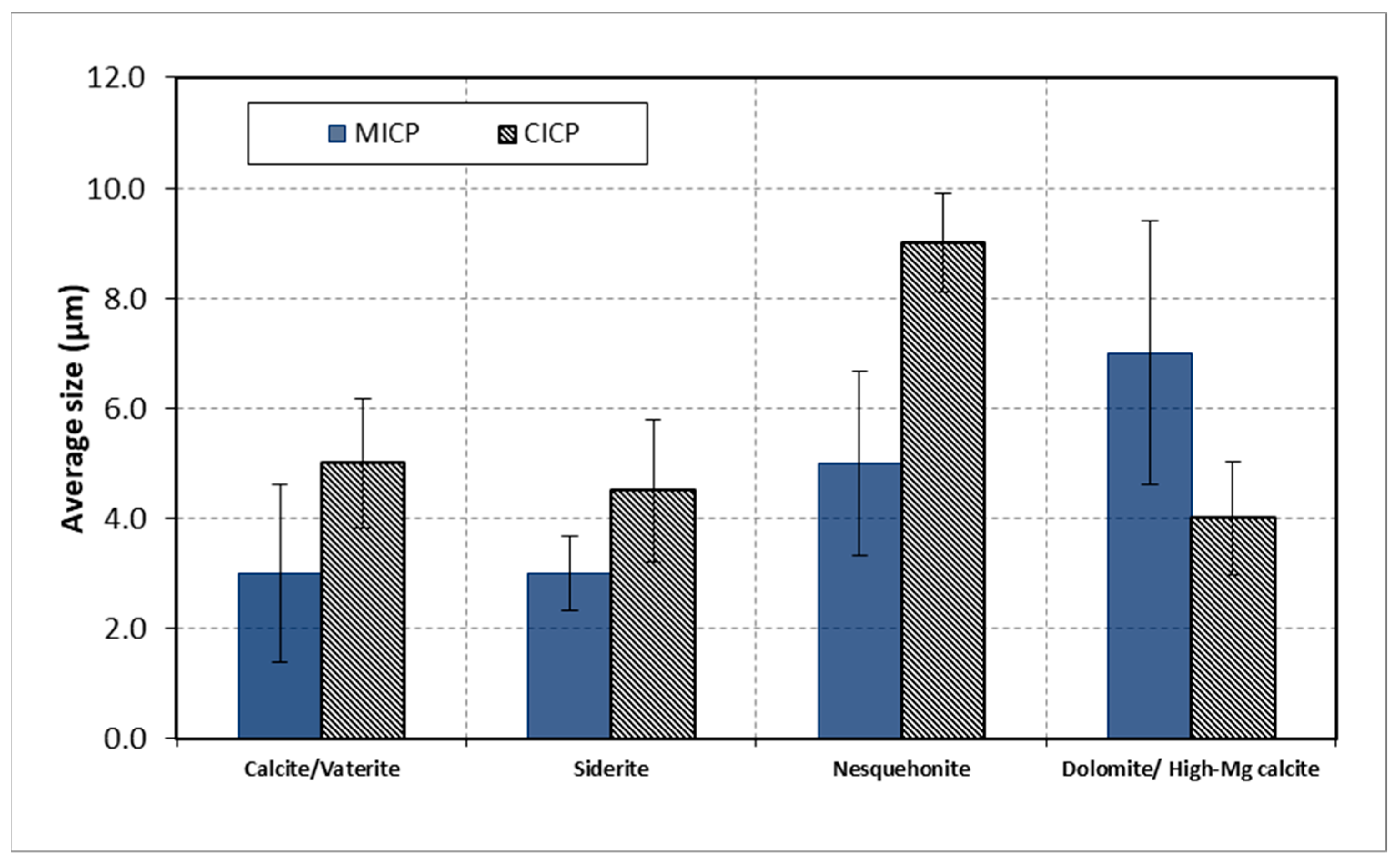
The mean size distribution of minerals was measured in SEM images. Figure 14 shows the average size of carbonate minerals produced by MICP and CICP methods. As can be seen from the results, the average size of crystals was larger in CICP, which was smaller than in MICP. For the Ca-Mg experiment, the result showed an exception. The results showed that the highest average crystal size was for nesquehonite (CICP), 8 µm in length and the lowest size was for calcite and siderite (MICP), 3 µm. In this work, CICP demonstrated a great capacity for precipitating carbonate minerals with more minerals and larger crystals. The results of the measurement of precipitated mineral sizes showed differences in distribution size in mineral precipitation. It seems that a wide range of mineral sizes in some carbonate depositions was due to laboratory conditions and reagents. The minerals that develop during biomineralization are frequently characterized by low crystallinity, structural well-ordering, and a limited size range [41]. Under the same environmental conditions, biologically induced mineralization is not always equal to inorganic mineralization, and the minerals are expected to contain crystal–chemical characteristics that are indistinguishable from those of minerals formed by inorganic chemical reactions. However, cell walls of bacterial surfaces, including biofilms, dormant spores, and slime sheaths, can act as important sites for the adsorption of ions for growth and nucleation minerals [53].
Bacteria operate as nucleation sites in the MICP process, whereby calcium carbonate precipitates alongside the bacteria. Because different bacterial species are capable of precipitating varying numbers, sizes, forms, and types of carbonate crystals from the same synthetic medium, the composition of the growth media or culture may also have an impact on the crystal morphology. Indeed, many factors affect urease activity and calcium precipitation including bacteria type, bacteria cell concentrations, pH, temperature, urea, and calcium concentrations [54]. The carbonation rate is affected by variables in the CICP process, including temperature, carbon dioxide pressure, and reaction time that are systematically changed [55].
A summary of the characterization of carbonate minerals produced by MICP and CICP techniques is shown in Table 2. In both procedures, all carbonate minerals were precipitated in various sizes, shapes, and types. As mentioned, MICP is largely dependent on metabolic processes and cell surface structure of ureolytic bacteria that influence crystal growth and shape, providing a favorable way to accumulate minerals to be recovered for applied purposes, and may modify carbonate mineral structures. The different products of their metabolism, such as proteins (especially enzymes), polysaccharides, and other EPS, and the charged groups embedded in their microbial cell walls, by interacting with anions and cations, affect the crystal properties and chemical composition of the minerals [56]. As mentioned in the text, previous studies showed that the amino acid in the urease enzyme favored the different formations of carbonate minerals such as calcite and vaterite [38]. It is also reported that bacteria can adsorb magnesium on their membranes during dolomite formation [47], while calcite precipitation is induced by preferential adsorption of calcium that affects the shape of deposited calcite.
Under the same environmental conditions, chemical mineralization, such as CO2-induced carbonate precipitation, is simple and just influenced by chemical ingredients, temperature, and pH [57] which can also be the main factors in biologically induced mineralization.
Less information is available concerning the use of other carbonate minerals for soil improvement. The solubility of carbonate minerals decreases by more than two orders of magnitude on transformation from Ca to Mg-rich [58]. Mg carbonates such as magnesite and dolomite are semi-soluble and hydrophilic [59]. Long et al. [60] showed that the increase of Mg ions in Mg-containing calcite improves the mechanical properties of biogenic minerals. As well, ferrous carbonate has low solubility in an aqueous solution [61]. It seems that carbonate minerals with substitution elements such as Mg and Fe affect the behavior of treated soil. Depending on the polymorph of CaCO3 crystals (i.e., calcite, vaterite, or aragonite), the strength of biocemented soil can be affected [20,21]. It appears that calcite crystals with rhombohedral shapes and botryoidal high-Mg calcite in CICP have the capability of interlocking due to their surface roughness.

3.4. Unconfined Compressive Strength

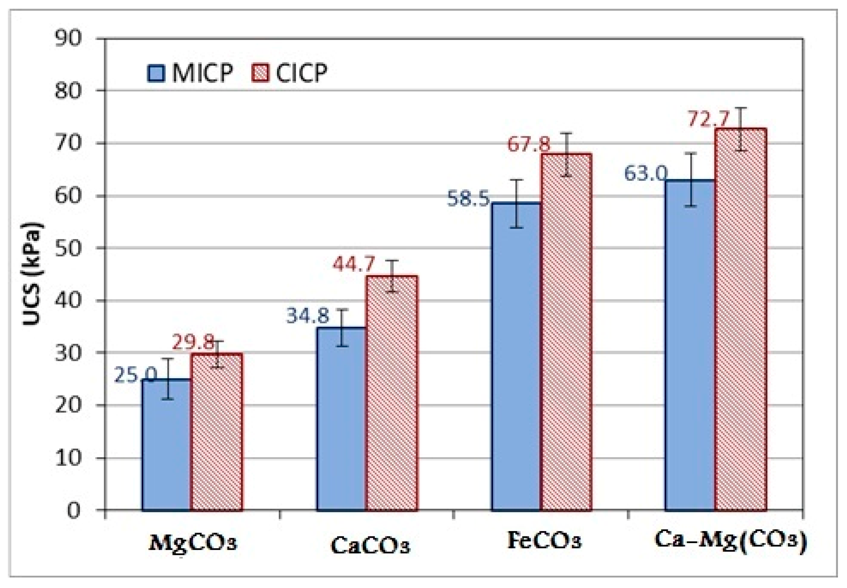
Figure 15 shows the results of the UCS tests carried out on carbonate mineral-treated soil. As can be seen from the figure, the treated specimens with condensed Ca-Mg(CO3) and FeCO3 minerals, produced by the MICP method gained about 58.5 kPa and 63.0 kPa UCS strength, respectively. Moreover, MgCO3 minerals, produced by the MICP method, had the lowest strength of about 25 kPa. The treated specimens with CaCO3 content gained higher compressive strength compared to the specimens with MgCO3 content in this study. The treated specimens with the CICP method gained higher compressive strength compared to the specimens treated with the MICP method. According to the data, the minerals produced by the CICP method have a 15 to 28 percent higher compressive strength than those produced by the MICP method.
The results of UCS showed that siderite and dolomite/high-Mg calcite had more efficiency in soil strength, as it seems to be because of the stability of Fe and Ca–Mg carbonate crystals. The siderite-treated soil with CICP had slightly more strength than MICP, which may be due to the larger particle size of the crystals. Similarly, it was observed for nesquehonite and calcite-treated soils. On the other hand, the higher strength of high-Mg calcite in CICP would be related to the shape and particle size of the minerals [62]. The strength of calcite-treated soil in CICP was slightly higher than that of vaterite-treated soil in MICP, which may be related to the mineral phase. The UCS result of treated soil with nesquehonite minerals showed a lower strength than other carbonate minerals in this study. However, Nesquehonite (because of its needle structure) had higher efficiency in clay soil in a previous study [3]. Moreover, the increased thickness of nesquehonite minerals precipitated by CICP would lead to a slightly larger strength of the treated soil samples.
According to earlier research, the strength of MICP-treated soil is significantly influenced by the characteristics of CaCO3 crystals, such as size and shape [63,64,65]. The internal friction angle in treated soil will gradually change to reflect the roughness of the particle surfaces. Additionally, it appeared that rhombohedral crystals were more stable than spherical crystals (or polycrystalline), which, in turn, were more stable than precipitates with irregular shapes [66]. The differences in the size of the crystals would, in turn, affect the engineering properties of MICP-treated samples, such as permeability, stiffness, and strength [64]. The stiffness and strength of MICP-treated soils may be improved more effectively by the formation of bigger crystals at narrow pore throats that can bind soil particles [65]. Further work involving using different carbonate minerals in real soil applications will be useful for determining a relationship between the treatment process and the engineering properties of CICP and MICP-treated soils.
As in the case of the MICP and CICP techniques, CO2 and bacteria can quickly produce significant concentrations of carbonate minerals. Applying these methods to stabilize soil as a building material by the precipitation of carbonate minerals [1,3,7,8] will thus be a substantial advancement of this research. However, the investigation of different aspects of these mechanisms should be assumed. Applying CO2 injection for in situ carbonate precipitation may have some disadvantages. Consuming a large volume of water for CO2 capture is the most disadvantageous. Also, to obtain a benefit in situ soil improvement needs to precipitate appropriate carbonate precipitation by CO2. To improve soil quality, the direct injection of CO2 through precipitation cycles may be necessary, although it can be time-consuming and costly. MICP processes involve a series of biochemical reactions that are affected by many factors, including bacterial species, the concentration of bacteria, the concentration of cementation solution, pH, temperature, grouting method, and soil properties that can make soil improvement applications difficult. In addition, MICP is not 100% environmentally friendly, as ureolysis plays a significant role in precipitation-generating by-products, including ammonium and nitrate. These compounds are toxic and thus hazardous both to human health and indigenous microbial communities, especially at high concentrations.
Therefore, using the produced carbonate minerals as a powder for mixing and injection application for ground improvement can be suggested. In this process, the carbonate minerals are made from both mechanisms (i.e., MICP and CICP) and then used for soil improvement. The application of carbonate mineral powder has advantages, while in situ operations may be difficult and have some disadvantages.

4. Conclusions

Microbial-induced carbonate precipitation and CO2-induced carbonate precipitation methods were used to create free carbonate ions (CO32−). The various carbonate minerals were precipitated by adding Fe2+, Mg2+, and Ca2+ ions to carbonate ion (CO32−) solutions. The amount of carbonate minerals by CICP was higher than those precipitated by MICP, according to the findings. The most carbonate minerals value FeCO3 (55 g·L−1) and Ca-Mg (CO3) (58 g·L−1) by CICP method were obtained while, these value were for FeCO3 (40 g.L−1) and Ca-Mg (CO3) (25 g·L−1) in MICP method.
Vaterite (CaCO3), siderite (FeCO3), nesquehonite (MgCO3(H2O)3), and dolomite (CaMg(CO3)2) were produced in MICP. Calcite (CaCO3), siderite (FeCO3), nesquehonite (MgCO3(H2O)3), and high-Mg calcite (CaMg(CO3)2) were produced in CICP. Vaterite and calcite had spherical and rhombohedral shapes, respectively. Moreover, siderite was precipitated in a spherical shape in both methods. Dolomite and high-Mg calcite showed botryoidal shape in MICP and CICP. Nesquehonite was deposited as needle-shaped crystals. Furthermore, larger crystals were observed in the CICP method. As well, crystals were smaller with the CICP method than with the MICP method in the Ca–Mg experiment, as an exception. The results showed the largest average size of crystals was for Nesquehonite (i.e., 8 µm length) by CICP, and the lowest size was for calcite and siderite (i.e., 3 µm) by MICP.
The UCS results demonstrated an Increase in strength for soils treated with siderite and high-Mg calcite/dolomite over calcite/vaterite and nesquehonite-treated soils. Mineral-treated soils in CICP were marginally stronger than MICP, likely due to larger crystal particle sizes or particle interlocking.
The novelty of this study is an investigation of comparing carbonate precipitations in CICP and MICP methods. While previous studies focused on microbial-induced carbonate minerals, the results of this research showed a strong potential for CO2-induced carbonate minerals in new characterizations and an environmentally friendly deposition.

Author Contributions

Conceptualization, methodology, validation, investigation, data curation, and writing—original draft preparation, visualization, supervision, and project administration, H.A.K.; Formal analysis, investigation, and visualization, A.Z.; Conceptualization, validation, formal analysis, data curation, and writing—review and visualization, H.M.R.; Conceptualization, methodology, review, and editing, A.A.; validation, investigation, and resources, S.K.; Formal analysis and investigation, N.R. All authors have read and agreed to the published version of the manuscript.

Funding

This research received no external funding.

Data Availability Statement

Data are contained within the article.

Acknowledgments

This work has been supported by the Centre for International Scientific Studies and Collaboration (CISSC), Ministry of Science Research and Technology.

Conflicts of Interest

The authors declare no conflict of interest.

References

  1. Keykha, H.A.; Asadi, A.; Zareian, M. Environmental factors affecting the compressive strength of microbiologically induced calcite precipitation-treated soil. Geomicrobiol. J. 2017, 34, 889–894. [Google Scholar] [CrossRef]
  2. Keykha, H.A.; Asadi, A.; Huat, B.B.; Kawasaki, S. Microbial induced calcite precipitation by Sporosarcina pasteurii and Sporosarcina aquimarina. Environ. Geotech. 2018, 6, 562–566. [Google Scholar] [CrossRef]
  3. Romiani, H.M.; Keykha, H.A.; Talebi, M.; Asadi, A.; Kawasaki, S. Green soil improvement: Using carbon dioxide to enhance the behaviour of clay. Proc. Inst. Civ. Eng.-Ground Improv. 2021, 9, 1–9. [Google Scholar] [CrossRef]
  4. Wang, L.; Cheng, W.C.; Xue, Z.F.; Xie, Y.X.; Lv, X.J. Feasibility study of applying electrokinetic technology coupled with enzyme-induced carbonate precipitation treatment to Cu-and Pb-contaminated loess remediation. J. Clean. Prod. 2023, 401, 136734. [Google Scholar] [CrossRef]
  5. Xue, Z.F.; Cheng, W.C.; Xie, Y.X.; Wang, L.; Hu, W.; Zhang, B. Investigating immobilization efficiency of Pb in solution and loess soil using bio-inspired carbonate precipitation. Environ. Pollut. 2023, 322, 121218. [Google Scholar] [CrossRef]
  6. Ali, A.; Li, M.; Su, J.; Li, Y.; Wang, Z.; Bai, Y.; Ali, E.F.; Shaheen, S.M. Brevundimonas diminuta isolated from mines polluted soil immobilized cadmium (Cd2+) and zinc (Zn2+) through calcium carbonate precipitation: Microscopic and spectroscopic investigations. Sci. Total Environ. 2022, 813, 152668. [Google Scholar] [CrossRef] [PubMed]
  7. Keykha, H.A.; Romiani, H.M.; Zebardast, E.; Asadi, A.; Kawasaki, S. CO2-induced carbonate minerals as soil stabilizing agents for dust suppression. Aeolian Res. 2021, 52, 100731. [Google Scholar] [CrossRef]
  8. Wang, Z.; Zhang, N.; Jin, Y.; Li, Q.; Xu, J. Application of microbially induced calcium carbonate precipitation (MICP) in sand embankments for scouring/erosion control. Mar. Georesour. Geotechnol. 2021, 39, 1459–1471. [Google Scholar] [CrossRef]
  9. Lowenstam, H.A.; Weiner, S. On Biomineralization; Oxford University Press: New York, NY, USA, 1989. [Google Scholar]
  10. Sánchez-Román, M.; Romanek, C.S.; Fernández-Remolar, D.C.; Sánchez-Navas, A.; McKenzie, J.A.; Pibernat, R.A.; Vasconcelos, C. Aerobic biomineralization of Mg-rich carbonates: Implications for natural environments. Chem. Geol. 2011, 281, 143–150. [Google Scholar] [CrossRef]
  11. Ferris, F.G.; Stehmeier, L.G.; Kantzas, A.; Mourits, F.M. Bacteriogenic mineral plugging. J. Can. Pet. Technol. 1997, 36, 56–61. [Google Scholar] [CrossRef]
  12. Stocks-Fischer, S.; Galinat, J.K.; Bang, S.S. Microbiological precipitation of CaCO3. Soil Biol. Biochem. 1999, 31, 1563–1571. [Google Scholar] [CrossRef]
  13. Ivanov, V.; Chu, J. Applications of microorganisms to geotechnical engineering for bioclogging and biocementation of soil in situ. Rev. Environ. Sci. Bio/Technol. 2008, 7, 139–153. [Google Scholar] [CrossRef]
  14. Tang, C.S.; Yin, L.Y.; Jiang, N.J.; Zhu, C.; Zeng, H.; Li, H.; Shi, B. Factors affecting the performance of microbial-induced carbonate precipitation (MICP) treated soil: A review. Environ. Earth Sci. 2020, 79, 94. [Google Scholar] [CrossRef]
  15. Chen, J.; Liu, B.; Zhong, M.; Jing, C.; Guo, B. Research status and development of microbial induced calcium carbonate mineralization technology. PLoS ONE 2022, 17, e0271761. [Google Scholar] [CrossRef] [PubMed]
  16. Xu, X.; Guo, H.; Li, M.; Deng, X. Bio-cementation improvement via CaCO3 cementation pattern and crystal polymorph: A review. Constr. Build. Mater. 2021, 297, 123478. [Google Scholar] [CrossRef]
  17. Mortensen, B.M.; Haber, M.J.; DeJong, J.T.; Caslake, L.F.; Nelson, D.C. Effects of environmental factors on microbial induced calcium carbonate precipitation. J. Appl. Microbiol. 2011, 111, 338–349. [Google Scholar] [CrossRef] [PubMed]
  18. Chen, L.; Shen, Y.; Xie, A.; Huang, B.; Jia, R.; Guo, R.; Tang, W. Bacteria-mediated synthesis of metal carbonate minerals 552 with unusual morphologies and structures. Cryst. Growth Des. 2009, 9, 743–754. [Google Scholar] [CrossRef]
  19. Hammes, F.; Verstraete, W. Key roles of pH and calcium metabolism in microbial carbonate precipitation. Rev. Environ. Sci. Biotechnol. 2002, 1, 3–7. [Google Scholar] [CrossRef]
  20. Dhami, N.K.; Reddy, M.S.; Mukherjee, A. Biomineralization of calcium carbonate polymorphs by the bacterial strains isolated from calcareous sites. J. Microbiol. Biotechnol. 2013, 23, 707–714. [Google Scholar] [CrossRef] [Green Version]
  21. Al-Thawadi, S.M. Consolidation of sand particles by aggregates of calcite nanoparticles synthesized by ureolytic bacteria under non-sterile conditions. J. Chem. Sci. Technol. 2013, 2, 141–146. [Google Scholar]
  22. Chang, R.; Kim, S.; Lee, S.; Choi, S.; Kim, M.; Park, Y. Calcium carbonate precipitation for CO2 storage and utilization: A review of the carbonate crystallization and polymorphism. Front. Energy Res. 2017, 5, 17. [Google Scholar] [CrossRef] [Green Version]
  23. Yoo, Y.; Kim, I.; Lee, D.; Choi, W.Y.; Choi, J.; Jang, K.; Park, J.; Kang, D. Review of contemporary research on inorganic CO2 utilization via CO2 conversion into metal carbonate-based materials. J. Ind. Eng. Chem. 2022, 116, 60–74. [Google Scholar] [CrossRef]
  24. Liu, W.; Teng, L.; Rohani, S.; Qin, Z.; Zhao, B.; Xu, C.C.; Ren, S.; Liu, Q.; Liang, B. CO2 mineral carbonation using industrial solid wastes: A review of recent developments. Chem. Eng. J. 2021, 416, 29093. [Google Scholar] [CrossRef]
  25. Smit, B.; Reimer, J.A.; Oldenburg, C.M.; Bourg, I.C. Introduction to Carbon Capture and Sequestration; World Scientific, Imperial College Press: London, UK, 2014; Volume 1. [Google Scholar]
  26. Department of Economic and Social Affairs. Transforming Our World: The 2030 Agenda for Sustainable Development; United Nations: New York, NY, USA, 2015. [Google Scholar]
  27. Inoue, R.; Ueda, S.; Wakuta, K.; Sasaki, K.; Ariyama, T. Thermodynamic consideration on the absorption properties of carbon dioxide to basic oxide. ISIJ Int. 2010, 50, 1532–1538. [Google Scholar] [CrossRef] [Green Version]
  28. ASTM D2166; Standard Test Method for Unconfined Compressive Strength of Cohesive Soil. ASTM International: West Conshohocken, PA, USA, 2010.
  29. Zhong, X.; Qi, J.; Li, H.; Dong, L.; Gao, D. Seasonal distribution of microbial activity in bioaerosols in the outdoor environment of the Qingdao coastal region. Atmos. Environ. 2016, 140, 506–513. [Google Scholar] [CrossRef]
  30. Bazylinski, D.A.; Moskowitz, B.M. Microbial biomineralization of magnetic iron minerals: Microbiology, magnetism and environmental significance. Geomicrobiology 2018, 17, 181–224. [Google Scholar] [CrossRef]
  31. Murugan, R.; Suraishkumar, G.K.; Mukherjee, A.; Dhami, N.K. Insights into the influence of cell concentration in design and development of microbially induced calcium carbonate precipitation (MICP) process. PLoS ONE 2021, 16, e0254536. [Google Scholar] [CrossRef]
  32. Lead, C. Mineral carbonation and industrial uses of carbon dioxide. Carbon Dioxide Capture Storage 2005, 10, 319. [Google Scholar]
  33. Kralj, D.; Kontrec, J.; Brečević, L.; Falini, G.; Nöthig-Laslo, V. Effect of inorganic anions on the morphology and structure of magnesium calcite. Chem.-A Eur. J. 2004, 10, 1647–1656. [Google Scholar] [CrossRef]
  34. Rahman, M.A.; Oomori, T. In vitro regulation of CaCO3 crystal growth by the highly acidic proteins of calcitic sclerites in soft coral, Sinularia polydactyla. Connect. Tissue Res. 2009, 50, 285–293. [Google Scholar] [CrossRef]
  35. Marvasi, M.; Gallagher, K.L.; Martinez, L.C.; Molina Pagan, W.C.; Rodríguez Santiago, R.E.; Castilloveitía Vega, G.; Visscher, P.T. Importance of B4 medium in determining organomineralization potential of bacterial environmental isolates. Geomicrobiol. J. 2012, 29, 916–924. [Google Scholar] [CrossRef]
  36. Wei, S.; Cui, H.; Jiang, Z.; Liu, H.; He, H.; Fang, N. Biomineralization processes of calcite induced by bacteria isolated from marine sediments. Braz. J. Microbiol. 2015, 46, 455–464. [Google Scholar] [CrossRef]
  37. Bielak, K.; Różycka, M.O.; Zoglowek, A.; Ożyhar, A.; Dobryszycki, P. Counter-Diffusion System as an in Vitro Model in the Investigation of Proteins Involved in the Formation of Calcium Carbonate Biominerals. Cryst. Growth Des. 2021, 21, 1389–1400. [Google Scholar] [CrossRef]
  38. Sondi, I.; Salopek-Sondi, B. Influence of the Primary Structure of Enzymes on the Formation of CaCO3 Polymorphs: A Comparison of Plant (Canavalia e nsiformis) and Bacterial (Bacillus p asteurii) Ureases. Langmuir 2005, 21, 8876–8882. [Google Scholar] [CrossRef]
  39. French, B.M. Stability relations of siderite (FeCO3) in the system Fe-CO. Am. J. Sci. 1971, 271, 37–78. [Google Scholar]
  40. Isambert, A.; Valet, J.P.; Gloter, A.; Guyot, F. Stable Mn-magnetite derived from Mn-siderite by heating in air. J. Geophys. Res. Solid Earth 2003, 108, 2283. [Google Scholar] [CrossRef]
  41. Frankel, R.B.; Bazylinski, D.A. Biologically induced mineralization by bacteria. Rev. Mineral. Geochem. 2003, 54, 95–114. [Google Scholar] [CrossRef] [Green Version]
  42. Dong, H.; Jaisi, D.P.; Kim, J.; Zhang, G. Microbe-clay mineral interactions. Am. Mineral. 2009, 94, 1505–1519. [Google Scholar] [CrossRef]
  43. Sánchez-Román, M.; Vasconcelos, C.; Warthmann, R.; Rivadeneyra, M.; McKenzie, J.A. Microbial dolomite precipitation under aerobic conditions: Results from Brejo do Espinho Lagoon (Brazil) and culture experiments. Perspect. Carbonate Geol. A Tribut. Career Robert Nathan Ginsburg 2009, 41, 167–178. [Google Scholar] [CrossRef] [Green Version]
  44. Al Disi, Z.A.; Jaoua, S.; Bontognali, T.R.; Attia, E.S.; Al-Kuwari, H.A.A.S.; Zouari, N. Evidence of a role for aerobic bacteria in high magnesium carbonate formation in the evaporitic environment of Dohat Faishakh Sabkha in Qatar. Front. Environ. Sci. 2017, 5, 1. [Google Scholar] [CrossRef] [Green Version]
  45. Kaczmarek, S.E.; Gregg, J.M.; Bish, D.L.; Machel, H.G.; Fouke, B.W.; MacNeil, A.; Lonnee, J.; Wood, R.J. Dolomite, very-high magnesium calcite, and microbes: Implications for the microbial model of dolomitization. In Characterization and Modeling of Carbonates–Mountjoy Symposium; SEPM (Society for Sedimentary Geology), 2017; Volume 1, pp. 7–20. Available online: https://www.researchgate.net/publication/320042867 (accessed on 1 June 2017).
  46. Dupraz, S.; Parmentier, M.; Ménez, B.; Guyot, F. Experimental and numerical modeling of bacterially induced pH increase and calcite precipitation in saline aquifers. Chem. Geol. 2009, 265, 44–53. [Google Scholar] [CrossRef]
  47. González-Muñoz, M.T.; Rodriguez-Navarro, C.; Martínez-Ruiz, F.; Arias, J.M.; Merroun, M.L.; Rodriguez-Gallego, M. Bacterial biomineralization: New insights from Myxococcus-induced mineral precipitation. Geol. Soc. Lond. Spec. Publ. 2010, 336, 31–50. [Google Scholar] [CrossRef]
  48. Morse, J.W.; Wang, Q.; Tsio, M.Y. Influences of temperature and Mg: Ca ratio on CaCO3 precipitates from seawater. Geology 1997, 25, 85–87. [Google Scholar] [CrossRef]
  49. Zhang, Z.; Zheng, Y.; Zhang, J.; Zhang, Q.; Chen, J.; Liu, Z.; Liang, X. Synthesis and shape evolution of monodisperse basic magnesium carbonate microspheres. Cryst. Growth Des. 2007, 7, 337–342. [Google Scholar] [CrossRef]
  50. Chen, Q.; Hui, T.; Sun, H.; Peng, T.; Ding, W. Synthesis of magnesium carbonate hydrate from natural talc. Open Chem. 2020, 18, 951–961. [Google Scholar] [CrossRef]
  51. Zhao, Y.; Zhao, H. Biomineralization of Calcium and Magnesium Carbonate Minerals Induced by Bacillus licheniformis and Its Application in Water Softening. Preprints.org 2017, 2017010096. [Google Scholar] [CrossRef] [Green Version]
  52. Glasser, F.P.; Jauffret, G.; Morrison, J.; Galvez-Martos, J.L.; Patterson, N.; Imbabi, M.S.E. Sequestering CO2 by mineralization into useful nesquehonite-based products. Front. Energy Res. 2016, 4, 3. [Google Scholar] [CrossRef] [Green Version]
  53. Bäuerlein, E. Biomineralization of unicellular organisms: An unusual membrane biochemistry for the production of inorganic nano- and microstructures. Angew. Chem. Int. Ed. 2003, 42, 614–641. [Google Scholar] [CrossRef]
  54. Anbu, P.; Kang, C.H.; Shin, Y.J.; So, J.S. Formations of calcium carbonate minerals by bacteria and its multiple applications. Springerplus 2016, 5, 250. [Google Scholar] [CrossRef] [Green Version]
  55. Huijgen, W.J.; Witkamp, G.J.; Comans, R.N. Mineral CO2 sequestration by steel slag carbonation. Environ. Sci. Technol. 2005, 39, 9676–9682. [Google Scholar] [CrossRef]
  56. Robles-Fernández, A.; Areias, C.; Daffonchio, D.; Vahrenkamp, V.C.; Sánchez-Román, M. The Role of Microorganisms in the Nucleation of Carbonates. Environ. Implic. Appl. Miner. 2022, 12, 1562. [Google Scholar]
  57. Turchyn, A.V.; Bradbury, H.J.; Walker, K.; Sun, X. Controls on the precipitation of carbonate minerals within marine sediments. Front. Earth Sci. 2021, 9, 618311. [Google Scholar] [CrossRef]
  58. Farsang, S.; Louvel, M.; Zhao, C.; Mezouar, M.; Rosa, A.D.; Widmer, R.N.; Feng, X.; Liu, J.; Redfern, S.A. Deep carbon cycle constrained by carbonate solubility. Nat. Commun. 2021, 12, 4311. [Google Scholar] [CrossRef]
  59. Wonyen, D.G.; Kromah, V.; Gibson, B.; Nah, S.; Chelgani, S.C. A review of flotation separation of Mg carbonates (dolomite and magnesite). Minerals 2018, 8, 354. [Google Scholar] [CrossRef] [Green Version]
  60. Long, X.; Ma, Y.; Qi, L. Biogenic and synthetic high magnesium calcite—A review. J. Struct. Biol. 2014, 185, 1–14. [Google Scholar] [CrossRef]
  61. Sun, W.; Nešić, S.; Woollam, R.C. The effect of temperature and ionic strength on iron carbonate (FeCO3) solubility limit. Corros. Sci. 2009, 51, 1273–1276. [Google Scholar] [CrossRef]
  62. Xiao, Y.; Stuedlein, A.W.; Ran, J.; Evans, T.M.; Cheng, L.; Liu, H.; Van Paassen, L.A.; Chu, J. Effect of particle shape on strength and stiffness of biocemented glass beads. J. Geotech. Geoenviron. Eng. 2019, 145, 06019016. [Google Scholar] [CrossRef]
  63. Van Paassen, L.A. Biogrout, Ground Improvement by Microbial Induced Carbonate Precipitation. Ph.D. Thesis, Department of Civil Engineering and Geosciences, Delft University of Technology, Delft, The Netherlands, 2009. [Google Scholar]
  64. Al Qabany, A.; Soga, K. Effect of chemical treatment used in MICP on engineering properties of cemented soils. Géotechnique 2013, 63, 331–339. [Google Scholar] [CrossRef]
  65. DeJong, J.T.; Mortensen, B.M.; Martinez, B.C.; Nelson, D.C. Bio-mediated soil improvement. Ecol. Eng. 2010, 36, 197–210. [Google Scholar] [CrossRef]
  66. Rodriguez-Blanco, J.D.; Shaw, S.; Benning, L.G. The kinetics and mechanisms of amorphous calcium carbonate (ACC) crystallization to calcite, via vaterite. Nanoscale 2011, 3, 265–271. [Google Scholar] [CrossRef]
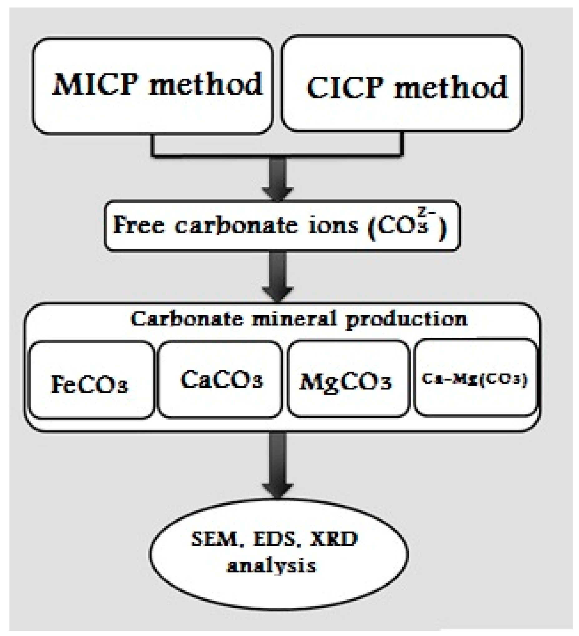
Figure 1. Schematic diagram of carbonate mineral production.
Minerals 13 00976 g001
Figure 2. Particle size distribution of Firoozkuh silica sand.
Minerals 13 00976 g002
Figure 3. The concentration of CO32− for MICP and CICP.
Minerals 13 00976 g003
Figure 4. The concentration of ammonium per different zeolite percentage.
Minerals 13 00976 g004
Figure 5. Carbonate minerals precipitation by MICP and CICP methods.
Minerals 13 00976 g005
Figure 6. SEM morphologies of CaCO3 mineral (a) spherical shape by MICP (b) rhombohedral shape by CICP.
Minerals 13 00976 g006
Figure 7. EDS and XRD of CaCO3 minerals (a) Vaterite by MICP (b) Calcite by CICP.
Minerals 13 00976 g007
Figure 8. SEM morphologies of FeCO3 mineral with spherical shape in (a) MICP and (b) CICP.
Minerals 13 00976 g008
Figure 9. EDS and XRD of FeCO3 mineral: (a) Siderite by MICP and (b) Siderite by CICP.
Minerals 13 00976 g009
Figure 10. SEM morphologies of Ca-Mg (CO3) mineral with botryoidal shape by (a) MICP and (b) CICP.
Minerals 13 00976 g010
Figure 11. EDS and XRD of Ca-Mg (CO3) minerals (a) Dolomite by MICP (b) High-Mg calcite by CICP.
Minerals 13 00976 g011
Figure 12. SEM morphologies of MgCO3 minerals with needle-shaped crystals by (a,c) MICP and (b,d) CICP.
Minerals 13 00976 g012
Figure 13. EDS and XRD of MgCO3 minerals (a) Nesquehonite by MICP (b) Nesquehonite by CICP.
Minerals 13 00976 g013
Figure 14. The average size of precipitated carbonate minerals by MICP and CICP methods.
Minerals 13 00976 g014
Figure 15. Unconfined compressive strength of treated specimens with different carbonate minerals.
Minerals 13 00976 g015
Table 1. Chemical properties of used reagents.
ReagentsConcentrationCation:Carbonate Ratios
FeSO4·7H2O1 MFe2+:CO32−1:1
MgSO4·7H2O1 MMg2+:CO32−1:1
CaCl2·2H2O1 MCa2+:CO32−1:1
MgSO4/CaCl21 MMg2+/Ca2+:CO32−1:1
Table 2. Characterization of produced carbonate minerals by MICP and CICP.
NameMethodType of MineralSize (µm)Mineral Shape
Calcium carbonateMICPVaterite 2–7Spherical
CICPCalcite2–6Rhombohedral
Ferrous carbonate MICPSiderite0.5–3Spherical
CICPSiderite2–6Spherical
Magnesium calcium carbonateMICPDolomite5–16Botryoidal
CICPHigh-Mg calcite2–5Botryoidal
Magnesium carbonateMICPNesquehonite1–10Radial-needle
CICPNesquehonite5–20Radial-needle
Disclaimer/Publisher’s Note: The statements, opinions and data contained in all publications are solely those of the individual author(s) and contributor(s) and not of MDPI and/or the editor(s). MDPI and/or the editor(s) disclaim responsibility for any injury to people or property resulting from any ideas, methods, instructions or products referred to in the content.