
Trace Elements in Dental Enamel Can Be a Potential Factor of Advanced Tooth Wear


Abstract
1. Introduction
2. Material and Methods
2.1. Samples
- (1)
- A total of 50 enamel samples were taken from upper central incisors derived from patients with advanced tooth wear. The greatest destruction of tooth hard tissue was observed on occlusal surfaces and/or incisal margins, which is why the tooth wear index for these surfaces was used in the further comparative analysis. The mean value of tooth wear was 2.27 ± 0.52 according to the Smith and Knight index (TWI), and mean patient age was 49.5 ± 9 years [21]. To include patients in the study, the following criteria were applied: visible features of advanced tooth wear on teeth, no dental caries or periodontal disease, no conservative treatment and preventive professional application of fluoride in a dental clinic prior to recruitment to the study. The patients were referred to the Department of Prosthetic Dentistry due to a clinically apparent decrease in occlusal vertical dimension (more than 4 mm) and a consequent reduced self-esteem of face aesthetics that required to be restored.
- (2)
- A total of 20 enamel samples were taken from upper central incisors without signs of pathological tooth wear from healthy volunteers aged 48.5 ± 6 years. They were asked to participate voluntarily in the study, as they presented at the Department to complete a prosthetic procedure relating only to a single tooth (e.g., crown or inlay).
- (3)
- A total of 15 permanent human central upper incisors with completed formation and without any visible pathological changes (donors between 18 and 21 years of age, who expressed their written informed consent for using their extracted teeth for studies) were used in the study. Mechanical damage in the area of alveolar process or changes in periodontium were an indication for tooth extraction. All teeth used in this research were obtained from the Bank of Teeth, University of Bern, School of Dental Medicine, Department of Preventive, Restorative and Pediatric Dentistry, Switzerland. They were prepared for the study in accordance with ISO/TS 11405:2015 [22].
2.2. Ethical Approval
2.3. Study Design
- (1)
- Teeth preparation and acid biopsy in vivo;
- (2)
- Teeth preparation and acid biopsy in vitro;
- (3)
- Biochemical analysis of samples using the AAS (atomic absorption spectrometry) method.
2.3.1. Clinical Procedure for Tooth Wear Patients and Volunteers
2.3.2. Extracted Teeth Preparation
2.3.3. Biochemical Analysis
2.4. Statistical Analysis
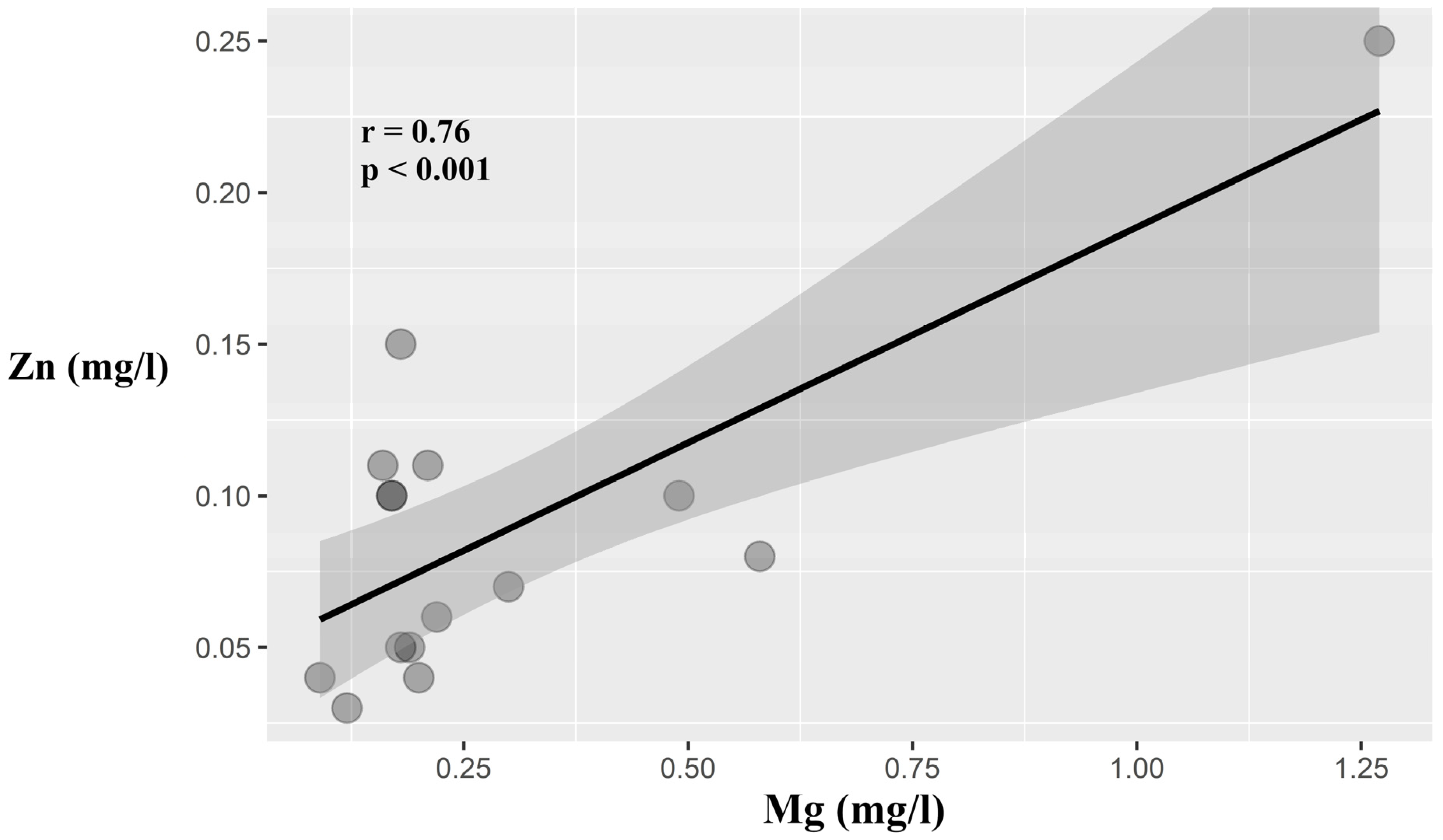
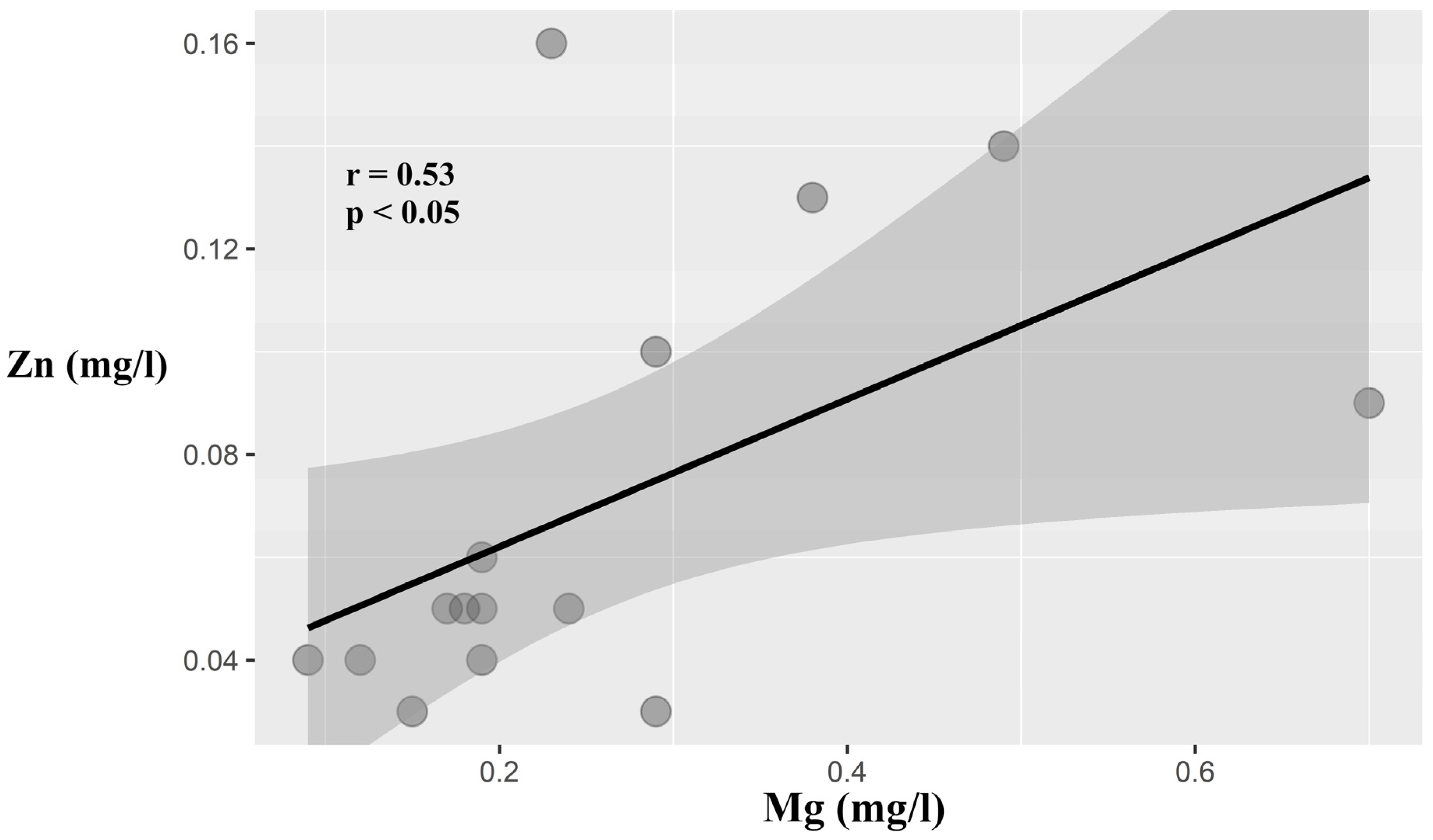
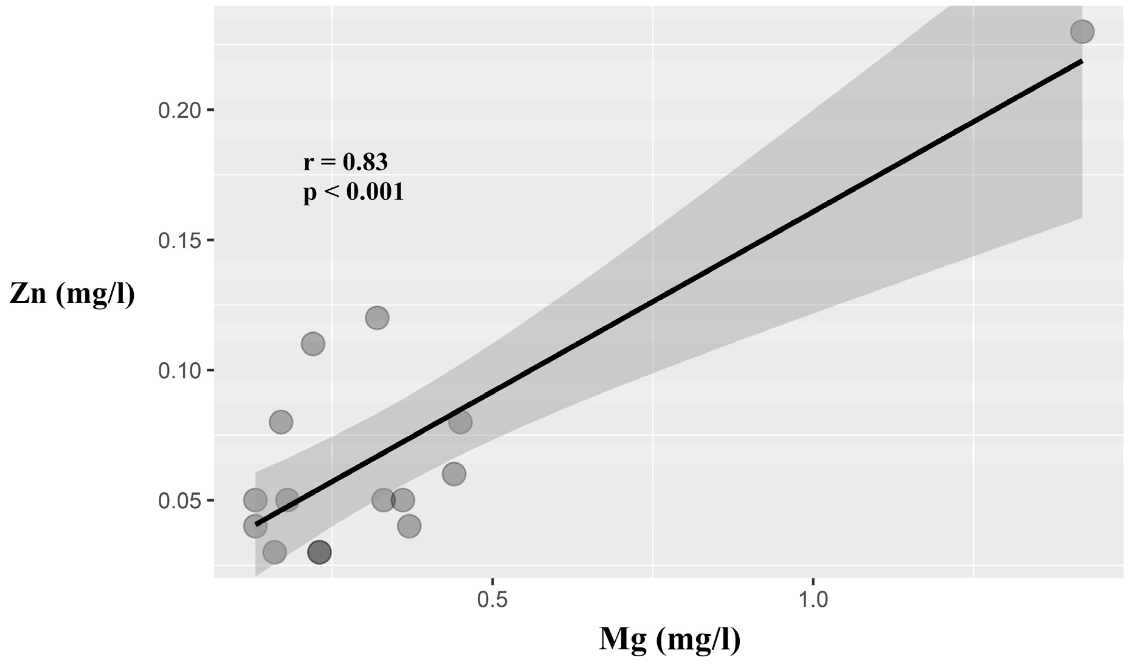
3. Results
4. Discussion
Author Contributions
Funding
Data Availability Statement
Conflicts of Interest
References
- Fatima, T.; Rahim, Z.H.A.; Chew, W.L.; Qamar, Z. Zinc: A precious trace element for oral health care? J. Pak. Med. Assoc. 2016, 66, 1019–1023. [Google Scholar] [PubMed]
- Sabel, N. Enamel of primary teeth--morphological and chemical aspects. Swed. Dent. J. 2012, 120 (Suppl. 1), 1–77. [Google Scholar]
- Bartlett, J.D.; Skobe, Z.; Nanci, A.; Smith, C.E. Matrix metalloproteinase 20 promotes a smooth enamel surface, a strong dentino-enamel junction, and a decussating enamel rod pattern. Eur. J. Oral Sci. 2011, 119 (Suppl. 1), 199–205. [Google Scholar] [CrossRef] [PubMed]
- Sa, Y.; Liang, S.; Ma, X.; Lu, S.; Wang, Z.; Jiang, T.; Wang, Y. Compositional, structural and mechanical comparisons of normal enamel and hypomaturation enamel. Acta Biomater. 2014, 10, 5169–5177. [Google Scholar] [CrossRef]
- Yahyazadehfar, M.; Bajaj, D.; Arola, D.D. Hidden contributions of the enamel rods on the fracture resistance of human teeth. Acta Biomater. 2013, 9, 4806–4814. [Google Scholar] [CrossRef]
- Weatherel, J. Composition of dental enamel. Br. Med. Bull. 1975, 31, 115–119. [Google Scholar] [CrossRef] [PubMed]
- Bartlett, D.W.; Anggiansah, A.; Owen, W.; Evans, D.F.; Smith, B.G.N. Dental erosion: A presenting feature of gastro-oesophegeal reflux disease. Eur. J. Gastoenterol. Hepatol. 1994, 6, 895–900. [Google Scholar] [CrossRef]
- Bialek, M.; Zyska, A. The biomedical role of zinc in the functioning of the human organism. Pol. J. Public Health 2014, 124, 160–163. [Google Scholar] [CrossRef]
- Gapys, B.; Raszeja-Specht, A.; Bielarczyk, H. Role of zinc in physiological and pathological processes of the body. Diagn. Lab. 2014, 50, 45–52. [Google Scholar]
- Lee, S.K.; Kim, Y.S.; Lee, S.S.; Lee, Y.J.; Song, I.S.; Park, S.C. Molecular cloning, chromosomal mapping, and characteristic expression in tooth organ of rat and mouse Krox-25. Genomics 2004, 83, 243–253. [Google Scholar] [CrossRef]
- Andreini, C.; Banci, L.; Bertini, I.; Luchinat, C.; Rosato, A. Bioinformatic comparison of structures and homology-models of matrix metalloproteinases. J. Proteome Res. 2004, 3, 21–31. [Google Scholar] [CrossRef] [PubMed]
- Li, Y.; Shi, X.; Li, W. Zinc-containing hydroxyapatite enhances cold-light-activated tooth bleaching treatment in vitro. Biomed. Res. Int. 2017, 6261248. [Google Scholar] [CrossRef] [PubMed]
- Sierpinska, T.; Orywal, K.; Kuc, J.; Golebiewska, M.; Szmitkowski, M. Enamel mineral content in patients with severe tooth wear. Int. J. Prosth. 2013, 26, 423–428. [Google Scholar] [CrossRef]
- Brookes, S.J.; Shore, R.C.; Robinson, C.; Wood, S.R.; Kirkham, J. Copper ions inhibit the demineralization of human enamel. Arch. Oral Biol. 2003, 48, 25–30. [Google Scholar] [CrossRef]
- Abdullach, A.Z.; Strafford, S.M.; Brookes, S.J.; Duggal, M.S. The effect of copper on demineralization of dental enamel. J. Dent. Res. 2006, 85, 1011–1015. [Google Scholar] [CrossRef]
- Bartlett, J.D.; Lee, Z.; Eright, J.T.; Li, Y.; Kulkarni, A.B.; Gibsin, C.W. A developmental comparison of matrix metalloproteinase-20 and amelogenin null mouse enamel. Eur. J. Oral Sci. 2006, 114 (Suppl. 1), 18–23. [Google Scholar] [CrossRef]
- Loomans, B.; Optam, N.; Attin, T.; Bartlett, D.; Edelhoff, D. Severe tooth wear: European consensus statemen on managment guidelines. J. Adhesive Dent. 2017, 19, 111–119. [Google Scholar]
- Grippo, J.O.; Simring, M.; Schreiner, S. Attrition, abrasion, corrosion and abfraction revisited. A new perspective on tooth surface lesions. JADA 2004, 135, 1109–1118. [Google Scholar]
- Wetselaar, P.; Lobbezoo, F. The tooth wear evaluation system: A modular clinical guideline for the diagnosis and management planning of worn dentition. J. Oral Rehab. 2016, 43, 69–80. [Google Scholar] [CrossRef]
- Sovik, J.B.; Vieira, A.R.; Tveit, A.B.; Mulic, A. Enamel formation genes associated with dental erosive wear. Caries Res. 2015, 49, 236–242. [Google Scholar] [CrossRef]
- Smith, B.; Knight, J. An Index for measuring the wear of teeth. Br. Dent. J. 1984, 156, 435–438. [Google Scholar] [CrossRef] [PubMed]
- ISO/TS 11405; Technical Specification. Testing of Adhesion to Tooth Structure. International Organization of Standardization: Geneve, Switzerland, 2015.
- Milosevic, A.; Dawson, L.J. Salivary factors in vomiting bulimics with and without pathological tooth wear. Caries Res. 1996, 30, 361–366. [Google Scholar] [CrossRef] [PubMed]
- Prajapati, S.; Tao, J.; Ruan, Q.; De Yoreo, J.J.; Moradian-Oldak, J. Matrix metalloproteinase-20 mediates dental enamel biomineralization by preventing protein occlusion inside apatite crystals. Biomaterials 2016, 75, 260–270. [Google Scholar] [CrossRef]
- Yamakoshi, Y.; Tanabe, T.; Fukae, M.; Shimizu, M. Porcine amelogenins. Calcif. Tissue Int. 1994, 54, 69–75. [Google Scholar] [CrossRef] [PubMed]
- Fang, M.M.; Lei, K.Y.; Kilgore, L.T. Effects of zinc deficiency on dental caries in rats. J. Nutr. 1980, 110, 1032–1036. [Google Scholar] [CrossRef] [PubMed]
- Stötzel, C.; Müller, F.A.; Reinert, F.; Niederdraenk, F.; Barralet, J.E.; Gbureck, U. Ion adsorption behaviour of hydroxyapatite with different crystallinities. Colloids Surf. Biointerfaces 2009, 74, 91–95. [Google Scholar] [CrossRef] [PubMed]
- Mneimne, M.; Hill, R.G.; Al-Jawad, M.; Lynch, R.J.; Mohammed, N.R.; Anderson, P. Physical chemical effects of zinc on in vitro enamel demineralization. J. Dent. 2014, 42, 1096–1104. [Google Scholar]
- Rahman, M.T.; Hossain, A.; Pin, C.H.; Yahya, N.A. Zinc and metallothionein in the development and progression of dental caries. Biol. Trace Elem. Res. 2008, 5, 12011–12018. [Google Scholar] [CrossRef]
- Davey, H.P.; Embery, G.; Cummins, C. Interaction of zinc with a synthetic calcium phosphate mineral. Caries Res. 1997, 31, 434–440. [Google Scholar] [CrossRef]
- Aoba, T. Recent observations on enamel crystal formation during mammalian amelogenesis. Anat. Rec. 1996, 245, 208–218. [Google Scholar] [CrossRef]
- Smith, C.E. Cellular and chemical events during enamel formation. Crit. Rev. Oral Biol. Med. 1998, 9, 128–161. [Google Scholar] [CrossRef] [PubMed]
- Nikolayeva, O.; Pysmenna, O. The influence of unbalanced nutrition of pregnant rats on the content of biogenic elements in the enamel and blood serum of their offspring. Georgian Med. News 2018, 278, 163–168. [Google Scholar]
- Krasuska-Slawinska, E.; Dembowska-Baginska, B.; Brozyna, A.; Olczak-Kowalczyk, D.; Czarnowska, E.; Sowinska, A. Changes in the chemical composition of mineralised teeth in children after antineoplastic treatment. Contempor. Oncol. 2018, 22, 37–41. [Google Scholar] [CrossRef]
- Medeiros, D.M.; Ilich, J.; Ireton, J.; Matkovic, V.; Shiry, L.; Wildman, R. Femurs from rats fed diets deficient in copper or iron have decreased mechanical strength and altered mineral composition. J. Trace Elem. Exp. Med. 1997, 10, 197–203. [Google Scholar] [CrossRef]
- Souza, A.P.; Gerlach, R.F.; Line, S.R.P. Inhibition of human gingival gelatinases (MMP-2 and 9) by metal salts. Dent. Mater. 2000, 16, 103–108. [Google Scholar] [CrossRef] [PubMed]
- Zampetti, P.; Scribante, A. Historical and bibliometric notes on the use of fluoride in caries prevention. Eur. J. Pediatr. Dent. 2020, 21, 148–152. [Google Scholar]
- Khanduri, N.; Kurup, D.; Mitra, M. Quantitative evaluation of remineralizing potential of three agents on artificially demineralized human enamel using scanning electron microscopy imaging and energy-dispersive analytical X-ray element analysis: An in vitro study. Dent. Res. J. Isfahan 2020, 17, 366–372. [Google Scholar] [CrossRef]
- Butera, A.; Pascadopoli, M.; Gallo, S.; Lelli, M.; Tarterini, F.; Giglia, F.; Scribante, A. SEM/EDS Evaluation of the Mineral Deposition on a Polymeric Composite Resin of a Toothpaste Containing Biomimetic Zn-Carbonate Hydroxyapatite (microRepair®) in Oral Environment: A Randomized Clinical Trial. Polymers 2021, 13, 2740. [Google Scholar] [CrossRef] [PubMed]


| Mineral | Worn Teeth (n = 50) | Healthy Teeth In Vivo (20) | 00 | 0 (0–150 µm) | 1 (150–300 µm) | 2 (300–450 µm) | 3 (450–600 µm) | 4 (600–750 µm) | 5 (750–900 µm) | 6 (900–1050 µm) |
|---|---|---|---|---|---|---|---|---|---|---|
| Ca [mg/L] | 1.88 ± 1.38 | 1.85 ± 1.24 | 1.42 ± 0,39 | 1.86 ± 1.54 | 1.59 ± 1.22 | 1.89 ± 1.63 | 1.96 ± 0.86 | 1.56 ± 0.96 | 1.47 ± 1.39 | 1.45 ± 1.29 |
| Mg [mg/L] | 0.30 ± 0.14 | 0.33 ± 0.15 | 0.18 ± 0.08 | 0.2 ± 0.07 | 0.31 ± 0.19 | 0.25 ± 0.15 | 0.26 ± 0.16 | 0.26 ± 0.09 | 0.31 ± 0.13 | 0.34 ± 0.13 |
| Zn [mg/L] | 0.14 ± 0.04 | 0.08 ± 0.06 * | 0.04 ± 0.01 # | 0.06 ± 0.02 # | 0.09 ± 0.05 # | 0.07 ± 0.03 # | 0.07 ± 0.05 # | 0.06 ± 0.03 # | 0.05 ± 0.02 # | 0.07 ± 0.05 # |
| Cu [µg/L] | 22.03 ± 17.45 | 36.67 ± 22.66 * | 10.42 ± 5.56 # | 17.87 ± 6.59 | 16.45 ± 3.54 | 16.22 ± 8.63 | 20.98 ± 12.2 | 17.65 ± 7.71 | 17.21 ± 7.16 | 14.55 ± 4.27 |
Disclaimer/Publisher’s Note: The statements, opinions and data contained in all publications are solely those of the individual author(s) and contributor(s) and not of MDPI and/or the editor(s). MDPI and/or the editor(s) disclaim responsibility for any injury to people or property resulting from any ideas, methods, instructions or products referred to in the content. |
© 2023 by the authors. Licensee MDPI, Basel, Switzerland. This article is an open access article distributed under the terms and conditions of the Creative Commons Attribution (CC BY) license (https://creativecommons.org/licenses/by/4.0/).
Share and Cite
Zamojda, E.; Orywal, K.; Mroczko, B.; Sierpinska, T. Trace Elements in Dental Enamel Can Be a Potential Factor of Advanced Tooth Wear. Minerals 2023, 13, 125. https://doi.org/10.3390/min13010125
Zamojda E, Orywal K, Mroczko B, Sierpinska T. Trace Elements in Dental Enamel Can Be a Potential Factor of Advanced Tooth Wear. Minerals. 2023; 13(1):125. https://doi.org/10.3390/min13010125
Chicago/Turabian StyleZamojda, Elzbieta, Karolina Orywal, Barbara Mroczko, and Teresa Sierpinska. 2023. "Trace Elements in Dental Enamel Can Be a Potential Factor of Advanced Tooth Wear" Minerals 13, no. 1: 125. https://doi.org/10.3390/min13010125
APA StyleZamojda, E., Orywal, K., Mroczko, B., & Sierpinska, T. (2023). Trace Elements in Dental Enamel Can Be a Potential Factor of Advanced Tooth Wear. Minerals, 13(1), 125. https://doi.org/10.3390/min13010125